Review

Injectable Hydrogel Systems for Targeted Drug Delivery: From Site-Specific Application to Design Strategy

1
Department of Bionanotechnology, Gachon University, Seongnam 13120, Republic of Korea
2
Department of Chemical Engineering, Delft University of Technology, 2628 CD Delft, The Netherlands
*
Authors to whom correspondence should be addressed.
Appl. Sci. 2025, 15(21), 11599; https://doi.org/10.3390/app152111599
Submission received: 30 September 2025 / Revised: 16 October 2025 / Accepted: 28 October 2025 / Published: 30 October 2025
(This article belongs to the Special Issue Anticancer Drugs: New Developments and Discoveries)

Abstract

Injectable hydrogels are adaptable drug delivery systems capable of forming localized depots that align with the anatomical and physiological constraints of administration sites. Their performance depends on both the injection environment and the properties of the therapeutic cargo. Applications span ocular, intra-articular, subcutaneous, intramuscular, tumoral, central nervous system, and mucosal delivery, where hydrogels address challenges of clearance, retention, and compatibility. Beyond bulk depots, particulate hydrogel formats such as microgels and nanogels improve syringeability, modularity, and integration with nanoparticle carriers. Functional versatility arises from stimuli responsiveness, including pH, enzymatic, thermal, redox, and light triggers, and from hybrid designs that integrate multiple cues for precision control. Loading strategies range from passive encapsulation to affinity binding and covalent conjugation, with release governed by diffusion, degradation, and stimuli-modulated kinetics. Translational progress depends on reproducible fabrication, scalable manufacturing, and device integration, while site-dependent constraints and regulatory hurdles remain significant challenges.

1. Introduction

Injectable therapeutics are one of the most widely used and clinically impactful routes of drug administration, enabling localized treatment, sustained exposure, and patient compliance in contexts ranging from oncology and immunology to ophthalmology and pain management [1,2,3,4,5,6,7,8,9,10,11]. However, despite their versatility, the formulation of injectable systems that balance biological compatibility, spatial control, manufacturability, and cargo versatility remains a critical engineering challenge [12,13,14]. The ideal injectable system should be physically deliverable through clinically practical routes, including subcutaneous, intra-articular, and intravitreal, while capable of forming a stable depot to maintain the therapeutic payload as well as adaptable to site-specific physiological constraints [1,15,16,17].
Hydrogels have long been proposed as a promising material candidate to meet many of these needs. Their high water content, polymeric network structure, biocompatibility and biodegradability emphasize their utility for soft tissue interfaces [18,19]. Several clinically approved systems, as represented by thermo-gelling copolymers and PEG-based in situ gelling platforms, demonstrate the feasibility of hydrogel-based injectables in therapeutic applications [20,21,22,23]. However, their structural scale and limited processability often constrain their applicability. Conventional bulk-forming gels are poorly suited for applications that require multi-compartmental release, precise spatial distribution, or adaptive delivery within anatomically confined environments. Also, the inability to tune mechanical properties or responsiveness independently from bulk viscosity poses formulation barriers in real-world scenarios [9,24,25].
Particulate hydrogel formulations, commonly referred to as microgels and nanogels, have emerged as a complementary class of injectable carriers. Defined across the micro- to nanoscale, they maintain the fundamental network properties of hydrogels while functioning as discrete, flowable units [5,11,26,27]. This form enables administration through narrow-gauge needles and dispersion into tissues without the viscosity limitations of bulk gels. Once delivered, hydrogel particles can occupy defined volumes, assemble into depots, or distribute within interstitial spaces, depending on particle size and mechanical softness. These behaviors provide control over retention and local drug release that is not achievable with bulk-forming platforms. Across the broader spectrum of injectable formulations, including polymeric depots, nanoparticle suspensions (Lupron Depot®, Doxil®), in situ forming hydrogels (ReSure®), each system offers specific advantages in administration or release control but often with trade-offs in mechanical adaptability or tissue conformity [28,29,30]. Microgels and nanogels extend these capabilities by uniting the structural cohesion of hydrogel with the versatility of particulate carriers, offering a continuum between bulk gels and nanosystems.
At miniaturized scales, hydrogel particles allow properties such as size, deformability, degradation, and responsiveness to be tuned independently from macroscopic viscosity, which expands formulation possibilities for anatomically constrained sites such as the posterior eye, intra-articular space, and tumor margins (Figure 1) [11,31,32]. Stimuli-responsive variants, incorporating thermosensitive domains, enzyme-cleavable linkers, or pH-sensitive moieties, further enable release kinetics to match pathological features including inflammation, dysregulated protease activity, and acidic microenvironments [23,33,34,35]. Functionally, particulate hydrogels overlap with several existing drug carriers including nanoparticles, liposomes, microsphere, and emulsions, but are distinguished by their mechanical compliance with soft tissues and their ability to form shear-thinning or in situ-setting suspensions [16,36,37]. Notably, their capabilities can be modulated and adapted to specific therapeutic sites, offering a degree of programmability that is extremely challenging in traditional formulations.
However, the advancement of injectable hydrogel delivery systems has been uneven in contrast to their clear technical practicality and importance. This has created technological gaps or discontinuity across material engineering, pharmacology, and clinical translation, often leading to inefficient struggles instead of stimulating innovative, creative progress. Particularly, the absence of clear design strategies and limited benchmarking against other delivery systems, in terms of injectability, anatomical fit, and release behavior, has hindered systematic evaluation even when experimental data are promising.
In this review, we aim to address this disconnect by offering a functional framework for the rational design and application of injectable hydrogel systems, with emphasis on particulate formulations where their modularity and depot-like behavior offer distinct therapeutic opportunities. Key administration routes, such as ocular delivery, intra-articular injection, subcutaneous or intramuscular implantation, and localized tumor therapy, are reviewed with respect to anatomical and physiological constraints that guide formulation choices. Next, design principles across diverse applications including control of size, deformability, responsiveness, and relevant fabrication constraints are discussed to aid in realizing reliable and accessible platforms. Finally, hybrid systems that integrate injectable hydrogels with nanoscale carriers are highlighted as an underutilized pathway toward multi-functional injectable delivery systems.
By bridging therapeutic constraints, materialistic aspects, and translational context, this review positions injectable hydrogels as strategically adaptable systems that merit deliberate and intensive development. We aim to provide a conceptual scaffold with updated recent works for researchers to more deliberately formulate, compare, and advance hydrogel-based delivery systems across the interface of biomaterials, nanotechnology, drug delivery, therapeutic engineering, and clinical need.

2. Hydrogels for Site-Specific Injectable Drug Delivery

Injectable formulations remain a cornerstone of therapeutic delivery, enabling direct access to target tissue and avoiding systemic dispersion. However, their performance is often limited by site-specific constraints such as mechanical stress, fluid turnover, and narrow injection tolerance. Traditional formulations, ranging from small molecules to bulk-forming gels, offer partial solutions, often at the expense of depot stability, bio-responsiveness, or patient comfort, as summarized in Table 1.

2.1. Ocular Delivery

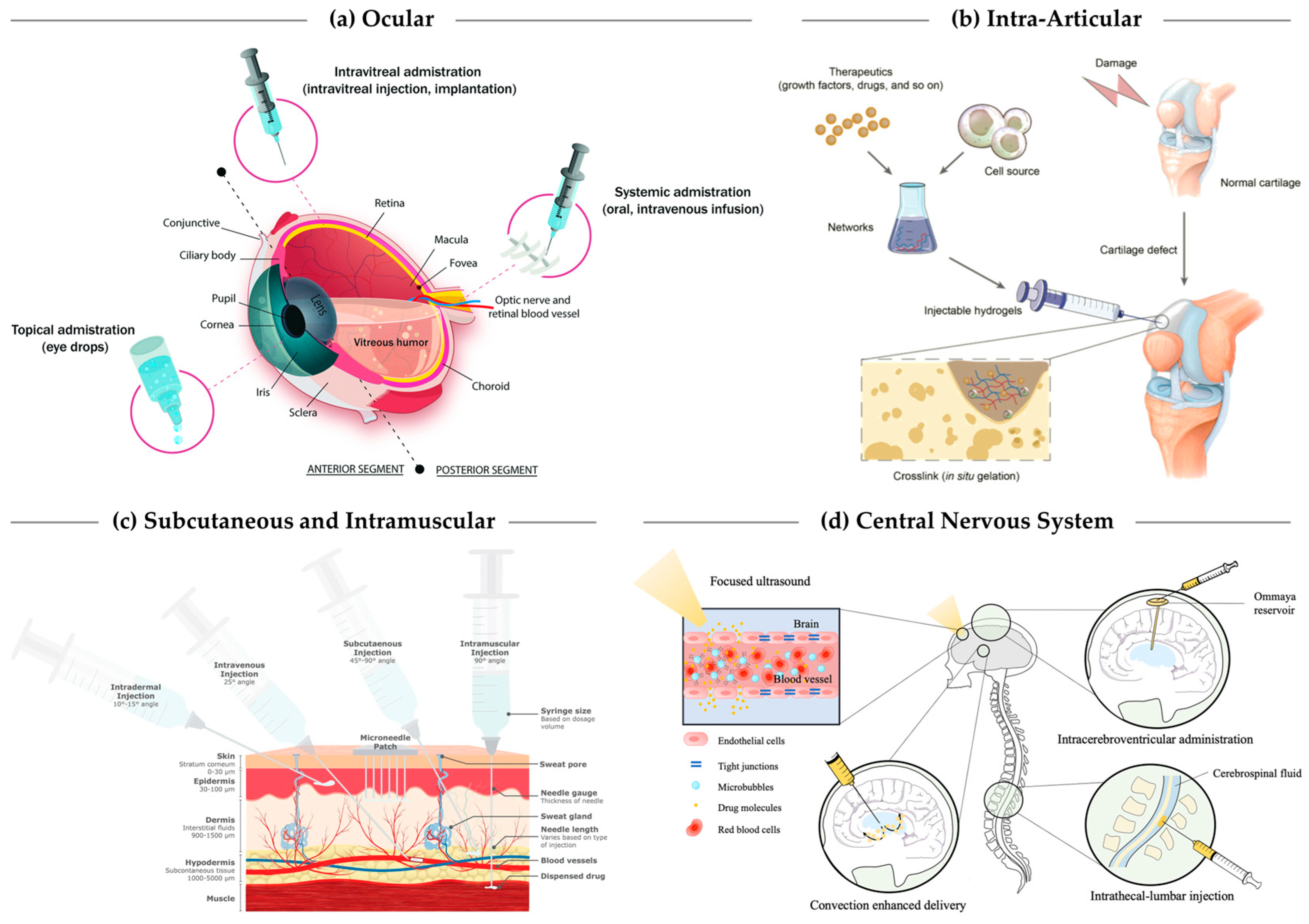
The eye presents a uniquely complex anatomical and physiological landscape for drug delivery. Drug access is spatially restricted by multilayered defense systems that serve to maintain optical transparency and immune privilege but sharply limit bioavailability. The anterior segment is shielded by the corneal epithelium, conjunctiva, tear film, and dynamic clearance mechanisms such as blinking and nasolacrimal drainage, all of which severely limit drug residence time and bioavailability. In contrast, the posterior segment is enclosed by structural and active barriers such as the sclera, choroid, and the blood–aqueous and blood–retinal barriers. These protective systems are highly effective at excluding exogeneous substances, particularly large or hydrophilic molecules, thereby reducing the efficacy of systemically or topically administered agents (Figure 2a) [69,70,71,72].
Conventional ocular drug delivery strategies are defined by these spatial constraints. Topical formulations including eye drops and ointments are widely used for anterior segment conditions such as conjunctivitis or dry eye but rarely achieve more than transient surface contact [69]. Posterior segment diseases, such as age-related macular degeneration and diabetic retinopathy, require intravitreal injection of biologics to ensure target site access, despite procedural risks and patient burden [76]. Periocular injections, including subconjunctival and sub-Tenon’s routes, are often used for inflammatory conditions but offer limited drug penetration and rapid clearance [11,69,70]. Across administration routes, achieving sufficient and sustained drug levels at target sites remains a core challenge.
The difficulty of sustaining therapeutic concentrations at ocular target sites, without compromising safety or patient compliance, has prompted a shift toward depot-forming, tissue-compatible formulations. This includes advances in micelle-based solubilization [77], in situ gelling polymers [39,78], and microparticle-based systems [79,80], each aiming to improve local drug exposure while mitigating clearance and dosing frequency. Among these, hydrogels have gained particular attention for their tunability in both mechanical and pharmacokinetic properties, offering an attractive alternative to rigid implants or bolus injections.
Current investigations in hydrogel-based ocular delivery span both anterior and posterior applications. In situ forming hydrogels have been studied as depots for biologics in the vitreous cavity [11,39,81], while mucoadhesive and stimuli-responsive hydrogel films have been explored for extending drug residence on the ocular surface [38,40]. Notably, a thermosensitive chitosan/β-glycerophosphate hydrogel enabled sustained dexamethasone release for up to three weeks after subconjunctival injection, effectively reducing ocular inflammation [42]. Likewise, a PTMC–F127–PTMC copolymer hydrogel loaded with mitomycin C exhibited controlled in situ gelation and fibrosis inhibition following glaucoma filtration surgery [41]. These systems remain in various stages of preclinical or early clinical development, but the convergence of materials engineering and anatomical targeting also reflects a shift from passive formulation to actively tunable, adaptable systems. In this context, hydrogels represent a versatile and evolving platform that responds to the unique constraints of ophthalmic administration.

2.2. Intra-Articular Injection

Intra-articular injections remain a clinically routine intervention for joint disorders, but their efficacy is often constrained by rapid clearance dynamics and mechanical instability within the joint cavity [43,82,83]. Synovial fluid, a viscous ultrafiltrate of plasma that lubricates joint surfaces and nourishes cartilage, is continuously renewed and drained through lymphatic and vascular pathways, resulting in short intra-articular residence for solutes. In addition, joint articulation introduces substantial shear stress that can destabilize depots and accelerate dispersion. The dense, avascular cartilage further restricts passive diffusion of therapeutics, limiting drug penetration into chondrocyte-rich and subchondral zones (Figure 2b) [73,82,84].
Current intra-articular drug delivery strategies rely on corticosteroids, non-steroidal anti-inflammatory drugs (NSAIDs), and hyaluronic acid (HA) for anti-inflammatory and pain-relieving effects. However, these agents are typically characterized by short joint residence times and limited bioavailability [44]. Corticosteroids often undergo burst release and rapid systemic clearance, necessitating repeated injections. HA, although commonly used as a viscosupplement, is susceptible to enzymatic degradation in inflamed joints and lacks long-term mechanical stability. Small-molecule agents like Celecoxib and Disulfiram exhibit poor aqueous solubility and low retention within joint tissues, while nanoparticles under 300 nm are often cleared via lymphatic drainage before meaningful accumulation [69,83].
In the same context, hydrogel-based formulations have been increasingly highlighted, benefiting from mechanical compliance, tunable degradation, and extended local retention. Hydrogel matrices have been adapted to encapsulate anti-inflammatory drugs, microspheres, and nanoparticle systems, allowing modulations of release dynamics while being compatible with load-bearing environments. Several material platforms have demonstrated prolonged joint retention, sustained suppression of inflammatory markers, and in some cases, enhancement of cartilage repair and protection [45,83,85,86]. Formulations include chemically crosslinked networks, thermo-responsive hydrogels, and bioadhesive composites that improve tissue residency and therapeutic durability. Injectable scaffolds combining hydrogels with stem cells or bioactive particles are also being explored for cartilage regeneration under physiological load, reflecting the dual potential of hydrogel platforms in intra-articular therapy [46,47].
While promising, challenges remain in degradation kinetics with joint-specific enzymatic and mechanical conditions, ensuring compatibility of drug cargos with immune response, and translating preclinical progress into clinically viable products.

2.3. Subcutaneous, Dermal, and Intramuscular Depots

Subcutaneous (SC) and intramuscular (IM) routes are among the most employed parenteral approaches for sustained drug delivery [87]. Both sites enable local depot formation and gradual systemic uptake but differ markedly in tissue architecture, extracellular matrix (ECM) composition, and pharmacokinetic characteristics.
The SC injections are typically administered into the hypodermis, commonly referred to as the bottom layer of skin, at depths of 5 to 10 mm using short, fine-gauge needles (Figure 2c) [14,74]. The site accommodates injection volumes up to 1.5 mL without significant discomfort, though higher volumes may lead to pain, tissue distension, or altered absorption kinetics [49]. The SC space presents a permissive environment for depot formation, with moderate immune surveillance and sufficient interstitial volume to accommodate injectable materials [8]. However, the injected materials are subject to enzymatic degradation and clearance via lymphatic drainage, leading to limited retention and requiring formulation strategies that resist early breakdown and promote controlled diffusion [48]. To address these challenges, injectable hydrogels have been developed to improve subcutaneous drug retention and therapeutic efficacy through sustained release and preservation of bioactivity. Notably, crosslinked polyethylene glycol and hyaluronic acid-based systems have demonstrated controlled release of both macromolecular and small-molecule drugs, while self-healing antibacterial hydrogels further expand their potential for localized infection treatment [50,51,52].
IM delivery involves injection into the deltoid, vastus lateralis, or gluteal muscles at depths of 25 to 38 mm, commonly using 22–25 gauge needles. This route allows for larger injection volumes, up to 5 mL depending on site and patient characteristics but introduces additional biomechanical complexity. The IM compartment is structurally defined by dense ECM, composed primarily of collagen I and hyaluronic acid, with injected materials distributed through the endomysial and perimysial layers surrounding muscle fibers [88,89,90]. Drug transport within this compartment is often anisotropic, directed along ECM-defined planes with limited radial spread. The stiffness and heterogeneity of the muscle ECM further contribute to high inter-individual variability in depot behavior [53,89].
Clinically, both SC and IM routes are used for sustained delivery of small molecules, peptides, and biologics [87]. SC injections are commonly used for monoclonal antibodies, insulin analogs, and other biologics where patient self-administration is desirable, while IM injections are generally utilized for long-acting therapies such as hormone analogs and antipsychotics [87,91]. However, the physicochemical constraints imposed by each site, such as lymphatic clearance for SC and fibrous compartmentalization for IM, limit the consistency and duration of release achievable with conventional formulation strategies [48,53].
Hydrogel-based depots have gained attention as adaptive systems that combine syringeability with localized drug retention [92,93]. For SC administration, in situ gelling systems composed of PEG, alginate, or hyaluronic acid can form soft depots that match local tissue mechanics while resisting enzymatic breakdown. Thermo-responsive hydrogels allow for rapid gelation upon injection and can be engineered to sustain drug release over days to weeks [94]. For IM administration, biomimetic hydrogel matrices that replicate ECM stiffness and porosity have been explored to improve reproducibility and maintain depot under contractile and mechanical stress [22,53,55].
The translation of these systems to clinical use will require further refinement of their degradation profiles, injection performance, and scalability [92]. Nevertheless, the shift toward hydrogel-based system reflects a broader strategy of tailoring materials to the unique characteristics of tissue injection environments, where both retention and mechanical compatibility are essential for better therapeutic efficacy.

2.4. Tumoral and Peritumoral Injection

While intravenous injections remain the dominant route for advanced antitumor drug delivery, particularly for nanoparticle formulations targeting solid tumors via systemic circulation, localized injection strategies such as intratumoral and peritumoral delivery offer a more direct and spatially confined alternative [33,58]. These approaches bypass vascular barriers, reduce systemic exposure, and achieve high local drug concentrations, yet have been relatively underexplored in recent DDS development despite their established clinical utility [10,58]. Conventional formulations include ethanol, bleomycin, or lipiodol emulsions for intratumoral ablation, and simple drug solutions or protein suspensions for peritumoral immunotherapy. However, clearance and depot stability remain limited, which has motivated the adaptation of hydrogel-based systems to enhance therapeutic efficacy [95,96,97,98]. Tumoral injection deposits therapeutic agents directly into the tumor mass, allowing circumvention of circulatory clearance and systemic dilution [15,99]. While this strategy achieves high intratumoral concentrations, its performance is often hindered by poor diffusion into dense tumor matrices, irregular perfusion, and limited access to deep or metastatic lesions [95,96,100,101]. Moreover, the lack of structural confinement in conventional solutions or suspensions often leads to burst release and rapid efflux, diminishing local efficacy [102]. Peritumoral injections, in contrast, target the surrounding stroma or tumor boundary, enabling drug infiltration into the tumor microenvironment (TME) and adjacent vasculature [103]. This approach is particularly important for immunomodulatory agents or therapies targeting perivascular niches and lymphatic drainage pathways [15].
Hydrogel-based depots have been explored to overcome the inherent instability of these local formulations [104]. Their ability to maintain mechanical integrity, match the viscoelasticity of peritumoral tissue, and provide sustained release makes them attractive for both tumoral and peritumoral settings [2,57,104]. These systems have been formulated from both natural and synthetic polymers, with tunable degradation and responsiveness to environmental cues such as pH, temperature, or enzymatic activity [105]. When injected, they form localized depots that can maintain therapeutic concentration gradients at the vicinity over extended periods. Several hydrogel systems have demonstrated strong efficacy in localized cancer therapy. Nanocomposite, dual-nanoparticle, and immunomodulatory hydrogels improved intratumoral drug penetration, enabled sustained chemo-photothermal synergy, and reprogrammed the tumor microenvironment to promote durable antitumor immunity [59,60,61].
The range of therapeutics administered via localized injection has been broadened to include inhibitors, immunomodulatory proteins, antibodies, and nucleic acids, often formulated to accommodate tumor-specific microenvironments. However, the distribution of injected agents remains influenced by factors varying across tumor types, such as interstitial fluid pressure, ECM density, and enzymatic degradation [15,56]. These variables highlight the importance of new DDS design that accounts for material properties, controlled release, spatial confinement, and effective and stable encapsulation of therapeutic cargos [2,57,103,106].

2.5. Central Nervous System Delivery

Direct drug administration into the central nervous system (CNS) is clinically practiced through intrathecal, intracerebroventricular, or intraparenchymal injection routes, each designed to bypass the blood–brain barrier and deliver therapeutic agents to neural compartments with minimal systemic exposure (Figure 2d) [63,64,75]. These approaches are used in conditions where localized drug effect is desired, such as spinal cord injury, neuroinflammation, glioma, or chronic neuropathic pain. However, the physical and physiological constraints of CNS tissue still impose strict requirements on formulation volume, injection pressure, and spatial retention, because of limited extracellular space and sensitivity of neural tissues to mechanical stresses [107].
Standard injectables for CNS use include corticosteroids for post-traumatic inflammation, riluzole for excitotoxicity modulation, and neurotrophic factors such as brain-derived neurotrophic factor (BDNF) or nerve growth factor (NGF) for regenerative therapies [108]. While intrathecal or intraparenchymal injection is effective in temporarily elevating local drug concentration, their clinical efficacy is often attenuated by rapid clearance via cerebrospinal fluid (CSF) flow and insufficient residence time at target sites [64]. Similarly, direct injection of cell suspensions is often hindered by low engraftment and rapid loss of viability, particularly in damaged or inflamed tissue.
Injectable hydrogels have been investigated to address these limitations, offering structural conformity and localized release [109]. HA, collagen, and dextran-based carriers have enabled the delivery of stem cells, interleukins, or small molecule inhibitors with improved tissue compatibility. For example, collagen microgels encapsulating neural progenitors promoted axonal repair in spinal cord models, while dextran microgels releasing conotoxins suppressed excitotoxicity with extended local retention [62,65,66].
Translational efforts are still challenged by several practical and physiological factors. The narrow volume tolerance of cerebrospinal and parenchymal space limits the total dose, and local inflammation or mechanical disruption at injection sites. Furthermore, the degradation behavior of hydrogels within neural tissue is often unpredictable, which underscores the necessity of precise formulation design and drug delivery strategy for their clinical utility.

2.6. Mucosal Delivery

Although not conventionally classified as injectable routes, several mucosal surfaces, including nasal, rectal, and vaginal compartments [68,110], provide accessible entry points for localized drug administration. In case of mucosal administrations, particularly when the formulation enters a confined tissue space and functions as a local depot, the system shares key design considerations with injectable drug delivery. Within the scope of this review, hydrogel-based strategies for mucosal application are considered closely relevant where physical placement and local retention approximate parenteral administration in their functional goals.
Mucosal surfaces offer an accessible and richly vascularized interface for localized drug delivery, often with reduced enzymatic activity and partial circumvention of hepatic first-pass metabolism. However, these tissues are protected by dynamic clearance mechanisms, including fluid turnover, ciliary motion, and mucus shedding, that present challenges to retention and absorption. The physicochemical variability of mucus, along with site-specific conditions such as pH, enzymatic activity, and immune surveillance, contributes to heterogeneity in drug residence and therapeutic efficacy [13,67].
Vaginal compartment has been the most widely explored for hydrogel-based drug carriers due to its anatomical capacity, hormonal responsiveness, and relatively mild enzymatic environment. Thermo-responsive and pH-responsive microgel systems have been developed to provide in situ depot formation, enhance mucosal adherence, and modulate release kinetic [111]. Examples include Pluronic-based metronidazole-loaded depots that extend local drug residence and dual-responsive gel matrices incorporating mucoadhesive lipids for controlled delivery [112]. Injectable HA microgels have also been applied clinically for treating vulvovaginal atrophy, supporting tissue hydration and extracellular matrix remodeling [68,112,113].
The nasal route, while primarily employed for small molecule or systemic peptide delivery, has also seen growing interest in hydrogel-based suspensions or semi-solid formulations that improve drug retention in the upper respiratory tract. Ciliary clearance, narrow anatomical space, and rapid absorption through the olfactory epithelium require that any gel-forming system balance bioadhesion with flow compatibility. Hydrogels tailored for nasal administration remain at a formative stage, but pilot studies have demonstrated their potential to enhance delivery of vaccines or macromolecules by improving mucosal contact time and reducing premature drainage.
Rectal delivery, though less emphasized in hydrogel-centered strategies, remains an important mucosal route for pediatric, geriatric, and perioperative drug administration. Similarly to vaginal delivery, the rectal mucosa can accommodate depot-forming systems introduced via cannula or applicator. Thermo-responsive or shear-thinning microgel suspensions may benefit localized drug release in the distal colon, especially for inflammatory or oncological indications. However, variability in retention due to peristalsis and the need for patient-friendly delivery system limits remain as important limitations.

3. Design Principles Governing Hydrogel-Based DDS Formulations for Injectable Applications

Hydrogel-based DDS incorporated with particulate drug carriers are uniquely positioned at the intersection of hydrogel chemistry and particulate engineering, enabling high spatial control, injectability, and stimulus-responsive functionality [3,11,114,115,116]. From a practical perspective, these formulations represent an intermediate formulation class within injectable delivery technologies, sharing the fluidic characteristics of gel or polymeric solutions and the structural characteristics of particulate suspensions. Conventional polymer-based solutions often encounter premature gelation or insufficient retention, whereas solid micro- and nanoparticles may show limited integration with soft tissues or limited dispersion. By bridging these contrasting behaviors, particulate forms of hydrogel could achieve formulations with precision over injectability, structural coherence, and local pharmacokinetics. However, their therapeutic effectiveness is ultimately shaped by a set of inter-related design principles. Key parameters such as particle size, matrix responsiveness, drug incorporation method, and diffusional behavior collectively determine performance outcomes, thereby must be carefully balanced in relation to clinical constraints and delivery goals (Table 2).

3.1. Fabrication Techniques for Nanogels and Microgels

Fabrication of hydrogel microparticle, commonly known as microgel, relies on gelation mechanisms similar to those established in bulk hydrogel systems, but the translation to microscale or sub-microscale architecture introduces additional constraints that are not addressed by chemistry alone [117]. At reduced dimensions, physical principles such as phase separation like droplet formation, surface tension and interfacial stability, interparticle interactions exert strong influence over particle morphology, size, and size uniformity. Even when identical gelation chemistries are used, the need to spatially confine the reaction within discrete, dispersed units introduces complexity that are often overlooked in macro-scale systems. These differences underscore that microgel fabrication involves more than simple miniaturization of bulk protocols, requiring coordinated control over reaction conditions, material interfaces, and scale-dependent behaviors [17]. The choice of fabrication method thus plays a central role in determining particle uniformity, scalability, and suitability for specific drug delivery applications.
Table 2. Comparative overview of fabrication methods, size scale, advantages, and limitations in hydrogel particulate DDS.
Size Category/FormsTypical Size RangeFabrication ApproachesDrug Delivery AdvantagesInjectability ConstraintsChallenges/LimitationsExamples
Formulated Agent
Refs.
Nanogels50–200 nmInverse microemulsion, nanoprecipitation, self-assembly, ionic gelationSystemic or targeted delivery, intracellular uptake Compatible with fine needles, require stable dispersionRapid clearance if <20 nm, surfactant/solvent residue, narrow crosslinking windowSmall-molecule drugs (Doxorubicin, Ozoile)
Nucleic acids
(DNA, antisense oligonucleotides)
[118,119,120,121]
Small microgelssub-µm to 5 µmMicrofluidics, batch emulsification, ionic gelationMucosal/ocular/topical retention, deformable and stimuli-responsiveFine needle (27–30 G) compatible, shear-induced deformation during injectionPolydispersity, batch variability, formulation sensitivityProteins (FITC–BSA)
Stem cells (mESCs)
[122,123,124,125]
Medium microgels5–50 µmMicrofluidics, W/O emulsification, electrospraying, jetting, micro-moldingBalanced retention–injectability, tunable depot formationSmall gauge needles, risk of clogging, moderate injection forceShear/enzymatic degradation, moderate throughputSmall-molecule drugs
(Ozoile)
Metallic ions (Fe2+/Fe3+)
[120,126,127,128]
Large microgels50–200 µmJetting, extrusion dripping, top-down fragmentation, micro-moldingRobust depot formation, intra-articular and dermal administrationsRequires larger needles, limited dispersion, higher injection forceBroad size distribution, diffusion-limited release, mechanical heterogeneityStem cells, Nucleic acid
(DNA, oligonucleotides)
Small-molecule drugs
(Doxorubicin)
[124,129,130,131,132]
Hybrid/modular compositesMulti-scaleNanogel–polymer hybrid, microgel–nanoparticle conjugates, ionic coupling, etc.Multi-drug treatment, modular and stimuli-responsive architecturesMatrix-dependent rheology, multi-phase stabilityComplex multi-step synthesis, long-term storage stabilitySmall-molecule drugs (Amoxicillin, Ketoprofen)
Proteins (FITC–BSA)
Cells (MSCs, HepG2, HUVEC)
[123,131,133,134]

3.1.1. Emulsion-Based Polymerization

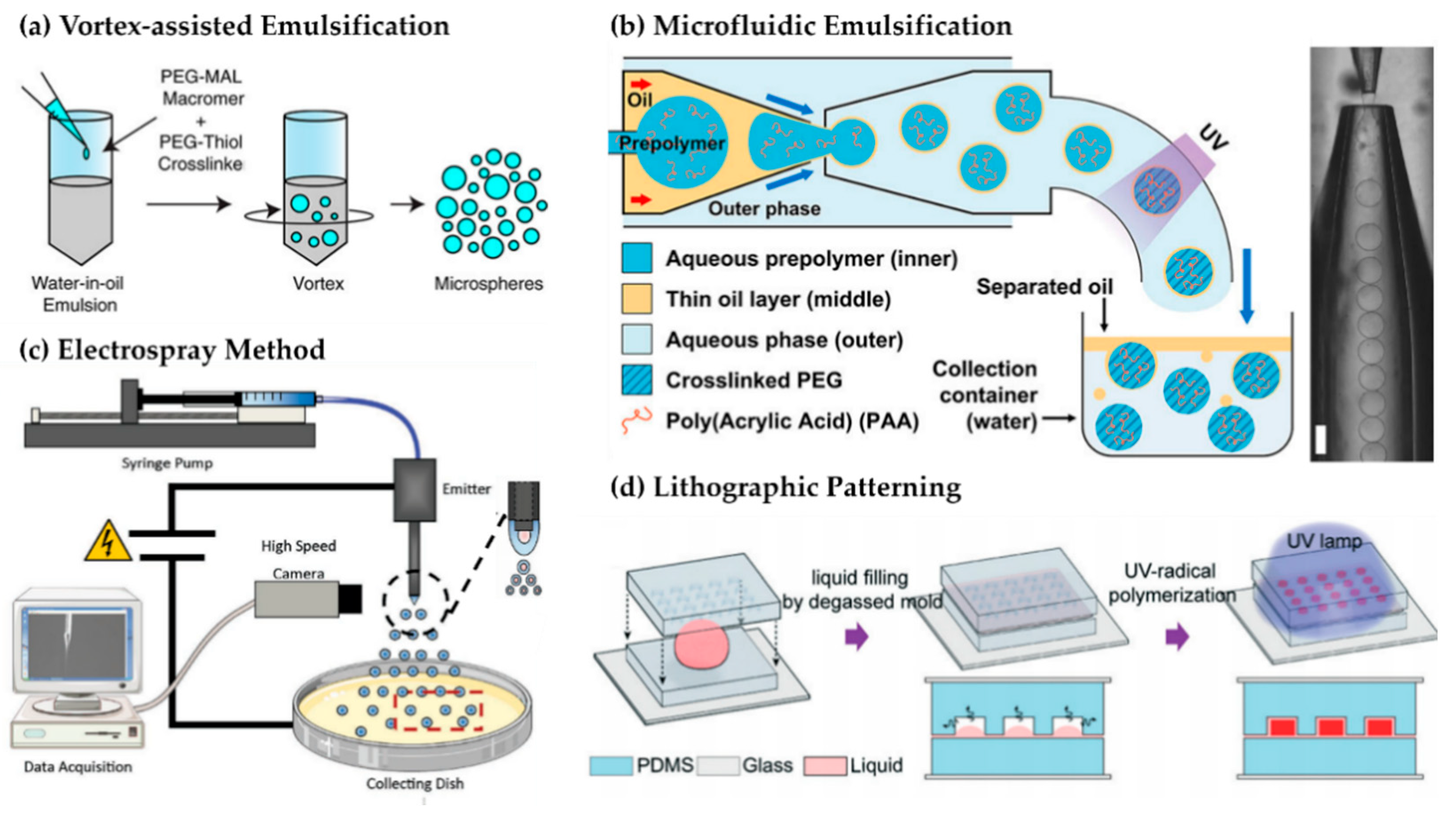
Emulsion-based polymerization is a widely used strategy for producing microgels across a broad size range, owing to its simplicity and compatibility with both natural and synthetic hydrogels. This approach relies on the generation of dispersed droplets in an immiscible phase, which serve as localized domains for gelation via chemical or physical crosslinking (Figure 3a) [135,136]. The spatially confined environment provides a basis for particle-level control, distinguishing this strategy from bulk gelation. Depending on the emulsification conditions, the resulting hydrogel particles can span from nanogels (typically below 200 nm) to larger microgels exceeding 100 μm [137].
In conventional microscale emulsification, aqueous hydrogel precursors are dispersed into a continuous oil phase using mechanical shear or stirring, forming water-in-oil (W/O) droplets [136,138]. These are subsequently crosslinked by ionic, photoinitiated, or thermal triggers to produce microgels. Surfactants or stabilizers are typically added to reduce interfacial tension and prevent premature coalescence, but the achievable size uniformity remains limited by the stochastic nature of shear-based droplet formation. As a result, W/O emulsification often yields microgels with broad size distributions, typically ranging from sub-micron to over 100 μm depending on formulation viscosity, surfactant concentration, and mixing parameters [127,139]. This method is operationally simple and scalable, making it suitable for exploratory studies or applications tolerant of heterogeneity, though post-processing steps such as filtration or centrifugation are often needed for size refinement [133].
Figure 3. Representative microgel fabrication techniques. (a) PEG-based microgels formed via emulsion polymerization using water-in-oil mixing and thiol–maleimide crosslinking [136]. (b) Monodisperse PAA-functionalized PEG microgels generated through coaxial-flow microfluidics and UV polymerization (scale bar = 200 μm) [140]. (c) Size-tunable microgels produced by electrospray of prepolymer solutions under high voltage, the dashed circle indicates the nozzle of the setup [131]. (d) Shape-defined hydrogel microparticles fabricated through micro-molding lithography with vacuum-assisted filling and photopolymerization. Red region represents the gelated phase upon UV irradiation [123].
Applsci 15 11599 g003
At the nanoscale, inverse (or water-in-oil) microemulsion polymerization enables the fabrication of nanogels by dispersing aqueous monomer solutions as sub-micron droplets within a continuous oil phase [118,141]. In contrast to mechanically driven emulsification, these systems are stabilized by carefully chosen surfactant and co-surfactant combinations that form thermodynamically stable nanoscale domains, typically via the formation of swollen reverse micelles. Here, droplet size is governed not by shear force but by interfacial curvature, Laplace pressure, and the packing constraints of the surfactant layer. The high surface-area-to-volume ratio of nanoscale droplets makes them inherently unstable, but the use of surfactants with low critical packing parameters and sufficient steric or electrostatic repulsion can kinetically hinder coalescence and Ostwald ripening. These physical effects enable relatively monodisperse nanodroplet populations suitable for confined polymerization reactions [142,143,144,145].
When polymerizable precursors are introduced into these droplets and crosslinked in situ, nanogels with sizes typically in the 20–200 nm range can be obtained, making them particularly attractive for systemic delivery applications where size-dependent biodistribution and enhanced permeation and retention (EPR) effect-mediated tumor accumulation are key [118].
Despite their advantages in producing well-defined nanogels, inverse emulsions present challenges for scale-up due to solvent toxicity, surfactant removal, and batch volume limitations. Consequently, while suitable for precision synthesis, their utility for clinical-scale production remains limited.

3.1.2. Microfluidic Droplet Generation

Microfluidics offer a high-precision approach to microgel fabrication by mechanically segmenting immiscible fluid streams within confined microchannel geometries [122,146]. In typical flow-focusing or T-junction devices, an aqueous hydrogel precursor is introduced as the dispersed phase and pinched into discrete droplet by a continuous oil phase under laminar flow. These droplets act as templated microreactors, subsequently crosslinked through ionic interactions, photopolymerization, or thermal triggers either within the microfluidic chip or post-collection [119,147].
Droplet size in these systems is governed by interfacial tension, flow rate ratios, and channel geometry, rather than the stochastic shear forces that dominate bulk emulsification. This results in monodisperse microgels with narrow size variation, with typical diameters ranging from 5 μm to over 200 μm depending on design parameters (Figure 3b) [140,148]. Consistent size distribution directly impacts dosage precision, cellular encapsulation, and release kinetics, particularly in contexts where particle-dependent clearance or tissue penetration is a concern [147,149]. As demonstrated in prior work using flow focusing devices, tightly controlled microgel populations have been produced for cell-laden formulations and depot injections, supporting reproducible therapeutic outcomes.
The customizable nature of microfluidic design also permits complex structural engineering of microgels. Multiple inlet systems incorporating more than a single oil and water combination can fabricate anisotropic microgels, including core–shell or Janus morphologies, with spatially encoded composition or degradation behavior. These capabilities have expanded the functional scope of microgels beyond simple carriers to include compartmentalized or programmable delivery systems [122,150,151].
Despite the advances toward higher throughput, including parallelized architectures and high-frequency droplet generation schemes, the broader scalability of microfluidic systems remains constrained by formulation-specific tuning [152]. Droplet formulation is highly sensitive to both material characteristics and fluid dynamic factors, which are often dictated by the hydrogel precursors and their compositions [153]. Therefore, each new material system may require device-specific and throughput-specific optimizations, limit operational flexibility and slow development. Although microfluidics offers exceptional control over droplet size and structure, generally it is most feasible when formulation parameters are narrowly defined and reliably synthesis is prioritized over throughput.

3.1.3. Electrospraying and Jetting

Electrospray and related jetting techniques represent extrusion-based approaches to microgel fabrication, where a polymeric solution is broken into droplets via electric or hydrodynamic instabilities and then crosslinked into discrete gel particles [120]. In electrospraying, high voltage is applied to a polymer-loaded nozzle to generate a find spray of charged droplets, which are collected into a gelling bath or irradiated with light to induce crosslinking [154]. The resulting particles typically fall within the 10–200 μm range, with size determined by the applied voltage, needle diameter, flow rate, and collection distance. Ionic crosslinking and photopolymerization are both commonly employed, enabling compatibility with a range of hydrogel precursors [155,156].
This approach is attractive for its simplicity, rapid gelation under mild conditions, and potential for multiplexing. The method does not necessitate organic solvents and operates under low shear, therefore it is well-suited for sensitive biological agents, including proteins or cells. Also, unlike microfluidics, this technique does not require well-defined microarchitecture or immiscible fluid systems, allowing for relatively straightforward setup and operation, as well as scalability [157].
However, the variability in droplet formation remains a major challenge. Electrosprayed microgels typically exhibit size coefficient of variation (CV) in the range of 10–20% under standard conditions. Polydispersity arises from sensitivity to environmental factors such as temperature, humidity, and voltage stability, as well as from nozzle clogging and fluctuations in solution viscosity [156]. Passive jetting methods, such as vibrating nozzle- or syringe-driven extrusion, can operate without electric fields and rely on Rayleigh instabilities to induce droplet breakup, producing microgels of comparable size but similar variability. These methods offer high throughput and operational simplicity, but controllability over particle characteristics is more limited than electrospraying (Figure 3c) [131].
Jetting methods are frequently utilized for scaled production, especially when size uniformity is not the primary constraint. Medium-sized microgels in the range of 10–200 μm are particularly advantageous for injectable depots, where particles must remain at the injection site yet pass through standard-gauge needles [158]. An example of this size class in clinical use is Zilretta® (Pacira BioSciences, Inc., San Diego, CA, USA), an FDA-approved formulation of triamcinolone acetonide encapsulated in PLGA microspheres of approximately 50 μm diameter. These particles are injected intra-articularly and provide sustained drug release over several weeks. While PGLA is not considered as hydrogel, the formulation illustrates the therapeutic utility of drug-loaded particles in this size range for depot-based delivery [159]. Similar fabrication strategies have been adapted to generate microgel-based formulations, offering improved hydration, biocompatibility, and tunable degradation for analogous applications [160].

3.1.4. Photolithography and Micro-Molding

Photolithography and soft lithographic molding offer a top-down strategy for fabricating microgels with high uniformity and customized geometries. These methods are particularly suited for applications requiring precise spatial control over size, shape, and architecture [161]. In photolithography, hydrogel precursors such as polyethylene glycol diacrylate (PEGDA) or gelatin methacrylate (GelMA) are crosslinked by patterned light exposure in the presence of photoinitiator such as Irgacure 2959 or lithium phynyl-2,4,6-trimethylbenzoylphosphinate (LAP), typically through a photomask or via digital projection [124,125,162,163]. The resulting microgels adopt the geometry of the illustrated region, producing well-defined particles ranging from tens to several hundreds of microns. In parallel, micro-molding techniques cast hydrogel solutions into pre-fabricated molds, often made from polydimethylsiloxane (PDMS), to form discrete particles upon curing (Figure 3d) [123,164]. Both approaches rely on spatially confined gelation and yield comparable levels of dimensional fidelity.
The principal advantage of these methods is the ability to produce exceptionally monodisperse microgels with custom shapes. Unlike droplet-based systems that predominantly yield spherical particles, lithographic techniques allow fabrication of disks, cubes, rods, or other anisotropic geometries [165]. These shape variations directly impact fluid behavior, cellular interactions, and mechanical properties, and have been leveraged in scaffold engineering, cell culture, and drug release studies [124,163]. In some cases, layer-by-layer lithography enables the construction of Janus or multi-domain microgels, expanding functional complexity [166].
However, these methods also have practical drawbacks of limited throughput and operational complexity. Photolithography typically requires batch processing, limited by the sizes of the exposure area and substates, and necessitates cleanroom-compatible equipment such as UV exposure tools and mask aligners [167]. Recollecting cured microgels from substrates or molds may be time-intensive and prone to mechanical damage. Furthermore, photoinitiator toxicity and UV exposure limit compatibility with sensitive drug cargos, although visible-light and bio-orthogonal systems are under development to mitigate these concerns [129]. As a result, lithographic microgel fabrication is predominantly used in exploratory settings where shape fidelity, resolution are prioritized over yield [124,130,167].

3.1.5. Top–Down Strategies

Top–down fragmentation offers a simple and accessible route to microgel production by mechanically reducing bulk hydrogels into microscale fragmentation [25,168]. Common wet-processing techniques include blending, grinding, or extrusion through a mesh, each capable of yielding particles with sizes typically ranging from 50 μm to over 200 μm depending on processing parameters and material properties [126]. For example, a crosslinked gelatin hydrogel can be fragmented in a household blender, with duration modulating the resulting particle size [169]. Likewise, extrusion through porous membranes or meshes enables size control mainly governed by mesh pore dimensions [168,170]. These methods are applicable to a wide variety of hydrogel materials, including those that cannot easily form stable droplets for emulsion-based techniques [171,172]. Notably, wet fragmentation can operate with minimal equipment and can process gram-scale batches within minutes of time [168,170].
In pharmaceutical manufacturing, dry-state fragmentation methods such as ball milling are commonly employed to reduce lyophilized or dehydrated materials into fine powders or granules [173]. For hydrogel-based drug delivery, ball milling may be applied to freeze-dried gels, but this technique is not generally suitable for microgel fabrication due to the viscoelastic and deformable nature of crosslinked hydrogel materials, which resists brittleness-oriented mechanical fracture [25,170]. Furthermore, excessive mechanical energy may compromise structural integrity or payload stability without further optimization in formulation compositions [170,171].
The primary limitations of top–down strategies are their heterogeneity in size and shape, and the difficulty in forming spherical particles [126,168]. The heterogeneity caused during mechanical disruption process complicates drug release modeling and may lead to inconsistent in vivo behavior, especially when diffusion or degradation rates. Additionally, the process can introduce shear or compressive stresses within polymeric network, potentially affecting encapsulated agents [25,174].

3.1.6. Physical Ionic Crosslinking (Ionotropic Crosslinking)

Ionotropic crosslinking refers to the gelation mechanism of polyelectrolytes through non-covalent interactions with multivalent counterions, forming a hydrogel network stabilized ionic coordination [134]. Unlike covalent crosslinking methods, which typically require initiators, energy input, or catalysts to form irreversible linkage, ionotropic gelation proceeds under ambient conditions through spontaneous electrostatic attraction between oppositely charged species. This mechanism enables gelation in aqueous environments without introducing organic solvents or reactive chemical agents, making it more compatible with sensitive biological cargos [93,132,175].
Ionic gelation has been widely applied for nanoscale gel fabrication, most notably in systems based on natural polymers such as chitosan, alginate, or gellan gums. For example, polycationic chitosan solution could undergo instantaneous gelation upon addition of multivalent phosphate anions, producing nanogels as small as 50 nm. This mild and aqueous process enables the encapsulation of proteins, nucleic acids, or even live cells, due to minimal mechanical stress and the absence of toxic reagents [121,132,176].
Despite the advantages, conventional ionotropic gelation in a batch system exhibits relatively limited size controllability and reproducibility. The gelation process is governed by rapid and kinetically unstable complexation between charged species, which can be highly sensitive to variables such as molecular weight distribution, degree of substitution, ionic strength, or the sequence of reagent addition. Mixing rate or local heterogeneity in pH can shift the outcome from uniform nanoparticles to randomly aggregated macrogel particles, which complicate scale-up and standardization [121,177]. These systems often require downstream processing such as filtration or dilution to narrow size distribution, while stabilizers or surfactants can improve colloidal stability and size uniformity.
In terms of material selection, ionotropic gelation aligns naturally with bio-derived polymers with functional groups capable of ionic complexation, such as carboxylates in alginate or amines in chitosan [128]. These materials are considered biocompatible and biodegradable but may lack mechanical stability or defined degradation kinetics compared to synthetic hydrogel systems.

3.1.7. Macro-Scale Hydrogel Beads

Macro-scale gel beads, typically exceeding several hundred microns, merit brief discussion as they provide valuable insights despite falling outside the scope of injectable hydrogel systems. These larger structures are typically fabricated by larger-sized droplet-based methods such as gravity-driven dripping or vibration-assisted jetting [178,179]. One of the most demonstrated setups is alginate beads, where aqueous solution of hydrogel precursors is added drop-wise into a gelling solution, such as aqueous solution of calcium chloride, to form spherical beads in sub-mm to several millimeters’ range [3,180]. More refined approaches, such as electrostatic dripping or syringe pump-controlled dripping can significantly improve size uniformity and allow moderate level of size controllability, but the overall technique remains an extension of the droplet formation principle discussed in the previous sections [181].
The primary advantage of macrogel beads is high loading capacity of drugs or cells. Their large volume allows prolonged residence at the administration site, as well represented by embolized beads for chemoembolization or gastro-retentive swellable hydrogel beads. However, their physical bulkiness limits the applicable delivery routes to non-injectable types, such as surgical implants [31,182]. Although macrogels are often excluded from injectable formulations, their fabrication methods and functional performance are closely related to drug encapsulation, clearance behavior, and material compatibility of hydrogel-based DDS, providing useful insights in defining practical limits and design priorities for injectable hydrogel-based DDS.

3.2. Stimuli-Responsive Material Behavior

Stimuli-responsiveness renders DDSs to react to specific physiological triggers, enabling on-demand and site-specific drug release. By incorporating functional chemical groups into the polymer network, hydrogel particles can be engineered to be sensitive to surrounding environmental cues including pH, enzymes, temperature, redox conditions, or even multiple cues simultaneously [183,184,185,186,187]. Material responsiveness in gel particulate is enabled through chemical transitions that translate environmental cues into changes in network structure or drug affinity, thereby serving as a key consideration in stimulus-specific design. The magnitude and reversibility of such responses are further influenced by intrinsic structural parameters such as crosslinking density, polymer chain mobility, and network topology, which collectively define how effectively the material couples environmental stimuli to macroscopic behavior.

3.2.1. pH-Responsiveness

pH-responsive microgels rely on polymers bearing ionizable groups that undergo protonation or deprotonation in response to the surrounding pH, inducing changes in polymer chain conformation, charge distribution, and network swelling. In acidic environments, protonation of acidic or basic pendant groups suppresses electrostatic repulsion and may lead to microgel collapse, whereas in alkaline conditions, deprotonation results in charge development and subsequent swelling. This shift in network hydration alters diffusion characteristics, thereby modulating drug retention and release [188].
Such responsiveness is particularly relevant for injection sites exhibiting pH gradients. For example, oral drug delivery must traverse the acidic gastric environment (pH 1.5–2.0) before reaching the more neutral-to-basic intestinal lumen (pH 6.5–7.4). Similarly, tumor microenvironments and sites of inflammation are typically mildly acidic (pH~6.5), in contrast to normal physiological pH (~7.4). Hydrogel particles designed to respond selectively to these differences can enable spatially targeted release while minimizing systemic exposure (Figure 4a) [189].
Commonly used materials include poly(acrylic acid) (PAA), polymethacrylic acid, alginate, HA, and chitosan, which contain carboxyl or amine groups with tunable ionization behavior [190]. For example, PAA-based hydrogel particles are utilized to protect insulin at gastric pH and then swell to release it in intestinal conditions [21]. Similarly, inclusion of weak acids such as itaconic acid has enabled esomeprazole delivery systems with high retention in acidic environments and rapid swelling under intestinal pH [191]. On the basic polymer side, cationic hydrogel particles composed of poly(2-(diethylamino)ethyl methacrylate) (PDEAEMA) exhibit swelling under mildly acidic tumor-like conditions due to protonation of amine groups. To extend function, pH-responsive polymers are often combined with hydrophilic or thermosensitive components, and acid-labile crosslinkers such as acetal or hydrazone moieties allow additional degradation-based release mechanisms [192,193].
The resulting hydrogel particles can be engineered to release drugs either through pH-induced swelling, leading to enhanced permeability, or through network disintegration triggered by chemical bond cleavage under acidic conditions [194]. This has been effectively applied in oral delivery systems designed to release cargo in the intestine while protecting it from gastric degradation, and in tumor-targeted systems where mild acidosis serves as the release trigger [195].
One of the advantages of pH-responsive hydrogel particles is their autonomous operation using intrinsic physiological cues, eliminating the need for external activation [35]. However, physiological pH differences are often modest, requiring polymers with carefully tuned pKa values to achieve selective responsiveness [193]. Additionally, non-specific off-target release can occur in tissues with fluctuating pH, and rapid osmotic swelling may result in burst release [196]. In short, pH-responsive hydrogel particles represent a mature and versatile strategy for environment-specific release, particularly in oral and tumor-targeted applications where pH gradients are reliably established.
Figure 4. Representative stimuli-responsive hydrogel-based particulate DDS. (a) pH-responsive microgels exhibiting swelling behavior driven by -COOH to -COO transition, enabling enhanced ciprofloxacin release at neutral pH [189]. (b) Enzyme-responsive microgels conjugated with MMP-2-cleavable peptides and cartilage-binding ligands for targeted, enzyme-triggered celecoxib release [197]. (c) Thermo-responsive nanogels composed of PNIPAM-co-AAc polymers showing increased β-LP release at physiological temperature. (c1,c2) represent electron microscopic images of the nanogels (scale bars = 500 nm) [198]. (d) Redox-responsive supramolecular microgels incorporating disulfide crosslinks for intracellular, reduction-triggered drug release [199]. (e) Light-responsive microgels functionalized with donor–acceptor Stenhouse adducts (DASAs) enabling visible light-mediated drug release control [200].
Applsci 15 11599 g004

3.2.2. Enzyme-Responsiveness

Enzyme-responsive microgels are designed to undergo network degradation, swelling, or drug detachment in response to disease-associated enzymatic activity. This strategy relies on integrating degradable substrates, such as cleavable peptide linkers or enzyme-labile polysaccharide backbones, into the microgel network [201]. Upon encountering a target enzyme, typically overexpressed at pathological sites, specific cleavage of these substrates alters the microgel’s structural integrity or drug retention, initiating release of entrapped drug. Representative triggers include proteases like matrix metalloproteinases (MMPs) in cancer and arthritis, glycosidases such as hyaluronidase in connective tissue disorders, and esterases in infection or metabolic disease. These enzyme cues offer a biochemical specificity that enables release behavior to align with pathological activity [202,203,204].
Microgel responsiveness can be encoded through several material strategies. Natural polymers such as gelatin, collagen, chitosan, and HA possess intrinsic susceptibility to enzyme-mediated cleavage by collagenases, lysozyme, or hyaluronidase [205,206]. Synthetic crosslinkers bearing protease-cleavable peptide sequences have also been incorporated to precisely regulate responsiveness. One example is HA microgels crosslinked with a GGPLGLAGGC peptide, which is selectively cleaved by MMP-2. These microgels were used to tether celecoxib via the same MMP-cleavable peptide, enabling release upon exposure to enzyme-rich osteoarthritic joints (Figure 4b) [197]. Other platforms have incorporated elastase-sensitive linkers or glycosidase-degradable polysaccharides to achieve inflammation- or infection-triggered release [206]. Enzyme-responsive design can also include modular domains for targeting or retention; for instance, cartilage-binding peptides have been used to anchor microgels within joint tissue, thereby prolonging local exposure to MMPs.
Drug release typically follows either network erosion or direct cleavage of drug–polymer linkages. In the case of celecoxib-loaded HA microgels, drug molecules were released upon enzymatic cleavage of the tether peptide, while degradation further contributed to matrix loosening [83]. Importantly, enzyme-responsive systems exhibit concentration-dependent release, which allows for disease-stage-correlated drug exposure. This has been leveraged in osteoarthritis models, where upregulated MMP-2 activity facilitated on-demand anti-inflammatory release without significant off-target leakage. Other systems have exploited similar responsiveness to bacterial enzymes in wound infection models, releasing antimicrobial agents in response to local enzymatic activity [197,207].
The primary advantage of enzyme responsiveness lies in its pathophysiological specificity. Unlike pH or redox triggers, enzymatic cues are often more directly associated with disease progression or immune activation [201,208]. However, this specificity also introduces several design constraints. The system must ensure that the target enzyme is sufficiently expressed and accessible at the site of action. For enzymes with intracellular localization or limited extracellular diffusion, the microgel must first reach the relevant compartment [209,210]. Off-target release remains a concern if the same enzyme is present in multiple tissues, and interpatient variability in enzyme expression may affect release consistency [211]. Stability of cleavable linkers during formulation and storage is another critical factor. Nonetheless, enzyme-responsive microgels represent a promising potential for achieving disease-synchronized drug delivery, particularly in arthritic, neoplastic, or inflamed tissues where enzyme activity is robust and spatiotemporally correlated with therapeutic need [212].

3.2.3. Thermo-Responsiveness

Thermo-responsive microgels undergo reversible volume transitions in response to temperature changes, typically governed by a polymer’s lower critical solution temperature (LCST) [213]. Below the LCST, the polymer chains are hydrated and hydrophilic, allowing the microgel to remain swollen. Above the LCST, hydrophobic interactions dominate, leading to dehydration and network collapse. This phase transition can be leveraged to modulate drug loading, retention, and release. Poly(N-isopropylacrylamide) (PNIPAM) remains the most studied thermo-responsive polymer, with an LCST near 32 °C that can be adjusted toward physiological temperatures through copolymerization. Above this threshold, PNIPAM microgels rapidly shrink and expel water, facilitating drug release, while upon cooling, they rehydrate and reswell [214]. Other thermo-responsive polymers such as poly(oligoethylene glycol) methacrylates, elastin-like polypeptides, and polysaccharide derivatives offer alternative architectures with tunable thermal responses [215].
PNIPAM is typically considered as a primary model for thermal responsiveness, but their poor degradability and potential accumulation in vivo have led to efforts to incorporate degradable linkers or blend with natural polymers. Strategies include introducing disulfide or ester-based crosslinkers, or integrating biocompatible components such as gelatin, collagen, or keratin to improve both degradability and tissue compatibility [215,216]. Dual-responsive designs are widely studied as well, for example, PNIPAM copolymerized with acrylic acid yields microgels that respond to both temperature and pH, expanding the scope of environmental sensitivity [217]. In addition to chemical modification, physical hybridization with photothermal materials such as gold nanorods, Prussian blue nanoparticles, or graphene oxide, enables external control via light-induced heating. This allows localized thermal triggering of drug release without systemic temperature elevation [218].
Drug release is typically governed by changes in microgel volume and internal mesh structure [219]. Upon heating above the LCST, the microgel network collapses, decreasing porosity and often forcing expulsion of entrapped solutes [34]. Alternatively, for hydrophobic drugs, increased hydrophobicity of the matrix at elevated temperatures may temporarily retain the cargo, delaying its release until reswelling occurs during cooling (Figure 4c) [198]. The dynamic control of release via temperature cycling has been demonstrated in composite microgels incorporating photothermal agents. In one such design, Prussian blue-loaded PNIPAM microgels released doxorubicin in response to near-infrared (808 nm) laser irradiation [220]. Heating induced by the photothermal core raised the local temperature above the LCST, triggering gel collapse and drug release, which could be repeated across multiple cycles. Another practical implementation involves thermally induced gelation or clustering, where certain PNIPAM formulations remain stable at ambient temperature but aggregate at 37 °C, enabling in situ formation of injectable depots that slowly dissolve to release therapeutic agents over time [221].
Thermal responsiveness offers spatial and temporal precision in drug release, especially when paired with externally applied heat sources [222]. However, the narrow range of physiologically safe temperatures imposes design constraints. LCSTs close to 37 °C may lead to unintended release from systemic fluctuations [26], while higher thresholds require precise and safe external stimulation [21]. Furthermore, burst release from network collapse may not be suitable for applications requiring prolonged, steady dosing [34]. Material safety also remains a concern, as PNIPAM is not biodegradable, and the toxicity of degradation products must be addressed as well. While these limitations persist, thermo-responsive microgels remain a promising platform for externally triggered or environment-sensitive drug delivery, particularly in contexts where localized heating or temporal control enhances therapeutic outcomes [221,222].

3.2.4. Redox-Responsiveness

Redox-responsive microgels are constructed using chemical linkages that are selectively cleaved or destabilized in response to the ambient reduction–oxidation potential. It leverages endogenous redox gradients within the body, particularly the pronounced difference in glutathione (GSH) concentrations between extracellular fluids (~2–20 μM) and the cytosol (~5–10 mM). Disulfide bonds, the most widely used redox-labile moieties, remain stable in circulation but undergo rapid thiol–disulfide exchange in reducing environments, leading to network cleavage and triggered drug release). Oxidation-sensitive chemistries, including thioketal and boronic ester linkages, respond to reactive oxygen species (ROS) such as hydrogen peroxide, offering an orthogonal route to achieve environment-selective activation under oxidative stress [223,224,225,226].
Redox-responsive strategies are particularly relevant for intracellular delivery, where the elevated reducing potential of the cytosol enables selective bond cleavage following cellular uptake. This is especially useful in tumor or inflammatory tissues, which exhibit both elevated intracellular GSH and localized oxidative stress. By incorporating degradable redox-sensitive bonds into the polymer matrix or drug linkage, microgels can be engineered to remain stable during systemic circulation and undergo disintegration or cargo release only after endocytosis or upon reaching ROS-enriched sites [223,224].
Common materials include synthetic or semi-synthetic polymers crosslinked via disulfide-containing monomers. For example, sodium alginate-based microgels crosslinked with disulfide bridges have demonstrated stable encapsulation of doxorubicin under oxidizing conditions, followed by accelerated release in simulated intracellular reductive environments [227]. Redox-labile drug linkers can also be incorporated for prodrug strategies, where conjugated drugs are released only upon bond cleavage. Additional chemistries include diselenide bonds, which offer higher redox sensitivity but require stabilization, and thioketal or boronic ester groups that degrade under ROS, although these are more frequently applied in macrogels or nanoparticle systems. Hybrid systems that combine disulfide and ROS-cleavable elements have also been proposed for dual-responsiveness in redox-dynamic environments such as tumors [223,224,228].
Drug release is initiated by chemical bond cleavage that results in network degradation, pore expansion, or conjugate breakdown. In prodrug-type designs, bond cleavage directly liberates the active pharmaceutical ingredient inside target cells. These mechanisms have proven effective in enhancing intracellular delivery, with minimal release observed under extracellular conditions and rapid activation upon endocytosis. In alginate-based disulfide-crosslinked microgels, for instance, doxorubicin release remained low in oxidizing buffers but increased markedly in the presence of 10 mM GSH. This responsiveness allows redox microgels to serve as intracellular carriers that exploit cellular biochemistry to initiate controlled drug discharge (Figure 4d) [199,229,230].
While redox-triggered delivery provides intracellular specificity, several design considerations remain. The near-universal reducing conditions within cells indicates that redox cleavage could not be cell-specific, necessitating upstream targeting strategies. Disulfide-containing systems must also balance circulation stability with responsiveness, as low concentrations of plasma thiols may cause gradual degradation. For ROS-sensitive systems, the heterogeneity and transient nature of oxidative stress in vivo can complicate prediction and tuning of release behavior. Yet, the simplicity and predictability of disulfide cleavage, along with increasing interest in tumor and inflammation-associated redox landscapes, make redox-responsive microgels a compelling platform for intracellular and site-activated drug delivery [225,231].

3.2.5. Light-Responsiveness

Light-responsive hydrogels incorporate photoactive chemistries to enable remote, temporally precise control of drug release. These systems operate through three principal mechanisms: light-induced isomerization, photothermal activation, and photochemical bond cleavage or crosslinking. Unlike endogenous triggers, light provides an orthogonal stimulus that can be externally applied to modulate microgel behavior with high spatial selectivity [232,233,234].
Azobenzene, spiropyran, and coumarin derivatives are widely used photoswitches in hydrogel design. Azobenzenes undergo reversible trans–cis isomerization under UV or visible light, altering molecular dipole moment, polarity, and steric configuration. These changes shift polymer–water interactions and can induce swelling or contraction depending on the system’s composition. For instance, azobenzene-modified PNIPAM microgels have shown size modulation contingent on isomer ratio and surfactant concentration [235,236,237,238]. Azobenzene groups can be introduced as pendant chains, crosslinkers, or non-covalent guest moieties, such as cyclodextrin complexes that reversibly dissociate upon isomerization [239]. Spiropyrans convert from nonpolar to polar merocyanines under UV exposure, affecting network hydration, while coumarin dimers enable reversible [2+2] photocycloadditions that alter crosslinking density in response to light exposure [240].
Photothermal strategies embed chromophores such as gold nanorods, polydopamine, or chlorophyll within thermo-responsive matrices like PNIPAM. Upon irradiation, these agents convert light to localized heat, surpassing the LCST and inducing rapid microgel collapse and drug expulsion. This has been used to trigger near-instantaneous release of small molecule drugs such as doxorubicin [234,241]. In addition, o-nitrobenzyl-based photolabile linkers allow irreversible light-induced bond cleavage, offering another mechanism for drug detachment or matrix degradation [242].
Drug release kinetics vary by mechanism. Light-triggered polarity shifts alter drug–matrix affinity, while photothermal collapse promotes convective expulsion. In photocleavable systems, bond breakage directly liberates covalently bound drugs or destabilizes the network to permit diffusion. Temporal control can be achieved through pulsed irradiation, enabling reversible or staged release profiles under cyclic activation [232,233,234].
The principal advantage of light-responsive microgels is their external controllability. Because the trigger is decoupled from biological variability, release can be temporally synchronized with therapeutic need and spatially localized to target tissues. These systems also allow repeated activation and are tunable through chromophore design. However, tissue penetration of UV and visible light remains limited, reducing utility in deep tissues unless combined with near-infrared responsive agents. Furthermore, some chromophores exhibit photo-fatigue or introduce synthetic complexity, necessitating careful optimization for translational applications (Figure 4e) [200,234,241].

3.2.6. Multi-Responsive and Hybrid Strategies

Hydrogel particles designed with multi-responsive features incorporate two or more stimulus-sensitive chemistries into a single polymer network to enable more precise drug release or to respond to complex physiological environments [18,243,244]. The underlying design logic typically follows combinatorial principles. In one case, coexisting triggers (e.g., pH and temperature) must be present simultaneously to induce release, functioning analogously to a logical AND gate [1,187]. In another, responsiveness to any one of multiple triggers (e.g., pH or redox) provides flexibility under uncertain or heterogeneous conditions. These strategies allow delivery systems to filter environmental signals and respond more selectively to pathological sites [27,185].
A commonly explored combination is pH and thermo-responsiveness. This can be achieved by copolymerizing temperature-sensitive monomers such as poly(N-isopropylacrylamide) (PNIPAM) or oligo(ethylene glycol) methacrylates with weak acids like acrylic acid or itaconic acid. For example, a dual-responsive microgel was fabricated using a semi-interpenetrating network of carboxylated polysaccharide and thermo-responsive PEGMA, enabling shrinking behavior in acidic environments and collapse above the lower critical solution temperature [245]. Such dual-triggered release mechanisms can improve site specificity: one trigger (e.g., acidic pH in tumor sites) initiates swelling, while a second (e.g., externally applied hyperthermia) accelerates drug release [187,246,247].
Other systems incorporate redox and pH responsiveness within the same carrier. Hydrogels containing disulfide crosslinkers combined with acid-cleavable moieties can first respond to acidic endosomal conditions, followed by intracellular glutathione-triggered disassembly [247,248]. These sequential responses allow the network to adapt as it transitions through tissue, endosomal, and cytosolic environments. In certain cases, light-responsive elements are added as a third control layer, such as o-nitrobenzyl linkages that cleave under UV exposure, to enable staged or remotely triggered release [27,249,250].
Hybrid material designs further expand multi-responsive strategies by incorporating inorganic or supramolecular elements into the microgel matrix. Photothermal nanoparticles such as Prussian blue or AuNP embedded within the microgel enable light-to-heat conversion, coupling an external optical trigger to internal temperature-sensitive collapse. Other hybrids involve coupling microgels to hydrogel matrices or forming microgel–nanoparticle conjugates, where each component contributes a distinct type of responsiveness or functionality. In one reported system, a bulk hydrogel scaffold contained embedded microgels engineered for local swelling and drug release in response to combined redox and enzymatic signals, while the macrostructure maintained overall mechanical stability [18,32,185,251].
Such multi-functional architectures allow hydrogel particles to perform coordinated tasks, for examples, aggregating upon environmental changes, generating heat in situ, or delivering multiple drugs with distinct release kinetics. However, layering stimuli also imposes nontrivial synthetic challenges, particularly in ensuring that each trigger remains orthogonal and does not interfere with others during fabrication, storage, or circulation [27,243,244,246].
Designing micro- and nanogels with multi-stimuli responsiveness imposes material-level constraints that are distinct from general hydrogel development. To accommodate multiple triggers, polymer backbones must support orthogonal reactivity and allow integration of independently tunable domains without compromising injectability or colloidal stability. This often necessitates blocky or modular architecture, selective crosslinking chemistry, and predictable network degradation kinetics. In microparticle formats, responsiveness must also be confined within a defined particle size and distribution, which adds complexity in emulsion processing or photopatterning methods [27,244,252]. Furthermore, the mechanical integrity of each gel particle must be maintained under physiological shear while preserving responsiveness under the intended trigger conditions. These requirements differ from planar or bulk hydrogels, where spatial constraints and shear exposure are less severe. In addition, the mechanical and structural parameters of multi-responsive networks introduce inherent trade-offs that define their practical behavior. Higher crosslinking density or incorporation of rigid domains improves shape retention and resistance to injection-induced shear but can diminish responsiveness or slow degradation. In contrast, softer networks with greater chain mobility enhance diffusion and reversible deformation but inevitably risk losing structural fidelity or cue selectivity. One straightforward strategy for these competing effects can be selective crosslinking chemistries that decouple mechanical stability from stimulus reactivity, which potentially allowing syringeability and responsiveness to be individually optimized.
Overall, achieving multi-stimuli responsiveness in injectable hydrogel particles requires a convergence of synthetic precision, formulation strategy, and microscale structural fidelity, ultimately enabling a finely tuned system for symptom- or disease-specific drug delivery [18,32,185,243,251].

3.3. Strategies for Drug Loading and Release Modulation

3.3.1. Drug−Particle Interactions and Loading Strategies

Drug incorporation into hydrogels relies on the physicochemical compatibility between therapeutic agents and the polymeric network. Three principal strategies govern this process: passive encapsulation, electrostatic or affinity-driven complexation, and covalent conjugation. Each approach presents distinct advantages and limitations, depending on drug size, solubility, charge, and required release profile.
Passive encapsulation involves physical entrapment of drugs within the gel matrix, either during polymerization (in situ loading) or by post-synthesis diffusion. In the former, monomers or polymer precursors are mixed with the drug during microgel formation, ensuring homogeneous distribution throughout the network [105]. This method is particularly useful for hydrophilic macromolecules and has demonstrated high loading efficiencies when mesh size and network hydrophilicity are optimized. Post-synthetic loading involves incubating pre-formed microgels in concentrated drug solutions to allow diffusion into the polymer network. Swelling–deswelling cycles, often pH- or temperature-mediated, enhance loading by controlling mesh accessibility [21,211]. Although conceptually simple, passive methods depend heavily on network porosity and drug–matrix partitioning, which may lead to early burst release if interactions are weak or if surface-adsorbed drug dominates the payload [194].
Electrostatic complexation leverages charge interactions between ionizable groups on the microgel and oppositely charged drug molecules. This approach is particularly relevant for nucleic acids, peptides, and charged small molecules. Anionic gels with carboxyl or sulfonate groups effectively bind cationic drugs or polymers, while cationic microgels can form stable complexes with siRNA or plasmid DNA [253,254]. Loading capacity and network conformation can be tuned through ionic strength, pH, or multivalent counterions [189,255,256]. Additionally, host–guest interactions, such as cyclodextrin–drug inclusion complexes, introduce affinity-based mechanisms that stabilize hydrophobic drugs within the gel [257]. These non-covalent strategies not only enhance loading but can also modulate microgel structure, as drug–polymer binding may alter swelling, rigidity, or charge distribution.
Covalent conjugation offers robust retention by chemically linking drugs to the microgel network via cleavable bonds. The gel must be functionalized with reactive moieties such as aldehydes, maleimides, or activated esters, while the drug should present complementary handles (amines, thiols, hydrazides) [258,259]. Cleavable linkers, such as hydrazones or disulfides, are selected to release the drug under specific physiological triggers [260]. This approach is particularly advantageous for hydrophobic or potent drugs requiring strict spatial and temporal control. However, conjugation efficiency, site selectivity, and maintenance of drug bioactivity require careful optimization, and high grafting density may alter gel properties such as increasing hydrophobicity or degradability, which affects release kinetics [261,262].

3.3.2. Release Kinetics and Stimulus-Modulated Delivery

Drug release from hydrogel particles is governed by a complex interplay between diffusion, matrix degradation, and external or endogenous stimuli. These mechanisms rarely act solely, rather they coexist and influence one another depending on the gel’s crosslinking density, swelling behavior, degradation kinetics, and environmental context [116].
Diffusion-controlled release is predominant in microgels with intact networks and relies on the concentration gradient between the interior of the gel and the surrounding medium. The mesh size, degree of swelling, and drug–polymer compatibility dictate how readily drug molecules traverse the matrix. Hydrophilic microgels with loose networks allow rapid release of small molecules, while tighter meshes can delay release or exclude larger agents. Crosslinking density and polymer chain mobility further tune permeability and injectability, as rigid networks resist deformation but slow diffusion, whereas flexible ones enhance release at the expense of structural stability. Release profiles in such systems typically follow Fickian diffusion or anomalous kinetics and are susceptible to initial burst effects if surface-associated drug dominates [262,263].
Degradation-mediated release occurs when the gel matrix undergoes chemical or enzymatic breakdown, resulting in disintegration of the network or cleavage of covalent linkages. This mechanism enables sustained release over longer timescales or conditional release in response to specific environments, such as acidic pH, elevated reductant levels, or disease-associated enzymes [211,259,264]. Incorporation of labile crosslinks such as ester, disulfide, or peptide bonds allows tailoring of degradation rates. Network topology and viscoelasticity also influence degradation behavior and tissue compatibility, where highly crosslinked matrices resist cleavage but may fragment under stress, while more elastic networks degrade uniformly and adapt to surrounding tissues. When combined with diffusional resistance, matrix degradation offers a robust control mechanism that governs the timing and completeness of release [3].
Stimulus-triggered release enables on-demand delivery by environmental or external signals to physical or chemical transitions in the microgel. Thermo-responsive systems, for instance, undergo deswelling upon heating, expelling loaded cargo. Redox-sensitive microgels respond to intracellular glutathione by cleaving disulfide bonds, while enzyme-responsive gels degrade selectively in the presence of target-specific proteases. Light, ultrasound, or magnetic fields can also be used to activate photothermal or mechanical transformations within the gel matrix. These systems permit spatiotemporal precision in release, reduce off-target effects, and are increasingly used for combination or sequential therapies [197,213,215,223,227,236,237,265].
Advanced designs integrate multiple triggers to construct logical control systems, such as AND-gated release that occurs only when multiple environmental criteria are met. Examples include pH–redox dual-sensitive gels or triple-responsive platforms combining redox, pH, and ultrasound trigger. Such architectures enable stepwise or synergistic release, reduce non-specific leakage, and better match the complexity of pathological environments such as tumors or infection sites. Altogether, fine-tuning the interactions between drug and microgel, with a fundamental understanding of diffusion, degradation, and responsiveness, enables rational design of delivery systems that meet increasingly demanding therapeutic standards [1,185,247].

4. Advanced Formulation Strategies for Programmable and Multi-Agent Microgel Delivery

Hydrogel platforms, including micro- and nanogel derivatives, now enable spatially programmed release, multi-agent encapsulation, and adaptive therapeutic responses, continuing to evolve beyond simple depots. These advanced functions are governed not only by the material characteristic but also by the nature of the therapeutic cargo, the configuration of delivery stimuli, and the interplay between agents in combination therapy. Among the various therapeutic classes, proteins and peptides, small molecules, stem cells, and nucleic acids represent the four major classes of modern drug development, as highlighted for their clinical relevance and formulation diversity (Table 3) [17].

4.1. Drug Cargos

The effectiveness of a drug delivery platform is fundamentally shaped by the physicochemical and pharmacokinetic properties of its therapeutic payload. Compatibility and formulation requirements of different drug types are essential for designing and optimizing their encapsulation, retention, and release. As highlighted in the previous sections, microgel platforms offer distinct advantages that can be leveraged for structurally diverse cargos.

4.1.1. Protein/Peptides

Protein and peptide drugs act primarily by binding specific molecular targets including receptors, enzymes, and signaling proteins involved in immune regulation, endocrine function, and tissue remodeling. Agents such as insulin, monoclonal antibodies, or cytokine modulators are widely utilized across immunotherapy, endocrinology, oncology, and regenerative medicine [115,266]. Despite their potency and selectivity, their biologics are prone to degradation and aggregation under physiological stress, requiring carefully designed delivery systems to preserve their functionality [267].
Clinically, most protein and peptide therapeutics are delivered by subcutaneous or intravenous injections. Oral and mucosal routes are largely excluded due to extensive enzymatic degradation in the gastrointestinal tract and limited epithelial permeability. However, systemic administration results in rapid plasma clearance, non-specific biodistribution, and frequent dosing, complicating treatment for chronic or localized conditions. Transdermal strategies using microneedles or iontophoresis have been explored but remain limited in payload versatility and depth of penetration [266].
Hydrogel-based systems offer a physically protective and chemically inert environment for protein and peptide encapsulation. Non-covalent affinity interactions, such as electrostatic attraction or ligand binding, can be exploited to prolong release while maintaining bioactivity. Thermo-responsive or pH-sensitive microgels allow drug release in response to tissue-specific cues, reducing the risk of burst release and systemic exposure [268]. Composite systems such as protein-loaded microgels embedded in bulk hydrogels enable spatial segregation of multiple agents, which can be designed for co-delivery or staged release in complex tissue environments such as wounds or tumors [269]. These approaches address critical formulation challenges including aggregation, denaturation, and proteolytic instability while enhancing the localization and therapeutic persistence of protein-based drugs [115,267].

4.1.2. Small Molecules

Small molecules remain the cornerstone of pharmacotherapy due to their broad bioactivity, synthetic tractability, and ability to modulate intracellular targets such as enzymes, ion channels, and transcription factors. Their therapeutic scope spans inflammation, infection, cancer, and cardiovascular disease, with agents like doxorubicin, diclofenac, and moxifloxacin [270]. Their efficacy depends heavily on achieving sufficient intracellular concentrations at the target site, which is often constrained by solubility, metabolism, and tissue distribution [23,271,272].
Conventionally administered via oral or intravenous routes, small molecules often suffer from rapid systemic clearance and dose-limiting side effects. Hydrophobic compounds exhibit poor aqueous solubility, requiring cosolvents or surfactants that can irritate tissues or induce burst release. Even topically or locally administered small molecules frequently show short retention and broad diffusion away from the intended site [12,272].
Hydrogel systems provide an adaptable framework for controlling the pharmacokinetics of small molecule drugs. By tuning the hydrophilic–hydrophobic balance, crosslinking density, and mesh size, microgels can encapsulate poorly soluble agents and modulate their release over clinically relevant time frames. Responsive microgels designed to swell or degrade under specific pH or redox conditions have demonstrated preferential release in tumor or inflamed tissues. For example, doxorubicin has been successfully incorporated into core–shell microgels with cationic outer layers that shield premature release but respond to intracellular triggers [23,271,272]. Similarly, chitosan-based microgels have been developed to prolong the action of NSAIDs in arthritic joints by sustaining release and minimizing systemic dispersion. Such systems extend the therapeutic window while lowering the risk of toxicity, especially for drugs requiring high local concentrations or repeated dosing [44].
Table 3. Therapeutic cargos and their integration with hydrogel-based drug delivery systems.
CategorySubtypeUnique ChallengesIntegration with Hydrogel SystemsRepresentative ApplicationsRefs.
Drug
Cargo
Proteins/PeptidesAggregation, proteolysis, loss of bioactivityAffinity binding; pH-/enzyme-responsive gels; composite depotsInsulin, cytokines, monoclonal antibodies[267,269]
Small MoleculesRapid clearance, burst release, poor solubilityEntrapment in hydrophobic domains; host–guest complexesDOX, NSAIDs, antibiotics[12,23,273]
Nucleic Acids (DNA, siRNA, mRNA)Nuclease degradation, poor uptake, immune activationCationic gels, LNP–hydrogel hybrids, DNA hydrogelssiRNA, mRNA vaccines, plasmid DNA[7,16,37,274]
Stem CellsLow viability, shear stress, poor engraftmentECM-mimetic hydrogels; peptide-functionalized microgelsCartilage, spinal cord, myocardial regeneration[275,276,277]
DDSLiposomesLow viability, shear stress, poor engraftmentEntrapment in hydrogels; mucoadhesive or injectable compositesOcular, dermal, and cancer therapies[276,278]
Lipid Nanoparticles (LNPs)Liver accumulation, transient expression, immune activationEmbedding LNPs in gels for local release and spatial targetingmRNA cancer vaccines, regenerative medicine[16,37]
Polymeric Nanoparticles/MicellesRapid clearance, non-specific biodistributionPhysical embedding, hybrid gels for staged releaseAnticancer agents, antibiotics[44,270]

4.1.3. Stem Cells

Stem cell therapeutics are uniquely positioned to promote tissue regeneration and functional repair through their dual capacity for self-renewal and differentiation into lineage-specific cells. Their effects are mediated by both engraftment into damaged tissue and the secretion of trophic factors that modulate inflammation, angiogenesis, and extracellular matrix remodeling. Clinical targets include myocardial infarction, spinal cord injury, and cartilage degeneration, yet clinical outcomes have been suffered by poor cell survival and integration [275,277].
Traditional administration of stem cells involves suspension injections through intravenous, intrathecal, or intramyocardial routes. However, these approaches often expose cells to shear stress, hypoxia, and poor mechanical support, which significantly reduce viability and limit long-term engraftment. The absence of structural cues also impairs the ability of stem cells to retain their phenotype or differentiate appropriately in vivo [279].
Hydrogel-based matrices offer a biomimetic alternative that better supports stem cell viability and therapeutic function. Injectable microgels composed of gelatin, alginate, or hyaluronic acid can encapsulate cells in a soft, hydrated, and adhesive microenvironment that mimics the native ECM. These platforms reduce anoikis and mechanical stress during injection and have been shown to enhance cell survival under oxidative or ischemic conditions. In addition to mechanical protection, microgels can be functionalized with bioactive peptides or growth factors to guide differentiation and integration. Spatially organized microgels also enable the patterning of heterogeneous environments, such as gradients of stiffness or ligand density, which are critical for in situ programming of stem cell behavior. Such architectures are particularly promising in tissue interfaces like cartilage-bone or nerve-muscle junctions, where zonal control of cell fate is essential [275,276,279].

4.1.4. Nucleic Acids

Nucleic acids function as therapeutic agents by directly altering gene expression at the transcriptional or translational level. This class includes plasmid DNA, mRNA for protein replacement or vaccination, and siRNA or antisense oligonucleotides for gene silencing. The recent clinical success of mRNA vaccines and the growing interest in RNA interference therapies underscore the potential of these agents for precision medicine across oncology, rare diseases, and immunotherapy [1,3,11,274].
Delivery of nucleic acids is complicated by their large size, negative charge, and susceptibility to nuclease degradation. Systemic administration using lipid nanoparticles (LNPs) or cationic polymers has enabled some clinical translation, yet is often accompanied by liver accumulation, immune activation, and transient expression profiles. Moreover, targeted or sustained delivery to tissues outside the liver remains difficult, limiting the broader applicability of existing systems [16].
Microgel and hydrogel-based formulations offer a complementary approach by enabling local retention, bio-responsive release, and co-delivery with immunomodulatory or regenerative agents. DNA hydrogels formed through rolling circle amplification or enzymatic ligation provide a structural matrix that also serves as a delivery vehicle, allowing high loading and slow degradation. For mRNA-based therapies, composite systems combining microgels with nanocarriers (e.g., LNPs or polyplexes) have demonstrated prolonged protein expression and enhanced T cell activation at tumor sites. Also, chitosan–alginate microgels have shown efficacy in protecting RNA payloads and extending expression in inflamed or ischemic tissues. These systems reduce exposure to systemic circulation, improve cellular uptake, and mitigate burst release, offering a more refined alternative to conventional carriers for local gene modulation [7,37,280].

4.2. Stimuli-Responsive and Controlled Release Systems

Therapeutic efficacy in many clinical contexts depends not only on the activity of the drug but also on its temporally and spatially controlled release to the target site. In this regard, stimuli-responsive systems, those capable of control release kinetics in response to environmental changes or externally applied cues, have emerged as a critical strategy for enhancing precision in drug delivery. The responsiveness of bulk hydrogel materials to a wide range of stimuli, including pH, temperature, enzymatic activity, and redox potential, has been extensively studied and is well summarized in prior literature, as represented in Figure 5 [19]. Building upon these foundational insights, recent studies are now applying these principles to hydrogel architecture, enabling greater control over spatial deployment and injection compatibility.

4.2.1. Single-Trigger Responsive Systems
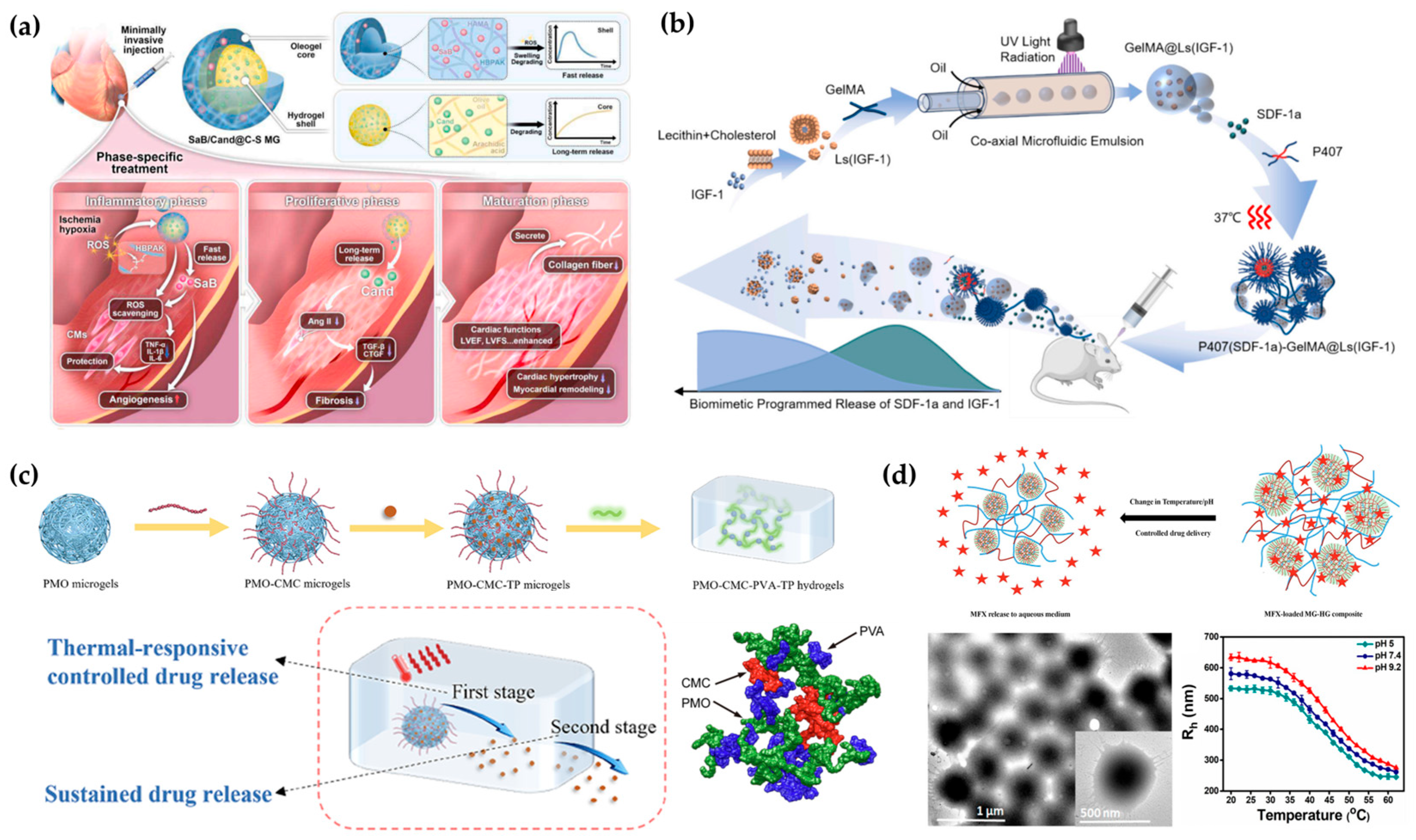
Stimulus-specific platforms remain a foundational strategy in responsive delivery, with many microgel and hydrogel systems designed to respond to a single pathological cue such as acidic pH, elevated temperature, or reduced conditions. These triggers are particularly relevant in cancer, inflammation, and wound healing, where local environments deviate significantly from homeostasis [23,271].
One example is polyampholyte-based core–shell microgels designed for pH- and redox-sensitive doxorubicin (DOX) delivery. This architecture consists of a carboxyl-rich poly(acrylic acid) (PAA) core and a disulfide-crosslinked PEG-based cationic shell. At physiological pH (7.4), strong electrostatic interaction retains DOX within the core; however, exposure to acidic and glutathione (GSH)-rich conditions (pH 5.0, 40 mM GSH) weakens these interactions and reduces crosslinking density, releasing over 85% of the loaded drug. This system demonstrated cytotoxic selectivity toward MCF-7 cells while sparing healthy MCF-10A cells, supporting its tumor-selective behavior [271,273].
Similarly, thermos- and pH-responsive microgels composed of PNIPAM-co-methacrylic acid embedded within chitosan hydrogel matrices have been used to prolong moxifloxacin release. This stimuli-responsive system demonstrated extended drug availability over 68 h, outperforming either microgels or hydrogels alone, while preserving cytocompatibility and minimizing hemolysis, indicating suitability for soft tissue applications (Figure 5) [23,281].
Figure 5. Schematic illustrations of smart hydrogel-based delivery systems. (a) A dual-compartment microgel designed for time-controlled release of SaB and Cand during the inflammatory and proliferative phases, respectively [270]. (b) Injectable thermo-responsive P407 hydrogel with GelMA microgels encapsulating IGF-1-loaded liposomes, showing gelation at 37 °C (red) for sequential SDF-1α and IGF-1 release to enhance bone regeneration [278]. (c) A two-phase drug delivery system featuring a thermally triggered initial release followed by a sustained release from the hydrogel matrix [282]. (d) A composite hydrogel–microgel (MG-HG) platform responsive to pH and temperature, enabling controlled release of moxifloxacin (MFX) [23].
Applsci 15 11599 g005
While single-stimulus systems are conceptually straightforward and have been extensively studied in bulk hydrogel literature, most operate on well-established mechanisms and design principles [271]. Given the extensive prior works, this subsection selectively highlights representative microgel-based examples, these systems form the conceptual and technical baseline upon which more complex, multi-responsive or spatially programmed designs are built, as discussed in the following subsections.

4.2.2. Multi-Stimuli Responsive Systems

To achieve greater selectivity in complex physiological environments, delivery platforms have increasingly adopted multi-stimuli responsiveness or logic-gated architectures. These systems are designed to release therapeutic agents only when exposed to a defined combination of triggers, including pH, temperature, redox potential, or metabolic cues, thereby reducing off-target effects and minimizing premature leakage. Compared to single-trigger systems, multi-responsive designs more closely mimic the multifactorial nature of pathological microenvironments, such as those found in tumors, inflamed tissues, or diabetic lesions.
One notable example is a cystamine-crosslinked PAA microgel integrated with gold nanoparticles (AuNPs) for glucose-triggered and pH-sensitive release of doxorubicin (DOX). In this design, the AuNPs exhibit catalytic activity, converting glucose into gluconic acid, which locally lowers the pH. The resulting acidification protonates the carboxyl groups on the PAA backbone, weakens electrostatic attractions and initiating DOX release. This cascade process aligns with the elevated glucose concentrations and localized acidosis commonly observed in tumor tissues and inflamed or diabetic sites. The system demonstrated a linear release profile over the first 40 min under hyperglycemic conditions (25 mM glucose), while maintaining minimal drug leakage under normoglycemia, thus leveraging a pathologically relevant combination to refine spatial and temporal delivery [272].
In another approach, folic acid-conjugated palygorskite/AuNP hybrid microgels were designed to integrate three stimuli: acidic pH, mild hyperthermia, and light. Each stimulus corresponds to features of the tumor microenvironment or can be externally controlled. Tumor tissues are typically acidic (pH~6.5 or lower) and hyperthermic (due to elevated metabolic activity), while light irradiation provides spatial selectivity benefitted by long penetration depth of near-infrared (NIR) light. In this system, acidic and thermal conditions enhanced DOX release up to 74%, and NIR laser exposure further accelerated the process through plasmonic heating by the embedded AuNPs. Folic acid conjugation targeted folate receptors, which are frequently overexpressed in various solid tumors including breast and ovarian cancers, thereby increasing cellular uptake. Together, the system exhibited multi-modal responsiveness with improved tumoral specificity and minimized off-target toxicity [273].
These examples highlight how combinatorial responsiveness, when anchored to clinically relevant environmental cues, enables highly selective and pathologically accurate drug release. The use of metabolite-induced pH shifts, redox gradients, or receptor-mediated uptake is not only mechanistically effective but also therapeutically strategic. By requiring the co-occurrence of two or more pathological conditions, such systems reduce the risk of premature leakage and allow more synchronized drug availability relative to disease activity. As such, multi-stimuli microgels serve as a critical steppingstone toward disease-responsive, logic-driven therapeutic design [272,273].

4.2.3. Spatially Compartmentalized Systems

Spatially compartmentalized delivery systems are designed to achieve coordinated release of therapeutic agents through structural segregation within the matrix. These architectures decouple the physical localization of cargo from its release dynamics, allowing different payloads, or even the same agent in different phases, to be selectively activated in response to spatial or temporal cues. Such designs are particularly valuable for multi-stage therapies or immune modulation, where precise sequence, co-localization, or physical containment are required.
One strategy integrates mRNA-loaded lipid nanoparticles (LNPs) into hybrid scaffolds consisting of electrospun nanofibers embedded within hydrogel matrices. In this architecture, the nanofiber mesh serves as a mechanical backbone that stabilizes the scaffold and limits bulk fluid diffusion, while the surrounding hydrogel provides a hydration environment and governs molecular diffusion of the mRNA payload. This physical confinement allows mRNA to be presented locally and persistently, enabling synchronization with immune cell recruitment and activation. Moreover, hydrogel properties can be tuned to respond to environmental pH or enzymatic degradation, further regulating temporal dispersion. Such systems have shown improved antigen expression and CD8+ T cell activation at tumor sites, outperforming LNPs alone in both retention and immunogenic response [37].
In another example, DNA-based hydrogels assembled via strand hybridization were engineered to function as both the matrix and the cargo. Here, the oligonucleotide sequences encode not only the hydrogel structure but also therapeutic information, such as CpG motifs or siRNA. Degradation of the matrix by tumor-associated nucleases releases the functional strands into its vicinity, achieving therapeutic effect. The matrix thus serves as a programmable depot with degradation kinetics intrinsically tied to the pathological microenvironment, providing molecular-level spatial logic [7].
A further strategy utilizes ionically crosslinked biopolymer systems, such as chitosan–alginate hybrids, to entrap nucleic acids through charge interactions while controlling their diffusion via matrix porosity. These systems offer simplicity, biocompatibility, and soft tissue compliance, and have been employed to extend mRNA expression in localized tissues without relying on synthetic carriers. The chitosan-rich domains promote gel formation and mRNA binding, while alginate controls the release kinetics and provides structural integrity under physiological conditions [280].
In all these designs, compartmentalization is not merely architectural but functional. Nanofiber regions define retention and mechanical stability, hydrogel domains govern diffusivity and responsiveness, and certain sequences or ionic domains control cargo stability and release. Such systems enable layered control over drug localization and release timing, which is critical for applications such as tissue regeneration, immune priming, or gene modulation where therapeutic context defines its efficacy [7,37,280].

4.2.4. Translational Pathways from Bulk to Modular Hydrogels

Many programmable delivery principles, particularly those specialized for nucleic acids, stem cells, and protein drugs, have been first validated in bulk hydrogel platforms. These systems offer flexible synthesis and sufficient volume for controlled-release studies but often lack the spatial resolution and injectability required for minimally invasive or precision-targeted therapies.
Despite this, the underlying logic of these systems is directly translatable to microgel-scale architectures. For instance, IGF-1 mimetic delivery from a Laponite–alginate composite hydrogel demonstrated prolonged release in acidic injury environments. This design, though developed in bulk, could be reconfigured into microparticulate formats to allow injectable application and localized tissue adaptation [269]. Similarly, chitosan–alginate matrices for mRNA delivery and enzyme-responsive scaffolds for stem cell support have shown favorable outcomes in vivo, yet remain constrained by their form factor. Transitioning these systems to modular microgels may preserve their bioactivity while enhancing formulation versatility and clinical usability [280].
Translational feasibility also depends on how fabrication strategies align with pharmaceutical manufacturing and regulatory expectations. Methods such as microfluidic droplet generation and electrospraying offer exceptional control over particle size and composition but require advances in parallelization and fouling-proof system design to reach industrial throughput. Emulsion-based polymerization and ionic crosslinking generally perceived as more scalable technologies and compatible with existing bioprocess infrastructure, though batch-to-batch variability and limited drug encapsulation control still poses challenges. Across the fabrication techniques discussed in this review, clinical translation will largely depend on integrating process reproducibility, in-line monitoring, and post-fabrication quality control into the design stage.
Thus, while hydrogel-based programmable delivery remains in its early stages for certain therapeutic classes, the design rationale, materials, and biological principles have already been explored in bulk systems. Future translation will depend on balancing this molecular scale complexity with scalable fabrication fidelity, ensuring the new architectures can meet the standards expected in pharmaceutical manufacturing. Ongoing translation efforts should also focus on downsizing these architectures without compromising the complex responsiveness or stability required for next-generation therapeutics.

4.3. Multi-Drug Delivery and Combination Therapy

Hydrogel-based DDS have provided a modular platform for administering multiple therapeutic agents with distinct release profiles. These systems are particularly suited for addressing diseases characterized by sequential pathological phases or requiring combinatorial treatment strategies [186,273,278]. This section categorizes recent designs based on three formulation logics: phase-specific release aligned with disease progression, controlled kinetic modulation within a single drug class, and spatial segregation of structurally distinct agents.
The first approach involves aligning drug release timing with specific stages of the healing or disease response. In bone regeneration, an injectable platform combining Poloxamer 407 (P407) bulk hydrogel and GelMA microgels was designed to deliver two protein-based agents with temporally distinct roles. SDF-1α, a chemokine known for its role in stem cell homing, was directly mixed into the P407 hydrogel to support early recruitment of mesenchymal stem cells during the inflammatory phase. IGF-1, associated with osteogenic differentiation, was encapsulated in liposome-loaded GelMA microgels to sustain its release over two to three weeks. In a rat calvarial defect model, this dual-protein system enhanced osteogenic marker expression and achieved approximately 66.9 percent bone volume recovery by week 8 (Figure 4) [278]. A related application in myocardial infarction employed a core–shell microgel architecture to deliver two small molecule drugs in sequence. The hydrophilic anti-inflammatory agent salvianolic acid B was released rapidly from the outer ROS-responsive hydrogel shell, while the inner oleogel core composed of arachidic acid and olive oil retained the hydrophobic antifibrotic drug candesartan for delayed release. In vivo results showed cumulative candesartan release over 21 days under oxidative conditions, reduced expression of fibrotic markers, and improved cardiac remodeling and function [270].
A second strategy leverages tunable release kinetics within a single therapeutic class by varying the physical state and encapsulation environment of the cargo. In one platform, dextran-based microgels were embedded within a PEG matrix and loaded with the same model protein, hen egg white lysozyme, in two distinct forms: freely dissolved and coacervated. The resulting release behavior could be precisely tuned by adjusting the ratio of fast- and slow-releasing microgel populations. This approach enabled predictable control over protein delivery without altering the chemical structure of the protein or the matrix. Dual-protein formulations further demonstrated that the platform could synchronize or invert release sequences. A similar principle was demonstrated in thermal-responsive PMO–CMC microgels incorporated into PVA hydrogels, where tea polyphenols exhibited a two-stage release profile governed by microgel responsiveness and matrix diffusion (Figure 4) [282]. In a representative case, coacervated lysozyme showed less than 20 percent release after two hours, while its soluble form released over 70 percent in ten minutes. In mixed systems, PEGylated BSA was released more rapidly than coacervated lysozyme, with crossover observed around 12 h. These outcomes were modeled using the Korsmeyer–Peppas equation, providing a quantitative framework for tuning kinetic profiles [268].
The third category focuses on chemical and structural compartmentalization of therapeutics with divergent properties. This approach supports the co-delivery of drugs with incompatible solubility, stability, or activity requirements. In the myocardial infarction study described above, the spatial separation of hydrophilic and hydrophobic agents into hydrogel and oleogel domains enabled their respective release under condition-specific cues [270]. Similarly, in protein-based systems, embedding distinct protein cargos into separately prepared microgels permitted individualized release control while maintaining compatibility and bioactivity. These architectures reduce the risk of cross-interactions between agents, simplify formulation logistics, and facilitate the development of combination therapies targeting multiple biological pathways [268].
While conventional injectable systems, such as polymeric depots, liposomes or other nanoparticles, have established the groundwork for combination delivery, their design flexibility and capacity for coordinated control remain limited. Hydrogel-based and hydrogel particulate systems extend this paradigm by allowing multi-drug release to be guided not only by chemical composition but also by network architecture as well as environmental responsiveness. Future developments could focus on programmable compartmentalization, hierarchical assembly, or stimulus-coupled feedback mechanisms that dynamically adjust therapeutic ratios in response to local cues. These direction position microgel and nanogels as adaptive therapeutic matrices capable of integrating pharmacological logic directly into material design.
This categorization highlights how microgel-based and hybrid systems can be rationally constructed to match the temporal, kinetic, and chemical demands of combination therapy. Examples drawn from regenerative medicine, cardiovascular treatment, and protein delivery illustrate the feasibility of implementing multi-drug strategies with formulation precision.

5. Translational Outlook for Hydrogel Drug Delivery

The promising potentials of hydrogel-based DDS are well represented by their modularity, injectability, and capacity for controlled multi-drug release. However, their translation from laboratory research to clinical application is constrained by a set of inter-related technical and regulatory hurdles. Among the foremost challenges is achieving consistent and reproducible fabrication of hydrogel particles at scale, particularly under Good Manufacturing Practice (GMP) conditions. Many of the methods used to produce uniform gel populations, including the techniques discussed here such as microfluidic droplet generation or emulsion polymerization, offer high precision at bench scale but require substantial adaptation for scalable, sterile, and cost-effective production. Furthermore, terminal sterilization remains a critical barrier, as many formulations are sensitive to conventional gamma irradiation or autoclave methods and may require aseptic manufacturing, which imposes additional complexity and regulatory challenges.
Also, it should be emphasized that scalability and reproducibility largely depend on the fabrication route. Emulsion-based and electrospraying techniques are suitable for large scale batch processing but could inherently introduce additional variability, whereas microfluidics-centered techniques allow high uniformity and size controllability but at limited throughput. Photolithographic strategies require cleanroom environment that complicates GMP compliance. Moreover, sterilization remains a critical challenge especially for microgels, since heat or irradiation can alter its polymer chemistry or payload integrity.
Beyond manufacturing, translational feasibility depends largely on the route of administration. For ocular delivery, constraints include limited injection volume, shear-induced dispersion, and rapid clearance via aqueous humor turnover, all of which demand high retention and minimal burst release. In contrast, peritumoral or subcutaneous systems must tolerate mechanical compression, variable extracellular matrix density, and, in some cases, immunological reactivity against depot materials. CNS applications, including intrathecal or intraparenchymal injections, require high precision in anatomical targeting and impose stringent requirements on injection force, depot viscosity, and neuroinflammatory compatibility. Each use case introduces distinct physical, anatomical, and regulatory boundaries, indicating that no single microgel architecture can be universally applied across tissues.
While no FDA-approved product currently exists that directly corresponds to particulate hydrogel-based therapeutics, relevant precedents can be found in injectable scaffolds, thermosensitive hydrogels, and long-acting microsphere depots. Products such as Lupron Depot® (AbbVie Inc., Mettawa, IL, USA) and Risperdal Consta® (Janssen Pharmaceuticals, Inc., Raritan, NJ, USA) demonstrate that injectable particulate systems can gain approval when reproducibility, depot behavior, and pharmacokinetics are well-characterized. For microgels to enter this space, developers must consider not only pharmacological performance criteria but also delivery device compatibility, terminal packaging standards, and pathway-specific regulatory classification. Moving forward, advancing standardization in characterization protocols, in vivo depot behavior modeling, and administration-site-aware design will be essential for integrating micro- and nanogel systems into the therapeutic landscape.

6. Conclusions

Injectable hydrogel systems represent a versatile convergence point between hydrogel chemistry, microscale engineering, and drug delivery design. As more than functioning miniaturized hydrogels, micro- and nanogels enable spatiotemporally controlled drug release, anatomical site compatibility, and integration with multi-agent or stimuli-responsive strategies. This review has examined how hydrogel particle platforms are uniquely positioned to meet the constraints of various injection sites, including ocular, subcutaneous, mucosal, tumoral, and CNS tissues, and how their structural environmental properties intersect with the demands of specific drug cargos.
By aligning material characteristics with administration route-specific challenges and therapeutic goals, hydrogel design has evolved toward increasingly modular, adaptive, and programmable systems. Importantly, advances in composite architecture and environmental responsiveness are progressively defining engineering strategies for next-generation therapeutics. As key translational barriers are systematically overcome, particulate hydrogel-based DDS hold strong potential toward clinically robust solutions, capable of delivering therapeutic agents with precision while simultaneously reconstructing local microenvironments that reflect the biological complexity of disease progression and tissue repair.

Author Contributions

Conceptualization, S.S. and J.Y.H.; investigation, Y.L., M.K., N.K. and S.B.; formal analysis, Y.L., M.K. and N.K.; writing—original draft preparation, Y.L., M.K., N.K., S.B., S.S. and J.Y.H.; review and editing, J.Y.H.; visualization, Y.L., M.K., N.K., S.B. and J.Y.H.; supervision, S.S. and J.Y.H.; funding acquisition, S.S. and J.Y.H. All authors have read and agreed to the published version of the manuscript.

Funding

This work was supported by the Basic Science Research program through the National Research Foundation of Korea (NRF) funded by the Ministry of Education (RS-2023-00253941) and the Gachon University research fund of GCU-202110350001.

Institutional Review Board Statement

Not applicable.

Informed Consent Statement

Not applicable.

Data Availability Statement

No new data were created or analyzed in this study. Data sharing is not applicable to this article.

Conflicts of Interest

The authors declare no conflicts of interest.

References

  1. Jia, R.; Teng, L.; Gao, L.; Su, T.; Fu, L.; Qiu, Z.; Bi, Y. Advances in Multiple Stimuli-Responsive Drug-Delivery Systems for Cancer Therapy. Int. J. Nanomed. 2021, 16, 1525–1551. [Google Scholar] [CrossRef]
  2. Ahmed, M.S.; Yun, S.; Kim, H.Y.; Ko, S.; Islam, M.; Nam, K.W. Hydrogels and Microgels: Driving Revolutionary Innovations in Targeted Drug Delivery, Strengthening Infection Management, and Advancing Tissue Repair and Regeneration. Gels 2025, 11, 179. [Google Scholar] [CrossRef]
  3. Li, J.; Mooney, D.J. Designing Hydrogels for Controlled Drug Delivery. Nat. Rev. Mater. 2016, 1, 27–36. [Google Scholar] [CrossRef]
  4. Smeets, N.M.B.; Hoare, T. Designing Responsive Microgels for Drug Delivery Applications. J. Polym. Sci. A Polym. Chem. 2013, 51, 3027–3043. [Google Scholar] [CrossRef]
  5. Oh, J.K.; Drumright, R.; Siegwart, D.J.; Matyjaszewski, K. The Development of Microgels/Nanogels for Drug Delivery Applications. Prog. Polym. Sci. 2008, 33, 448–477. [Google Scholar] [CrossRef]
  6. Hajebi, S.; Rabiee, N.; Bagherzadeh, M.; Ahmadi, S.; Rabiee, M.; Roghani-Mamaqani, H.; Tahriri, M.; Tayebi, L.; Hamblin, M.R. Stimulus-Responsive Polymeric Nanogels as Smart Drug Delivery Systems. Acta Biomater. 2019, 92, 1–18. [Google Scholar] [CrossRef]
  7. Wang, Q.; Qu, Y.; Zhang, Z.; Huang, H.; Xu, Y.; Shen, F.; Wang, L.; Sun, L. Injectable DNA Hydrogel-Based Local Drug Delivery and Immunotherapy. Gels 2022, 8, 400. [Google Scholar] [CrossRef] [PubMed]
  8. Chen, W.; Yung, B.C.; Qian, Z.; Chen, X. Improving Long-Term Subcutaneous Drug Delivery by Regulating Material-Bioenvironment Interaction. Adv. Drug Deliv. Rev. 2018, 127, 20–34. [Google Scholar] [CrossRef] [PubMed]
  9. Sivakumaran, D.; Maitland, D.; Hoare, T. Injectable Microgel-Hydrogel Composites for Prolonged Small-Molecule Drug Delivery. Biomacromolecules 2011, 12, 4112–4120. [Google Scholar] [CrossRef] [PubMed]
  10. Paresishvili, T.; Kakabadze, Z. Challenges and Opportunities Associated with Drug Delivery for the Treatment of Solid Tumors. Oncol. Rev. 2023, 17, 10577. [Google Scholar] [CrossRef]
  11. Lee, H.; Noh, H. Advancements in Nanogels for Enhanced Ocular Drug Delivery: Cutting-Edge Strategies to Overcome Eye Barriers. Gels 2023, 9, 718. [Google Scholar] [CrossRef]
  12. Kidane, A.; Bhatt, P.P. Recent Advances in Small Molecule Drug Delivery. Curr. Opin. Chem. Biol. 2005, 9, 347–351. [Google Scholar] [CrossRef]
  13. Brako, F.; Boateng, J. Transmucosal Drug Delivery: Prospects, Challenges, Advances, and Future Directions. Expert Opin. Drug Deliv. 2025, 22, 525–553. [Google Scholar] [CrossRef]
  14. Gandhat, S.; Ugale, S.; Kumavat, S.; Gadge, K.; Dandawate, N.; Bhor, V. Microneedles in Transdermal Drug Delivery: A Comprehensive Review. J. Drug Deliv. Ther. 2025, 15, 201–209. [Google Scholar] [CrossRef]
  15. Subhan, M.A.; Parveen, F.; Filipczak, N.; Yalamarty, S.S.K.; Torchilin, V.P. Approaches to Improve EPR-Based Drug Delivery for Cancer Therapy and Diagnosis. J. Pers. Med. 2023, 13, 389. [Google Scholar] [CrossRef]
  16. Cullis, P.R.; Hope, M.J. Lipid Nanoparticle Systems for Enabling Gene Therapies. Mol. Ther. 2017, 25, 1467–1475. [Google Scholar] [CrossRef] [PubMed]
  17. Choi, Y.; Koh, H.Y.; Han, J.Y.; Seo, S. Synthesis of Hydrogel-Based Microgels and Nanogels Toward Therapeutic and Biomedical Applications. Appl. Sci. 2025, 15, 1368. [Google Scholar] [CrossRef]
  18. Rana, M.M.; De la Hoz Siegler, H. Evolution of Hybrid Hydrogels: Next-Generation Biomaterials for Drug Delivery and Tissue Engineering. Gels 2024, 10, 216. [Google Scholar] [CrossRef] [PubMed]
  19. Coulter, S.M.; Pentlavalli, S.; An, Y.; Vora, L.K.; Cross, E.R.; Moore, J.V.; Sun, H.; Schweins, R.; McCarthy, H.O.; Laverty, G. In Situ Forming, Enzyme-Responsive Peptoid-Peptide Hydrogels: An Advanced Long-Acting Injectable Drug Delivery System. J. Am. Chem. Soc. 2024, 146, 21401–21416. [Google Scholar] [CrossRef]
  20. Lv, C.; Wang, Q.; Li, Z.; Jiang, X.; Liu, T.; Xu, X.; Feng, X. An Injectable Thermos-Sensitive Hydrogel for Sustained Release of α-Mangostin Promotes MRSA-Infected Wound Healing in Mice. Macromol. Res. 2025. [Google Scholar] [CrossRef]
  21. Bai, Y.; Zhang, Z.; Deng, M.; Chen, L.; He, C.; Zhuang, X.; Chen, X. Thermo- and PH-Responsive Microgels for Controlled Release of Insulin. Polym. Int. 2012, 61, 1151–1157. [Google Scholar] [CrossRef]
  22. Wang, X.; Zhang, Y.; Xue, W.; Wang, H.; Qiu, X.; Liu, Z. Thermo-Sensitive Hydrogel PLGA-PEG-PLGA as a Vaccine Delivery System for Intramuscular Immunization. J. Biomater. Appl. 2017, 31, 923–932. [Google Scholar] [CrossRef]
  23. Singh, N.; Aery, S.; Juneja, S.; Kumari, L.; Lone, M.S.; Dar, A.A.; Pawar, S.V.; Mehta, S.K.; Dan, A. Chitosan Hydrogels with Embedded Thermo- and PH-Responsive Microgels as a Potential Carrier for Controlled Release of Drugs. ACS Appl. Bio Mater. 2022, 5, 3487–3499. [Google Scholar] [CrossRef]
  24. Okuno, Y.; Iwasaki, Y. Microgel-Based Smart Materials: How Do You Design a Microgel? Langmuir 2025, 41, 7946–7964. [Google Scholar] [CrossRef] [PubMed]
  25. Alzanbaki, H.; Moretti, M.; Hauser, C.A.E. Engineered Microgels—Their Manufacturing and Biomedical Applications. Micromachines 2021, 12, 45. [Google Scholar] [CrossRef] [PubMed]
  26. Tehrani, S.M.; Lu, Y.; Winnik, M.A. PEGMA-Based Microgels: A Thermoresponsive Support for Enzyme Reactions. Macromolecules 2016, 49, 8711–8721. [Google Scholar] [CrossRef]
  27. Kumar, N.; Singh, S.; Sharma, P.; Kumar, B.; Kumar, A. Single-, Dual-, and Multi-Stimuli-Responsive Nanogels for Biomedical Applications. Gels 2024, 10, 61. [Google Scholar] [CrossRef]
  28. Thambi, T.; Li, Y.; Lee, D.S. Injectable Hydrogels for Sustained Release of Therapeutic Agents. J. Control. Release 2017, 267, 57–66. [Google Scholar] [CrossRef]
  29. Al Fatease, A.; Abdelkader, H. Recent Advances in Long-Acting Drug Delivery and Formulations. Pharmaceutics 2023, 15, 2519. [Google Scholar] [CrossRef]
  30. Lee, J.H. Injectable Hydrogels Delivering Therapeutic Agents for Disease Treatment and Tissue Engineering. Biomater. Res. 2018, 22, 27. [Google Scholar] [CrossRef]
  31. Marchenko, I.V.; Trushina, D.B. Local Drug Delivery in Bladder Cancer: Advances of Nano/Micro/Macro-Scale Drug Delivery Systems. Pharmaceutics 2023, 15, 2724. [Google Scholar] [CrossRef]
  32. Merino, S.; Martín, C.; Kostarelos, K.; Prato, M.; Vázquez, E. Nanocomposite Hydrogels: 3D Polymer-Nanoparticle Synergies for on-Demand Drug Delivery. ACS Nano 2015, 9, 4686–4697. [Google Scholar] [CrossRef]
  33. Lee, S.Y.; Yang, M.; Seo, J.H.; Jeong, D.I.; Hwang, C.; Kim, H.J.; Lee, J.; Lee, K.; Park, J.; Cho, H.J. Serially PH-Modulated Hydrogels Based on Boronate Ester and Polydopamine Linkages for Local Cancer Therapy. ACS Appl. Mater. Interfaces 2021, 13, 2189–2203. [Google Scholar] [CrossRef]
  34. Ahiabu, A.; Serpe, M.J. Rapidly Responding pH-and Temperature-Responsive Poly (N-Isopropylacrylamide)-Based Microgels and Assemblies. ACS Omega 2017, 2, 1769–1777. [Google Scholar] [CrossRef] [PubMed]
  35. Garcia, A.; Marquez, M.; Cai, T.; Rosario, R.; Hu, Z.; Gust, D.; Hayes, M.; Vail, S.A.; Park, C. Do Photo-, Thermally, and PH-Responsive Microgels. Langmuir 2007, 23, 224–229. [Google Scholar] [CrossRef]
  36. Mehraji, S.; DeVoe, D.L. Microfluidic Synthesis of Lipid-Based Nanoparticles for Drug Delivery: Recent Advances and Opportunities. Lab Chip 2024, 24, 1154–1174. [Google Scholar] [CrossRef] [PubMed]
  37. Zhu, Y.; Yao, Z.-C.; Li, S.; Ma, J.; Wei, C.; Yu, D.; Stelzel, J.L.; Ni, B.Y.X.; Miao, Y.; Van Batavia, K.; et al. An MRNA Lipid Nanoparticle-Incorporated Nanofiber-Hydrogel Composite for Cancer Immunotherapy. Nat. Commun. 2025, 16, 5707. [Google Scholar] [CrossRef] [PubMed]
  38. Cheng, Y.H.; Fung, M.P.; Chen, Y.Q.; Chiu, Y.C. Development of Mucoadhesive Methacrylic Anhydride-Modified Hydroxypropyl Methylcellulose Hydrogels for Topical Ocular Drug Delivery. J. Drug Deliv. Sci. Technol. 2024, 93, 105450. [Google Scholar] [CrossRef]
  39. Lee, S.; Park, J.Y.; Hong, H.K.; Son, J.Y.; Kim, B.; Chung, J.Y.; Woo, S.J.; Park, K.D. Intravitreal Long-Term Sustained Ranibizumab Delivery Using Injectable Microgel-Embedded Hydrogel. Asian J. Pharm. Sci. 2024, 19, 100947. [Google Scholar] [CrossRef]
  40. Anumolu, S.N.S.; DeSantis, A.S.; Menjoge, A.R.; Hahn, R.A.; Beloni, J.A.; Gordon, M.K.; Sinko, P.J. Doxycycline Loaded Poly(Ethylene Glycol) Hydrogels for Healing Vesicant-Induced Ocular Wounds. Biomaterials 2010, 31, 964–974. [Google Scholar] [CrossRef]
  41. Xi, L.; Wang, T.; Zhao, F.; Zheng, Q.; Li, X.; Luo, J.; Liu, J.; Quan, D.; Ge, J. Evaluation of an Injectable Thermosensitive Hydrogel as Drug Delivery Implant for Ocular Glaucoma Surgery. PLoS ONE 2014, 9, e100632. [Google Scholar] [CrossRef]
  42. Annala, A.; Ilochonwu, B.C.; Wilbie, D.; Sadeghi, A.; Hennink, W.E.; Vermonden, T. Self-Healing Thermosensitive Hydrogel for Sustained Release of Dexamethasone for Ocular Therapy. ACS Polym. Au 2023, 3, 118–131. [Google Scholar] [CrossRef]
  43. Cao, Y.; Ma, Y.; Tao, Y.; Lin, W.; Wang, P. Intra-Articular Drug Delivery for Osteoarthritis Treatment. Pharmaceutics 2021, 13, 2166. [Google Scholar] [CrossRef] [PubMed]
  44. Liu, Y.; Yao, J.; Deng, G.; Zhong, G.; Zhao, J.; Lan, Q.; Meng, J.; Yu, Y.; Chen, F. Microgel Encapsulated Nanoparticles for Intra-Articular Disulfiram Delivery to Treat Osteoarthritis. Mol. Pharm. 2024, 21, 87–101. [Google Scholar] [CrossRef]
  45. Mancipe Castro, L.M.; Sequeira, A.; García, A.J.; Guldberg, R.E. Articular Cartilage and Synoviocyte-Binding Poly(Ethylene Glycol) Nanocomposite Microgels as Intra-Articular Drug Delivery Vehicles for the Treatment of Osteoarthritis. ACS Biomater. Sci. Eng. 2020, 6, 5084–5095. [Google Scholar] [CrossRef]
  46. Said, M.; Tavakoli, C.; Dumot, C.; Toupet, K.; Olivier, C.; Gilles, A.; Maumus, M.; Dong, Y.C.; Collomb, N.; Auxenfans, C.; et al. A Self-Healing Radiopaque Hyaluronic Acid Hydrogel as a New Injectable Biomaterial for Precision Medicine in Osteoarthritis. Theranostics 2025, 15, 4054–4073. [Google Scholar] [CrossRef] [PubMed]
  47. Vinikoor, T.; Dzidotor, G.K.; Le, T.T.; Liu, Y.; Kan, H.M.; Barui, S.; Chorsi, M.T.; Curry, E.J.; Reinhardt, E.; Wang, H.; et al. Injectable and Biodegradable Piezoelectric Hydrogel for Osteoarthritis Treatment. Nat. Commun. 2023, 14, 6257. [Google Scholar] [CrossRef] [PubMed]
  48. Dubbelboer, I.R.; Sjögren, E. Overview of Authorized Drug Products for Subcutaneous Administration: Pharmaceutical, Therapeutic, and Physicochemical Properties. Eur. J. Pharm. Sci. 2022, 173, 106181. [Google Scholar] [CrossRef]
  49. Yadav, J.; Uddin, S.; Civati, F.; Ma, W.; Liebminger, A.; Teschner, W.; André, G.; Trout, B.L.; Braatz, R.D.; Myerson, A.S. Developing Ultra-High Concentration Formulations of Human Immune Globulins for Subcutaneous Injectables. J. Pharm. Sci. 2025, 114, 1605–1614. [Google Scholar] [CrossRef]
  50. Kang, N.W.; Yoon, S.Y.; Kim, S.; Yu, N.Y.; Park, J.H.; Lee, J.Y.; Cho, H.J.; Kim, D.D. Subcutaneously Injectable Hyaluronic Acid Hydrogel for Sustained Release of Donepezil with Reduced Initial Burst Release: Effect of Hybridization of Microstructured Lipid Carriers and Albumin. Pharmaceutics 2021, 13, 864. [Google Scholar] [CrossRef]
  51. Tian, Y.; Zhang, R.; Cui, J.; Zhu, Y.; Sun, M.; Hamley, I.W.; Xiao, C.; Chen, L. An Injectable Antibacterial Hydrogel with Bacterial-Targeting Properties for Subcutaneous Suppuration Treatment. Chem. Eng. J. 2024, 488, 151137. [Google Scholar] [CrossRef]
  52. Lo, Y.W.; Sheu, M.T.; Chiang, W.H.; Chiu, Y.L.; Tu, C.M.; Wang, W.Y.; Wu, M.H.; Wang, Y.C.; Lu, M.; Ho, H.O. In Situ Chemically Crosslinked Injectable Hydrogels for the Subcutaneous Delivery of Trastuzumab to Treat Breast Cancer. Acta Biomater. 2019, 86, 280–290. [Google Scholar] [CrossRef] [PubMed]
  53. McCartan, A.; Mackay, J.; Curran, D.; Mrsny, R.J. Modelling Intramuscular Drug Fate in Vitro with Tissue-Relevant Biomimetic Hydrogels. Int. J. Pharm. X 2022, 4, 100125. [Google Scholar] [CrossRef]
  54. Wei, J.; Xue, W.; Yu, X.; Qiu, X.; Liu, Z. PH Sensitive Phosphorylated Chitosan Hydrogel as Vaccine Delivery System for Intramuscular Immunization. J. Biomater. Appl. 2017, 31, 1358–1369. [Google Scholar] [CrossRef] [PubMed]
  55. Din, F.U.; Kim, J.S.; Lee, H.C.; Cheon, S.; Woo, M.R.; Woo, S.; Ku, S.K.; Yoo, H.H.; Kim, J.O.; Jin, S.G.; et al. Injectable Dual Thermoreversible Hydrogel for Sustained Intramuscular Drug Delivery. J. Control. Release 2024, 374, 590–605. [Google Scholar] [CrossRef]
  56. Soltani, M.; Chen, P. Effect of Tumor Shape and Size on Drug Delivery to Solid Tumors. J. Biol. Eng. 2012, 6, 4. [Google Scholar] [CrossRef]
  57. Chen, X.; Qian, H.; Qiao, H.; Dong, B.; Chen, E.; Huang, D.; Wang, T.; Chen, W. Tumor-Adhesive and PH-Degradable Microgels by Microfluidics and Photo-Cross-Linking for Efficient Antiangiogenesis and Enhanced Cancer Chemotherapy. Biomacromolecules 2020, 21, 1285–1294. [Google Scholar] [CrossRef]
  58. Liu, G.; Yang, L.; Chen, G.; Xu, F.; Yang, F.; Yu, H.; Li, L.; Dong, X.; Han, J.; Cao, C.; et al. A Review on Drug Delivery System for Tumor Therapy. Front. Pharmacol. 2021, 12, 735446. [Google Scholar] [CrossRef] [PubMed]
  59. Luo, F.Q.; Xu, W.; Zhang, J.Y.; Liu, R.; Huang, Y.C.; Xiao, C.; Du, J.Z. An Injectable Nanocomposite Hydrogel Improves Tumor Penetration and Cancer Treatment Efficacy. Acta Biomater. 2022, 147, 235–244. [Google Scholar] [CrossRef]
  60. Sabino, I.J.; Lima-Sousa, R.; Alves, C.G.; Melo, B.L.; Moreira, A.F.; Correia, I.J.; de Melo-Diogo, D. Injectable in Situ Forming Hydrogels Incorporating Dual-Nanoparticles for Chemo-Photothermal Therapy of Breast Cancer Cells. Int. J. Pharm. 2021, 600, 120510. [Google Scholar] [CrossRef]
  61. Zhao, Z.; Li, Q.; Qin, X.; Zhang, M.; Du, Q.; Luan, Y. An Injectable Hydrogel Reshaping Adenosinergic Axis for Cancer Therapy. Adv. Funct. Mater. 2022, 32, 2200801. [Google Scholar] [CrossRef]
  62. Zhao, X.; Jin, L.; Zhu, Z.; Lu, H.; Shi, H.; Zhong, Q.; Oliveira, J.M.; Reis, R.L.; Gao, C.; Mao, Z. Conotoxin Loaded Dextran Microgel Particles Alleviate Effects of Spinal Cord Injury by Inhibiting Neuronal Excitotoxicity. Appl. Mater. Today 2021, 23, 101064. [Google Scholar] [CrossRef]
  63. Wang, Z.; Zhao, H.; Tang, X.; Meng, T.; Khutsishvili, D.; Xu, B.; Ma, S. CNS Organoid Surpasses Cell-Laden Microgel Assembly to Promote Spinal Cord Injury Repair. Research 2022, 2022, 9832128. [Google Scholar] [CrossRef]
  64. Kung, Y.; Chen, K.Y.; Liao, W.H.; Hsu, Y.H.; Wu, C.H.; Hsiao, M.Y.; Huang, A.P.H.; Chen, W.S. Facilitating Drug Delivery in the Central Nervous System by Opening the Blood-Cerebrospinal Fluid Barrier with a Single Low Energy Shockwave Pulse. Fluids Barriers CNS 2022, 19, 3. [Google Scholar] [CrossRef]
  65. Tseng, T.C.; Tao, L.; Hsieh, F.Y.; Wei, Y.; Chiu, I.M.; Hsu, S.H. An Injectable, Self-Healing Hydrogel to Repair the Central Nervous System. Adv. Mater. 2015, 27, 3518–3524. [Google Scholar] [CrossRef] [PubMed]
  66. Tsintou, M.; Dalamagkas, K.; Seifalian, A. Injectable Hydrogel versus Plastically Compressed Collagen Scaffold for Central Nervous System Applications. Int. J. Biomater. 2018, 2018, 1–10. [Google Scholar] [CrossRef] [PubMed]
  67. Yeruva, T.; Yang, S.; Doski, S.; Duncan, G.A. Hydrogels for Mucosal Drug Delivery. ACS Appl. Bio Mater. 2023, 6, 1684–1700. [Google Scholar] [CrossRef]
  68. Nie, L.; Zou, P.; Dong, J.; Sun, M.; Ding, P.; Han, Y.; Ji, C.; Zhou, Q.; Yuan, H.; Suo, J. Injectable Vaginal Hydrogels as a Multi-Drug Carrier for Contraception. Appl. Sci. 2019, 9, 1638. [Google Scholar] [CrossRef]
  69. Liu, L.C.; Chen, Y.H.; Lu, D.W. Overview of Recent Advances in Nano-Based Ocular Drug Delivery. Int. J. Mol. Sci. 2023, 24, 15352. [Google Scholar] [CrossRef] [PubMed]
  70. Lynch, C.R.; Kondiah, P.P.D.; Choonara, Y.E.; du Toit, L.C.; Ally, N.; Pillay, V. Hydrogel Biomaterials for Application in Ocular Drug Delivery. Front. Bioeng. Biotechnol. 2020, 8, 228. [Google Scholar] [CrossRef]
  71. Cassano, R.; Di Gioia, M.L.; Trombino, S. Gel-Based Materials for Ophthalmic Drug Delivery. Gels 2021, 7, 130. [Google Scholar] [CrossRef]
  72. Ilochonwu, B.C.; Urtti, A.; Hennink, W.E.; Vermonden, T. Intravitreal Hydrogels for Sustained Release of Therapeutic Proteins. J. Control. Release 2020, 326, 419–441. [Google Scholar] [CrossRef]
  73. Wu, J.; Chen, Q.; Deng, C.; Xu, B.; Zhang, Z.; Yang, Y.; Lu, T. Exquisite Design of Injectable Hydrogels in Cartilage Repair. Theranostics 2020, 10, 9843–9864. [Google Scholar] [CrossRef]
  74. Sunstrom, N.; Sunstrum, F.N. Wearable Devices for Subcutaneous Delivery of Large-Volume Biologics: Design, Use, and Regulatory Perspective. Biomed. Mater. Devices 2025. [Google Scholar] [CrossRef]
  75. Bellotti, E.; Schilling, A.L.; Little, S.R.; Decuzzi, P. Injectable Thermoresponsive Hydrogels as Drug Delivery System for the Treatment of Central Nervous System Disorders: A Review. J. Control. Release 2021, 329, 16–35. [Google Scholar] [CrossRef]
  76. Wang, K.; Han, Z. Injectable Hydrogels for Ophthalmic Applications. J. Control. Release 2017, 268, 212–224. [Google Scholar] [CrossRef]
  77. Assiri, A.A.; Glover, K.; Mishra, D.; Waite, D.; Vora, L.K.; Thakur, R.R.S. Block Copolymer Micelles as Ocular Drug Delivery Systems. Drug Discov. Today 2024, 29, 104098. [Google Scholar] [CrossRef] [PubMed]
  78. Gupta, H.; Jain, S.; Mathur, R.; Mishra, P.; Mishra, A.K.; Velpandian, T. Sustained Ocular Drug Delivery from a Temperature and PH Triggered Novel in Situ Gel System. Drug Deliv. 2007, 14, 507–515. [Google Scholar] [CrossRef] [PubMed]
  79. Bin Choy, Y.; Park, J.H.; Prausnitz, M.R. Mucoadhesive Microparticles Engineered for Ophthalmic Drug Delivery. J. Phys. Chem. Solids 2008, 69, 1533–1536. [Google Scholar] [CrossRef] [PubMed]
  80. Fitaihi, R.; Abukhamees, S.; Orlu, M.; Craig, D.Q.M. Transscleral Delivery of Dexamethasone-Loaded Microparticles Using a Dissolving Microneedle Array. Pharmaceutics 2023, 15, 1622. [Google Scholar] [CrossRef]
  81. Singh, M.; Bharadwaj, S.; Lee, K.E.; Kang, S.G. Therapeutic Nanoemulsions in Ophthalmic Drug Administration: Concept in Formulations and Characterization Techniques for Ocular Drug Delivery. J. Control. Release 2020, 328, 895–916. [Google Scholar] [CrossRef] [PubMed]
  82. Zhou, D.; Zhou, F.; Sheng, S.; Wei, Y.; Chen, X.; Su, J. Intra-Articular Nanodrug Delivery Strategies for Treating Osteoarthritis. Drug Discov. Today 2023, 28, 103482. [Google Scholar] [CrossRef]
  83. Kalairaj, M.S.; Pradhan, R.; Saleem, W.; Smith, M.M.; Gaharwar, A.K. Intra-Articular Injectable Biomaterials for Cartilage Repair and Regeneration. Adv. Healthc. Mater. 2024, 13, e2303794. [Google Scholar] [CrossRef]
  84. Qi, X.; Qin, X.; Yang, R.; Qin, J.; Li, W.; Luan, K.; Wu, Z.; Song, L. Intra-Articular Administration of Chitosan Thermosensitive in Situ Hydrogels Combined with Diclofenac Sodium-Loaded Alginate Microspheres. J. Pharm. Sci. 2016, 105, 122–130. [Google Scholar] [CrossRef]
  85. Stefani, R.M.; Lee, A.J.; Tan, A.R.; Halder, S.S.; Hu, Y.; Guo, X.E.; Stoker, A.M.; Ateshian, G.A.; Marra, K.G.; Cook, J.L.; et al. Sustained Low-Dose Dexamethasone Delivery via a PLGA Microsphere-Embedded Agarose Implant for Enhanced Osteochondral Repair. Acta Biomater. 2020, 102, 326–340. [Google Scholar] [CrossRef]
  86. Fang, W.; Yang, F.; Li, W.; Hu, Q.; Chen, W.; Yang, M.; Chen, J.; Qiu, L. Dexamethasone Microspheres and Celecoxib Microcrystals Loaded into Injectable Gels for Enhanced Knee Osteoarthritis Therapy. Int. J. Pharm. 2022, 622, 121802. [Google Scholar] [CrossRef]
  87. Shah, J.C.; Hong, J. Model for Long Acting Injectables (Depot Formulation) Based on Pharmacokinetics and Physical Chemical Properties. AAPS J. 2022, 24, 44. [Google Scholar] [CrossRef]
  88. Gordon, M.K.; Hahn, R.A. Collagens. Cell Tissue Res. 2010, 339, 247–257. [Google Scholar] [CrossRef]
  89. Purslow, P.P. The Structure and Functional Significance of Variations in the Connective Tissue within Muscle. Comp. Biochem. Physiol. A Mol. Integr. Physiol. 2002, 133, 947–966. [Google Scholar] [PubMed]
  90. Zhang, W.; Liu, Y.; Zhang, H. Extracellular Matrix: An Important Regulator of Cell Functions and Skeletal Muscle Development. Cell Biosci. 2021, 11, 65. [Google Scholar] [CrossRef] [PubMed]
  91. Desai, K.G. Subcutaneous Administration of Therapeutic Monoclonal Antibody Drug Products Using a Syringe in Blinded Clinical Trials: Advances and Key Aspects Related to Blinding/Matching/Masking Strategies for Placebo Formulation. Mol. Pharm. 2025, 22, 620–637. [Google Scholar] [CrossRef] [PubMed]
  92. Kittel, Y.; Kuehne, A.J.C.; De Laporte, L. Translating Therapeutic Microgels into Clinical Applications. Adv. Healthc. Mater. 2022, 11, e2101989. [Google Scholar] [CrossRef] [PubMed]
  93. Datta, D.; Colaco, V.; Bandi, S.P.; Dhas, N.; Janardhanam, L.S.L.; Singh, S.; Vora, L.K. Stimuli-Responsive Self-Healing Ionic Gels: A Promising Approach for Dermal and Tissue Engineering Applications. ACS Biomater. Sci. Eng. 2025, 11, 1338–1372. [Google Scholar] [CrossRef] [PubMed]
  94. Zhang, K.; Shi, X.; Lin, X.; Yao, C.; Shen, L.; Feng, Y. Poloxamer-Based in Situ Hydrogels for Controlled Delivery of Hydrophilic Macromolecules after Intramuscular Injection in Rats. Drug Deliv. 2015, 22, 375–382. [Google Scholar] [CrossRef] [PubMed]
  95. Liang, X.; Gao, J.; Jiang, L.; Luo, J.; Jing, L.; Li, X.; Jin, Y.; Dai, Z. Nanohybrid Liposomal Cerasomes with Good Physiological Stability and Rapid Temperature Responsiveness for High Intensity Focused Ultrasound Triggered Local Chemotherapy of Cancer. ACS Nano 2015, 9, 1280–1293. [Google Scholar] [CrossRef]
  96. She, W.; Li, N.; Luo, K.; Guo, C.; Wang, G.; Geng, Y.; Gu, Z. Dendronized Heparin-Doxorubicin Conjugate Based Nanoparticle as PH-Responsive Drug Delivery System for Cancer Therapy. Biomaterials 2013, 34, 2252–2264. [Google Scholar] [CrossRef]
  97. Wang, Q.; Gu, Z.; Jamal, S.; Detamore, M.S.; Berkland, C. Hybrid Hydroxyapatite Nanoparticle Colloidal Gels Are Injectable Fillers for Bone Tissue Engineering. Tissue Eng. Part A 2013, 19, 2586–2593. [Google Scholar] [CrossRef]
  98. Gu, Z.; Aimetti, A.A.; Wang, Q.; Dang, T.T.; Zhang, Y.; Veiseh, O.; Cheng, H.; Langer, R.S.; Anderson, D.G. Injectable Nano-Network for Glucose-Mediated Insulin Delivery. ACS Nano 2013, 7, 4194–4201. [Google Scholar] [CrossRef]
  99. Ozeki, T.; Kaneko, D.; Hashizawa, K.; Imai, Y.; Tagami, T.; Okada, H. Improvement of Survival in C6 Rat Glioma Model by a Sustained Drug Release from Localized PLGA Microspheres in a Thermoreversible Hydrogel. Int. J. Pharm. 2012, 427, 299–304. [Google Scholar] [CrossRef]
  100. Davis, A.J.; Tannock, I.F.; Tannock, I. Repopulation and Chemotherapy Review Repopulation of Tumour Cells between Cycles of Chemotherapy: A Neglected Factor. Lancet Oncol. 2000, 1, 86–93. [Google Scholar] [CrossRef]
  101. Fukumura, D.; Jain, R.K. Tumor Microenvironment Abnormalities: Causes, Consequences, and Strategies to Normalize. J. Cell Biochem. 2007, 101, 937–949. [Google Scholar] [CrossRef] [PubMed]
  102. Ni, G.; Yang, G.; He, Y.; Li, X.; Du, T.; Xu, L.; Zhou, S. Uniformly Sized Hollow Microspheres Loaded with Polydopamine Nanoparticles and Doxorubicin for Local Chemo-Photothermal Combination Therapy. Chem. Eng. J. 2020, 379, 122317. [Google Scholar] [CrossRef]
  103. Yan, J.; Wang, Y.; Ran, M.; Mustafa, R.A.; Luo, H.; Wang, J.; Smått, J.H.; Rosenholm, J.M.; Cui, W.; Lu, Y.; et al. Peritumoral Microgel Reservoir for Long-Term Light-Controlled Triple-Synergistic Treatment of Osteosarcoma with Single Ultra-Low Dose. Small 2021, 17, e2100479. [Google Scholar] [CrossRef] [PubMed]
  104. Wang, Y.; Wang, L.; Yan, M.; Feng, L.; Dong, S.; Hao, J. Drug Implants of Hydrogels via Collective Behavior of Microgel Colloids for On-Demand Cancer Therapy. ACS Appl. Bio Mater. 2019, 2, 1531–1541. [Google Scholar] [CrossRef]
  105. Huang, D.; Zhu, J.; Xu, M.F.; Chen, J.; Gao, X.; Zhao, L.; Ding, F.; Wu, C.Z. One-Pot Preparation of PH- and Redox-Responsive Polymeric Microgel as an Efficient Carrier for Improved Breast Cancer Therapy. Colloids Surf. A Physicochem. Eng. Asp. 2024, 685, 133320. [Google Scholar] [CrossRef]
  106. Zhai, M.; Wu, P.; Liao, Y.; Wu, L.; Zhao, Y. Polymer Microspheres and Their Application in Cancer Diagnosis and Treatment. Int. J. Mol. Sci. 2024, 25, 6556. [Google Scholar] [CrossRef]
  107. Gao, Y.; Zhang, T.L.; Zhang, H.J.; Gao, J.; Yang, P.F. A Promising Application of Injectable Hydrogels in Nerve Repair and Regeneration for Ischemic Stroke. Int. J. Nanomed. 2024, 19, 327–345. [Google Scholar] [CrossRef] [PubMed]
  108. Hyun, J.K.; Kim, H.W. Clinical and Experimental Advances in Regeneration of Spinal Cord Injury. J. Tissue Eng. 2010, 1, 650857. [Google Scholar] [CrossRef]
  109. Rose, J.C.; Cámara-Torres, M.; Rahimi, K.; Köhler, J.; Möller, M.; De Laporte, L. Nerve Cells Decide to Orient inside an Injectable Hydrogel with Minimal Structural Guidance. Nano Lett. 2017, 17, 3782–3791. [Google Scholar] [CrossRef]
  110. Niu, G.; Jin, Z.; Zhang, C.; He, D.; Gao, X.; Zou, C.; Zhang, W.; Ding, J.; Das, B.C.; Severinov, K.; et al. An Effective Vaginal Gel to Deliver CRISPR/Cas9 System Encapsulated in Poly (β-Amino Ester) Nanoparticles for Vaginal Gene Therapy. EBioMedicine 2020, 58, 102897. [Google Scholar] [CrossRef]
  111. Permana, A.D.; Asri, R.M.; Amir, M.N.; Himawan, A.; Arjuna, A.; Juniarti, N.; Utami, R.N.; Mardikasari, S.A. Development of Thermoresponsive Hydrogels with Mucoadhesion Properties Loaded with Metronidazole Gel-Flakes for Improved Bacterial Vaginosis Treatment. Pharmaceutics 2023, 15, 1529. [Google Scholar] [CrossRef]
  112. Kenechukwu, F.C.; Momoh, M.A.; Nnamani, P.O.; Umeyor, C.E.; Uronnachi, E.M.; Dias, M.L.; Ibezim, E.C.; Attama, A.A. Dual-Responsive Micellar Microgels Matrixed with Surface-Engineered Lipids: A New Approach for Controlled Vaginal Drug Delivery. J. Pharm. Innov. 2022, 17, 821–839. [Google Scholar] [CrossRef]
  113. Gosecka, M.; Gosecki, M. Antimicrobial Polymer-Based Hydrogels for the Intravaginal Therapies—Engineering Considerations. Pharmaceutics 2021, 13, 1393. [Google Scholar] [CrossRef]
  114. Feng, Q.; Li, D.; Li, Q.; Cao, X.; Dong, H. Microgel Assembly: Fabrication, Characteristics and Application in Tissue Engineering and Regenerative Medicine. Bioact. Mater. 2022, 9, 105–119. [Google Scholar] [CrossRef] [PubMed]
  115. Banga, A.K.; Chien, Y.W. Hydrogel-Based Lontotherapeutic Delivery Devices for Transdermal Delivery of peptide/protein Drugs. Pharm. Res. 1993, 10, 697–702. [Google Scholar] [CrossRef] [PubMed]
  116. Dave, R.; Randhawa, G.; Kim, D.; Simpson, M.; Hoare, T. Microgels and Nanogels for the Delivery of Poorly Water-Soluble Drugs. Mol. Pharm. 2022, 19, 1704–1721. [Google Scholar] [CrossRef]
  117. Farjami, T.; Madadlou, A. Fabrication Methods of Biopolymeric Microgels and Microgel-Based Hydrogels. Food Hydrocoll. 2017, 62, 262–272. [Google Scholar] [CrossRef]
  118. McAllister, K.; Sazani, P.; Adam, M.; Cho, M.J.; Rubinstein, M.; Samulski, R.J.; DeSimone, J.M. Polymeric Nanogels Produced via Inverse Microemulsion Polymerization as Potential Gene and Antisense Delivery Agents. J. Am. Chem. Soc. 2002, 124, 15198–15207. [Google Scholar] [CrossRef]
  119. Sartipzadeh, O.; Naghib, S.M.; Haghiralsadat, F.; Shokati, F.; Rahmanian, M. Microfluidic-Assisted Synthesis and Modeling of Stimuli-Responsive Monodispersed Chitosan Microgels for Drug Delivery Applications. Sci. Rep. 2022, 12, 8382. [Google Scholar] [CrossRef]
  120. Ciarleglio, G.; Russo, T.; Toto, E.; Santonicola, M.G. Fabrication of Alginate/Ozoile Gel Microspheres by Electrospray Process. Gels 2024, 10, 52. [Google Scholar] [CrossRef]
  121. Ahmed, H.; Stokke, B.T. Fabrication of Monodisperse Alginate Microgel Beads by Microfluidic Picoinjection: A Chelate Free Approach. Lab Chip 2021, 21, 2232–2243. [Google Scholar] [CrossRef]
  122. Chen, M.; Bolognesi, G.; Vladisavljević, G.T. Crosslinking Strategies for the Microfluidic Production of Microgels. Molecules 2021, 26, 3752. [Google Scholar] [CrossRef]
  123. Kim, H.U.; Lim, Y.J.; Lee, H.J.; Lee, N.J.; Bong, K.W. Degassed Micromolding Lithography for Rapid Fabrication of Anisotropic Hydrogel Microparticles with High-Resolution and High Uniformity. Lab Chip 2020, 20, 74–83. [Google Scholar] [CrossRef]
  124. Panda, P.; Ali, S.; Lo, E.; Chung, B.G.; Hatton, T.A.; Khademhosseini, A.; Doyle, P.S. Stop-Flow Lithography to Generate Cell-Laden Microgel Particles. Lab Chip 2008, 8, 1056–1061. [Google Scholar] [CrossRef] [PubMed]
  125. Wolff, H.J.M.; Linkhorst, J.; Göttlich, T.; Savinsky, J.; Krüger, A.J.D.; De Laporte, L.; Wessling, M. Soft Temperature-Responsive Microgels of Complex Shape in Stop-Flow Lithography. Lab Chip 2020, 20, 285–295. [Google Scholar] [CrossRef] [PubMed]
  126. Asadikorayem, M.; Weber, P.; Surman, F.; Puiggalí-Jou, A.; Zenobi-Wong, M. Foreign Body Immune Response to Zwitterionic and Hyaluronic Acid Granular Hydrogels Made with Mechanical Fragmentation. Adv. Healthc. Mater. 2025, 14, e2402890. [Google Scholar] [CrossRef] [PubMed]
  127. Hei, X.; Li, S.; Liu, Z.; Wu, C.; Ma, X.; Jiao, B.; Hu, H.; Zhu, J.; Adhikari, B.; Wang, Q.; et al. Characteristics of Pickering Emulsions Stabilized by Microgel Particles of Five Different Plant Proteins and Their Application. Food Chem. 2024, 449, 139187. [Google Scholar] [CrossRef]
  128. Duffy, C.; O’Sullivan, M.; Jacquier, J.C. Preparation of Novel Chitosan Iron Microgel Beads for Fortification Applications. Food Hydrocoll. 2018, 84, 608–615. [Google Scholar] [CrossRef]
  129. Wang, H.; Liu, H.; Liu, H.; Su, W.; Chen, W.; Qin, J. One-Step Generation of Core–Shell Gelatin Methacrylate (GelMA) Microgels Using a Droplet Microfluidic System. Adv. Mater. Technol. 2019, 4, 1800632. [Google Scholar] [CrossRef]
  130. Huang, F.; Zhang, J.; Li, T.; Duan, R.; Xia, F.; Willner, I. Two-Photon Lithographic Patterning of DNA-Coated Single-Microparticle Surfaces. Nano Lett. 2019, 19, 618–625. [Google Scholar] [CrossRef]
  131. Naqvi, S.M.; Vedicherla, S.; Gansau, J.; McIntyre, T.; Doherty, M.; Buckley, C.T. Living Cell Factories—Electrosprayed Microcapsules and Microcarriers for Minimally Invasive Delivery. Adv. Mater. 2016, 28, 5662–5671. [Google Scholar] [CrossRef]
  132. Wang, H.; Deng, H.; Gao, M.; Zhang, W. Self-Assembled Nanogels Based on Ionic Gelation of Natural Polysaccharides for Drug Delivery. Front. Bioeng. Biotechnol. 2021, 9, 703559. [Google Scholar] [CrossRef]
  133. Lin, D.; Kelly, A.L.; Miao, S. Alginate-Based Emulsion Micro-Gel Particles Produced by an External/Internal O/W/O Emulsion-Gelation Method: Formation, Suspension Rheology, Digestion, and Application to Gel-in-Gel Beads. Food Hydrocoll. 2021, 120, 106926. [Google Scholar] [CrossRef]
  134. Gadziński, P.; Froelich, A.; Jadach, B.; Wojtyłko, M.; Tatarek, A.; Białek, A.; Krysztofiak, J.; Gackowski, M.; Otto, F.; Osmałek, T. Ionotropic Gelation and Chemical Crosslinking as Methods for Fabrication of Modified-Release Gellan Gum-Based Drug Delivery Systems. Pharmaceutics 2023, 15, 108. [Google Scholar] [CrossRef]
  135. Butté, A.; Storti, G.; Morbidelli, M. Microgel Formation in Emulsion Polymerization. Macromol. Theory Simul. 2007, 16, 441–457. [Google Scholar] [CrossRef]
  136. Widener, A.E.; Duraivel, S.; Angelini, T.E.; Phelps, E.A. Injectable Microporous Annealed Particle Hydrogel Based on Guest–Host-Interlinked Polyethylene Glycol Maleimide Microgels. Adv. Nanobiomed Res. 2022, 2, 2200030. [Google Scholar] [CrossRef]
  137. Torres, O.; Murray, B.; Sarkar, A. Design of Novel Emulsion Microgel Particles of Tuneable Size. Food Hydrocoll. 2017, 71, 47–59. [Google Scholar] [CrossRef]
  138. Torres, O.; Andablo-Reyes, E.; Murray, B.S.; Sarkar, A. Emulsion Microgel Particles as High-Performance Bio-Lubricants. ACS Appl. Mater. Interfaces 2018, 10, 26893–26905. [Google Scholar] [CrossRef] [PubMed]
  139. Torres, O.; Murray, B.; Sarkar, A. Emulsion Microgel Particles: Novel Encapsulation Strategy for Lipophilic Molecules. Trends Food Sci. Technol. 2016, 55, 98–108. [Google Scholar] [CrossRef]
  140. Choi, Y.; Park, S.R.; Lee, S.J.; Choi, C.H. Microfluidic Production of Polyacrylic Acid Functionalized PEG Microgels for Efficient Biomolecular Conjugation. Front. Sens. 2022, 3, 1016791. [Google Scholar] [CrossRef]
  141. Torres, O.; Tena, N.M.; Murray, B.; Sarkar, A. Novel Starch Based Emulsion Gels and Emulsion Microgel Particles: Design, Structure and Rheology. Carbohydr. Polym. 2017, 178, 86–94. [Google Scholar] [CrossRef]
  142. Ngai, T.; Auweter, H.; Behrens, S.H. Environmental Responsiveness of Microgel Particles and Particle-Stabilized Emulsions. Macromolecules 2006, 39, 8171–8177. [Google Scholar] [CrossRef]
  143. Gong, J.; Su, Y.; Lei, J.; Zhu, S.; He, Y.; Tan, C.P.; Liu, Y.; Xu, Y.J. Construction and Characterization of Pickering Emulsion Gels Stabilized by β-Glucans Microgel Particles. Food Hydrocoll. 2024, 151, 109778. [Google Scholar] [CrossRef]
  144. Torres, O.; Murray, B.S.; Sarkar, A. Overcoming in Vitro Gastric Destabilisation of Emulsion Droplets Using Emulsion Microgel Particles for Targeted Intestinal Release of Fatty Acids. Food Hydrocoll. 2019, 89, 523–533. [Google Scholar] [CrossRef]
  145. Shi, Z.; Chen, Z.; Meng, Z. Study on Oil Body Emulsion Gels Stabilized by Composited Polysaccharides through Microgel Particles Compaction and Natural Gelation. Food Hydrocoll. 2023, 135, 108146. [Google Scholar] [CrossRef]
  146. Lu, B.; Tarn, M.D.; Pamme, N.; Georgiou, T.K. Microfluidically Fabricated PH-Responsive Anionic Amphiphilic Microgels for Drug Release. J. Mater. Chem. B 2016, 4, 3086–3093. [Google Scholar] [CrossRef] [PubMed]
  147. Wang, J.; Wang, C.; Wang, Q.; Zhang, Z.; Wang, H.; Wang, S.; Chi, Z.; Shang, L.; Wang, W.; Shu, Y. Microfluidic Preparation of Gelatin Methacryloyl Microgels as Local Drug Delivery Vehicles for Hearing Loss Therapy. ACS Appl. Mater. Interfaces 2022, 14, 46212–46223. [Google Scholar] [CrossRef] [PubMed]
  148. de Oliveira Bianchi, J.R.; Carvalho, B.G.; Carvalho, H.F.; de la Torre, L.G. Microfluidic-Based Gelatin Methacrylate Microgel as a Scaffold to Create Reverse-Polarity HT29 Spheroids. Int. J. Biol. Macromol. 2025, 305, 140824. [Google Scholar] [CrossRef]
  149. Chung, C.H.Y.; Cui, B.; Song, R.; Liu, X.; Xu, X.; Yao, S. Scalable Production of Monodisperse Functional Microspheres by Multilayer Parallelization of High Aspect Ratio Microfluidic Channels. Micromachines 2019, 10, 592. [Google Scholar] [CrossRef] [PubMed]
  150. Sharma, A.; Rohne, F.; Vasquez-Muñoz, D.; Jung, S.H.; Lomadze, N.; Pich, A.; Santer, S.; Bekir, M. Selective Segregation of Thermo-Responsive Microgels via Microfluidic Technology. Small Methods 2024, 8, e2400226. [Google Scholar] [CrossRef]
  151. Seyfoori, A.; Seyyed Ebrahimi, S.A.; Samandari, M.; Samiei, E.; Stefanek, E.; Garnis, C.; Akbari, M. Microfluidic-Assisted CTC Isolation and In Situ Monitoring Using Smart Magnetic Microgels. Small 2023, 19, 2205320. [Google Scholar] [CrossRef]
  152. Seiffert, S.; Weitz, D.A. Microfluidic Fabrication of Smart Microgels from Macromolecular Precursors. Polymer 2010, 51, 5883–5889. [Google Scholar] [CrossRef]
  153. Utech, S.; Prodanovic, R.; Mao, A.S.; Ostafe, R.; Mooney, D.J.; Weitz, D.A. Microfluidic Generation of Monodisperse, Structurally Homogeneous Alginate Microgels for Cell Encapsulation and 3D Cell Culture. Adv. Healthc. Mater. 2015, 4, 1628–1633. [Google Scholar] [CrossRef]
  154. Liu, S.; Fang, Z.; Ng, K. Electrospray Alginate-Konjac Glucomannan Microgel as Efficient Colon-Targeted Delivery Vehicle for Quercetin. Food Biosci. 2023, 56, 103307. [Google Scholar] [CrossRef]
  155. He, J.; Wang, Z.; Wei, L.; Ye, Y.; Din, Z.U.; Zhou, J.; Cong, X.; Cheng, S.; Cai, J. Electrospray-Assisted Fabrication of Dextran–Whey Protein Isolation Microcapsules for the Encapsulation of Selenium-Enriched Peptide. Foods 2023, 12, 1008. [Google Scholar] [CrossRef] [PubMed]
  156. Mousavi, S.F.; Koocheki, A.; Ghorani, B.; Mohebbi, M. Formation of Alginate/Alyssum Homolocarpum Seed Gum (AHSG) Microgels through Electrospraying Technique to Encapsulate and Release Curcumin. Food Hydrocoll. Health 2024, 5, 100177. [Google Scholar] [CrossRef]
  157. Wang, H.; Zhao, Z.; Liu, Y.; Shao, C.; Bian, F.; Zhao, Y. Biomimetic Enzyme Cascade Reaction System in Microfluidic Electrospray Microcapsules. Sci. Adv. 2018, 4, eaat2816. [Google Scholar] [CrossRef]
  158. Liu, S.; Loo, Y.T.; Zhang, Y.; Ng, K. Electrospray Alginate Microgels Co-Encapsulating Degraded Konjac Glucomannan and Quercetin Modulate Human Gut Microbiota in Vitro. Food Chem. 2024, 434, 137508. [Google Scholar] [CrossRef]
  159. Paik, J.; Duggan, S.T.; Keam, S.J. Triamcinolone Acetonide Extended-Release: A Review in Osteoarthritis Pain of the Knee. Drugs 2019, 79, 455–462. [Google Scholar] [CrossRef]
  160. Zhang, C.; Yao, Z.C.; Ding, Q.; Choi, J.J.; Ahmad, Z.; Chang, M.W.; Li, J.S. Tri-Needle Coaxial Electrospray Engineering of Magnetic Polymer Yolk-Shell Particles Possessing Dual-Imaging Modality, Multiagent Compartments, and Trigger Release Potential. ACS Appl. Mater. Interfaces 2017, 9, 21485–21495. [Google Scholar] [CrossRef]
  161. Paulsen, K.S.; Di Carlo, D.; Chung, A.J. Optofluidic Fabrication for 3D-Shaped Particles. Nat. Commun. 2015, 6, 6976. [Google Scholar] [CrossRef]
  162. Martino, C.; Vigolo, D.; Solvas, X.C.I.; Stavrakis, S.; deMello, A.J. Real-Time PEGDA-Based Microgel Generation and Encapsulation in Microdroplets. Adv. Mater. Technol. 2016, 1, 1600028. [Google Scholar] [CrossRef]
  163. Habasaki, S.; Lee, W.C.; Yoshida, S.; Takeuchi, S. Vertical Flow Lithography for Fabrication of 3D Anisotropic Particles. Small 2015, 11, 6391–6396. [Google Scholar] [CrossRef]
  164. Chai, N.; Zhang, J.; Zhang, Q.; Du, H.; He, X.; Yang, J.; Zhou, X.; He, J.; He, C. Construction of 3D Printed Constructs Based on Microfluidic Microgel for Bone Regeneration. Compos. B Eng. 2021, 223, 109100. [Google Scholar] [CrossRef]
  165. Doan, D.; Kulikowski, J.; Gu, X.W. Diffusion of Anisotropic Colloidal Microparticles Fabricated Using Two-Photon Lithography. Part. Part. Syst. Charact. 2021, 38, 2100033. [Google Scholar] [CrossRef]
  166. Keutgen, A.; Klein, I.; Shi, F.; Kuehne, A.J.C. Mesoscopic Supramolecular Assembly of Stereolithographically Printed Microgels. Adv. Funct. Mater. 2024, 34, 2310835. [Google Scholar] [CrossRef]
  167. Chen, Y.S.; Ke, L.Y.; Wei, S.Y.; Poddar, M.S.; Liu, C.H. Optofluidic Thin-Film Lithography for Photocrosslinking Hydrogel-Based Microarchitectures and the Assembling of Modular Cell-Embedded Microarchitectures. Sens. Actuators B Chem. 2022, 352, 131048. [Google Scholar] [CrossRef]
  168. Stubley, S.J.; Cayre, O.J.; Murray, B.S.; Torres, I.C.; Farrés, I.F. Enzyme Cross-Linked Pectin Microgel Particles for Use in Foods. Food Hydrocoll. 2021, 121, 107045. [Google Scholar] [CrossRef]
  169. Han, Y.; Zhu, L.; Zhang, H.; Liu, T.; Wu, G. Fabrication of Soy Protein Microgels via Two Top-down Methods and Characterization of the Foaming Behavior. Food Biosci. 2024, 59, 103950. [Google Scholar] [CrossRef]
  170. Ishii, T.; Matsumiya, K.; Aoshima, M.; Matsumura, Y. Microgelation Imparts Emulsifying Ability to Surface-Inactive Polysaccharides—Bottom-up vs Top-down Approaches. NPJ Sci. Food 2018, 2, 15. [Google Scholar] [CrossRef]
  171. Chen, K.; Jiang, Q.; Zhang, H. Microgel Particles of Chlorella Pyrenoidosa Protein to Fabricate Camellia Oil-Based Oleogels by Pickering Emulsion-Templated Approach. ACS Food Sci. Technol. 2024, 4, 1501–1510. [Google Scholar] [CrossRef]
  172. Lefroy, K.S.; Murray, B.S.; Ries, M.E.; Curwen, T.D. A Natural, Cellulose-Based Microgel for Water-in-Oil Emulsions. Food Hydrocoll. 2021, 113, 106408. [Google Scholar] [CrossRef]
  173. Siegel, R.A.; Nuxoll, E.E.; Hillmyer, M.A.; Ziaie, B. Top-Down and Bottom-Up Fabrication Techniques for Hydrogel Based Sensing and Hormone Delivery Microdevices. Annu. Int. Conf. IEEE Eng. Med. Biol. Soc. 2009, 2009, 232–235. [Google Scholar]
  174. Polley, N.; Werner, P.; Balderas-Valadez, R.F.; Pacholski, C. Bottom, Top, or in Between: Combining Plasmonic Nanohole Arrays and Hydrogel Microgels for Optical Fiber Sensor Applications. Adv. Mater. Interfaces 2022, 9, 2102312. [Google Scholar] [CrossRef]
  175. Sharratt, W.N.; Lopez, C.G.; Sarkis, M.; Tyagi, G.; O’connell, R.; Rogers, S.E.; Cabral, J.T. Ionotropic Gelation Fronts in Sodium Carboxymethyl Cellulose for Hydrogel Particle Formation. Gels 2021, 7, 44. [Google Scholar] [CrossRef] [PubMed]
  176. Froelich, A.; Jakubowska, E.; Jadach, B.; Gadziński, P.; Osmałek, T. Natural Gums in Drug-Loaded Micro- and Nanogels. Pharmaceutics 2023, 15, 759. [Google Scholar] [CrossRef]
  177. Smeraldo, A.; Ponsiglione, A.M.; Netti, P.A.; Torino, E. Tuning of Hydrogel Architectures by Ionotropic Gelation in Microfluidics: Beyond Batch Processing to Multimodal Diagnostics. Biomedicines 2021, 9, 1551. [Google Scholar] [CrossRef]
  178. Shen, C.; Liu, F.; Wu, L.; Yu, C.; Yu, W. Dripping, Jetting and Regime Transition of Droplet Formation in a Buoyancy-Assisted Microfluidic Device. Micromachines 2020, 11, 962. [Google Scholar] [CrossRef]
  179. Marra, F.; De Vivo, A.; Sarghini, F. Virtualization of Fluid-Dynamics in Micro-Air Assisted Extruders for Food Microfluidic Based Encapsulation. J. Food Eng. 2017, 213, 89–98. [Google Scholar] [CrossRef]
  180. Hosokawa, Y.; Goshima, T.; Kai, T.; Kobaru, S.; Ohzuno, Y.; Nii, S.; Kiyoyama, S.; Yoshida, M.; Takei, T. Preparation of Alginate Hydrogel Beads on a Superhydrophobic Surface with Calcium Salt Powder to Enhance the Mechanical Strength and Encapsulation Efficiency of Ingredients. Materials 2024, 17, 6027. [Google Scholar] [CrossRef]
  181. Chyzy, A.; Tomczykowa, M.; Plonska-Brzezinska, M.E. Hydrogels as Potential Nano-, Micro- and Macro-Scale Systems for Controlled Drug Delivery. Materials 2020, 13, 188. [Google Scholar] [CrossRef]
  182. Casalini, T.; Perale, G. From Microscale to Macroscale: Nine Orders of Magnitude for a Comprehensive Modeling of Hydrogels for Controlled Drug Delivery. Gels 2019, 5, 28. [Google Scholar] [CrossRef]
  183. Ko, J. Multi-Functional Hydrogel Electrodes for Emerging Electronic and Robotic Applications. Korean J. Chem. Eng. 2023, 40, 3106–3129. [Google Scholar] [CrossRef]
  184. Yoo, S.; Lee, H.J. Spheroid-Hydrogel-Integrated Biomimetic System: A New Frontier in Advanced Three-Dimensional Cell Culture Technology. Cells Tissues Organs 2025, 214, 128–147. [Google Scholar] [CrossRef]
  185. Khalil, A.K.A.; Teow, Y.H.; Takriff, M.S.; Ahmad, A.L.; Atieh, M.A. Recent Developments in Stimuli-Responsive Polymer for Emerging Applications: A Review. Results Eng. 2025, 25, 103900. [Google Scholar] [CrossRef]
  186. Kunene, S.C.; Lin, K.-S.; Weng, M.-T.; Carrera Espinoza, M.J.; Lin, Y.-S.; Wu, C.-M.; Tsai, W.-C. Dual Stimuli-Responsive Polymeric Microgels for Enhanced Doxorubicin Delivery to Hepatocellular Carcinoma. J. Drug Deliv. Sci. Technol. 2023, 87, 104776. [Google Scholar] [CrossRef]
  187. Morris, G.; Goodman, S.; Sorzabal Bellido, I.; Milanese, C.; Girella, A.; Pallavicini, P.; Taglietti, A.; Gaboardi, M.; Jäckel, F.; Diaz Fernandez, Y.A.; et al. Temperature and PH Stimuli-Responsive System Delivers Location-Specific Antimicrobial Activity with Natural Products. ACS Appl. Bio Mater. 2024, 7, 131–143. [Google Scholar] [CrossRef]
  188. Annegarn, M.; Dirksen, M.; Hellweg, T. Importance of Ph in Synthesis of Ph-Responsive Cationic Nano-and Microgels. Polymers 2021, 13, 827. [Google Scholar] [CrossRef] [PubMed]
  189. Huang, Z.; Dong, H.; Qiu, Y.; Chang, A.; Zhu, H. PH-Responsive Deacetylated Sphingan WL Gum-Based Microgels for the Oral Delivery of Ciprofloxacin Hydrochloride. ACS Omega 2024, 9, 46397–46407. [Google Scholar] [CrossRef] [PubMed]
  190. Bashir, S.; Hina, M.; Iqbal, J.; Rajpar, A.H.; Mujtaba, M.A.; Alghamdi, N.A.; Wageh, S.; Ramesh, K.; Ramesh, S. Fundamental Concepts of Hydrogels: Synthesis, Properties, and Their Applications. Polymers 2020, 12, 2702. [Google Scholar] [CrossRef]
  191. Suhail, M.; Li, X.R.; Liu, J.Y.; Hsieh, W.C.; Lin, Y.W.; Wu, P.C. Fabrication of Alginate Based Microgels for Drug-Sustained Release: In-Vitro and in-Vivo Evaluation. Int. J. Biol. Macromol. 2021, 192, 958–966. [Google Scholar] [CrossRef]
  192. Zengin, A.; Karakose, G.; Caykara, T. Poly(2-(Dimethylamino)Ethyl Methacrylate) Brushes Fabricated by Surface-Mediated RAFT Polymerization and Their Response to PH. Eur. Polym. J. 2013, 49, 3350–3358. [Google Scholar] [CrossRef]
  193. Amalvy, J.I.; Wanless, E.J.; Li, Y.; Michailidou, V.; Armes, S.P.; Duccini, Y. Synthesis and Characterization of Novel PH-Responsive Microgels Based on Tertiary Amine Methacrylates. Langmuir 2004, 20, 8992–8999. [Google Scholar] [CrossRef]
  194. Klinger, D.; Landfester, K. Stimuli-Responsive Microgels for the Loading and Release of Functional Compounds: Fundamental Concepts and Applications. Polymer 2012, 53, 5209–5231. [Google Scholar] [CrossRef]
  195. Rashid, Z.; Ranjha, N.M.; Rashid, F.; Raza, H. Pharmacokinetic Evaluation of Microgels for Targeted and Sustained Delivery of Acid Labile Active Pharmaceutical Agent in Animal Model. J. Drug Deliv. Sci. Technol. 2020, 57, 101770. [Google Scholar] [CrossRef]
  196. Ishak, W.H.W.; Jia, O.Y.; Ahmad, I. PH-Responsive Gamma-Irradiated Poly(Acrylic Acid)-Cellulose-Nanocrystal-Reinforced Hydrogels. Polymers 2020, 12, 1932. [Google Scholar] [CrossRef]
  197. Chen, K.; Wang, J.; Cao, J.; Liu, F.; Fang, J.; Zheng, W.; Liu, S.; Zhao, Y.; Shuai, X.; Huang, J.; et al. Enzyme-Responsive Microgel with Controlled Drug Release, Lubrication and Adhesion Capability for Osteoarthritis Attenuation. Acta Biomater. 2024, 190, 191–204. [Google Scholar] [CrossRef]
  198. Kim, Y.K.; Kim, E.J.; Lim, J.H.; Cho, H.K.; Hong, W.J.; Jeon, H.H.; Chung, B.G. Dual Stimuli-Triggered Nanogels in Response to Temperature and PH Changes for Controlled Drug Release. Nanoscale Res. Lett. 2019, 14, 77. [Google Scholar] [CrossRef] [PubMed]
  199. Jung, S.H.; Schneider, S.; Plamper, F.; Pich, A. Responsive Supramolecular Microgels with Redox-Triggered Cleavable Crosslinks. Macromolecules 2020, 53, 1043–1053. [Google Scholar] [CrossRef]
  200. Hu, C.; Sun, Y.; van Wissen, G.; Peng, Y.; Pich, A. Visible Light-Responsive Microgels Modified with Donor-Acceptor Stenhouse Adducts. Chem. Mater. 2022, 34, 4774–4784. [Google Scholar] [CrossRef]
  201. Chandrawati, R. Enzyme-Responsive Polymer Hydrogels for Therapeutic Delivery. Exp. Biol. Med. 2016, 241, 972–979. [Google Scholar] [CrossRef]
  202. Roy, R.; Yang, J.; Moses, M.A. Matrix Metalloproteinases as Novel Biomarkers and Potential Therapeutic Targets in Human Cancer. J. Clin. Oncol. 2009, 27, 5287–5297. [Google Scholar] [CrossRef]
  203. Schötz, S.; Griepe, A.K.; Goerisch, B.B.; Kortam, S.; Vainer, Y.S.; Dimde, M.; Koeppe, H.; Wedepohl, S.; Quaas, E.; Achazi, K.; et al. Esterase-Responsive Polyglycerol-Based Nanogels for Intracellular Drug Delivery in Rare Gastrointestinal Stromal Tumors. Pharmaceuticals 2023, 16, 1618. [Google Scholar] [CrossRef] [PubMed]
  204. Fan, Z.; Zhang, G.; Zhan, W.; Wang, J.; Wang, C.; Yue, Q.Y.; Huang, Z.; Wang, Y. Hyaluronidase-Responsive Hydrogel Loaded with Magnetic Nanoparticles Combined with External Magnetic Stimulation for Spinal Cord Injury Repair. Mater. Today Bio 2025, 30, 101378. [Google Scholar] [CrossRef]
  205. Brouns, J.E.P.; Dankers, P.Y.W. Introduction of Enzyme-Responsivity in Biomaterials to Achieve Dynamic Reciprocity in Cell-Material Interactions. Biomacromolecules 2021, 22, 4–23. [Google Scholar] [CrossRef]
  206. Luo, C.; Zhao, J.; Tu, M.; Zeng, R.; Rong, J. Hyaluronan Microgel as a Potential Carrier for Protein Sustained Delivery by Tailoring the Crosslink Network. Mater. Sci. Eng. C 2014, 36, 301–308. [Google Scholar] [CrossRef] [PubMed]
  207. Li, M.; Zhao, G.; Su, W.K.; Shuai, Q. Enzyme-Responsive Nanoparticles for Anti-Tumor Drug Delivery. Front. Chem. 2020, 8, 647. [Google Scholar] [CrossRef]
  208. Torres-Herrero, B.; Armenia, I.; Ortiz, C.; de la Fuente, J.M.; Betancor, L.; Grazú, V. Opportunities for Nanomaterials in Enzyme Therapy. J. Control. Release 2024, 372, 619–647. [Google Scholar] [CrossRef]
  209. Wei, Q.; Jiang, S.; Zhu, R.; Wang, X.; Wang, S.; Wang, Q. Injectable Peptide Hydrogel Enables Integrated Tandem Enzymes’ Superactivity for Cancer Therapy. iScience 2019, 14, 27–35. [Google Scholar] [CrossRef] [PubMed]
  210. Hu, M.; Tang, Y.; He, X.; Liu, K.; Qin, L.; Wang, X.; Wang, Q. Enzyme-Integrated Hydrogels for Advanced Biological Applications. Polym. Sci. Technol. 2025. [Google Scholar] [CrossRef]
  211. Kaniewska, K.; Mackiewicz, M.; Smutok, O.; Gonchar, M.; Katz, E.; Karbarz, M. Enzymatically Triggered Drug Release from Microgels Controlled by Glucose Concentration. ACS Biomater. Sci. Eng. 2024, 10, 6415–6424. [Google Scholar] [CrossRef] [PubMed]
  212. Ogawa, K.; Wang, B.; Kokufuta, E. Enzyme-Regulated Microgel Collapse for Controlled Membrane Permeability. Langmuir 2001, 17, 4704–4707. [Google Scholar] [CrossRef]
  213. Rosi, B.P.; Tavagnacco, L.; Comez, L.; Sassi, P.; Ricci, M.; Buratti, E.; Bertoldo, M.; Petrillo, C.; Zaccarelli, E.; Chiessi, E.; et al. Thermoresponsivity of Poly(N-Isopropylacrylamide) Microgels in Water-Trehalose Solution and Its Relation to Protein Behavior. J. Colloid. Interface Sci. 2021, 604, 705–718. [Google Scholar] [CrossRef]
  214. Sanzari, I.; Buratti, E.; Huang, R.; Tusan, C.G.; Dinelli, F.; Evans, N.D.; Prodromakis, T.; Bertoldo, M. Poly(N-Isopropylacrylamide) Based Thin Microgel Films for Use in Cell Culture Applications. Sci. Rep. 2020, 10, 6126. [Google Scholar] [CrossRef]
  215. Ward, M.A.; Georgiou, T.K. Thermoresponsive Polymers for Biomedical Applications. Polymers 2011, 3, 1215–1242. [Google Scholar] [CrossRef]
  216. Marcelo, G.; Areias, L.R.P.; Viciosa, M.T.; Martinho, J.M.G.; Farinha, J.P.S. PNIPAm-Based Microgels with a UCST Response. Polymer 2017, 116, 261–267. [Google Scholar] [CrossRef]
  217. Gheysoori, P.; Paydayesh, A.; Jafari, M.; Peidayesh, H. Thermoresponsive Nanocomposite Hydrogels Based on Gelatin/Poly (N–Isopropylacrylamide) (PNIPAM) for Controlled Drug Delivery. Eur. Polym. J. 2023, 186, 111846. [Google Scholar] [CrossRef]
  218. Shang, Y.; Yi, X.; Xiang, D.; Zhou, L. Nanoagent-Mediated Photothermal Therapy: From Delivery System Design to Synergistic Theranostic Applications. Int. J. Nanomed. 2025, 20, 6891–6927. [Google Scholar] [CrossRef]
  219. Welsch, N.; Wittemann, A.; Ballauff, M. Enhanced Activity of Enzymes Immobilized in Thermoresponsive Core-Shell Microgels. J. Phys. Chem. B 2009, 113, 16039–16045. [Google Scholar] [CrossRef]
  220. Fan, X.; Teng, C.P.; Yeo, J.C.C.; Li, Z.; Wang, T.; Chen, H.; Jiang, L.; Hou, X.; He, C.; Liu, J. Temperature and PH Responsive Light-Harvesting System Based on AIE-Active Microgel for Cell Imaging. Macromol. Rapid. Commun. 2021, 42, 2000716. [Google Scholar] [CrossRef] [PubMed]
  221. Mazzotta, E.; Tavano, L.; Muzzalupo, R. Thermo-Sensitive Vesicles in Controlled Drug Delivery for Chemotherapy. Pharmaceutics 2018, 10, 150. [Google Scholar] [CrossRef]
  222. Winning, D.; Wychowaniec, J.K.; Wu, B.; Heise, A.; Rodriguez, B.J.; Brougham, D.F. Thermoresponsiveness Across the Physiologically Accessible Range: Effect of Surfactant, Cross-Linker, and Initiator Content on Size, Structure, and Transition Temperature of Poly(N-Isopropylmethacrylamide) Microgels. ACS Omega 2024, 9, 36185–36197. [Google Scholar] [CrossRef]
  223. Guo, X.; Cheng, Y.; Zhao, X.; Luo, Y.; Chen, J.; Yuan, W.E. Advances in Redox-Responsive Drug Delivery Systems of Tumor Microenvironment. J. Nanobiotechnol. 2018, 16, 74. [Google Scholar] [CrossRef]
  224. Mackiewicz, M.; Dagdelen, S.; Marcisz, K.; Waleka-Bargiel, E.; Stojek, Z.; Karbarz, M. Redox-Degradable Microgel Based on Poly(Acrylic Acid) as Drug-Carrier with Very High Drug-Loading Capacity and Decreased Toxicity against Healthy Cells. Polym. Degrad. Stab. 2021, 190, 109652. [Google Scholar] [CrossRef]
  225. Abed, H.F.; Abuwatfa, W.H.; Husseini, G.A. Redox-Responsive Drug Delivery Systems: A Chemical Perspective. Nanomaterials 2022, 12, 3183. [Google Scholar] [CrossRef]
  226. Liu, L.; Liu, P. Synthesis Strategies for Disulfide Bond-Containing Polymer-Based Drug Delivery System for Reduction-Responsive Controlled Release. Front. Mater. Sci. 2015, 9, 211–226. [Google Scholar] [CrossRef]
  227. Shee, M.; Lal Banerjee, S.; Dey, A.; Das Jana, I.; Basak, P.; Mandal, M.; Mondal, A.; Kumar Das, A.; Das, N.C. PH-Induced Fluorescent Active Sodium Alginate-Based Ionically Conjugated and REDOX Responsive Multi-Functional Microgels for the Anticancer Drug Delivery. Int. J. Pharm. 2024, 662, 124490. [Google Scholar] [CrossRef] [PubMed]
  228. Du, Y.; Ling, L.; Ismail, M.; He, W.; Xia, Q.; Zhou, W.; Yao, C.; Li, X. Redox Sensitive Lipid-Camptothecin Conjugate Encapsulated Solid Lipid Nanoparticles for Oral Delivery. Int. J. Pharm. 2018, 549, 352–362. [Google Scholar] [CrossRef] [PubMed]
  229. Radecka, I.; Trzebicka, B.; Jiao, C.; Obst, F.; Geisler, M.; Che, Y.; Richter, A.; Appelhans, D.; Gaitzsch, J.; Voit, B. Reversible Protein Capture and Release by Redox-Responsive Hydrogel in Microfluidics. Polymers 2022, 14, 267. [Google Scholar] [CrossRef] [PubMed]
  230. Dagdelen, S.; Mackiewicz, M.; Osial, M.; Waleka-Bargiel, E.; Romanski, J.; Krysinski, P.; Karbarz, M. Redox-Responsive Degradable Microgel Modified with Superparamagnetic Nanoparticles Exhibiting Controlled, Hyperthermia-Enhanced Drug Release. J. Mater. Sci. 2023, 58, 4094–4114. [Google Scholar] [CrossRef]
  231. Meng, F.; Hennink, W.E.; Zhong, Z. Reduction-Sensitive Polymers and Bioconjugates for Biomedical Applications. Biomaterials 2009, 30, 2180–2198. [Google Scholar] [CrossRef]
  232. Zhang, Q.M.; Wang, W.; Su, Y.Q.; Hensen, E.J.M.; Serpe, M.J. Biological Imaging and Sensing with Multiresponsive Microgels. Chem. Mater. 2016, 28, 259–265. [Google Scholar] [CrossRef]
  233. Zuo, Y.; Yu, J.; Liu, X.; Cao, P.; Song, P.; Wang, R.; Xiong, Y. Poly(Ionic Liquid)-Based Nanogels and Their Reversible Photo-Mediated Association and Dissociation. Polym. Chem. 2017, 8, 1146–1154. [Google Scholar] [CrossRef]
  234. Tang, Y.; Wang, G. NIR Light-Responsive Nanocarriers for Controlled Release. J. Photochem. Photobiol. C Photochem. Rev. 2021, 47, 100420. [Google Scholar] [CrossRef]
  235. Schimka, S.; Lomadze, N.; Rabe, M.; Kopyshev, A.; Lehmann, M.; Von Klitzing, R.; Rumyantsev, A.M.; Kramarenko, E.Y.; Santer, S. Photosensitive Microgels Containing Azobenzene Surfactants of Different Charges. Phys. Chem. Chem. Phys. 2017, 19, 108–117. [Google Scholar] [CrossRef] [PubMed]
  236. Colaco, R.; Appiah, C.; Staubitz, A. Controlling the LCST-Phase Transition in Azobenzene-Functionalized Poly (N-Isopropylacrlyamide) Hydrogels by Light. Gels 2023, 9, 75. [Google Scholar] [CrossRef] [PubMed]
  237. Schimka, S.; Gordievskaya, Y.D.; Lomadze, N.; Lehmann, M.; Von Klitzing, R.; Rumyantsev, A.M.; Kramarenko, E.Y.; Santer, S. Communication: Light Driven Remote Control of Microgels’ Size in the Presence of Photosensitive Surfactant: Complete Phase Diagram. J. Chem. Phys. 2017, 147, 031101. [Google Scholar] [CrossRef] [PubMed]
  238. Fan, K.; Bradley, M.; Vincent, B. Photo-Responsive Properties of Poly(NIPAM-Co-AAc) Microgel Particles with Absorbed, Hydrophobically Modified Organic Salts. J. Colloid Interface Sci. 2012, 368, 287–291. [Google Scholar] [CrossRef]
  239. Dolgopolov, A.V.; Grafskaia, K.N.; Bovsunovskaya, P.V.; Melnikova, E.R.; Ivanov, D.A.; Pich, A.; Zhu, X.; Möller, M. Aqueous Microgels Modified with Photosensitive Wedge-Shaped Amphiphilic Molecules: Synthesis, Structure and Photochemical Behaviour. Photochem. Photobiol. Sci. 2019, 18, 1709–1715. [Google Scholar] [CrossRef]
  240. Lu, D.; Zhu, M.; Wu, S.; Wang, W.; Lian, Q.; Saunders, B.R. Triply Responsive Coumarin-Based Microgels with Remarkably Large Photo-Switchable Swelling. Polym. Chem. 2019, 10, 2516–2526. [Google Scholar] [CrossRef]
  241. Jangizehi, A.; Eckhardt, J.; Borkowska, K.I.; Dallos, Z.; Bauer, M.; Salehi, H.; Razavi, R.; Shakeri, A.; Hashemifard, S.A.; Seiffert, S. Thermo- and Light Responsive Microgels for Efficient Brackish and Seawater Forward Osmosis Desalination. Desalination 2025, 595, 118314. [Google Scholar] [CrossRef]
  242. Klinger, D.; Landfester, K. Photo-Sensitive PMMA Microgels: Light-Triggered Swelling and Degradation. Soft Matter 2011, 7, 1426–1440. [Google Scholar] [CrossRef]
  243. Liu, H.; Zhao, X.; Jia, N.; Sotto, A.; Zhao, Y.; Shen, J.; Gao, C.; Van Der Bruggen, B. Engineering of Thermo-/PH-Responsive Membranes with Enhanced Gating Coefficients, Reversible Behaviors and Self-Cleaning Performance through Acetic Acid Boosted Microgel Assembly. J. Mater. Chem. A Mater. 2018, 6, 11874–11883. [Google Scholar] [CrossRef]
  244. Ruiz, A.L.; Ramirez, A.; McEnnis, K. Single and Multiple Stimuli-Responsive Polymer Particles for Controlled Drug Delivery. Pharmaceutics 2022, 14, 421. [Google Scholar] [CrossRef] [PubMed]
  245. Peng, X.; Peng, Q.; Wu, M.; Wang, W.; Gao, Y.; Liu, X.; Sun, Y.; Yang, D.; Peng, Q.; Wang, T.; et al. A PH and Temperature Dual-Responsive Microgel-Embedded, Adhesive, and Tough Hydrogel for Drug Delivery and Wound Healing. ACS Appl. Mater. Interfaces 2023, 15, 19560–19573. [Google Scholar] [CrossRef] [PubMed]
  246. Zhuang, J.; Gordon, M.R.; Ventura, J.; Li, L.; Thayumanavan, S. Multi-Stimuli Responsive Macromolecules and Their Assemblies. Chem. Soc. Rev. 2013, 42, 7421–7435. [Google Scholar] [CrossRef] [PubMed]
  247. Augustine, R.; Kim, D.K.; Kalva, N.; Eom, K.H.; Kim, J.H.; Kim, I. Multi-Stimuli-Responsive Nanomicelles Fabricated Using Synthetic Polymer Polylysine Conjugates for Tumor Microenvironment Dependent Drug Delivery. J. Mater. Chem. B 2020, 8, 5745–5755. [Google Scholar] [CrossRef]
  248. Alkanawati, M.S.; Machtakova, M.; Landfester, K.; Thérien-Aubin, H. Bio-Orthogonal Nanogels for Multiresponsive Release. Biomacromolecules 2021, 22, 2976–2984. [Google Scholar] [CrossRef]
  249. Xiang, D.; Wu, X.; Cao, W.; Xue, B.; Qin, M.; Cao, Y.; Wang, W. Hydrogels with Tunable Mechanical Properties Based on Photocleavable Proteins. Front. Chem. 2020, 8, 7. [Google Scholar] [CrossRef]
  250. Huynh, C.T.; Nguyen, M.K.; Tonga, G.Y.; Longé, L.; Rotello, V.M.; Alsberg, E. Photocleavable Hydrogels for Light-Triggered SiRNA Release. Adv. Healthc. Mater. 2016, 5, 305–310. [Google Scholar] [CrossRef]
  251. Han, I.K.; Chung, T.; Han, J.; Kim, Y.S. Nanocomposite Hydrogel Actuators Hybridized with Various Dimensional Nanomaterials for Stimuli Responsiveness Enhancement. Nano Converg. 2019, 6, 18. [Google Scholar] [CrossRef] [PubMed]
  252. Cho, E.C.; Kim, J.W.; Fernández-Nieves, A.; Weitz, D.A. Highly Responsive Hydrogel Scaffolds Formed by Three-Dimensional Organization of Microgel Nanoparticles. Nano Lett. 2008, 8, 168–172. [Google Scholar] [CrossRef]
  253. Xiao, X.; Ji, J.; Wang, H.; Nangia, S.; Wang, H.; Libera, M. Self-Defensive Antimicrobial Surfaces Using Polymyxin-Loaded Poly(Styrene Sulfonate) Microgels. ACS Biomater. Sci. Eng. 2022, 8, 4827–4837. [Google Scholar] [CrossRef]
  254. Spencer, D.S.; Shodeinde, A.B.; Beckman, D.W.; Luu, B.C.; Hodges, H.R.; Peppas, N.A. Cytocompatibility, Membrane Disruption, and SiRNA Delivery Using Environmentally Responsive Cationic Nanogels. J. Control. Release 2021, 332, 608–619. [Google Scholar] [CrossRef]
  255. Ma, J.; He, X.; Wang, L.; Pang, J. Magnetic Fucoidan Aerogel Microspheres: Cationic Drug Loading Kinetics and Electrostatic-Magnetic Synergy. Colloids Surf. A Physicochem. Eng. Asp. 2025, 726, 137843. [Google Scholar] [CrossRef]
  256. Hriberšek, P.; Kogej, K. Effect of Multivalent Cations on Intermolecular Association of Isotactic and Atactic Poly(Methacrylic Acid) Chains in Aqueous Solutions. Polymers 2019, 11, 605. [Google Scholar] [CrossRef]
  257. Topuz, F.; Uyar, T. Advances in the Development of Cyclodextrin-Based Nanogels/Microgels for Biomedical Applications: Drug Delivery and Beyond. Carbohydr. Polym. 2022, 297, 120033. [Google Scholar] [CrossRef]
  258. Pillarisetti, S.; Vijayan, V.; Rangasamy, J.; Bardhan, R.; Uthaman, S.; Park, I.K. A Multi-Stimuli Responsive Alginate Nanogel for Anticancer Chemo-Photodynamic Therapy. J. Ind. Eng. Chem. 2023, 123, 361–370. [Google Scholar] [CrossRef]
  259. Lei, B.; Chen, M.; Wang, Y.; Zhang, J.; Xu, S.; Liu, H. Double Security Drug Delivery System DDS Constructed by Multi-Responsive (PH/Redox/US) Microgel. Colloids Surf. B Biointerfaces 2020, 193, 111022. [Google Scholar] [CrossRef] [PubMed]
  260. Lu, J.; Jiang, F.; Lu, A.; Zhang, G. Linkers Having a Crucial Role in Antibody–Drug Conjugates. Int. J. Mol. Sci. 2016, 17, 561. [Google Scholar] [CrossRef] [PubMed]
  261. Yan, Y.; Wu, Q.; Ren, P.; Liu, Q.; Zhang, N.; Ji, Y.; Liu, J. Zinc Ions Coordinated Carboxymethyl Chitosan-Hyaluronic Acid Microgel for Pulmonary Drug Delivery. Int. J. Biol. Macromol. 2021, 193, 1043–1049. [Google Scholar] [CrossRef]
  262. Al-Tikriti, Y.; Hansson, P. Drug-Eluting Polyacrylate Microgels: Loading and Release of Amitriptyline. J. Phys. Chem. B 2020, 124, 2289–2304. [Google Scholar] [CrossRef]
  263. Gao, Y.; Wong, K.Y.; Ahiabu, A.; Serpe, M.J. Sequential and Controlled Release of Small Molecules from Poly(N -Isopropylacrylamide) Microgel-Based Reservoir Devices. J. Mater. Chem. B 2016, 4, 5144–5150. [Google Scholar] [CrossRef] [PubMed]
  264. Birlik Demirel, G.; Bayrak, Ş. Ultrasound/Redox/PH-Responsive Hybrid Nanoparticles for Triple-Triggered Drug Delivery. J. Drug Deliv. Sci. Technol. 2022, 71, 103267. [Google Scholar] [CrossRef]
  265. Lin, X.; Deng, S.; Fu, T.; Lei, Y.; Wang, Y.; Yao, J.; Lu, Y.; Huang, Y.; Shang, J.; Chen, J.; et al. Hyaluronic Acid-Based Hydrogel Microspheres with Multi-Responsive Properties for Antibacterial Therapy and Bone Regeneration in Staphylococcus Aureus-Infected Skull Defects. Mater. Today Bio 2025, 32, 101676. [Google Scholar] [CrossRef] [PubMed]
  266. Bruno, B.J.; Miller, G.D.; Lim, C.S. Basics and Recent Advances in Peptide and Protein Drug Delivery. Ther. Deliv. 2013, 4, 1443–1467. [Google Scholar] [CrossRef] [PubMed]
  267. Abune, L.; Wang, Y. Affinity Hydrogels for Protein Delivery. Trends Pharmacol. Sci. 2021, 42, 300–312. [Google Scholar] [CrossRef]
  268. Chen, L.; Khan, S.A. Programmable Protein Delivery from Microgel/Hydrogel Composites (MHCs) via Discrete Combinations of Multi-State Protein-Loaded Unit Ingredients. RSC Pharm. 2024, 1, 689–704. [Google Scholar] [CrossRef]
  269. Li, J.; Weber, E.; Guth-Gundel, S.; Schuleit, M.; Kuttler, A.; Halleux, C.; Accart, N.; Doelemeyer, A.; Basler, A.; Tigani, B.; et al. Tough Composite Hydrogels with High Loading and Local Release of Biological Drugs. Adv. Healthc. Mater. 2018, 7, e1701393. [Google Scholar] [CrossRef]
  270. Song, L.; Zheng, W.; Wang, S.; Zhai, Z.; Li, S.; Ding, J.; Shen, L.; Zhang, J.; Zhu, Y.; Gao, C. ROS-Responsive Core–Shell Microgels for Phase-Specific Treatment of Myocardial Infarction via Programmed Drug Delivery. Chem. Eng. J. 2025, 507, 160295. [Google Scholar] [CrossRef]
  271. Mackiewicz, M.; Dagdelen, S.; Waleka-Bargiel, E.; Karbarz, M. A Polyampholyte Core-Shell Microgel as an Environmentally Sensitive Drug Carrier. Arab. J. Chem. 2024, 17, 105464. [Google Scholar] [CrossRef]
  272. Koscielniak, P.; Sawicka, M.; Sterin, I.; Marcisz, K.; Kaniewska, K.; Karbarz, M.; Katz, E.; Smutok, O. Cystamine-Crosslinked and Nanozyme Decorated Polyacrylic Acid-Based Composite Microgel of Dual Functionality: Delivery and Controlled Doxorubicin Release. Appl. Surf. Sci. Adv. 2025, 27, 100773. [Google Scholar] [CrossRef]
  273. Liu, L.; Liu, Y.X.; Zhong, H.; Li, X.R.; Jun, Y.L.; Wang, Q.L.; Ding, L.S.; Cheng, Z.P.; Qian, H.Y. Folic Acid Conjugated Palygorskite/Au Hybrid Microgels: Temperature, PH and Light Triple-Responsive and Its Application in Drug Delivery. Colloids Surf. B Biointerfaces 2023, 229, 113432. [Google Scholar] [CrossRef]
  274. Kaczmarek, J.C.; Kowalski, P.S.; Anderson, D.G. Advances in the Delivery of RNA Therapeutics: From Concept to Clinical Reality. Genome Med. 2017, 9, 60. [Google Scholar] [CrossRef]
  275. Tsou, Y.H.; Khoneisser, J.; Huang, P.C.; Xu, X. Hydrogel as a Bioactive Material to Regulate Stem Cell Fate. Bioact. Mater. 2016, 1, 39–55. [Google Scholar] [CrossRef] [PubMed]
  276. Guo, S.; Han, Y.; Li, J.; Huang, Y.; Yu, D.; Liu, Y.; Wang, Y.; Fan, C.; Huang, Y.; Guan, W.; et al. Topical Delivery of Cultivated Limbal Stem Cells via Porous Photocurable Hydrogel: A Noninvasive, Convenient, and Efficient Stem Cells Delivery Strategy for Limbal Stem Cell Deficiency. J. Control. Release 2025, 384, 113900. [Google Scholar] [CrossRef] [PubMed]
  277. Zhong, J.; Chan, A.; Morad, L.; Kornblum, H.I.; Fan, G.; Carmichael, S.T. Hydrogel Matrix to Support Stem Cell Survival after Brain Transplantation in Stroke. Neurorehabil. Neural Repair 2010, 24, 636–644. [Google Scholar] [CrossRef]
  278. Zhong, Q.; Wang, D.; Mai, H.; Chen, R.; Xu, Y.; Lei, M.; Xie, J.; Tang, Z.; Fu, J.; Chen, Y.; et al. Injectable Thermo-Responsive Poloxamer Hydrogel/Methacrylate Gelatin Microgels Stimulates Bone Regeneration through Biomimetic Programmed Release of SDF-1a and IGF-1. Int. J. Biol. Macromol. 2024, 271, 132742. [Google Scholar] [CrossRef]
  279. Sponchioni, M.; O’Brien, C.T.; Borchers, C.; Wang, E.; Rivolta, M.N.; Penfold, N.J.W.; Canton, I.; Armes, S.P. Probing the Mechanism for Hydrogel-Based Stasis Induction in Human Pluripotent Stem Cells: Is the Chemical Functionality of the Hydrogel Important? Chem. Sci. 2020, 11, 232–240. [Google Scholar] [CrossRef]
  280. Steinle, H.; Ionescu, T.M.; Schenk, S.; Golombek, S.; Kunnakattu, S.J.; Özbek, M.T.; Schlensak, C.; Wende, H.P.; Avci-Adali, M. Incorporation of Synthetic MRNA in Injectable Chitosan-Alginate Hybrid Hydrogels for Local and Sustained Expression of Exogenous Proteins in Cells. Int. J. Mol. Sci. 2018, 19, 1313. [Google Scholar] [CrossRef]
  281. Navaei, A.; Truong, D.; Heffernan, J.; Cutts, J.; Brafman, D.; Sirianni, R.W.; Vernon, B.; Nikkhah, M. PNIPAAm-Based Biohybrid Injectable Hydrogel for Cardiac Tissue Engineering. Acta Biomater. 2016, 32, 10–23. [Google Scholar] [CrossRef] [PubMed]
  282. Liu, Z.; Li, H.; Huang, Y.; Li, J.; Dong, R.; Yun, X.; Ren, Y.; Liu, X.; Hui, H.; Wu, L.; et al. Thermal-Responsive Microgels Incorporated PVA Composite Hydrogels: Integration of Two-Stage Drug Release and Enhanced Self-Healing Ability for Chronic Wound Treatment. Chem. Eng. J. 2025, 506, 159813. [Google Scholar] [CrossRef]
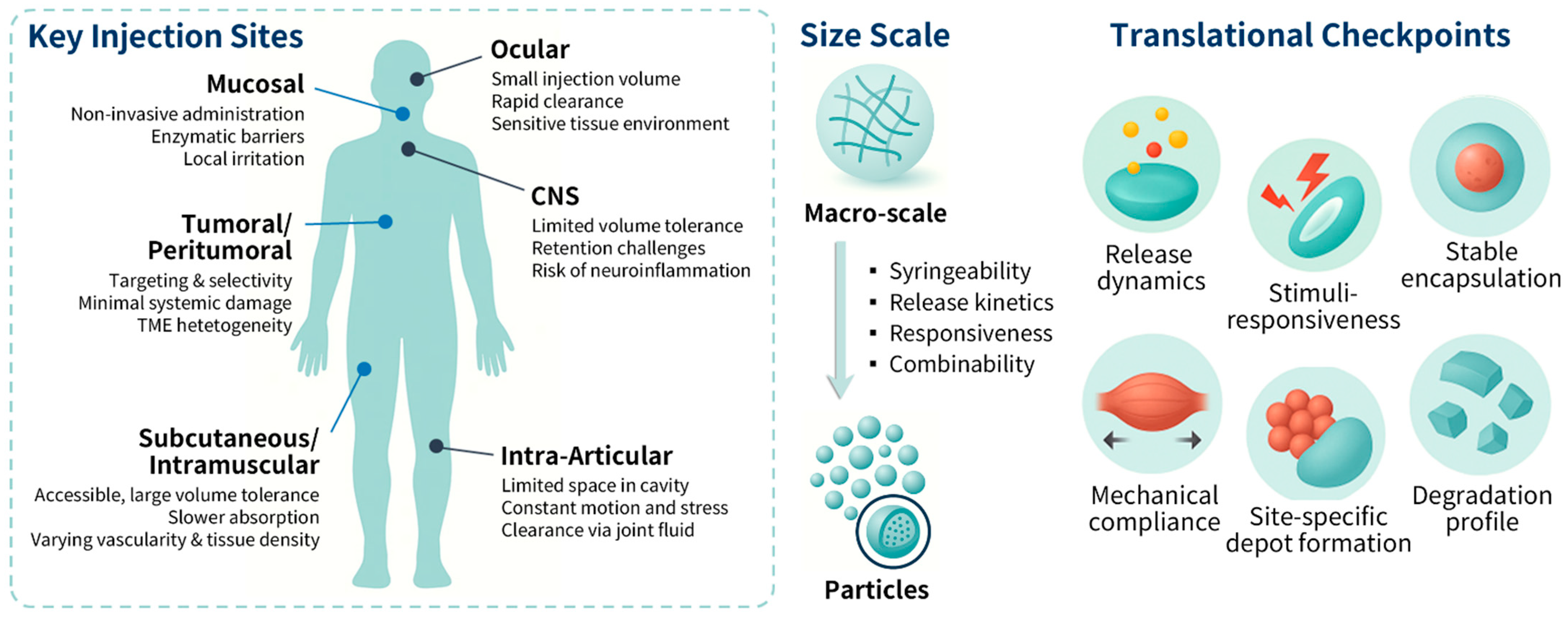
Figure 1. Representative injection sites for hydrogel-based drug delivery systems, and the transition from bulk hydrogel depots to particulate gel and nanoparticle-incorporated gel systems with improved injectability and control, and essential features of optimized formulations such as tunability, responsiveness, encapsulation, and mechanical compliance.
Applsci 15 11599 g001
Figure 2. Illustrations of major injection routes utilized in hydrogel-based drug delivery. (a) Ocular, (b) intra-articular, (c) subcutaneous and intramuscular, and (d) central nervous system routes are shown as representative examples of site-specific administration and localized depot formation [72,73,74,75].
Applsci 15 11599 g002
Table 1. Practical considerations for injectable hydrogel delivery by target site.
Target Site/RouteClinical/Application ContextInjection Volume/Depot LimitsFormulation ConsiderationsRefs.
Ocular
(topical, subconjunctival, intravitreal)
Dry eye, conjunctivitis, uveitis, age-related macular degeneration, diabetic retinopathyLimited ocular retention; intravitreal ≤ 50 µLMucoadhesion for surface retention; vitreous depot stability; optical clarity[38,39,40,41,42]
Intra-articularOsteoarthritis, rheumatoid arthritis, inflammatory joint diseases1–2 mL typical; high shear; rapid synovial turnoverShear-compliant, bioadhesive; tunable degradation; cartilage penetration[43,44,45,46,47]
Subcutaneous (SC)Monoclonal antibodies, insulin analogs, long-acting biologics≤1.5 mL; enzymatic degradation; lymphatic clearanceIn situ gelling depots; resistance to lymphatic clearance; soft mechanics[8,48,49,50,51,52]
Intramuscular (IM)Hormone analogs, antipsychotics, long-acting injectables, vaccines≤5 mL tolerated; dense ECM; anisotropic spreadECM-mimetic stiffness; reproducible distribution; stability under contractile stress[22,53,54,55]
Tumoral/Peritumoral injectionIntratumoral ablation; localized immunotherapy; stromal targetingTumor-size dependent; irregular ECM; proximity to vasculatureBio-responsive hydrogels; sustained retention; stromal-matched mechanics; prevent burst release[56,57,58,59,60,61]
Central Nervous SystemSpinal cord injury, glioma, neuropathic pain, neuroinflammation≤100 µL; reflux risk; rapid CSF turnoverSoft, conformal hydrogels; resist CSF clearance; controlled degradation[62,63,64,65,66]
Mucosal
(nasal, vaginal, rectal)
Anti-infective, anti-inflammatory, vaccine, supportive therapies≤1 mL; clearance by mucus, cilia, or peristalsisMucoadhesion; pH/enzymatic responsiveness; depot formation in confined space[13,67,68]
Disclaimer/Publisher’s Note: The statements, opinions and data contained in all publications are solely those of the individual author(s) and contributor(s) and not of MDPI and/or the editor(s). MDPI and/or the editor(s) disclaim responsibility for any injury to people or property resulting from any ideas, methods, instructions or products referred to in the content.