Article

Human Neural Stem Cells Are More Vulnerable to Damage from Pesticide-Induced Oxidative Stress After Differentiation

1
Clinical Toxicology Research Group, School of Medicine, University of Nottingham, Royal Derby Hospital Centre, Uttoxeter Road, Derby DE22 3DT, UK
2
Faculty of Agricultural Sciences, Sabaragamuwa University of Sri Lanka, Belihuloya 70140, Sri Lanka
3
School of Life Sciences, Faculty of Medicine and Health Sciences, University of Nottingham, Nottingham NG7 2RD, UK
*
Author to whom correspondence should be addressed.
Appl. Sci. 2025, 15(19), 10800; https://doi.org/10.3390/app151910800
Submission received: 18 August 2025 / Revised: 3 October 2025 / Accepted: 5 October 2025 / Published: 8 October 2025
(This article belongs to the Special Issue Exposure Pathways and Health Implications of Environmental Chemicals)

Abstract

Organophosphate (OP) and carbamate pesticides are widely employed in agriculture to facilitate the production of economically viable crops. However, pesticide contamination of food, water, and air leads to undesired human exposure. Neuronal tissue may be particularly vulnerable to pesticide toxicity during periods of neurodevelopment. Hence, this study aimed to investigate the neurotoxicity of three pesticide compounds, namely chlorpyrifos-oxon (CPO), azamethiphos (AZO), and aldicarb, on human neural progenitor cells (hNPCs) and whether toxicity differed between undifferentiated and differentiated stem cells. Undifferentiated and differentiated hNPCs were exposed to these neurotoxicants at concentrations of 0–200 µM for 24 h, and cell viability was evaluated using 3-(4,5 dimethylthiazol-2-yl)-2,5-diphenyl-tetrazolium bromide (MTT) and lactate dehydrogenase (LDH) assays. The impact of the neurotoxicants on cellular bioenergetics was determined by quantifying cellular ATP levels and the production of reactive oxygen species (ROS) using a 2′,7′-dichlorofluorescein diacetate (DCFDA) assay. Concentration–response curves were also generated to measure their relative inhibition of AChE. The neurotoxicants induced concentration-dependent reductions in cell viability (p < 0.0001), cellular ATP levels (p < 0.0001), and the inhibition of AChE (p < 0.0001). Notably, differentiated neurons displayed higher sensitivity than undifferentiated neural stem cells (NSCs), with a toxicity threshold of ≥1 µM. ROS levels were significantly increased (p < 0.0001) following neurotoxicant exposures, more so in differentiated cells, with levels that correlated with cytotoxicity, cell death, and the induction of oxidatively damaged proteins in surviving cells. These findings suggest a central role of oxidative stress and protein oxidation in mediating the neurotoxic effects of pesticide compounds on NSCs. Furthermore, the heightened susceptibility of NSCs to pesticide toxicity after differentiation is indicative of human vulnerability during periods of neurodevelopment.

1. Introduction

The economic viability and profitability of crop production are intrinsically linked to appropriate pest management. One common mechanism of pest control is the use of chemicals as pesticides. However, despite benefits that include economic productivity, the effects of pesticide usage in non-target species, including humans, and the broader impact on the environment and ecosystems, warrant the need for careful consideration of their application and sustainable usage [1,2,3]. Indeed, the long-term and somewhat indiscriminate use of insecticides has led to their ineffectual application and the emergence of populations of target and non-target insect species with evolved insecticide resistance [4]. Furthermore, the relatively low costs, availability, and acute toxicity of pesticides have resulted in extensive accidental and non-accidental human deaths, estimated at over 100,000 per year [5,6].
The recognition of the environmental impact and persistence of xenobiotic pesticides has helped drive a shift from the use of some organochlorines, such as dichlorodiphenyltrichloroethane (DDT), to organophosphates (OPs) and carbamates [7]; chemicals exploited for their broad-spectrum insecticidal activity as well as relatively low production costs. Although OPs and carbamates constitute a diverse chemical grouping, they share a common mode of action through the targeted inhibition of pest acetylcholinesterase (AChE) at synaptic junctions, triggering cholinergic toxicity [7,8,9]. However, the broad clinical sequelae reported for human OP pesticide exposures can be (at least partly) explained by off-target (non-cholinesterase) effects as well as the induction of oxidative stress [9,10,11,12,13,14].
Chlorpyrifos (CPF) (O,O-diethyl O-3,5,6-trichloropyridin-2-yl phosphorothioate) has been extensively utilized as an insecticide since its registration in 1965, but there are concerns regarding its persistence and damage to the environment, as well as potential for human harm [15,16]. Humans are exposed to CPF from dermal, inhalation, and ingestion routes, particularly the latter for non-occupational exposures via residues on foodstuffs [16]. CPF is an AChE inhibitor and, after metabolic desulphuration, forms chlorpyrifos-oxon (CPO), an even more potent AChE inhibitor, as well as other metabolites, including the major chemical-specific production of 3,5,6-trichloro-2-pyridinol (TCPy), often used as a urine biomarker of CPF exposures [8,9,10,11,15,16].
Azamethiphos (AZO) (S-[(6-chloro-2-oxo-1,3-oxazolo [4,5-b]pyridin-3(2H)-yl)methyl] O,O-dimethyl phosphorothioate) is a synthetic OP insecticide, commercially available as an oxon, and does not require biotransformation within the liver. AZO is an AChE inhibitor often applied in aquacultures, particularly for controlling ectoparasites in salmon and trout populations [17]. The environmental impacts of AZO are not well documented, and there are concerns regarding its discharge and toxicity to non-target species [18,19].
Carbamates, such as aldicarb ((EZ)-2-methyl-2-(methylthio)propionaldehyde O-(methylcarbamoyl)oxime), are derivatives of carbamic acid and are often used as insecticides [20]. Like OPs, carbamate pesticides target the inhibition of a pest’s AChE to elicit toxicity, and AChE inhibition can be rapid, albeit more transient than that for OPs [20,21].
Collectively, OP and carbamate pesticides constitute two of the most widely used neurotoxic insecticides. There is an established link between chronic exposure to certain neurotoxicants, such as OP and carbamate pesticides, and neuropsychological, neurodevelopmental and neurobehavioral deficits [21,22,23,24,25,26]. Moreover, prenatal exposure to OPs (including CPF) and carbamates can result in damage to the developing brain and reduced intellectual development and cognitive performance in children [27,28,29]. This could reflect the potential vulnerability of developing and newly differentiated cells to the toxic effects of pesticides [30].
Human neural progenitor ReNcell CX cells (hNPCs) provide a useful model of the developing brain. These cells are an immortalized NPC line derived from the cortex of a 14-week human fetus. They are multipotent, express key neural markers such as Nestin and the transcription factor SOX2 and can differentiate into a mixed co-culture of central nervous system (CNS) neurons, astrocytes, and oligodendrocytes, and form functional synapses [31,32]. hNPCs proliferate rapidly but retain a normal karyotype and are therefore useful for compound screening for developmental neurotoxicity [33].
Previously, we have shown that CPF is toxic to differentiated hNPCs, which is in part mediated by its activation and modulation of N-Methyl-D-Aspartate (NMDA) receptors [34]. Herein, we have extended this work, with an aim to consider further the mechanism of neurotoxicity of three pesticide compounds, CPO, AZA, and aldicarb, to these neural stem cells (NSCs), and whether neurotoxic effects differed between the undifferentiated and differentiated stem cells.

2. Materials and Methods

2.1. Chemicals

Immortalized human cortical neural progenitor cells (ReNcell CX cells, product SCC007) were purchased from Sigma-Aldrich (Poole, UK). Chlorpyrifos (O,O-diethyl O-3,5,6-trichloropyridin-2-yl phosphorothioate, C9H11Cl3NO4P, MW = 344.5 g/mol, purity 97.2–99.1%) was purchased in its oxon form from Greyhound Chromatography (Birkenhead, UK). Azamethiphos (S-[(6-chloro-2-oxo-1,3-oxazolo [4,5-b]pyridin-3(2H)-yl)methyl] O,O-dimethyl phosphorothioate, C9H10ClN2O5PS, MW = 324.7 mg/mol, purity at 95–99.5%) was obtained (as an oxon) from Greyhound Chromatography (Birkenhead, UK). Aldicarb (EZ)-2-methyl-2-(methylthio)propionaldehyde O-(methylcarbamoyl)oxime), C7H14N2O2S, MW = 190.27 g/mol) was from Chem Service Inc. (West Chester, PA, USA) as supplied by Greyhound Chromatography (Birkenhead, UK). Pesticide stock solutions were prepared at a concentration of 50 mM using 99.5% pure ethyl alcohol (product 459844, Sigma-Aldrich, Poole, UK). 3-(4,5 dimethylthiazol-2-yl)-2,5-diphenyl-tetrazolium bromide (MTT) (product M5655) was from Sigma-Aldrich (Poole, UK), along with acetylthiocholine iodide (ATCI) (product A5751) and DTNB (5, 5′-Dithiobis-(2-nitrobenzoic acid)) (product D8130) for AChE assays. Reactive oxygen species (ROS) measurements used 2′7′-dichlorofluorescein diacetate (DCFDA) (product D6883), and 30% H2O2 in H2O (product H1009) as a positive control, both obtained from Sigma-Aldrich (Poole, UK). Cell lysates were prepared using radioimmunoprecipitation assay (RIPA) buffer (Millipore, Burlington, MA, USA) supplemented with protease inhibitors (product 4693124001, Roche) and phosphatase inhibitor cocktail (product P0044, Sigma-Aldrich, Poole, UK). The protein carbonyl assay utilized 2,4-dinitrophenylhydrazine (DNPH) (product D199303) and trichloroacetic acid (TCA) (product T0699), ethyl acetate (product 270989), and guanidine hydrochloride (product 50950) sourced from Sigma-Aldrich (Poole, UK).

2.2. Cell Culture

ReNcell CX cells, which are derived from the cortical region of human fetal brain tissue, were cultured on laminin coated T25 or T75 (130189 or 130190, respectively, ThermoFisher Scientific, Rochester, UK) flasks to expand the ReNcell CX neural stem cell (NSC) population. The ReNCell CX NSC maintenance media (SCM005, EMD Millipore Cooperation, Temecula, CA, USA) was supplemented with freshly added epidermal growth factor (EGF) (20 ng/mL) (GF 001, EMD Millipore Cooperation, Temecula, CA, USA) and fibroblast growth factor-2 (FGF-2) (20 ng/mL) (GF 003, EMD Millipore Cooperation, Temecula, CA, USA). The culture was maintained at 37 °C with 5% CO2 and 95% humidity using passage #2 cells from a freeze-thawed vial. Cell passaging was carried out when the cells reached approximately 60% confluency, by carefully aspirating ReNCell CX NSC maintenance media and rinsing with cold phosphate-buffered saline (PBS) using accutase (SCR 005, EMD Millipore Cooperation, Temecula, CA, USA) for 3 min at 37 °C with an atmosphere of 5% CO2 and 95% humidity. Termination was achieved by the addition of ReNCell CX NSC maintenance media supplemented with 20 ng/mL of EGF and FGF-2. After centrifugation at 300× g for 5 min, the cells were resuspended in ReNcell CX NSC maintenance medium with freshly supplied EGF and FGF-2 (each at 20 ng/mL) and seeded onto laminin-coated cell culture plates. To maintain cell characteristics, all experiments were conducted using cells from passage #4 that were expanded on laminin-coated cell culture ware. Daily observations were made until the cells reached 80% confluence, after which the culture medium was changed every other day with freshly supplied growth factors. For differentiation experiments, the cells were allowed to reach 60% confluency before the removal of growth factors. The differentiation process was continued every other day for a 5-day duration (refer to Supplementary Figure S1). This protocol results in terminal differentiation of the cells, with gene and protein markers that overlap with human embryonic stem cell-derived NSCs [35].

2.3. MTT Assay

The impact of CPO, AZO, and aldicarb (0–200 µM) on undifferentiated and differentiated ReNcell CX cells was assessed using a thiazolyl blue tetrazolium bromide (MTT) reduction assay. Laminin-coated 96-well clear-bottom tissue culture plates were seeded with 1 × 10⁴ cells/well. For undifferentiated or differentiated cells, each agent was administered for 24 h at the specified concentrations by diluting the neurotoxicant into the cell culture medium. Spent medium was removed and replaced with medium containing 10% (w/v) of 5 mg/mL MTT and incubated for 4 h, since this produced an optimal (linear) signal for the human neuroprogenitor cells hNPCs (refer to Supplementary Figure S2). Background control wells were similarly treated with 10% MTT and respective growth media. MTT assays were then processed according to a previous publication [30].

2.4. Lactate Dehydrogenase Assay

The release of extracellular lactate dehydrogenase (LDH) in response to pesticide exposures was assessed using an LDH assay kit (ab65393, Abcam, Cambridge, UK). The method was followed according to a previous publication [30].

2.5. Intracellular ATP Quantification

Intracellular ATP levels were quantified using the ATP Bioluminescence Assay Kit CLS II (product 11 699 695 001, Roche, Mannheim, Germany). Cells were grown in 6-well plates and treated with concentrations known to induce a 10, 20, 50, and 80% loss of cell viability (as determined by MTT assays). ATP levels were quantified by luminescence and interpolation from a standard curve, as detailed previously [30].

2.6. Assessment of Acetylcholinesterase Activity

The inhibition of acetylcholinesterase (AChE) was evaluated through a customized Ellman’s assay [36] adapted for collected cellular homogenates from both undifferentiated and differentiated NSCs after a 24 h treatment with the selected neurotoxicants over the concentration range of 0–1 µM. After neurotoxicant treatments, cells were gathered into ice-cold potassium phosphate buffer, pH 8.0, and then centrifuged at 13,000× g for 5 min at 4 °C. The cellular pellet was preserved and suspended in 1 mL of potassium phosphate buffer, pH 8.0. Subsequently, a 100 µL portion of this suspension was combined in a 1:1 ratio with 5,5′-dithiobis (2-nitrobenzoic acid) (DTNB) and acetylthiocholine iodide (ATCI) substrate to initiate a kinetic reaction, which was monitored at 412 nm with a spectrophotometer (Multiskan Spectrum, ThermoFisher, Vantaa, Finland). Measurements were taken every minute at a temperature of 37 °C, in the dark, over a time span of 10 min. To counteract a drift in absorbance values over time, the corresponding absorbance values from buffer-only blanks were subtracted at each time point for data correction. The absorbance readings for each treatment were standardized based on the mean values of the vehicle control. The activity of AChE was then expressed as a percentage relative to the vehicle control.

2.7. Measurement of Reactive Oxygen Species

The quantification of reactive oxygen species (ROS) in cell extracts was conducted using a 2′,7′-dichlorofluorescein diacetate (DCFDA) assay. NSCs (undifferentiated and differentiated) were plated in 96-well plates with clear bottoms and black walls (165305, ThermoFisher Scientific, Rochester, UK), and then the neurotoxicants were applied for 6 and 24 h at concentrations that reduced cell viability by 10, 20, 50, and 80%, as determined by the MTT assay. DCFDA at a concentration of 50 µM was introduced to each well 30 min prior to the conclusion of the experimental period, allowing time for cellular integration. After the treatment procedures, cells were subjected to two ice-cold PBS washes and then the levels of fluorescence were quantified as described previously [30].

2.8. Cell Lysis and Fractionation

Following the cellular treatments with the pesticide compounds, the NSCs were gently detached and collected into 0.5 mL of radioimmunoprecipitation assay (RIPA) buffer. To achieve thorough mixing, the cell suspension was vigorously vortexed within the RIPA buffer, and then the mixture was passed through a 28-gauge needle 25 times. The resultant homogenates were kept at a temperature of −20 °C until further utilization. After thawing, homogenates were fractionated by differential centrifugation [37], and the crude cytosolic extract was used for protein carbonyl content and oxy-blot analyses.

2.9. Protein Quantification

The determination of protein concentrations within cell cytosolic extracts was based on a modified Lowry assay [38], using bovine serum albumin (BSA) protein standards (5000206, Bio-Rad, Hertfordshire, UK), as described previously [30].

2.10. Quantitation of Protein Carbonyl Content and Characterisation of Oxidatively Damaged Proteins

The quantitation of the levels of cellular protein carbonyl content (PCC) in cytosolic extracts generated from undifferentiated and differentiated NSCs after neurotoxicant challenges was conducted according to a previous publication [30]. Oxidatively damaged proteins were resolved by gel electrophoresis and detected using an OxyBlot Protein Oxidation Detection Kit (S71590, Millipore, Temecula, CA, USA), with immunoreactive bands captured with a ChemiDoc MP imager (BioRad, Hertfordshire, UK), configured for auto-exposure readings to ensure signal linearity [30]. Representative blots are included in the figures to illustrate the research findings.

2.11. Statistical Analysis

Statistical analysis was conducted utilizing GraphPad Prism 9.2.0 (GraphPad Prism, San Diego, CA, USA). Concentration–response curves were constructed to determine the concentrations that produced a 50% inhibition (IC50) using a non-linear regression curve with lines of optimal fit. To assess the impact of treatments, comparisons were made between control and treated groups using either one-way analysis of variance (ANOVA) or two-way ANOVA, along with Dunnett’s multiple comparison test for the former and Tukey’s multiple comparisons for the latter. Statistical significance was established from a threshold p-value of 0.05.

3. Results

Assessment of the Neurotoxicity of Chlorpyrifos-Oxon, Azamethiphos, and Aldicarb to Human Neural Progenitor Cells

Cultured undifferentiated and differentiated human neural progenitor cells (hNPCs) were exposed to the pesticide compounds, chlorpyrifos-oxon (CPO), azamethiphos-oxon (AZO), and aldicarb, over a concentration range of 0–200 µM for 24 h, and changes in cell morphology and cell number were observed using bright-field, phase-contrast microscopy and quantified using an MTT assay. At relatively high neurotoxicant concentrations, cells adopted a rounded morphology, particularly following treatment with CPO or AZO, and there was a decline in visible cell number that correlated with increasing neurotoxicant concentration (refer to Supplementary Figures S3 and S4). Remarkably, even at the highest concentration examined (200 µM), some of the stem cells were still resilient to the toxicological insult and survived; however, neuronal connections were progressively reduced with increasing neurotoxicant concentrations, with a reduction in neurite arborization for differentiated cells (refer to Supplementary Figure S3). Exposure to the pesticide compounds also diminished cell viability in a concentration-dependent trend. Notably, compound toxicity was more pronounced in differentiated hNPCs, which displayed relatively heightened vulnerability at lower agent concentrations (Figure 1A,B). A non-linear regression plot of neurotoxicity versus agent concentration was used to determine the concentrations that produced a 50% inhibition of cell viability (IC50 values), and these have been included in Table 1. The potency of toxicity in undifferentiated hNPCs was CPO > aldicarb > AZO, whereas in differentiated hNPCs, it was CPO > AZO > aldicarb. All compounds were more neurotoxic to newly differentiated hNPCs with lower IC50 values (refer to Table 1), and although morphological changes were not easily observed after an incubation at 1 µM (of neurotoxicant), this represented the threshold concentration for reduced viability for both hNPC phenotypes and with all neurotoxic agents; indicative of the acute sensitivity of hNPCs to a toxicological insult.
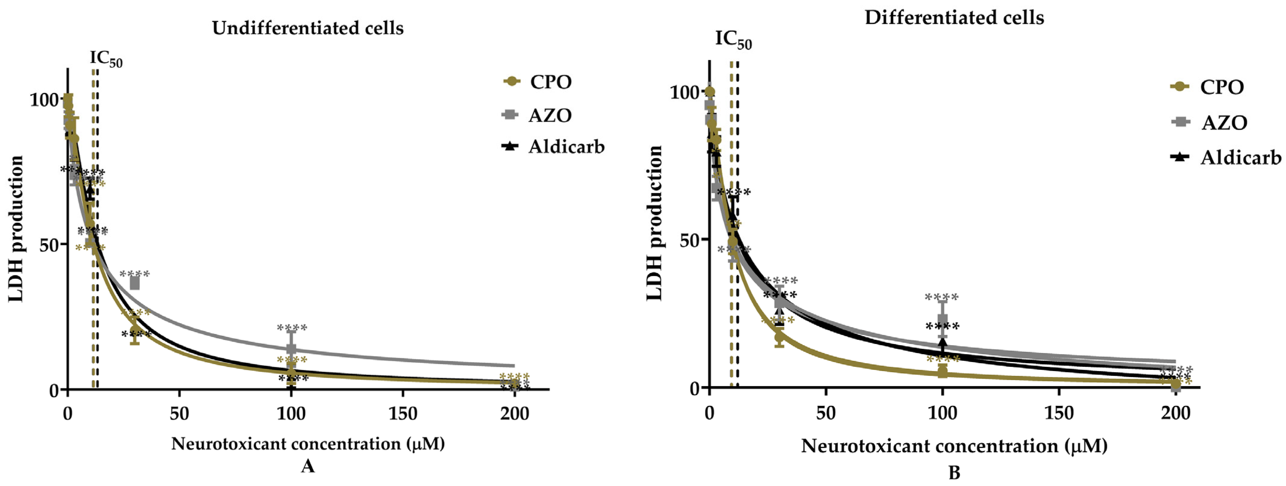
As an alternative and independent approach to assess neurotoxicant effects on cell viability, the liberation of extracellular lactate dehydrogenase (LDH) activity was measured after a 24 h exposure to the pesticide compounds. Both undifferentiated and differentiated hNPCs displayed a concentration-dependent decrease in cell viability after incubation with the toxic agents (Figure 2A,B). Like the MTT assays, the threshold for loss of cell viability was a concentration of 1 µM, and the OPs were more toxic than aldicarb to both cell phenotypes and more neurotoxic to differentiated hNPCs, as indicated by lower IC50 values (refer to Table 1). The potency of toxicity in undifferentiated hNPCs was CPO = AZO > aldicarb, whereas in differentiated hNPCs, it was CPO > AZO > aldicarb (refer to Table 1).
The impact of the neurotoxicants on the cellular bioenergetics of undifferentiated and differentiated hNPCs was assessed by quantification of intracellular ATP levels. In response to a 24 h exposure to the pesticide compounds, there was a progressive decline in ATP levels that was concentration-dependent and mirrored the concentration–response curves observed for MTT and LDH assays (Figure 3A,B). The neurotoxicant-induced decreases in ATP levels yielded IC50 values comparable to those derived from MTT and LDH assays (refer to Table 1).
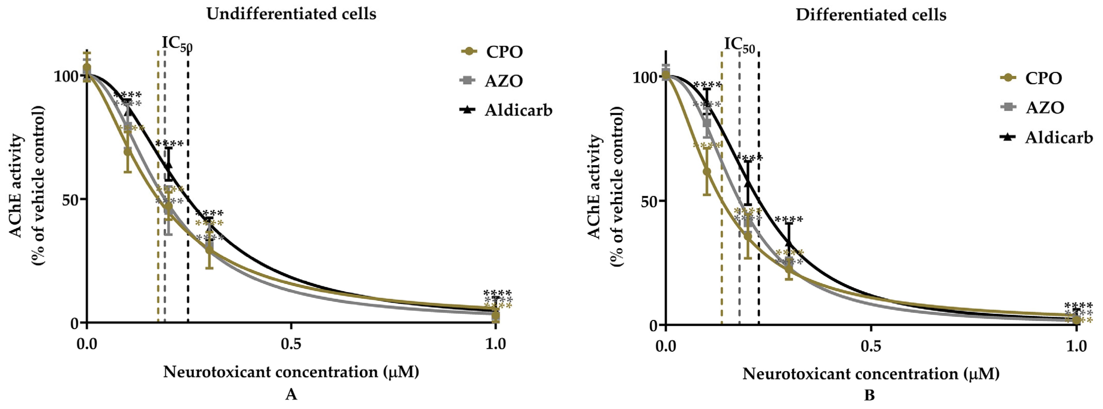
The ability of the pesticide compounds to inhibit AChE within undifferentiated and differentiated hNPCs was examined using a modified Ellman’s assay. After a 24 h incubation of hNPCs with the neurotoxicants, there was a concentration-dependent inhibition of AChE with compound efficacy in the low micromolar range (Figure 4). At a neurotoxicant concentration of 0.1 µM, CPO, AZO, and aldicarb all induced significant (p < 0.0001) inhibition of AChE in undifferentiated cells by approximately 31, 21, and 15%, respectively. Similarly, the application of 0.1 µM of the neurotoxicants (CPO, AZO, and aldicarb) to differentiated hNPCs triggered a higher AChE inhibition (than in undifferentiated cells) with an approximate 39, 39, and 25% inhibition of AChE, respectively. At higher neurotoxicant concentrations, AChE inhibition escalated and then plateaued over the concentration range of 0.3–1 µM, indicating near-complete inhibition at higher concentrations. The IC50 values, derived from concentration–response curves for both cell types, were consistently in the nanomolar range (100–230 nM) for all three pesticides, confirming their potency for incapacitating AChE activity (refer to Table 1).
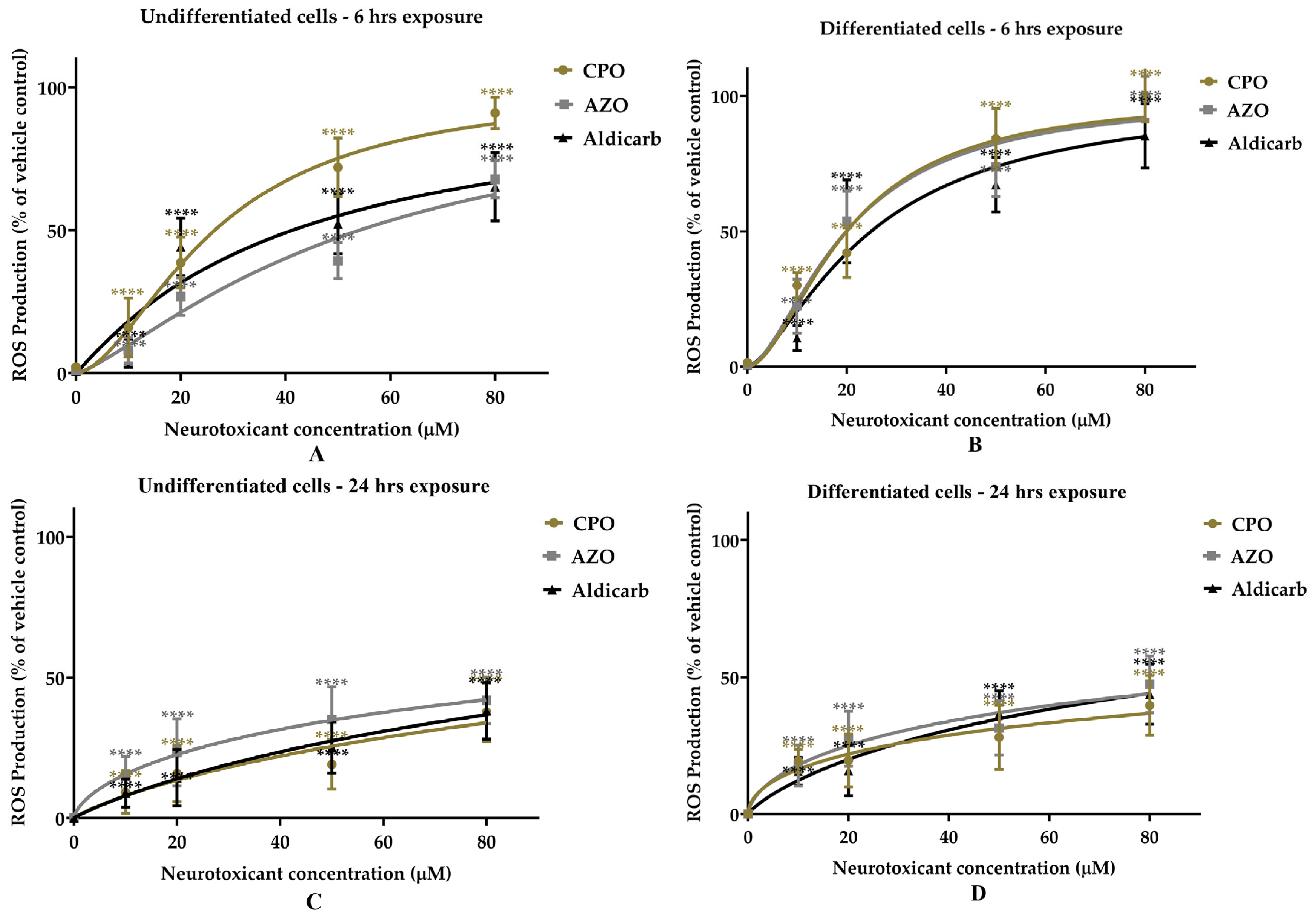
To consider an alternative mechanism of cell toxicity to that of AChE inhibition, the production of reactive oxygen species (ROS) following the application of CPO, AZO, or aldicarb to hNPCs was quantified using a DCFDA assay after 6 and 24 h neurotoxicant exposures. To establish a benchmark for cellular redox stress, the levels of ROS were measured relative to those produced by 500 µM H2O2, as a positive control cellular redox stressor. After the 6 h pesticide compound exposures, ROS levels increased in both undifferentiated and differentiated hNPCs. This rise reached a plateau at the concentration range of 50–80 µM (Figure 5A,B). Similarly, after 24 h exposure to the compounds, the ROS levels ascended proportionally with neurotoxicant concentrations, capping off at 50–80 µM (Figure 5C,D). Differentiated hNPCs exhibited elevated ROS levels compared to undifferentiated cells, particularly at higher pesticide compound concentrations (AZO and aldicarb) and at 6 h post-exposure. Elevated ROS levels were still detectable after 24 h, but were lower than those observed after 6 h. There was a positive correlation between the concentrations of pesticide compounds that induced reduced cell viability and the associated production of ROS after 6 and 24 h (Supplementary Table S2). CPO was the most extensive redox stressor with ROS production in the order CPO > AZO > aldicarb (Figure 5).
The level of oxidatively damaged proteins in response to neurotoxicant exposures was determined from a quantification of total protein carbonyl content (PCC). The neurotoxicants increased PCC in undifferentiated and differentiated hNPCs in a concentration-dependent fashion (Figure 6A,B). Consistent with the relatively high levels of ROS, CPO also induced the highest increase in PCC in hNPCs, and to levels that were higher in differentiated cells than in undifferentiated cells.
In order to characterize the oxidatively damaged proteins, protein homogenates from CPO, AZO, and aldicarb treated hNPCs were prepared and resolved by one-dimensional polyacrylamide gel electrophoresis, followed by probing by oxy-blotting (Figure 7). The levels of detectable protein bands with oxidatively damaged proteins were positively correlated with the concentration of neurotoxicant, with higher levels in differentiated cells. The most prominent band present in the oxyblots was at approximately 50–55 kDa, and this was common to all pesticide treatments.

4. Discussion

Experimental evidence has supported the posit that the developing brain may be particularly vulnerable to the toxic, damaging effects of xenobiotics such as pesticides and their metabolites [22,23,24,25,26,27,28,29,30]. Hence, this study investigated the neurotoxic properties of the bioactive forms of two OP pesticides, CPO and AZO, and the carbamate pesticide, aldicarb, on a population of human cortical neuronal progenitor cells. These multipotent cells can differentiate into a range of cell types that are representative of brain tissue, including neurons, astrocytes, and oligodendrocytes, and therefore provide a useful model of mixed brain cell populations that undergo differentiation during neurodevelopment [31,32]. Cytotoxic effects on the NSCs were observed, which were pesticide compound concentration-dependent and triggered altered cell morphology and reduced cell viability. The neurotoxicants induced damage to stem cell bioenergetics with depleted ATP production. The toxicity of the pesticide compounds was mediated by cholinesterase and non-cholinesterase mechanisms, with a pesticide induction of ROS and production and accumulation of oxidatively damaged proteins, particularly in newly differentiated cells. Noteworthy, was that the threshold for reduced cell viability (1 µM) was approximately 10-times lower than that for similarly treated SHSY-5Y cells, a neuroblastoma cell line often used for neurotoxicity studies [30,39]. Similarly, the IC50 values for reduced cell viability in response to these pesticide compounds for differentiated NSCs were 53–61% lower than those for differentiated SHSY-5Y cells (as determined by MTT assays) [30]; indicative of a higher vulnerability of NSCs to pesticide neurotoxicity.
Pesticide compound toxicity was initially established using MTT assays (Figure 1), a measure of cellular metabolic activity often used as a surrogate for cell viability [40,41]. These studies showed that OPs were more neurotoxic than aldicarb and were in the order CPO > AZO > aldicarb for differentiated hNPCs. Furthermore, differentiated hNPCs were more prone to neurotoxicant-induced cell death than undifferentiated hNPCs. This cell viability data was supported by LDH assays and by quantitation of cellular ATP levels (Figure 2 and Figure 3), alternative methodologies used to provide cell viability data [40,41], and these assays generated comparable IC50 values (refer to Table 1).
The acute neurotoxicity of OP and carbamate pesticides stems from their targeted inhibition of AChE, which results in a cholinergic crisis in the target pest. The three pesticide compounds all exhibited potent inhibition of endogenous AChE in undifferentiated and differentiated hNPCs (Figure 4), with similar IC50 values, and all lower than 250 nM (refer to Table 1). These IC50 values are lower (more potent) than eserine (physostigmine), a documented inhibitor of human AChE [42], and generally comparable to the transient cholinesterase inhibitor drugs, such as donepezil, used as a pharmacotherapy for Alzheimer’s disease [42,43]. We also consider non-cholinesterase toxicity and report that the pesticide compounds induced the production of deleterious levels of ROS in stem cells, which in part contributed to reduced cell viability (Figure 5). Furthermore, pesticide-induced oxidative stress resulted in the production and accumulation of oxidatively damaged (carbonylated) proteins (Figure 6 and Figure 7). The most prominent carbonylated protein band had a denatured molecular weight of 50–55 kDa (Figure 7). The levels of this protein band were elevated in response to increasing pesticide compound concentration and were also higher in differentiated NSCs than in undifferentiated ones. The vulnerability of this protein band to oxidative damage was observed after neurotoxicant treatment of SHSY-5Y cells [30] and has been proposed to be tubulin(s) [30], in keeping with other studies that have detailed the susceptibility of microtubular proteins to OP pesticide binding and damage, and their increased expression during neuronal cell differentiation [44,45,46,47]. Furthermore, our preliminary studies using SHSY-5Y cell proteins resolved by two-dimensional polyacrylamide gel electrophoresis after treatment with CPO identified several protein bands with corresponding carbonylated versions, and these included tubulin β-chain and tubulin α1B-chain (our unpublished data, results not included) consistent with the proposal that tubulin is a major target of oxidative damage.
The functional implications for oxidatively damaged cells that survive the pesticide insult have not been fully considered. In vitro studies have shown that prolonged exposure of rat hippocampal slices to CPO (0.1–10 µM) resulted in a progressive decline in cell viability and impaired tubulin polymerization [48]. Presumably, if OP and carbamate pesticides act as broad cytoskeleton-disrupting agents, they could contribute to a number of pathological conditions [46]. Moreover, in the context of neurodevelopment, our studies indicate exquisite sensitivity of multipotent cells to the neurotoxic effects of pesticide compounds, with reduced damage and viability from exposures of 1 µM (in vitro), but context is needed to translate these findings to pesticide exposures in vivo. However, the circulatory concentrations of OPs and carbamates encountered in vivo are difficult to predict for generalized non-occupational, chronic exposures. For mothers living in an agricultural community (Salinas Valley, California, USA) and therefore with proximity to pesticide exposures, their plasma levels at delivery varied extensively, and ranged from undetectable to ≈5 µM for CPF and from undetectable to only ≈1.6 nM for diazinon (another OP pesticide) [49]. From a combination of predictive measurements and quantitation of urinary metabolites, pregnant women in France were estimated to have similar levels to fetal levels for CPF exposures, but with brain levels predicted to be 10-fold higher than those for blood [50]. This cohort data was generated before CPF was subjected to a European Union (EU) ban (in 2020), but CPF is still applied in other agriculturally producing countries, resulting in CPF residues in food [51], and collectively, OPs and carbamates remain in widespread use throughout the EU and worldwide.
Ultimately, our data shows that redox stress is a common mechanism of neuronal cellular damage by two OPs and the carbamate, aldicarb, and is concurrent with inhibition of AChE. Hence, these cholinesterase and non-cholinesterase mechanisms (including redox stress, excitotoxicity, and engagement with secondary targets [9,10,11,12,13,14,34]), likely contribute to the potential for detrimental health after acute or chronic pesticide exposures. This highlights the need for commercially available OP and carbamate pesticides to undergo rigorous neurodevelopmental risk assessment, which at present may be absent and/or require more independent review, and to limit the possibility of study under-reporting [52,53,54].
At present, we can only speculate on the rationale for the heightened vulnerability of NSCs to the toxic effects of these pesticide compounds when compared with undifferentiated or differentiated SHSY-5Y cells. This could reflect that these are multipotent cells that develop into mixed neuronal populations, with unequal sensitivity to pesticide compounds. Differentiation of the NSCs could increase the expression of AChE, as observed in SHSY-5Y cells [55]. However, in contrast to an expected increased vulnerability of SHSY-5Y cells to toxic challenge from OP compounds, differentiated SHSY-5Y cells were less sensitive to toxicological challenge, and this was proposed to be due to non-cholinesterase effects of the OP compounds [55]. There may be altered expression of other proteins vulnerable to neurotoxic effects and redox stress, such as the 50–55 kDa protein band (Figure 7). Assuming this is tubulin(s), it has a central effect on cellular activity as well as mitosis, which was inhibited in primary progenitors in mice after prenatal CPF exposure [56]. Furthermore, the protein targets for pesticide adduction vary across tissues and the pesticide applied [10,11], and there are differences between the levels of pesticide-induced redox stress within brain tissues [57]. Hence, presumably, there will be differential pesticide effects within the mixed NSC population, and this will be considered in future studies. The neurotoxic effects and induction of redox stress may also relate to pesticide-induced damage of mitochondria [58]. This can promote the liberation of damaging and deleterious ROS [13,58,59]. Furthermore, redox stress can be intrinsically linked to the induction of an inflammatory response, leading to the release of inflammatory mediators and cellular damage [13,59,60].
Human NPCs provide a useful basis for studying the effects of neurotoxicants on the CNS, since these neural stem cells are capable of differentiation into a mixed population of neurons, astrocytes, and oligodendrocytes. However, there are limitations with this in vitro approach. The range of concentrations and 24 h duration is only reflective of acute exposures and intoxications, including those likely to be observed in overdose. For example, a human subject who experienced pesticide poisoning with chlorpyrifos had serum and gastric concentrations of 5.3 and 9.4 µg/mL of unmetabolized chlorpyrifos (approximately 15 and 27 µM, respectively) [61]. For another OP pesticide, the levels of parathion in a toxic overdose ranged from 0.21 to 19.64 mg/L (approximately 0.7 µM to 67 µM, respectively) [62]. Since we only consider acute studies, this reduces the applicability of the results and their translation to the repeated, chronic low-level exposures that typify the majority of human pesticide encounters. Furthermore, in vitro incubations of the NSCs with pesticides provide no indication of relative take-up or storage of the agent, and under conditions devoid of an intact blood–brain barrier (BBB). Nevertheless, an advantage of this approach compared to that of a continuous (cancer) cell line is that the mixed cell populations are more representative of brain tissue, and one that can undergo acute differentiation to model the impact of pesticides on neurodevelopmental changes.
In summary, this study considered the neurotoxic impact of pesticide compounds and their metabolites on multipotent stem cells and highlights their heightened vulnerability to pesticide toxicity. This sensitivity of NSCs has health implications for low-level neurotoxicant exposures that could elicit cellular damage below the threshold of cell death but with lasting effects on cellular functionality. Hence, there is a need to consider more broadly the potential toxic mechanisms induced by xenobiotics in order to provide a solid scientific foundation for evidence-based decision-making in pesticide regulation, risk assessment, and public health policy. Furthermore, this study highlights the need to consider more sustainable and environmentally friendly next-generation pest management strategies for ensuring global food security. Lastly, the potential for acute and potentially long-lasting health effects from the non-cholinesterase mechanisms of OPs and carbamates underscores the requirements for therapeutic interventions to tackle intentional and non-intentional poisonings.

Supplementary Materials

The following supporting information can be downloaded at: https://www.mdpi.com/article/10.3390/app151910800/s1, Figure S1: Morphological and protein expression characteristics of undifferentiated and differentiated ReNcell CX cells; Figure S2: Determination of the correlation between the optical density signal and cell seeding density for the MTT assay for human neural progenitor cells; Figure S3: The neurotoxic effects of CPO, AZO, and aldicarb to undifferentiated hNPCs examined using phase-contrast microscopy; Figure S4: The neurotoxic effects of CPO, AZO, and aldicarb to differentiated hNPCs examined using phase-contrast microscopy; Figure S5: Original Western (oxy-)blots of CPO, AZO, and aldicarb induction of carbonylated proteins in hNPCs; Table S1: MTT inhibition concentrations interpolated from concentration-response curves; Table S2: Correlation between pesticide concentrations used to induce reduced cell viability and ROS generation after 24 h in hNPCs.

Author Contributions

Conceptualization, A.W., I.R.M. and W.G.C.; methodology, A.W., B.W. and A.K.; validation, A.W., B.W. and A.K.; formal analysis, A.W., B.W. and A.K.; investigation, A.W., B.W. and A.K.; resources, I.R.M. and W.G.C.; writing—original draft preparation, A.W. and W.G.C.; writing—review and editing, A.W., B.W., A.K., I.R.M. and W.G.C.; visualization, A.W. and W.G.C.; supervision, I.R.M. and W.G.C.; project administration, I.R.M. and W.G.C.; funding acquisition, A.W. and W.G.C. All authors have read and agreed to the published version of the manuscript.

Funding

This research was funded by a UK Foreign, Commonwealth and Development Office (FCDO) Commonwealth Scholarship Commission (UK) PhD award (grant number LKCS-2016-678) to A.W.M. This research was also funded by “Funds for Women Graduates,” (Ref: GA-00171) to A.W.M.

Data Availability Statement

Data that supports this work are available as Supplementary Data files or directly by contacting the first author.

Conflicts of Interest

The authors declare no conflict of interest.

References

  1. Wilson, C.; Tisdell, C. Why farmers continue to use pesticides despite environmental, health and sustainability costs. Ecol. Economics 2001, 39, 449–462. [Google Scholar] [CrossRef]
  2. Popp, J.; Pető, K.; Nagy, J. Pesticide productivity and food security. A review. Agron. Sustain. Dev. 2013, 33, 243–255. [Google Scholar] [CrossRef]
  3. Kariyanna, B.; Senthil-Nathan, S.; Vasantha-Srinivasan, P.; Reddy, B.V.S.; Krishnaiah, A.; Meenakshi, N.H.; Han, Y.S.; Karthi, S.; Chakravarthy, A.K.; Park, K.B. Comprehensive insights into pesticide residue dynamics: Unraveling impact and management. Chem. Biol. Technol. Agric. 2024, 11, 182. [Google Scholar] [CrossRef]
  4. Pu, J.; Chung, H. New and emerging mechanisms of insecticide resistance. Curr. Opin. Insect Sci. 2024, 63, 101184. [Google Scholar] [CrossRef]
  5. Mew, E.J.; Padmanathan, P.; Konradsen, F.; Eddleston, M.; Chang, S.S.; Phillips, M.R.; Gunnell, D. The global burden of fatal self-poisoning with pesticides 2006-15: Systematic review. J Affect. Disord. 2017, 219, 93–104. [Google Scholar] [CrossRef]
  6. Moebus, S.; Boedeker, W. Case Fatality as an Indicator for the Human Toxicity of Pesticides-A Systematic Scoping Review on the Availability and Variability of Severity Indicators of Pesticide Poisoning. Int. J. Environ. Res. Public Health 2021, 18, 8307. [Google Scholar] [CrossRef] [PubMed]
  7. Jayaraj, R.; Megha, P.; Sreedev, P. Organochlorine pesticides, their toxic effects on living organisms and their fate in the environment. Interdiscip. Toxicol. 2016, 9, 90–100. [Google Scholar] [CrossRef] [PubMed]
  8. Colovic, M.B.; Krstic, D.Z.; Lazarevic-Pasti, T.D.; Bondzic, A.M.; Vasic, V.M. Acetylcholinesterase Inhibitors: Pharmacology and Toxicology. Curr. Neuropharmacol. 2013, 11, 315–335. [Google Scholar] [CrossRef] [PubMed]
  9. Costa, L.G. Organophosphorus Compounds at 80: Some Old and New Issues. Toxicol. Sci. 2018, 162, 24–35. [Google Scholar] [CrossRef]
  10. Carter, W.G.; Tarhoni, M.; Rathbone, A.J.; Ray, D.E. Differential protein adduction by seven organophosphorus pesticides in both brain and thymus. Hum. Exp. Toxicol. 2007, 26, 347–353. [Google Scholar] [CrossRef]
  11. Carter, W.G.; Tarhoni, M.H.; Ray, D.E. Analytical approaches to investigate protein-pesticide adducts. J. Chromatogr. B Anal. Technol. Biomed. Life Sci. 2010, 878, 1312–1319. [Google Scholar] [CrossRef]
  12. Terry, A.V., Jr. Functional consequences of repeated organophosphate exposure: Potential non-cholinergic mechanisms. Pharmacol. Ther. 2012, 134, 355–365. [Google Scholar] [CrossRef]
  13. Sule, R.O.; Condon, L.; Gomes, A.V. A Common Feature of Pesticides: Oxidative Stress-The Role of Oxidative Stress in Pesticide-Induced Toxicity. Oxid. Med. Cell Longev. 2022, 2022, 5563759. [Google Scholar] [CrossRef] [PubMed]
  14. Chen, Y.; Yang, Z.; Nian, B.; Yu, C.; Maimaiti, D.; Chai, M.; Yang, X.; Zang, X.; Xu, D. Mechanisms of Neurotoxicity of Organophosphate Pesticides and Their Relation to Neurological Disorders. Neuropsychiatr. Dis. Treat. 2024, 20, 2237–2254. [Google Scholar] [CrossRef] [PubMed]
  15. Kumar, P.; Arshad, M.; Gacem, A.; Soni, S.; Singh, S.; Kumar, M.; Yadav, V.K.; Tariq, M.; Kumar, R.; Shah, D.; et al. Insight into the environmental fate, hazard, detection, and sustainable degradation technologies of chlorpyrifos-an organophosphorus pesticide. Environ. Sci. Pollut. Res. Int. 2023, 30, 108347–108369. [Google Scholar] [CrossRef] [PubMed]
  16. Rahman, H.U.U.; Asghar, W.; Nazir, W.; Sandhu, M.A.; Ahmed, A.; Khalid, N. A comprehensive review on chlorpyrifos toxicity with special reference to endocrine disruption: Evidence of mechanisms, exposures and mitigation strategies. Sci. Total Environ. 2021, 755, 142649. [Google Scholar] [CrossRef]
  17. Barisic, J.; Cannon, S.; Quinn, B. Cumulative impact of anti-sea lice treatment (azamethiphos) on health status of Rainbow trout (Oncorhynchus mykiss, Walbaum 1792) in aquaculture. Sci. Rep. 2019, 9, 16217. [Google Scholar] [CrossRef]
  18. Couillard, C.M.; Burridge, L.E. Sublethal exposure to azamethiphos causes neurotoxicity, altered energy allocation and high mortality during simulated live transport in American lobster. Ecotoxicol. Environ. Saf. 2015, 115, 291–299. [Google Scholar] [CrossRef]
  19. Montory, J.A.; Cubillos, V.M.; Chaparro, O.R.; Gebauer, P.; Lee, M.R.; Ramírez-Kuschel, E.; Paredes-Molina, F.; Lara-Sandoval, V.; Cumillaf, J.P.; Salas-Yanquin, L.P.; et al. The Interactive Effects of the Anti-Sea Lice Pesticide Azamethiphos and Temperature on Oxidative Damage and Antioxidant Responses in the Oyster Ostrea chilensis. Antioxidants 2024, 13, 737. [Google Scholar] [CrossRef]
  20. Mdeni, N.L.; Adeniji, A.O.; Okoh, A.I.; Okoh, O.O. Analytical Evaluation of Carbamate and Organophosphate Pesticides in Human and Environmental Matrices: A Review. Molecules 2022, 27, 618. [Google Scholar] [CrossRef]
  21. Risher, J.F.; Mink, F.L.; Stara, J.F. The toxicologic effects of the carbamate insecticide aldicarb in mammals: A review. Environ. Health Perspect. 1987, 72, 267–281. [Google Scholar] [CrossRef] [PubMed]
  22. Ross, S.M.; McManus, I.C.; Harrison, V.; Mason, O. Neurobehavioral problems following low-level exposure to organophosphate pesticides: A systematic and meta-analytic review. Crit. Rev. Toxicol. 2013, 43, 21–44. [Google Scholar] [CrossRef] [PubMed]
  23. Roldan-Tapia, L.; Nieto-Escamez, F.A.; del Aguila, E.M.; Laynez, F.; Parron, T.; Sanchez-Santed, F. Neuropsychological sequelae from acute poisoning and long-term exposure to carbamate and organophosphate pesticides. Neurotoxicol. Teratol. 2006, 28, 694–703. [Google Scholar] [CrossRef] [PubMed]
  24. Grandjean, P.; Landrigan, P.J. Neurobehavioural effects of developmental toxicity. Lancet Neurol. 2014, 13, 330–338. [Google Scholar] [CrossRef]
  25. Burke, R.D.; Todd, S.W.; Lumsden, E.; Mullins, R.J.; Mamczarz, J.; Fawcett, W.P.; Gullapalli, R.P.; Randall, W.R.; Pereira, E.F.R.; Albuquerque, E.X. Developmental neurotoxicity of the organophosphorus insecticide chlorpyrifos: From clinical findings to preclinical models and potential mechanism. J. Neurochem. 2017, 142, 162–177. [Google Scholar] [CrossRef]
  26. Bouchard, M.F.; Chevrier, J.; Harley, K.G.; Kogut, K.; Vedar, M.; Calderon, N.; Trujillo, C.; Johnson, C.; Bradman, A.; Barr, D.B.; et al. Prenatal exposure to organophosphate pesticides and IQ in 7-year-old children. Environ. Health Perspect. 2011, 119, 1189–1195. [Google Scholar] [CrossRef]
  27. Rauh, V.A.; Perera, F.P.; Horton, M.K.; Whyatt, R.M.; Bansal, R.; Hao, X.; Liu, J.; Barr, D.B.; Slotkin, T.A.; Peterson, B.S. Brain anomalies in children exposed prenatally to a common organophosphate pesticide. Proc. Natl. Acad. Sci. USA 2012, 109, 7871–7876. [Google Scholar] [CrossRef]
  28. Rauh, V.; Arunajadai, S.; Horton, M.; Perera, F.; Hoepner, L.; Barr, D.B.; Whyatt, R. Seven-year neurodevelopmental scores and prenatal exposure to chlorpyrifos, a common agricultural pesticide. Environ. Health Perspect. 2011, 119, 1196–1201. [Google Scholar] [CrossRef]
  29. Rowe, C.; Gunier, R.; Bradman, A.; Harley, K.G.; Kogut, K.; Parra, K.; Eskenazi, B. Residential proximity to organophosphate and carbamate pesticide use during pregnancy, poverty during childhood, and cognitive functioning in 10-year-old children. Environ. Res. 2016, 150, 128–137. [Google Scholar] [CrossRef]
  30. Mudyanselage, A.W.; Wijamunige, B.C.; Kocon, A.; Carter, W.G. Differentiated Neurons Are More Vulnerable to Organophosphate and Carbamate Neurotoxicity than Undifferentiated Neurons Due to the Induction of Redox Stress and Accumulate Oxidatively-Damaged Proteins. Brain Sci. 2023, 13, 728. [Google Scholar] [CrossRef]
  31. Donato, R.; Miljan, E.A.; Hines, S.J.; Aouabdi, S.; Pollock, K.; Patel, S.; Edwards, F.A.; Sinden, J.D. Differential development of neuronal physiological responsiveness in two human neural stem cell lines. BMC Neurosci. 2007, 8, 36. [Google Scholar] [CrossRef] [PubMed]
  32. La Cruz, B.M.; Markus, R.; Malla, S.; Haig, M.I.; Gell, C.; Sang, F.; Bellows, E.; Sherif, M.A.; McLean, D.; Lourdusamy, A.; et al. Modifying the m6A brain methylome by ALKBH5-mediated demethylation: A new contender for synaptic tagging. Mol. Psychiatry 2021, 26, 7141–7153. [Google Scholar] [CrossRef] [PubMed]
  33. Breier, J.M.; Radio, N.M.; Mundy, W.R.; Shafer, T.J. Development of a high-throughput screening assay for chemical effects on proliferation and viability of immortalized human neural progenitor cells. Toxicol. Sci. 2008, 105, 119–133. [Google Scholar] [CrossRef] [PubMed]
  34. Sherif, M.A.; Carter, W.G.; Mellor, I.R. Chlorpyrifos Acts as a Positive Modulator and an Agonist of N-Methyl-D-Aspartate (NMDA) Receptors: A Novel Mechanism of Chlorpyrifos-Induced Neurotoxicity. J. Xenobiot. 2025, 15, 12. [Google Scholar] [CrossRef]
  35. Oh, J.H.; Jung, C.R.; Lee, M.O.; Kim, J.; Son, M.Y. Comparative analysis of human embryonic stem cell-derived neural stem cells as an in vitro human model. Int. J. Mol. Med. 2018, 41, 783–790. [Google Scholar] [CrossRef]
  36. Ellman, G.L.; Courtney, K.D.; Andres, V., Jr.; Featherstone, R.M. A new and rapid colorimetric determination of acetylcholinesterase activity. Biochem. Pharmacol. 1961, 7, 88–95. [Google Scholar] [CrossRef]
  37. Erdozain, A.M.; Morentin, B.; Bedford, L.; King, E.; Tooth, D.; Brewer, C.; Wayne, D.; Johnson, L.; Gerdes, H.K.; Wigmore, P.; et al. Alcohol-Related Brain Damage in Humans. PLoS ONE 2014, 9, e93586. [Google Scholar] [CrossRef]
  38. Lowry, O.H.; Rosebrough, N.J.; Farr, A.L.; Randall, R.J. Protein measurement with the Folin phenol reagent. J. Biol. Chem. 1951, 193, 265–275. [Google Scholar] [CrossRef]
  39. Lopez-Suarez, L.; Awabdh, S.A.; Coumoul, X.; Chauvet, C. The SH-SY5Y human neuroblastoma cell line, a relevant in vitro cell model for investigating neurotoxicology in human: Focus on organic pollutants. Neurotoxicology 2022, 92, 131–155. [Google Scholar] [CrossRef]
  40. Riss, T.L.; Moravec, R.A.; Niles, A.L.; Duellman, S.; Benink, H.A.; Worzella, T.J.; Minor, L. Cell Viability Assays. In Assay Guidance Manual; Markossian, S., Grossman, A., Baskir, H., et al., Eds.; Eli Lilly & Company and the National Center for Advancing Translational Sciences: Bethesda, MD, USA, 2004. Available online: https://www.ncbi.nlm.nih.gov/books/NBK144065/ (accessed on 18 September 2025).
  41. Kamiloglu, S.; Sari, G.; Ozdal, T.; Capanoglu, E. Guidelines for cell viability assays. Food Front. 2020, 1, 332–349. [Google Scholar] [CrossRef]
  42. Aljadaan, A.M.; AlSaadi, A.M.; Shaikh, I.A.; Whitby, A.; Ray, A.; Kim, D.-H.; Carter, W.G. Characterization of the Anticholinesterase and Antioxidant Properties of Phytochemicals from Moringa oleifera as a Potential Treatment for Alzheimer’s Disease. Biomedicines 2025, 13, 2148. [Google Scholar] [CrossRef]
  43. Green, K.D.; Fosso, M.Y.; Garneau-Tsodikova, S. Multifunctional Donepezil Analogues as Cholinesterase and BACE1 Inhibitors. Molecules 2018, 23, 3252. [Google Scholar] [CrossRef]
  44. Zhang, Q.; Zhang, M.; Huang, X.; Liu, X.; Li, W. Inhibition of cytoskeletal protein carbonylation may protect against oxidative damage in traumatic brain injury. Exp. Ther. Med. 2016, 12, 4107–4112. [Google Scholar] [CrossRef] [PubMed]
  45. Grigoryan, H.; Schopfer, L.M.; Peeples, E.S.; Duysen, E.G.; Grigoryan, M.; Thompson, C.M.; Lockridge, O. Mass spectrometry identifies multiple organophosphorylated sites on tubulin. Toxicol. Appl. Pharmacol. 2009, 240, 149–158. [Google Scholar] [CrossRef] [PubMed]
  46. Hernandez-Toledano, D.; Vega, L. The cytoskeleton as a non-cholinergic target of organophosphate compounds. Chem. Biol. Interact. 2021, 346, 109578. [Google Scholar] [CrossRef] [PubMed]
  47. Mudyanselage, A.W.; Wijamunige, B.C.; Kocoń, A.; Turner, R.; McLean, D.; Morentin, B.; Callado, L.F.; Carter, W.G. Alcohol Triggers the Accumulation of Oxidatively Damaged Proteins in Neuronal Cells and Tissues. Antioxidants 2024, 13, 580. [Google Scholar] [CrossRef]
  48. Prendergast, M.A.; Self, R.L.; Smith, K.J.; Ghayoumi, L.; Mullins, M.M.; Butler, T.R.; Buccafusco, J.J.; Gearhart, D.A.; Terry, A.V., Jr. Microtubule-associated targets in chlorpyrifos oxon hippocampal neurotoxicity. Neuroscience 2007, 146, 330–339. [Google Scholar] [CrossRef]
  49. Huen, K.; Bradman, A.; Harley, K.; Yousefi, P.; Boyd Barr, D.; Eskenazi, B.; Holland, N. Organophosphate pesticide levels in blood and urine of women and newborns living in an agricultural community. Environ. Res. 2012, 117, 8–16. [Google Scholar] [CrossRef]
  50. Thépaut, E.; Tebby, C.; Bisson, M.; Brochot, C.; Ratier, A.; Zaros, C.; Personne, S.; Chardon, K.; Zeman, F. Prenatal exposure to chlorpyrifos of French children from the Elfe cohort. Int. J. Hyg. Environ. Health 2025, 263, 114480. [Google Scholar] [CrossRef]
  51. Wołejko, E.; Łozowicka, B.; Jabłońska-Trypuć, A.; Pietruszyńska, M.; Wydro, U. Chlorpyrifos Occurrence and Toxicological Risk Assessment: A Review. Int. J. Environ. Res. Public Health 2022, 19, 12209. [Google Scholar] [CrossRef]
  52. Bjorling-Poulsen, M.; Andersen, H.R.; Grandjean, P. Potential developmental neurotoxicity of pesticides used in Europe. Environ. Health 2008, 7, 50. [Google Scholar] [CrossRef] [PubMed]
  53. Sachana, M.; Shafer, T.J.; Terron, A. Toward a Better Testing Paradigm for Developmental Neurotoxicity: OECD Efforts and Regulatory Considerations. Biology 2021, 10, 86. [Google Scholar] [CrossRef] [PubMed]
  54. Mie, A.; Rudén, C. Non-disclosure of developmental neurotoxicity studies obstructs the safety assessment of pesticides in the European Union. Environ. Health 2023, 22, 44. [Google Scholar] [CrossRef] [PubMed]
  55. Pulkrabkova, L.; Muckova, L.; Hrabinova, M.; Sorf, A.; Kobrlova, T.; Jost, P.; Bezdekova, D.; Korabecny, J.; Jun, D.; Soukup, O. Differentiated SH-SY5Y neuroblastoma cells as a model for evaluation of nerve agent-associated neurotoxicity. Arch. Toxicol. 2023, 97, 2209–2217. [Google Scholar] [CrossRef]
  56. Chen, X.P.; Wang, T.T.; Wu, X.Z.; Wang, D.W.; Chao, Y.S. An in vivo study in mice: Mother’s gestational exposure to organophosphorus pesticide retards the division and migration process of neural progenitors in the fetal developing brain. Toxicol. Res. 2016, 5, 1359–1370. [Google Scholar] [CrossRef]
  57. Elmorsy, E.; Al-Ghafari, A.; Al Doghaither, H.; Salama, M.; Carter, W.G. An Investigation of the Neurotoxic Effects of Malathion, Chlorpyrifos, and Paraquat to Different Brain Regions. Brain Sci. 2022, 12, 975. [Google Scholar] [CrossRef]
  58. Farkhondeh, T.; Mehrpour, O.; Forouzanfar, F.; Roshanravan, B.; Samarghandian, S. Oxidative stress and mitochondrial dysfunction in organophosphate pesticide-induced neurotoxicity and its amelioration: A review. Environ. Sci. Pollut. Res. 2020, 27, 24799–24814. [Google Scholar] [CrossRef]
  59. Banerjee, B.D.; Seth, V.; Ahmed, R.S. Pesticide-induced oxidative stress: Perspectives and trends. Rev. Environ. Health 2021, 16, 1–40. [Google Scholar] [CrossRef]
  60. Lopes-Ferreira, M.; Farinha, L.R.L.; Costa, Y.S.O.; Pinto, F.J.; Disner, G.R.; Rosa, J.G.D.S.; Lima, C. Pesticide-Induced Inflammation at a Glance. Toxics 2023, 11, 896. [Google Scholar] [CrossRef]
  61. Martínez, M.A.; Ballesteros, S.; Sánchez de la Torre, C.; Sanchiz, A.; Almarza, E.; García-Aguilera, A. Attempted suicide by ingestion of chlorpyrifos: Identification in serum and gastric content by GC-FID/GC-MS. J. Anal. Toxicol. 2004, 28, 609–615. [Google Scholar] [CrossRef]
  62. Park, M.J.; In, S.W.; Lee, S.K.; Choi, W.K.; Park, Y.S.; Chung, H.S. Postmortem blood concentrations of organophosphorus pesticides. Forensic Sci. Int. 2009, 184, 28–31. [Google Scholar] [CrossRef]
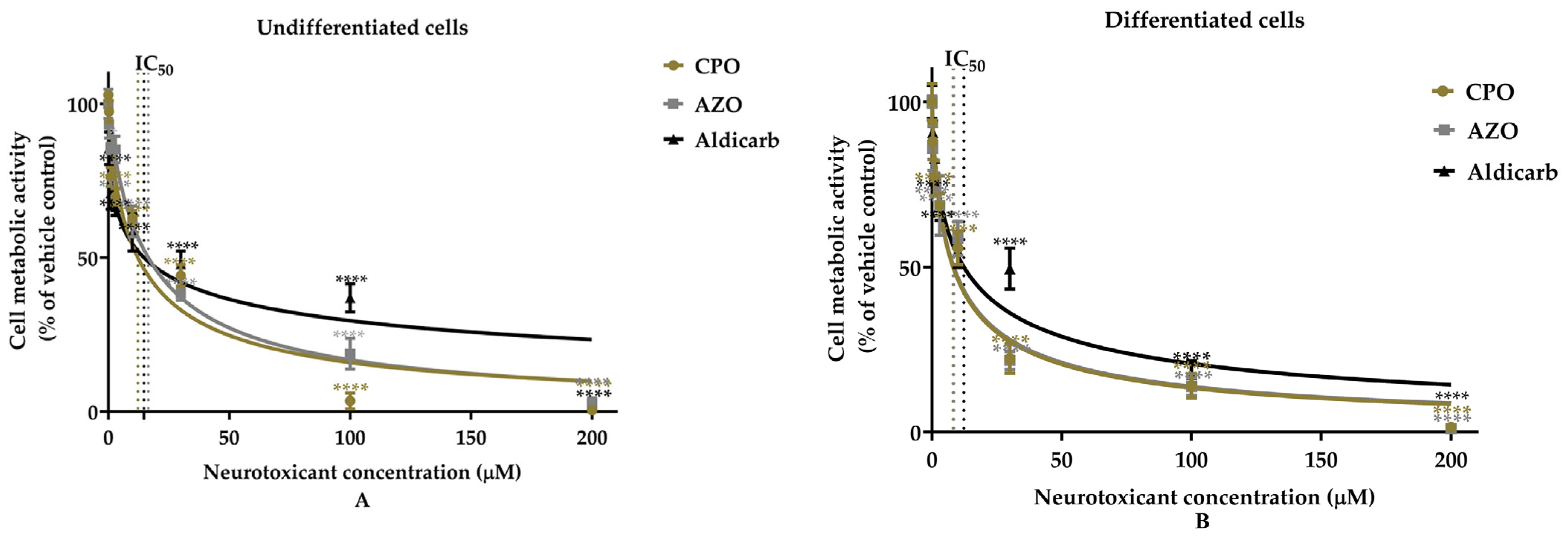
Figure 1. The neurotoxic effects of chlorpyrifos-oxon, azamethiphos, and aldicarb on undifferentiated and differentiated hNPCs examined using an MTT assay. Undifferentiated (A) and differentiated (B) hNPCs were treated with the pesticide compounds (0–200 mM) for 24 h, and toxicity was assessed using an MTT assay. The absorbance readings obtained were normalized to the viability of vehicle control-treated cells, and cell viability was calculated as a percentage relative to the control. Data were gathered from five independent experiments, each consisting of three replicates per treatment concentration. Statistical analysis was performed using one-way ANOVA followed by Dunnett’s multiple comparison tests. Data points are presented as means ± standard error of the mean (SEM), with indicated significance of **** p < 0.0001 from at least five separate experiments.
Applsci 15 10800 g001
Figure 2. The neurotoxic effects of chlorpyrifos-oxon, azamethiphos, and aldicarb on hNPCs examined using an LDH assay. Undifferentiated (A) and differentiated (B) hNPCs were treated with the pesticide compounds (0–200 mM) for 24 h, and toxicity was assessed using an LDH assay. After treatment with the neurotoxicants, absorbance readings underwent blank value correction, were normalized to vehicle controls, and then presented as a percentage relative to the control. Data points are presented as means ± standard error of the mean (SEM), from five separate experiments, each including three replicates per treatment condition. Statistical analysis was conducted using one-way ANOVA followed by Dunnett’s multiple comparison tests, with indicated significance of **** p < 0.0001.
Applsci 15 10800 g002
Figure 3. The neurotoxic effects of chlorpyrifos-oxon, azamethiphos, and aldicarb on hNPCs examined using an ATP assay. Undifferentiated (A) and differentiated (B) hNPCs were treated with the pesticide compounds for 24 h at their IC10, IC20, IC50, and IC80 concentrations (refer to Supplementary Table S1), and toxicity was assessed using an ATP assay. Absorbance readings underwent blank value correction, and the resultant values were normalized to the ATP level of vehicle controls, presenting ATP levels as a percentage relative to the control. Data were obtained from five independent experiments, each comprising three replicates per treatment. Statistical analysis employed one-way ANOVA with Dunnett’s multiple comparison tests. Data points are presented as means ± standard error of the mean (SEM), with indicated significance of **** p < 0.0001.
Applsci 15 10800 g003
Figure 4. Anti-cholinesterase effects of chlorpyrifos-oxon, azamethiphos, and aldicarb in hNPCs. Undifferentiated (A) and differentiated (B) hNPCs were treated with the pesticide compounds (0–3 µM) for 24 h, and inhibition of AChE was quantified using a modified Ellman’s assay. Absorbance readings underwent blank value correction, and the resultant values were normalized to vehicle control levels and then expressed as a percentage of the controls. Data were obtained from five independent experiments, each comprising three replicates per treatment. Statistical analysis employed one-way ANOVA with Dunnett’s multiple comparison tests. Data points are presented as means ± standard error of the mean (SEM), with indicated significance of **** p < 0.0001.
Applsci 15 10800 g004
Figure 5. The neurotoxic effects of chlorpyrifos-oxon, azamethiphos, and aldicarb in hNPCs examined using an assay for ROS levels. Undifferentiated (A,C) and differentiated (B,D) hNPCs were treated with the pesticide compounds at MTT IC10, IC20, IC50, and IC80 concentrations for 6 and 24 h, and the levels of ROS were quantified using a DCFDA assay. The cellular ROS levels were normalized to vehicle control treatments and expressed as a percentage relative to the control. Data were obtained from five independent experiments, each comprising three replicates per treatment. Statistical analysis employed one-way ANOVA with Dunnett’s multiple comparison tests. Data points are presented as means ± standard error of the mean (SEM), with indicated significance of **** p < 0.0001.
Applsci 15 10800 g005
Figure 6. The induction of protein carbonyl content in hNPCs by chlorpyrifos-oxon, azamethiphos, and aldicarb. Undifferentiated (A) and differentiated (B) hNPCs were treated with the pesticide compounds at MTT IC10, IC20, IC50, and IC80 concentrations (refer to Supplementary Table S1) for 24 h, and the levels of protein carbonyl content (PCC) in cytosolic fractions were quantified. The readings were normalized to vehicle control treatments after blank subtraction. Each data point represents triplicate assays, with data derived from five independent experiments. Statistical analysis employed one-way ANOVA with Dunnett’s multiple comparison tests, and outcomes are presented as means ± standard error of the mean (SEM), with indicated significance of **** p < 0.0001.
Applsci 15 10800 g006
Figure 7. The induction of carbonylated proteins in hNPCs by chlorpyrifos-oxon, azamethiphos, and aldicarb detected using an oxy-blot. Undifferentiated and differentiated hNPCs were treated with neurotoxicants at MTT IC10, IC20, IC50, and IC80 concentrations for 24 h, and carbonylated proteins were detected by oxy-blottng. Three independent blotting experiments were conducted for each of the neurotoxicants, with representative blots included to illustrate the findings.
Applsci 15 10800 g007
Table 1. Neurotoxicity of chlorpyrifos-oxon (CPO), azamethiphos-oxon (AZO), and aldicarb to hNPCs. IC50 concentrations (in µM) of the neurotoxicants were determined by interpolating from concentration–response curves and are displayed with their respective 95% confidence intervals, derived from five independent experiments. The goodness of fit of the curves to the expected non-linear model was determined using R2 values.
Cell TypePesticidesMTT AssayLDH AssayATP AssayAChE Inhibition
IC50 (µM)R2IC50 (µM)R2IC50 (µM)R2IC50 (µM)R2
UndifferentiatedCPO12.0 ± 1.90.93712.2 ± 0.60.99512.5 ± 1.40.9610.23 ± 0.020.949
Differentiated8.2 ± 0.80.9719.8 ± 0.30.9909.18 ± 0.90.9780.13 ± 0.010.949
UndifferentiatedAZO16.5 ± 1.40.97712.2 ± 1.20.97312.8 ± 0.20.9970.21 ± 0.020.925
Differentiated8.6 ± 1.00.96211.4 ± 1.00.97411.0 ± 0.30.9950.19 ± 0.010.964
UndifferentiatedAldicarb14.8 ± 2.90.91014.2 ± 1.00.97914.8 ± 1.20.9700.22 ± 0.010.993
Differentiated12.3 ± 1.70.94913.5 ± 0.70.99013.8 ± 1.40.9610.21 ± 0.010.997
Disclaimer/Publisher’s Note: The statements, opinions and data contained in all publications are solely those of the individual author(s) and contributor(s) and not of MDPI and/or the editor(s). MDPI and/or the editor(s) disclaim responsibility for any injury to people or property resulting from any ideas, methods, instructions or products referred to in the content.