


Abstract
Background: The aim of this systematic review was to critically evaluate the clinical indications, the techniques, and the effects of interproximal enamel reduction (IPR). Methods: A systematic review of the existing literature was carried out following the PRISMA guidelines in the following databases: PubMed-Medline, Scopus, Embase, Web of Science, and Cochrane. The search was conducted according to the established inclusion and exclusion criteria until March 2025. Results: A total of 420 articles were identified. Only 23 were selected for the analysis. The efficiency and validity of IPR as a non-extraction protocol adopted in several orthodontic cases was reported in all selected studies. No negative effects in terms of enamel demineralization, residual irregularities, caries incidence, and periodontal damages were reported. Using oscillating mechanical techniques was more efficient than using manual ones in terms of efficiency, predictability, and respect of enamel surfaces. Overall, the quality of the selected articles was low. More randomized controlled clinical trials with in vivo tests, research with longer follow-up times, and high-quality studies are needed to assess more valid statements. Conclusions: Higher accuracy of enamel reduction is achieved by means of oscillating mechanical techniques, which allows for more effective and predictable IPR procedures, as well as more preserved residual enamel surfaces.
1. Introduction
Interproximal enamel reduction (IPR), also known as interproximal reduction or stripping, was introduced in 1944 as a technique to manage tooth size discrepancies [1,2]. The concept of IPR has been discussed for decades [1,2,3,4,5,6,7], and over time several techniques have been proposed and refined to improve clinical efficiency and safety [8,9,10,11,12,13,14]. More recently, technological developments and new clinical investigations have focused on the biological effects and surface alterations associated with different IPR methods [12,15] highlighting both advancements and persistent controversies that justify the need for an updated systematic review. Over the years, its applications have expanded to address late secondary crowding, prevent relapse by stabilizing dental contacts, and serve as an alternative to extractions in borderline cases [3,4,5]. Additionally, IPR has been used to reshape teeth and reduce black triangles in periodontal patients [6,7,8]. Nowadays, IPR represents one of the primary space-gaining techniques in orthodontic treatment, particularly in cases treated with clear aligners [4,6]. Combined with proclination and transversal expansion, IPR serves as a valuable alternative to extractions for resolving mild-to-moderate crowding [7,8,9,10]. In these cases, the amount of enamel to be removed should be calculated considering the space required. Several IPR techniques have been introduced over the years, with mechanical oscillating abrasive strips gaining popularity because of their precision, efficiency, reduced chairside time, and minimally invasive effects on enamel surfaces [6,9]. Recent studies have demonstrated that mechanical oscillating diamond strips provide higher efficiency and produce more regular enamel surfaces compared to manual systems [6]. Furthermore, it has been widely established that the polishing phase after IPR is crucial in achieving smoother enamel surfaces and minimizing the risk of plaque accumulation [11]. Despite its long-standing clinical use, concerns have been raised regarding potential iatrogenic effects, such as an increased risk of caries, periodontal disease, and dentin hypersensitivity [12,13]. However, recent studies suggest that dentin hypersensitivity represents a rare long-term side effect, and that reduced interproximal width does not predispose teeth to accelerated periodontal breakdown [14,15]. While IPR may initially increase enamel surface roughness, this effect is mitigated by proper finishing and polishing techniques, as well as natural remineralization over time [6,13,15,16,17,18,19,20,21,22]. A clinically relevant aspect to consider is the need of a standardized protocol to ensure both accuracy and safety during IPR. A standardized clinical sequence—including initial space creation, enamel removal, quantitative evaluation, and final polishing—ensures the predictability of clinical results while preserving enamel integrity [12]. Regardless of the method used, adherence to a systematic approach is essential to control the amount of enamel removed and to prevent undesirable surface roughness. Despite the widespread use of IPR in orthodontic treatment, there are still controversies regarding its effectiveness and long-term effects on enamel structure. Indeed, previous systematic reviews have reported high heterogeneity among available studies, with conflicting conclusions as to whether IPR contributes to an increased risk of caries and enamel demineralization [7,12,23]. Given these uncertainties, this systematic review aims to critically evaluate the effectiveness of IPR, in terms of predictability and efficiency of the different techniques, and its safety, in terms of biological effects on enamel, dental sensitivity, caries risk, and periodontal outcomes, by analyzing the existing literature and assessing the risk of bias in the included studies.
2. Materials and Methods
2.1. Protocol and Registration
The herein review was based on the PRISMA (Preferred Reporting Items for Systematic Reviews and Meta-Analyses) guidelines [24]. The protocol was registered in the PROSPERO database (Registration number: CRD420251010105). A comprehensive search of existing literature was conducted to identify all the available data on the IPR techniques and the effects related thereto. Specifically, we followed this step to address the PICO questions: (1) Is IPR safe for treated enamel surfaces? (2) What are the effects of IPR on biological structures? (3) Which IPR technique features the highest predictability? (4) What is the most biologically respectful technique for IPR procedures?
2.2. Eligibility Criteria
The following eligibility criteria were applied: (1) Population: studies evaluating stripping in orthodontic treatment; in vitro/in vivo controlled studies; Randomized controlled trials (RCTs), cohort studies, and systematic reviews; studies including untreated enamel control groups; SEM and/or quantitative evaluation of enamel roughness; studies with a minimum follow-up period of 6 months, as shorter observation times may not adequately capture clinically relevant outcomes; articles published in peer-reviewed journals. (2) Interventions: Interproximal enamel reduction. (3) Comparison: comparative analysis with untreated enamel surfaces and different interproximal enamel reduction techniques. (4) Outcome measure: evaluation of enamel surfaces after IPR procedures and identification of the most efficient and predictable protocol of IPR. The exclusion criteria included: case reports, case series, case–control, and cross-sectional studies, uncontrolled studies, opinion articles, studies on animals’ teeth or unhealthy human teeth, lack of control group, other method of assessing enamel roughness evaluation.
2.3. Search Strategy
A comprehensive literature search was conducted on the following: PubMed-Medline, Scopus, Embase, Web of Science, and Cochrane. The search was carried out from January 2025 until March 2025 to include the most recent studies. The strategy included three “MeSH” terms (Medical Subject Headings): “Interproximal reduction” OR “stripping” OR “interproximal enamel reduction” AND “IPR techniques” OR “IPR methods” OR “IPR protocols” AND “enamel surfaces” OR “enamel effects” AND “orthodontic treatment” OR “alignment” OR “protocol”. Boolean operators AND/OR were used to refine search results. Moreover, the references of included studies were manually searched to identify articles not found in the database that might have met the inclusion criteria.
2.4. Screening Process and Data Collection
The articles selected on the database were screened in accordance with the Prisma Declaration Statement [24]. The inclusion in the final data collection required several steps. The articles were retrieved from several identified databases and all duplicates were removed. Then, two investigators (FG and RL) carried out the data extraction. The titles and the abstracts of the selected studies were examined. Arising inter-examiner discrepancies were tackled by means of discussion and consultation with a third author (CP). Successively, the full texts were carefully reviewed to ensure the accordance for the identified requirements to be included in the results. Several studies were excluded due to lack of necessary data or incompliance with PICO questions. Standard variables were used to extract information from each article. From each eligible study, the following variables were extracted: authors and year of publication, country, study design, sample size and characteristics, IPR technique used, follow-up duration, evaluated outcomes (enamel roughness, dental sensitivity, caries incidence, periodontal changes, efficiency/predictability), main results, and risk of bias assessment.
2.5. Risk of Bias and Quality Assessment of Individual Studies
The two investigators evaluated the risk of bias and the quality of the selected studies. For the risk of bias, the Newcastle- Ottawa quality assessment scale (NOS) [25] and the Risk of Bias 2 (RoB 2) [26] were applied. Specifically, the NOS provides nine points subdivided into three domains: (a) study group selection, (b) group comparability, and (c) exposure and outcome ascertainment. High, moderate, and low risk of bias correspond to scores 0–3, 4–6, and, 7–9 respectively [27]. RoB 2 evaluates the following: (1) bias derived from the randomization process; (2) bias related to the deviation from intended interventions; (3) bias due to missing outcome data; (4) bias in outcome measurement and selection of the reported results. Moreover, the quality of the selected studies was assessed by means of the Grading of Recommendations Assessment, Development and Evaluation (GRADE) criteria. In case of disagreement between the two investigators, the third author (CP) was consulted [28].
2.6. Summary Measures and Approach to Data Synthesis
Quantitative findings obtained from the included studies were summarized. The qualitative analysis was performed by grouping the study outcomes in the following categories: enamel surfaces, dental sensitivity, predictability and efficiency of different IPR techniques. For each outcome, the methodological heterogeneity of the included studies was thoroughly analyzed to assess the feasibility of conducting a quantitative synthesis.
3. Results
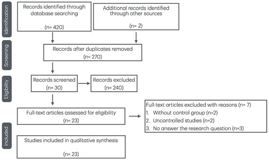
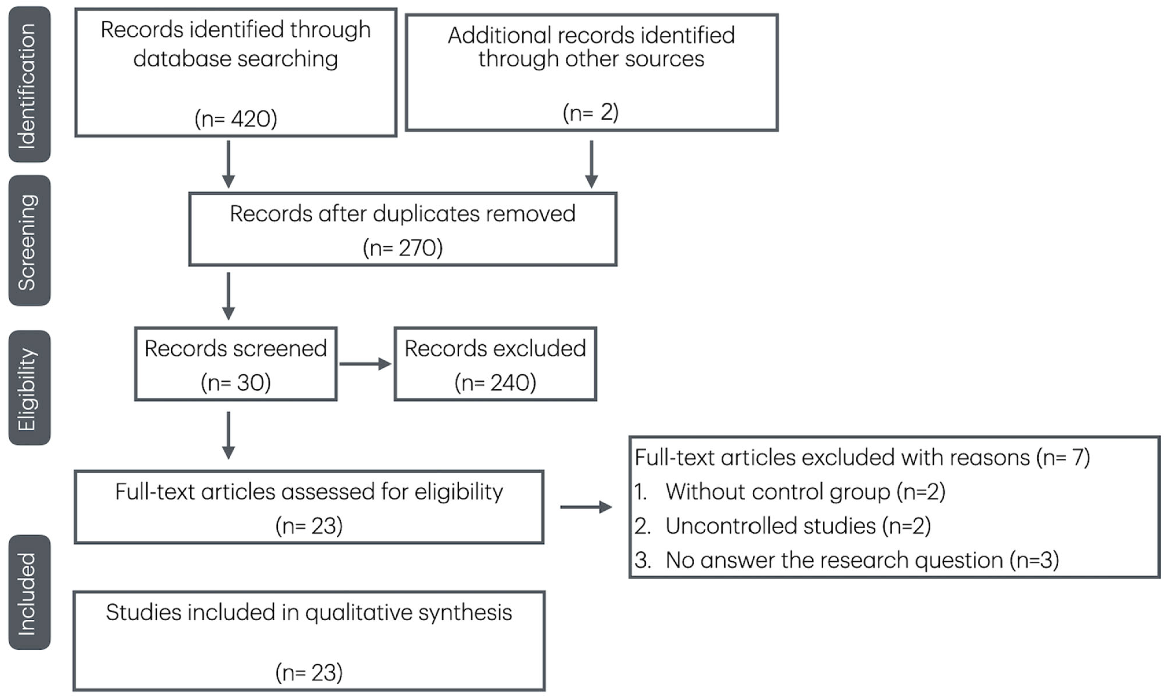
A total of 420 articles were found from the databases. After removing duplicates, 270 articles remained. Of these, 30 articles were selected for full-text eligibility assessment, and 7 were subsequently excluded with reasons. The screening of the reference lists of the included articles and relevant reviews identified two additional eligible [16,20], which were included in the review. Hence, 23 studies were included in the herein review: 6 observational studies, 4 randomized clinical trials (RCT), 10 non-randomized clinical trials (non-RCT), 3 systematic reviews. No further articles were retrieved through this manual search. The earliest study included was published in 1944 [1]. All included studies were available in English. One study [21] also provided a French abstract, but the full text was in English and was used for the analysis. The search strategy, including the databases consulted, the keywords used, the time frame, and the total number of records retrieved before duplicate removal, is summarized in Table 1. Table 2 revealed the characteristics of each selected study. The selection process is illustrated in the PRISMA flow diagram (Figure 1). Regarding the risk of bias, all the included RCT somewhat raised concerns towards the reported results. As for the observational studies, 50% showed low risk of bias, while 50% raised bias concerns. Given the variability in outcomes and measurement methods, the certainty of evidence was summarized in a narrative table (Table 3). The overall study quality ranged from low (8% of the studies) to high (20%). A meta-analysis was not conducted, and recommendations following the GRADE approach were not provided due to several factors: the heterogeneity in IPR procedures, differences in measurement units for assessing outcomes, and variable follow-up intervals across the studies. Thus, such limitations prevented data aggregation. The studies with comparable outcomes were categorized for the purposes of qualitative analysis.
Table 1.
Literature search strategy and number of records retrieved from each database.
Figure 1.
PRISMA flow diagram of the systematic review of the role of the interproximal enamel reduction in orthodontics.
3.1. Enamel Surfaces
11 articles assessed the enamel surfaces after IPR procedures. Obtained results concluded that all enamel reduction procedures somewhat caused surface changes without any adverse effects. Numerous authors agreed on the importance of the polishing and finishing phases to reduce the surface roughness after IPR procedures [11,12,16,17,18,19,20,29,30,31,32,33,34,35,36,37,38]. Moreover, several findings highlighted how the oscillating mechanical systems produce a more uniform enamel surface when compared to manual ones and diamond burs [6,11,31,32,33]. On the contrary, Katsigialou et al. [34] did not find any differences between manual and mechanical techniques, highlighting the importance of polishing procedures to restore enamel integrity. Two articles [13,39] emphasized the need for standardized protocols to optimize results minimizing enamel damages.
Table 2.
Characteristics of each selected study and main results.
Table 3.
Quality assessment of the selected studies.
3.2. Dental Sensitivity
Zachrisson et al. [8,36] concluded in two investigations that no increased tooth sensitivity was reported after IPR procedures. Bonetti et al. [13] highlighted the effect of the Casein Phosphopeptide-Amorphous Calcium Phosphate (CPP-ACP) on treated enamel surfaces in enhancing enamel resistance to acid attacks and hypersensitivity phenomena. Paganelli et al. [19] analyzed the Ca/P ratio and the amount of Ca in treated enamel. The authors showed that no demineralization of teeth had occurred and, thus, it was deemed unlikely for IPR to cause hypersensitivity or a higher tendency to develop carious lesions. Meredith et al. concluded that the existing literature would not indicate any risks of dental sensitivity subsequent to IPR, similarly to what was observed by Gómez-Aguirre et al. in a more recent systematic review [23,37]. In general, all the selected studies suggested that interproximal enamel reduction would not significantly increase tooth sensitivity or caries risk.
3.3. Predictability and Efficiency of Different IPR Techniques
As a result of this systematic review, it was found that a number of studies existing in the literature focused on the effects of IPR on the tooth surfaces. By contrast, only a few studies have analyzed the validity and efficacy of the various available IPR tech niques [6,38,39,40]. Gazzani et al. [6], evaluating the efficiency and the abrasive properties of oscillating and manual systems, concluded that oscillating strips revealed more controlled efficiency in terms of removed enamel when compared to manual ones. In a further investigation [38], the same group evaluated the wear and friction properties of oscillating strips stating the importance of a validated standardized IPR sequence to allow a more efficient wear performance of the strips with a significant impact on their abrasive properties and lifetime.
4. Discussion
This systematic review aimed to evaluate the role of IPR in orthodontics, with a focus on the impact on enamel surfaces, dental sensitivity, and the predictability and efficiency of various IPR techniques. A total of 23 studies were included, consisting of observational studies, randomized clinical trials (RCTs), non-randomized clinical trials (non-RCTs), and systematic reviews. The findings of this review underline several key aspects of IPR, though they also highlight the substantial variability across studies and the methodological limitations that hinder the aggregation of data [23]. The results regarding enamel surface changes after IPR procedures are generally reassuring [11,16,19,30,31,32,33,34,35,36,37,38].
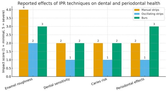
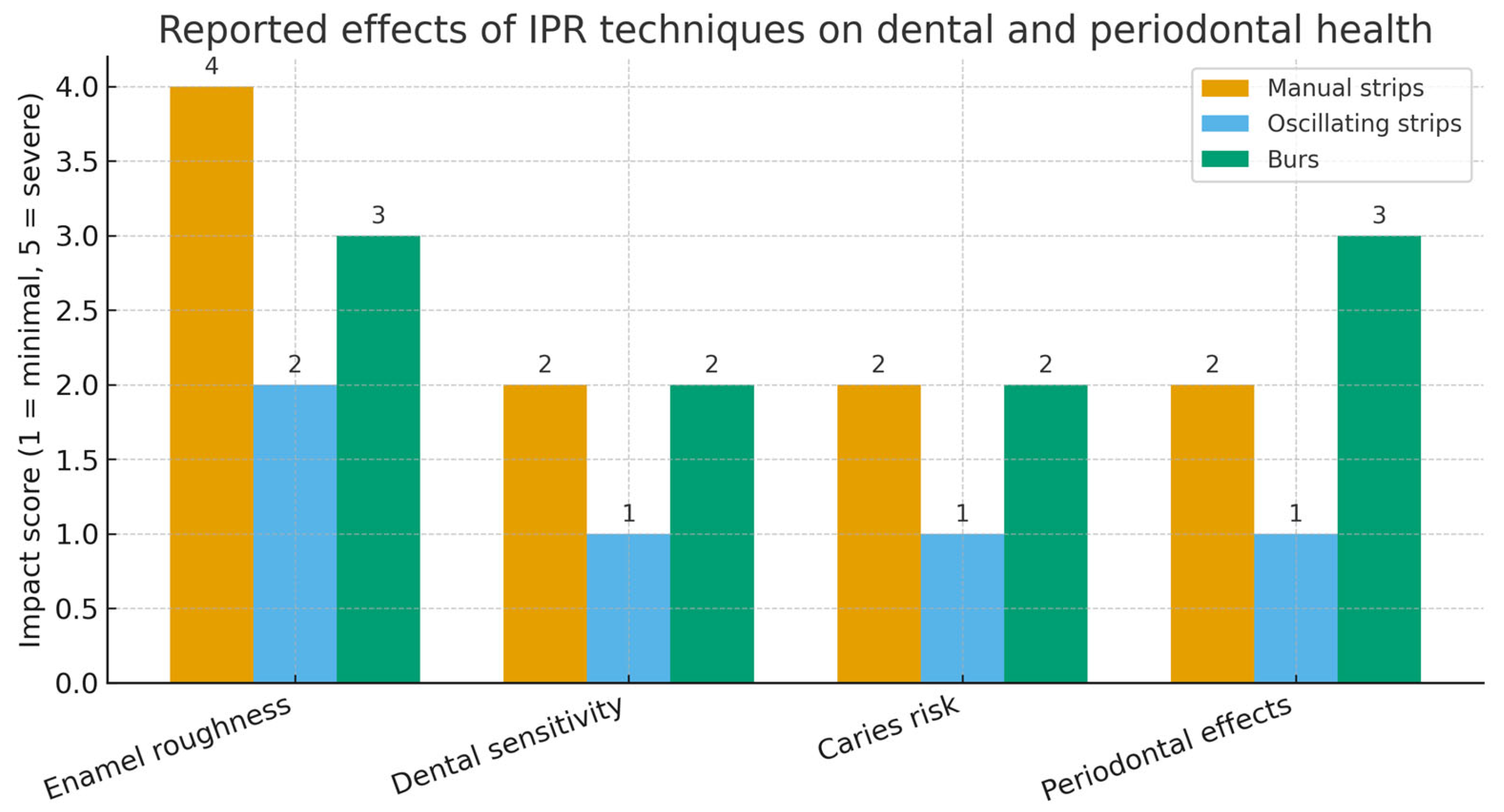
The studies consistently demonstrated that enamel reduction did not result in adverse effects, although all IPR techniques induced some degree of surface alteration (Figure 2).
Figure 2.
Biological effects of different enamel reduction (IPR) techniques. Oscillating strips are associated with lower enamel roughness, dental sensitivity, caries risk, and periodontal impact compared with manual strips and burs, supporting their safety and clinical applicability.
The polishing and finishing stages were emphasized as crucial for minimizing surface roughness and restoring enamel integrity [11,16,19,29,30,31,32,33,34]. Notably, mechanical oscillating systems were found to produce more uniform enamel surfaces compared to manual techniques and diamond burs [6,11,38].
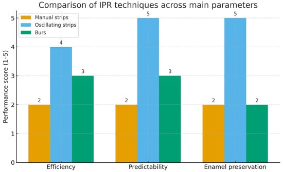
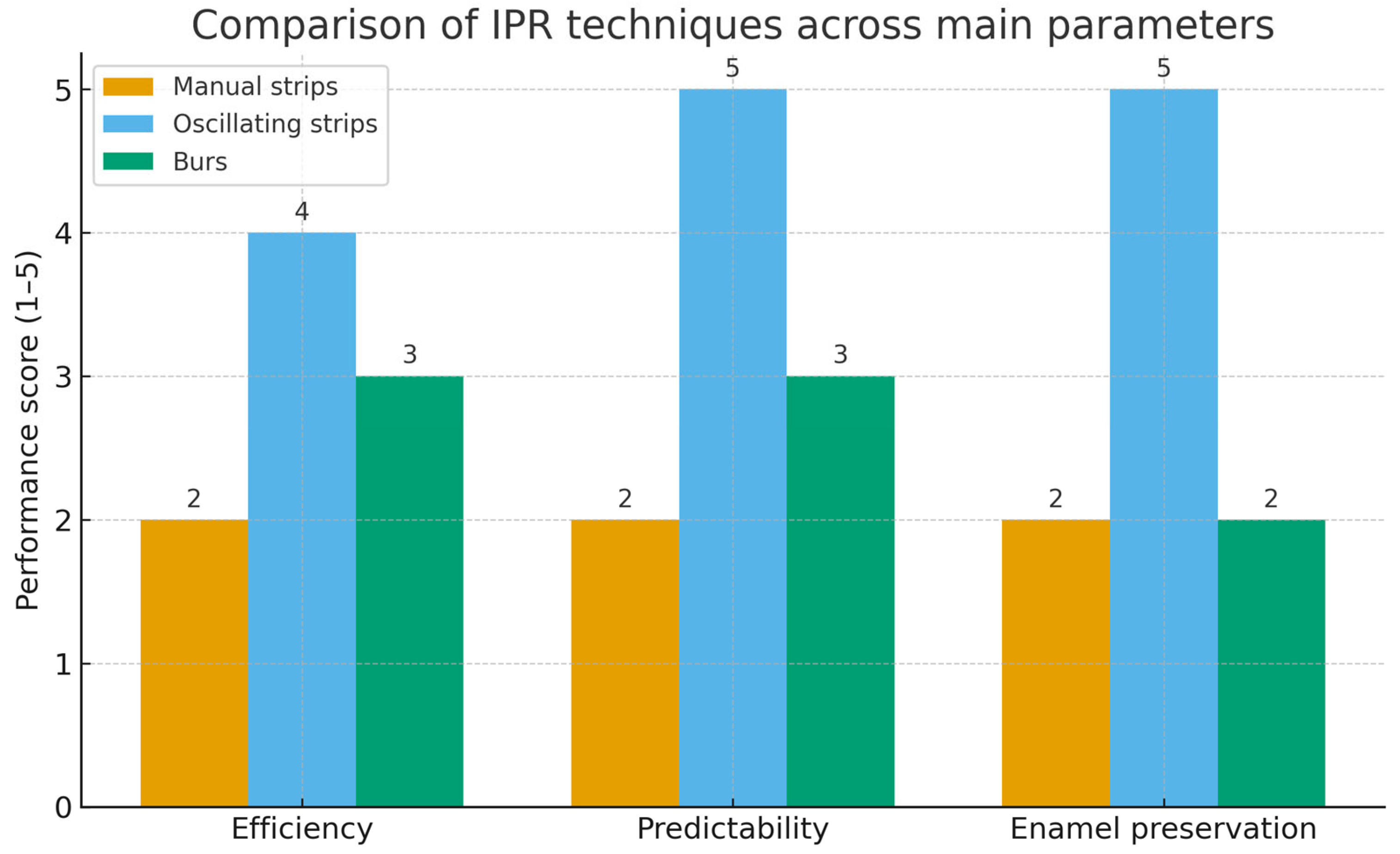
However, there was some disagreement, as Katsigialou et al. [34] did not report any significant differences between manual and mechanical methods, emphasizing the importance of proper polishing. These results underscore the need for standardized protocols to optimize the outcomes of IPR procedures and minimize enamel damage [12,34]. A key concern regarding IPR procedures is the potential increase in dental sensitivity. The studies reviewed here, however, consistently indicated that IPR did not result in increased tooth sensitivity or a higher risk of carious lesions [8,12,19,23,36,37]. Several studies pointed out the protective role of Casein Phosphopeptide-Amorphous Calcium Phosphate (CPP-ACP) in reducing enamel hypersensitivity, suggesting that IPR, when performed correctly, does not lead to detrimental effects on enamel health [12]. These findings are consistent with previous systematic reviews, supporting the notion that IPR is a safe procedure in terms of dental sensitivity [23]. The review also highlighted the efficiency and predictability of various IPR techniques (Figure 3).
Figure 3.
Performance comparison of different IPR techniques based on the main clinical parameters reported in the literature. Oscillating strips demonstrated superior efficiency, predictability, and enamel preservation compared with manual strips and burs, supporting their use as the preferred technique in clinical practice.
While many studies focused on the effects of IPR on enamel surfaces, fewer studies investigated the relative efficacy of different IPR available techniques [6,35,36,40]. The main findings reveal how oscillating strips allow a greater control and efficiency in enamel removal compared to manual strips. The same authors [8,36] also highlighted the need for standardized protocols to optimize the abrasive performance and longevity of oscillating strips. These insights are crucial for improving clinical outcomes and enhancing the predictability of IPR procedures. From a clinical standpoint, the evidence suggests that mechanical oscillating systems should be considered the technique of choice whenever available, as they provide greater precision, efficiency, and enamel preservation compared with manual strips. Nevertheless, regardless of the method employed, polishing and finishing remain indispensable to reduce surface roughness and prevent plaque accumulation. These findings underline the importance of standardized protocols and adequate operator training to ensure predictable and biologically respectful outcomes in daily practice.
Several limitations of the included studies must be mentioned. The overall reliability of the findings is affected by the risk of bias in many of the included RCTs. In addition, most studies relied on small sample sizes and relatively short follow-up periods, which further reduce the strength of the available evidence. Variability in IPR procedures, measurement units, and observational intervals also contributed to the difficulty in conducting a meta-analysis. As a result, the quality of evidence was assessed as low to very low for most studies, primarily due to inconsistencies and imprecision. Future research should focus on standardizing IPR protocols and measurement methods to enable more consistent and reliable results. Moreover, long-term studies assessing the durability of enamel surfaces post-IPR and the potential risks of caries development are needed to provide further insights into the safety of this procedure. Additionally, the development of more controlled RCTs with clearer methodologies could help mitigate the current risks of bias and improve the robustness of the evidence base.
5. Conclusions
The results suggest that IPR does not significantly impact dental sensitivity or increase caries risk and that different techniques present different efficiency in enamel reduction. Despite the overall low quality of evidence, the findings support the use of IPR as a safe and effective procedure in orthodontic treatment, especially when performed by an oscillating mechanical system. The standardization of the IPR clinical sequence enhances the accuracy of enamel reduction while respecting biological structures. Furthermore, proper polishing—defined as the use of progressively finer abrasive strips or disks, followed by rubber cups or silicone points—plays a crucial role in ensuring a good long-term prognosis and preserving enamel morphological integrity. However, further high-quality studies are necessary to refine IPR protocols and better assess the long-term effects on enamel health.
Author Contributions
Conceptualization, F.G. and R.L.; methodology, F.G.; software, F.C.D.R. and L.L.; validation, F.G. and R.L.; formal analysis, C.P.; investigation, G.L.; resources, F.G.; data curation, F.G.; writing—original draft preparation, F.G.; writing—review and editing, F.C.D.R.; supervision, P.C.; project administration, P.C. All authors have read and agreed to the published version of the manuscript.
Funding
This research received no external funding.
Institutional Review Board Statement
Not applicable.
Conflicts of Interest
The authors declare no conflicts of interest.
Abbreviations
The following abbreviations are used in this manuscript:
| IPR | Interproximal Enamel Reduction |
| NOS | Newcastle–Ottawa scale |
| RCT | Randomized Clinical Trial |
| CPP-ACP | Casein Phosphopeptide–Amorphous Calcium Phosphate |
| N/A | Not Available |
References
- Ballard, M.L. Asymmetry in tooth size: A factor in the etiology, diagnosis and treatment of malocclusion. Angle Orthod. 1944, 14, 67–70. [Google Scholar]
- Sheridan, J.J. Guidelines for contemporary air-rotor stripping. J. Clin. Orthod. 2007, 41, 315–320. [Google Scholar]
- Bolton, A. Disharmony in tooth size and its relation to the analysis and treatment of malocclusion. Angle Orthod. 1958, 28, 113–130. [Google Scholar]
- Borda, A.F.; Garfinkle, J.S.; Covell, D.A.; Wang, M.; Doyle, L.; Sedgley, C.M. Outcome assessment of orthodontic clear aligner vs fixed appliance treatment in a teenage population with mild malocclusions. Angle Orthod. 2020, 90, 485–490. [Google Scholar] [CrossRef] [Scilit] [PubMed]
- Rossouw, P.E.; Tortorella, A. Enamel reduction procedures in orthodontic treatment. J. Can. Dent. Assoc. 2003, 69, 378–383. [Google Scholar]
- Gazzani, F.; Lione, R.; Pavoni, C.; Mampieri, G.; Cozza, P. Comparison of the abrasive properties of two different systems for interproximal enamel reduction: Oscillating versus manual strips. BMC Oral Health 2019, 19, 247. [Google Scholar] [CrossRef] [Scilit]
- Vignoletti, F.; Di Martino, M.; Clementini, M.; Di Domenico, G.L.; De Sanctis, M. Prevalence and risk indicators of gingival recessions in an Italian school of dentistry and dental hygiene: A cross-sectional study. Clin. Oral Investig. 2020, 24, 991–1000. [Google Scholar] [CrossRef] [Scilit] [PubMed]
- Zachrisson, B.U.; Nyoygaard, L.; Mobarak, K. Dental health assessed more than 10 years after interproximal enamel reduction of mandibular anterior teeth. Am. J. Orthod. Dentofac. Orthop. 2007, 131, 162–169. [Google Scholar] [CrossRef] [Scilit]
- Laganà, G.; Malara, A.; Lione, R.; Danesi, C.; Meuli, S.; Cozza, P. Enamel interproximal reduction during treatment with clear aligners: Digital planning versus Ortho CAD analysis. BMC Oral Health 2021, 21, 199. [Google Scholar] [CrossRef] [Scilit]
- Lapenaite, E.; Lopatiene, K. Interproximal enamel reduction as a part of orthodontic treatment. Stomatologija 2014, 16, 19–24. [Google Scholar] [PubMed]
- Gazzani, F.; Bellisario, D.; Fazi, L.; Balboni, A.; Licoccia, S.; Pavoni, C.; Cozza, P.; Lione, R. Effects of IPR by mechanical oscillating strips system on biological structures: A quantitative and qualitative evaluation. Prog. Orthod. 2023, 24, 9. [Google Scholar] [CrossRef] [Scilit]
- Koretsi, V.; Chatzigianni, A.; Sidiropoulou, S. Enamel roughness and incidence of caries after interproximal enamel reduction: A systematic review. Orthod. Craniofac Res. 2014, 17, 1–13. [Google Scholar] [CrossRef] [Scilit]
- Bonetti, G.; Zanarini, M.; Incerti Parenti, S.; Marchionni, S.; Checchi, L. In vitro evaluation of casein phosphopeptide-amorphous calcium phosphate (CPP-ACP) effect on stripped enamel surfaces: A SEM investigation. J. Dent. 2009, 37, 228–232. [Google Scholar]
- Radlanski, R.J.; Jäger, A.; Schwestka, R.; Bertzbach, F. Plaque accumulations caused by interdental stripping. Am. J. Orthod. Dentofac. Orthop. 1988, 94, 416–420. [Google Scholar] [CrossRef] [Scilit] [PubMed]
- Bayram, M.; Kusgoz, A.; Yesilyurt, C.; Nur, M. Effects of casein phosphopeptide-amorphous calcium phosphate application after interproximal stripping on enamel surface: An in vivo study. Am. J. Orthod. Dentofac. Orthop. 2017, 151, 167–173. [Google Scholar] [CrossRef] [Scilit]
- Zingler, S.; Sommer, A.; Sen, S.; Saure, D.; Langer, J.; Guillon, O.; Lux, C.J. Efficiency of powered systems for interproximal enamel reduction (IER) and enamel roughness before and after polishing: An in vitro study. Clin. Oral Investig. 2016, 20, 933–942. [Google Scholar] [CrossRef] [Scilit] [PubMed]
- Piacentini, C.; Sfondrini, G. A scanning electron microscopy comparison of enamel polishing methods after air-rotor stripping. Am. J. Orthod. Dentofac. Orthop. 1996, 109, 57–63. [Google Scholar] [CrossRef] [Scilit]
- Arman, A.; Cehreli, S.B.; Ozel, E.; Arhun, N.; Cetinsahin, A.; Soyman, M. Qualitative and quantitative evaluation of enamel after various stripping methods. Am. J. Orthod. Dentofac. Orthop. 2006, 130, 131–136. [Google Scholar] [CrossRef] [Scilit]
- Paganelli, C.; Zanarini, M.; Pazzi, E.; Marchionni, S.; Visconti, L.; Bonetti, G. Interproximal enamel reduction: An in vivo study. Scanning 2015, 37, 73–81. [Google Scholar] [CrossRef] [Scilit]
- Baumgartner, S.; Iliadi, A.; Eliades, T.; Eliades, G. An in vitro study on the effect of an oscillating stripping method on enamel roughness. Prog. Orthod. 2015, 16, 1. [Google Scholar] [CrossRef] [Scilit] [PubMed]
- Lione, R.; Gazzani, F.; Moretti, S.; Danesi, C.; Cretella Lombardo, E.; Pavoni, C. Gingival margins’ modifications during orthodontic treatment with Invisalign First®: A preliminary study. Children 2022, 9, 1423. [Google Scholar] [CrossRef] [Scilit]
- Kaaouara, Y.; Mohind, H.B.; Azaroual, M.F.; Zaoui, F.; Bahije, L.; Benyahia, H. In vivo enamel stripping: A macroscopic and microscopic analytical study. Int. Orthod. 2019, 17, 235–242. [Google Scholar] [CrossRef] [Scilit]
- Gomez-Aguirre, J.N.; Argueta-Figueroa, L.; Castro-Gutierrez, M.E.M.; Torres-Rosas, R. Effects of interproximal enamel reduction techniques used for orthodontics: A systematic review. Orthod. Craniofac Res. 2022, 25, 304–319. [Google Scholar] [CrossRef] [Scilit]
- Moher, D.; Liberati, A.; Tetzlaff, J.; Altman, D.G.; PRISMA Group. Preferred reporting items for systematic reviews and meta-analyses: The PRISMA statement. BMJ 2009, 339, b2535. [Google Scholar] [CrossRef] [PubMed]
- Peterson, J.; Welch, V.; Losos, M.; Tugwell, P. The Newcastle-Ottawa Scale (NOS) for Assessing the Quality of Non-Randomised Studies in Meta-Analyses; Ottawa Health Research Institute: Ottawa, ON, Canada, 2011. [Google Scholar]
- Sterne, J.A.C.; Savović, J.; Page, M.J.; Elbers, R.G.; Blencowe, N.S.; Boutron, I.; Cates, C.J.; Cheng, H.Y.; Corbett, M.S.; Eldridge, S.M.; et al. RoB 2: A revised tool for assessing risk of bias in randomised trials. BMJ 2019, 366, l4898. [Google Scholar] [CrossRef] [Scilit] [PubMed]
- Lo, C.K.L.; Mertz, D.; Loeb, M. Newcastle-Ottawa Scale: Comparing reviewers’ to authors’ assessments. BMC Med. Res. Methodol. 2014, 14, 45. [Google Scholar] [CrossRef] [Scilit] [PubMed]
- Schünemann, H.; Brożek, J.; Guyatt, G.; Oxman, A. GRADE Handbook: Grading of Recommendations Assessment, Development and Evaluation; GRADE Working Group: Barcelona, Spain, 2014. [Google Scholar]
- Kalemaj, Z.; Levrini, L. Quantitative evaluation of implemented interproximal enamel reduction during aligner therapy. Angle Orthod. 2021, 91, 61–66. [Google Scholar] [CrossRef] [Scilit]
- Serbanoiu, D.C.; Vartolomei, A.C.; Ghiga, D.V.; Pop, S.I.; Panainte, I.; Moldovan, M.; Sarosi, C.; Petean, I.; Boileau, M.J.; Pacurar, M. Comparative evaluation of dental enamel microhardness following various methods of interproximal reduction: A Vickers hardness tester investigation. Biomedicines 2024, 20, 1132. [Google Scholar] [CrossRef] [Scilit]
- Butrus, D.J.; Chawshli, O.F.J. Evaluation of the enamel nano-topography influenced by different techniques of interproximal reduction: An atomic force microscopic study. J. Orthod. 2023, 50, 196–204. [Google Scholar] [CrossRef] [Scilit]
- Silvestrini Biavati, F.; Schiaffino, V.; Signore, A.; De Angelis, N.; Lanteri, V.; Ugolini, A. Evaluation of enamel surfaces after different techniques of interproximal enamel reduction. J. Funct. Biomater. 2023, 14, 110. [Google Scholar] [CrossRef] [Scilit]
- Shalchi, M.; Abdollahi, N.; Shafiei Haghshenas, E.; Khabbaz, S.; Olyaee, P. Effect of interdental enamel reduction on clinical attachment loss, bleeding on probing, and incidence of caries in treating Class I malocclusion cases: A retrospective cohort study. Cureus 2023, 15, e35018. [Google Scholar] [CrossRef] [Scilit]
- Katsigialou, N.; Sifakakis, I.; Zinelis, S.; Papageorgiou, S.N.; Eliades, T. Manual and mechanical stripping-induced enamel roughness and elemental composition in vivo. Eur. J. Orthod. 2023, 45, 250–257. [Google Scholar] [CrossRef] [Scilit] [PubMed]
- Dahas, F.Y.; Almutairi, N.S.; Almutairi, R.S.; Alshamrani, H.A.; Alshyai, H.S.; Almazyad, R.K.; Alsanouni, M.S.; Gadi, S.A. The role of interproximal reduction (IPR) in clear aligner therapy: A critical analysis of indications, techniques, and outcomes. Cureus 2024, 16, e56644. [Google Scholar] [CrossRef] [Scilit]
- Zachrisson, B.U.; Minster, L.; Ogaard, B.; Birkhed, D. Dental health assessed after interproximal enamel reduction: Caries risk in posterior teeth. Am. J. Orthod. Dentofac. Orthop. 2011, 139, 90–98. [Google Scholar] [CrossRef] [Scilit]
- Meredith, L.; Mei, L.; Cannon, R.D.; Farella, M. Interproximal reduction in orthodontics: Why, where, how much to remove? Australas. Orthod. J. 2017, 33, 150–157. [Google Scholar] [CrossRef] [Scilit]
- Gazzani, F.; Bellisario, D.; Fazi, L.; Balboni, A.; Licoccia, S.; Pavoni, C.; Cozza, P.; Lione, R. Friction and wear behavior of a mechanical oscillating strip system used for interproximal enamel reduction: A quantitative and qualitative scanning electron microscope evaluation. Angle Orthod. 2024, 94, 336–345. [Google Scholar] [CrossRef] [Scilit] [PubMed]
- Lione, R.; Gazzani, F.; Pavoni, C.; Guarino, S.; Tagliaferri, V.; Cozza, P. In vitro and in vivo evaluation of diamond-coated strips. Angle Orthod. 2017, 87, 455–459. [Google Scholar] [CrossRef] [Scilit]
- Lombardo, L.; Guarneri, M.P.; D’Amico, P.; Molinari, C.; Meddis, V.; Carlucci, A.; Siciliani, G. Orthofile®: A new approach for mechanical interproximal reduction: A scanning electron microscopic enamel evaluation. J. Orofac. Orthop. 2014, 75, 203–212. [Google Scholar] [CrossRef] [Scilit]

Disclaimer/Publisher’s Note: The statements, opinions and data contained in all publications are solely those of the individual author(s) and contributor(s) and not of MDPI and/or the editor(s). MDPI and/or the editor(s) disclaim responsibility for any injury to people or property resulting from any ideas, methods, instructions or products referred to in the content. |
© 2025 by the authors. Licensee MDPI, Basel, Switzerland. This article is an open access article distributed under the terms and conditions of the Creative Commons Attribution (CC BY) license (https://creativecommons.org/licenses/by/4.0/).