Systematic Review

Cold Plasma Treatment on Titanium Implants and Osseointegration: A Systematic Review

1
Oral Surgery Unit, Department of Biomedical and Neuromotor Sciences (DIBINEM), University of Bologna, 40125 Bologna, Italy
2
Department of Health Sciences, Magna Graecia University of Catanzaro, 88100 Catanzaro, Italy
3
Prosthodontic Unit, Department of Biomedical and Neuromotor Sciences (DIBINEM), University of Bologna, 40125 Bologna, Italy
*
Author to whom correspondence should be addressed.
Appl. Sci. 2025, 15(19), 10302; https://doi.org/10.3390/app151910302
Submission received: 30 July 2025 / Revised: 10 September 2025 / Accepted: 16 September 2025 / Published: 23 September 2025

Abstract

Background/Objectives: Osseointegration of titanium dental implants is essential for the long-term success of prosthetic treatments. Cold atmospheric pressure plasma (CAP) has recently emerged as a promising surface modification technique aimed at enhancing early osseointegration by improving implant surface properties and exerting antimicrobial effects. This systematic review aims to critically evaluate the in vivo preclinical evidence on the effects of CAP or similar cold plasma treatments on titanium dental implant surfaces with regard to osseointegration outcomes. Methods: A systematic literature search was conducted in PubMed and Scopus databases for preclinical in vivo studies published between 2005 and 2025 investigating the effects of cold plasma on titanium dental implant surfaces. The primary outcome assessed was the bone-to-implant contact (BIC), followed by secondary outcomes including implant stability quotient (ISQ), removal torque, bone area fraction occupancy (BAFO), peri-implant bone density (PIBD), interfacial bone density (IBD), bone-implant direct weight (BDWT) and bone loss measurements via histology and micro-CT. Risk of bias was evaluated using the SYRCLE Risk of Bias tool. Results: Nine eligible studies involving 310 titanium implants in 71 animal models (dogs, pigs and mice) were included. CAP-treated implants consistently demonstrated significant improvements in early osseointegration parameters compared to controls, with statistically significant increases in BIC (up to +20%), BAFO and biomechanical fixation metrics (removal torque and ISQ). Micro-CT analyses revealed enhanced peri-implant bone density and architecture. No adverse biological events or implant failures related to plasma treatment were reported. However, heterogeneity in plasma protocols, animal species and short follow-up durations (2–12 weeks) limited comparability and long-term interpretation. Conclusions: Preclinical evidence seems to support CAP as a safe and potentially effective surface treatment for enhancing early osseointegration of titanium dental implants. Further standardized long-term studies involving functional loading and clinical trials in humans are needed to confirm clinical efficacy and optimize treatment protocols.

1. Introduction

Osseointegration of titanium dental implants constitutes a pivotal biological process essential for the long-term success of prosthetic rehabilitations [1]. This phenomenon facilitates the formation of a direct, stable and functional interface between viable bone tissue and the implant surface, thereby ensuring mechanical stability, optimal distribution of masticatory loads and long-term prosthetic durability [2].
In light of the increasing prevalence of dental implant therapies worldwide and the demographic shift toward an aging population with complex systemic conditions, optimizing the biological integration of titanium implants has become a critical objective [3,4,5]. Early and stable osseointegration not only ensures long-term implant success but also helps reduce the incidence of complications that can lead to costly revision surgeries [6].
The quality and efficiency of osseointegration are modulated by a multitude of factors, notably the topographical, chemical and physical properties of the implant surface [7,8,9,10]. Surface modifications play a central role in regulating osteoblast adhesion, proliferation and differentiation, ultimately influencing the integrity and functionality of the bone-implant interface [11,12].
In this context, cold atmospheric pressure plasma (CAP), also referred to as non-thermal atmospheric pressure plasma (NTAPP), emerges as an innovative modality for surface modification aimed at enhancing osseointegration. Its composition allows interaction with biomaterial surfaces without compromising their structural or thermal integrity [13,14].
The mechanism of action of CAP is twofold: firstly, it induces controlled alterations of titanium surface properties, including increased hydrophilicity, surface energy and nanoscale roughness, leading to enhanced osteoblastic activity [15,16,17,18]; secondly, it exerts potent antimicrobial and decontaminating effects that contribute to the maintenance of a microenvironment conducive to bone regeneration by mitigating bacterial colonization, thereby potentially preventing peri-implantitis [19,20,21,22,23,24,25,26].
Moreover, CAP’s ability to modify surface chemistry and reach complex implant micro-architectures enhances early cell-surface interactions, a key step in initiating and advancing osseointegration [27]. Extensive investigations have substantiated that CAP treatment upregulates osteogenic markers such as alkaline phosphatase (ALP), osteocalcin and runt-related transcription factor 2 (Runx2), promotes the deposition of mineralized matrix and enhances osteoblastic metabolic functions [28,29].
Complementary in vivo studies have begun to corroborate these findings, showing statistically significant improvements in BIC and peri-implant bone density in animal models subjected to CAP treatment, suggesting a favorable influence on both the qualitative and temporal aspects of osseointegration.
Despite the compelling preclinical evidence, the absence of clinical trials in human subjects currently limits the extrapolation of these results to routine clinical practice.
Nonetheless, the aggregate data indicate that CAP represents a promising surface treatment technology capable of enhancing bone-implant interactions, particularly in clinical scenarios characterized by elevated risk profiles or where expedited healing is desirable [30,31,32,33,34,35].
It should be noted, however, that the majority of extant studies have employed generic implant surfaces or simplified experimental models rather than fully representative dental implants in clinically relevant contexts [36,37,38,39,40,41]. This limitation restricts the capacity to draw definitive conclusions concerning the clinical efficacy of CAP in implant dentistry.
Furthermore, systematic reviews rigorously evaluating in vivo evidence on CAP application in implantology remain scarce or nonexistent.
Accordingly, the present systematic review endeavors to address this gap by providing a comprehensive and up-to-date analysis of the in vivo literature regarding the application of cold plasma to titanium implant surfaces, with a focused assessment of its biological effects and clinical implications on osseointegration. This work aims to provide meaningful insights into the potential and future clinical applicability of this emerging surface modification technology in dental practice.

2. Materials and Methods

This systematic review aimed to critically evaluate the effects of cold plasma treatments on titanium dental implant surfaces in preclinical in vivo studies. Comparisons were made against untreated surfaces or those subjected to alternative surface modification techniques. The primary outcome of interest was bone-to-implant contact (BIC), while secondary outcomes included implant stability quotient (ISQ), bone area fraction occupancy (BAFO), removal torque, peri-implant bone density (PIBD), interfacial bone density (IBD), bone-implant direct weight (BDWT) and bone loss, as assessed through histological and micro-computed tomography (micro-CT) analyses.
The review protocol was prospectively specified in an internal document and subsequently registered in the PROSPERO database (CRD420251130327) after the initial searches. No protocol deviations occurred between the pre-specified protocol and the conduct of the review. The review was conducted in accordance with the PRISMA 2020 (Preferred Reporting Items for Systematic Reviews and Meta-Analyses) guidelines to ensure methodological rigor, transparency, and reproducibility [42].

2.1. Search Strategy

A comprehensive electronic search of the PubMed and Scopus databases was performed, with the final update conducted on 2 July 2025.
The following search strategy was employed to systematically retrieve relevant studies:
(“non-thermal plasma” OR “cold atmospheric plasma” OR “non-thermal atmospheric pressure plasma” OR “NTAPP” OR “argon plasma” OR “argon cold plasma” OR “cold atmospheric argon plasma” OR “oxygen plasma” OR “plasma gases” OR “gas plasma”) AND (“dental implants” OR “oral implants” OR “dental implant fixtures” OR “dental implant screws”) AND (“osseointegration” OR “bone-to-implant contact” OR “BIC” OR “implant stability quotient” OR “ISQ” OR “cell proliferation” OR “bacterial reduction” OR “antibacterial effect”) NOT (“zirconia implant” OR “zirconia implants” OR “zirconia surface” OR “zirconia”).
Reference lists of selected articles were manually screened to identify additional relevant studies.

2.2. Inclusion and Exclusion Criteria

Studies were considered eligible for inclusion if they were published between 2005 and 2025 and focused on the application of non-thermal atmospheric pressure plasma (also known as cold plasma) on titanium dental implant surfaces prior to their placement. Only preclinical in vivo primary research studies were included, specifically those that compared plasma-treated titanium implants to either untreated implants or implants treated with alternative surface modification methods. To be eligible, studies were required to assess osseointegration using one or more recognized evaluation methods, such as bone-to-implant contact (BIC), implant stability quotient (ISQ), removal torque, bone area fraction occupancy (BAFO), peri-implant bone density (PIBD), interfacial bone density (IBD), bone-implant direct weight (BDWT) and bone loss assessed through histological or micro-CT analysis.
In order to maintain a clear focus on titanium dental implant surfaces, studies that investigated prosthetic components like abutments or non-implant titanium substrates (such as discs or plates) were excluded. Furthermore, only studies published in English with full-text availability were considered for inclusion, to ensure the accessibility and quality of data.
Studies were excluded if they utilized thermal plasma or plasma generated under non-atmospheric pressure conditions, as these do not correspond to the non-thermal atmospheric pressure plasma technology of interest. Likewise, studies applying plasma treatments directly to soft tissues, such as the oral mucosa, were excluded. Research that failed to specify the type of plasma used, or that focused on zirconia implants or zirconia surfaces, were also excluded to maintain homogeneity of the implant material.
Studies that did not assess osseointegration were excluded from this systematic review. Additionally, in vitro studies, preliminary reports, narrative and systematic reviews, meta-analyses, editorials, and case reports were not considered eligible, as the focus was strictly on primary preclinical in vivo research.
Finally, articles published in languages other than English or available only as abstracts were excluded to ensure comprehensive data extraction and analysis.

2.3. Article Selection Process

The initial database search yielded 140 records, 55 from Pubmed and 85 on Scopus.
A total of 44 studies were removed as duplicates identified across the two databases.
One study was excluded for being published before 2005 and 3 were excluded due to language (non-English). Thus, 92 studies were screened by title and abstract. In total, 4 studies were excluded due to lack of full-text availability.
Of the full-text articles assessed for eligibility, 29 were excluded based on study type. Of the remaining 59 articles, 20 were excluded because they did not involve dental implants, 14 because they did not investigate cold atmospheric plasma and 16 due to being in vitro studies.
Ultimately, nine preclinical in vivo studies met all inclusion criteria and were included in the systematic review.
The selection process was performed independently by two expert reviewers. Any disagreements were resolved through discussion with a third reviewer. The overall selection workflow is presented in a PRISMA flow diagram (Figure 1).

2.4. Data Extraction and Quality Assessment

Two independent reviewers performed data extraction using a standardized Excel spreadsheet.
The following information was collected from each included study: first author, year of publication, journal, study design, animal model, number of implants, type of plasma treatment, follow-up duration, comparison and the main outcomes evaluated through various indicators of osseointegration such as implant stability quotient (ISQ), removal torque, bone-to-implant contact (BIC), bone area fraction occupancy (BAFO), histological and micro-CT analyses.
Any discrepancies between the two reviewers regarding study selection or risk of bias assessment were resolved through discussion with a third reviewer.
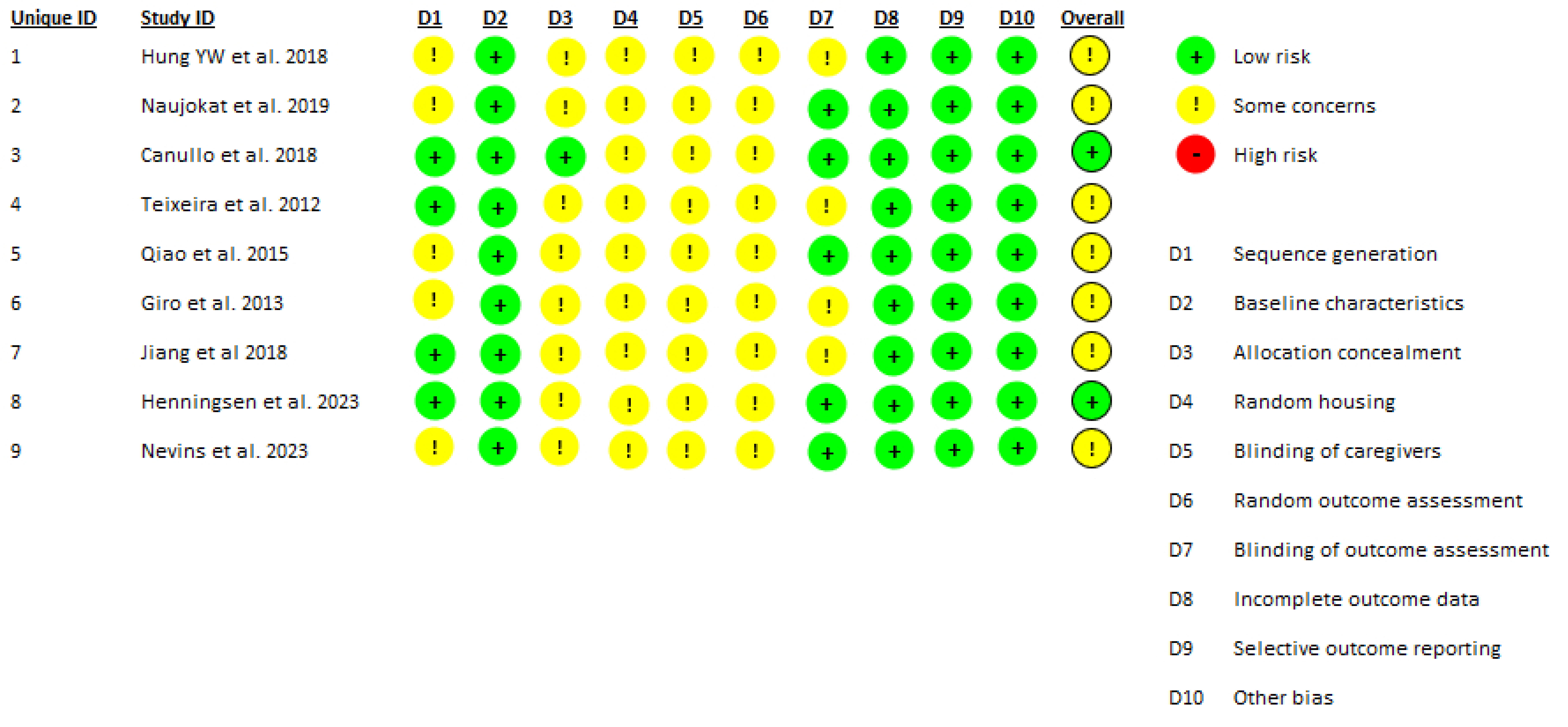
The methodological quality and risk of bias of the included in vivo studies were assessed using the SYRCLE Risk of Bias tool, which is specifically tailored for animal research [43]. This tool evaluates ten domains: sequence generation, baseline characteristics, allocation concealment, random housing, blinding of caregivers/investigators, random outcome assessment, blinding of outcome assessors, incomplete outcome data, selective reporting and other sources of bias.
Each domain was rated as “low risk”, “high risk” or “unclear risk” according to the available information. A summary table (“traffic light” format) was generated to visualize the risk of bias across studies (Figure 2).

3. Results

This systematic review included nine preclinical in vivo studies involving a total of 71 animals, comprising 41 dogs (Beagles, Labrador, Foxhounds), 4 Göttingen minipigs, 6 pigs and 20 mice in which 310 titanium dental implants were placed. Among these, 164 implants underwent surface modification using cold atmospheric pressure plasma (CAP) or silver-ion plasma immersion ion implantation (Ag-PIII), while the remaining 146 implants served as controls or received alternative treatments such as UV light activation. Follow-up periods ranged from 2 to 12 weeks. No adverse clinical events such as inflammation, implant instability, or soft tissue complications were reported, confirming the biological safety of plasma surface treatments under experimental conditions (Table 1).

3.1. Histomorphometric Bone Response

Most studies assessed bone-to-implant contact (BIC) and bone area fraction occupancy (BAFO) as primary indicators of early osseointegration, generally reporting significant improvements in plasma-treated implants compared to controls or alternative treatments. For example, Canullo et al. (2018) [46] in a Beagle model with 32 implants found that at 2 months, plasma-treated implants exhibited a BIC of 72.5% ± 12.4% compared to 64.7% ± 17.3% in controls, with a statistically significant difference (p = 0.012) suggesting enhanced mineralized bone fraction. Similarly, Giro et al. (2013) [49], working with six Beagle dogs and calcium phosphate-coated implants, reported that at 3 weeks plasma treatment doubled BIC and increased BAFO by 82% relative to calcium phosphate coating alone. In Göttingen minipigs, Naujokat et al. (2019) [45] reported BIC values of 90.4% ± 1.24% in plasma-treated implants versus 86.5% ± 1.23% in controls at 8 weeks (p = 0.053). Inter-thread bone density (IBD) was significantly higher in the plasma group (72.47% ± 5.21% vs. 63.36% ± 6.21%, p = 0.002).In Labrador dogs, Qiao et al. (2015) [48] placed 48 implants and found that at 8 weeks the Ag-PIII plasma-treated implants had significantly greater BIC (73.2% ± 5.2% for 30 min and 69.9% ± 4.1% for 60 min) than controls (62.0% ± 4.7%, p < 0.01), with bone density within threads (BDWT) similarly improved. Nevins et al. (2023) [52] working with Foxhounds reported significantly higher BIC (88.3% ± 4.8%) in plasma-treated implants compared to controls at 4 weeks (p = 0.046). Henningsen et al. (2023) [51], in minipigs, documented BAFO values of 80.4% ± 7.0% in plasma-treated implants versus 73.9% ± 12.2% in controls at 8 weeks (p = 0.027), alongside significantly higher BIC at 4 and 8 weeks. In a murine model, Jiang et al. (2018) [50] found that at 4 weeks plasma-treated implants showed a BIC of 47.8% ± 9.6% versus 39.4% ± 9.0% in SLA controls (p < 0.05), with BAFO also significantly improved. Conversely, Hung et al. (2018) [44] reported no statistically significant differences in BIC or crestal bone levels at 4, 8, and 12 weeks, although plasma-treated implants trended towards better values.

3.2. Biomechanical Fixation and Implant Stability

Mechanical implant stability was evaluated by several studies through removal torque testing and Implant Stability Quotient (ISQ) measurements. Teixeira et al. (2012) [47], using plateau-root form implants in Beagle dogs, found significantly higher removal torque values in both 20 s and 60 s plasma-treated groups compared to controls at 2 and 4 weeks (p = 0.001), with no significant difference between the two exposure times. Qiao et al. (2015) [48] observed significantly higher ISQ values in plasma-treated implants relative to SLA controls at 8 weeks (p < 0.01). Nevins et al. (2023) [52] measured ISQ at baseline and found no significant difference between plasma-treated implants and controls, nor changes over time. Henningsen et al. (2023) [51] reported an initial dip in ISQ values at 2 weeks across all groups, followed by a recovery to baseline levels by 8 weeks, with no significant differences between plasma and control groups. Similarly, Hung et al. (2018) [44] found no statistically significant differences in ISQ values between groups at any time point up to 12 weeks.

3.3. Bone Quality and Radiographic Outcomes

Selected studies also evaluated bone quality and architecture using micro-CT and histological imaging. Qiao et al. (2015) [48] reported significantly higher bone volume fraction, bone mineral density, trabecular thickness, and trabecular number in plasma-treated groups compared to controls (p < 0.01). Naujokat et al. (2019) [45] observed similar fluorescent labeling patterns between plasma and control groups, yet plasma-treated implants exhibited higher BIC and IBD (Interthread Bone Denisity) values. Henningsen et al. (2023) [51] noted BAFO exceeding 75% at 2 weeks in all groups, with plasma-treated implants achieving 80.4% ± 7.0% at 8 weeks (p = 0.027), supported by micro-CT scans showing dense mature bone around all implants. Nevins et al. (2023) [52] showed significantly reduced bone loss at 6 weeks in plasma-treated implants (0.56 mm ± 0.24) compared to controls (0.79 mm ± 0.20, p = 0.016), alongside surface chemistry analyses indicating a 31% reduction in carbon contamination and increased oxygen and titanium peaks, suggesting enhanced surface bioactivity.

3.4. Safety and Adverse Events

Across all nine studies, no adverse clinical events related to plasma treatment were reported. All animals tolerated the implant placement and surface treatment procedures without signs of peri-implant inflammation, wound dehiscence or systemic complications. Henningsen et al. (2023) [51] noted significant weight gain in animals and absence of implant loosening or inflammation, with micro-CT confirming consistent osseointegration. Nevins et al. (2023) [52] reported only one minor event in a control implant that lost its healing abutment without infection; all other implants remained stable. Jiang et al. (2018) [50], Qiao et al. (2015) [48] and Teixeira et al. (2012) [47] also confirmed clinical stability and mature bone formation without complications. Similarly, Hung et al. (2018) [44], Canullo et al. (2018) [46], Giro et al. (2013) [49] and Naujokat et al. (2019) [45] reported no adverse events, supporting the biocompatibility and safety of plasma-based surface treatments. Surface analyses from Nevins et al. [52] and Giro et al. [49] showed that plasma treatments effectively reduced carbon contamination and increased oxygen content without introducing harmful residues or causing structural damage.

3.5. Limitations

Despite the encouraging results, some limitations must be acknowledged. The short follow-up periods of 2 to 12 weeks limit the evaluation of long-term osseointegration durability, peri-implant bone stability and implant performance under physiological loading. None of the animal models included functional occlusal loading, which is critical to assess implant behavior in clinical contexts. Considerable heterogeneity in plasma treatment protocols across studies precluded a quantitative meta-analysis, as pooling data under such diverse conditions could have led to misleading conclusions. The use of different animal species, implant sites, and implant numbers per subject further introduces variability and may reduce translational applicability to humans. Inconsistent outcome measures and timing across studies also hinder meta-analytical synthesis. This review was based on PubMed and Scopus databases, a deliberate choice that ensured coverage of the main biomedical literature, although it may still represent a limiting factor. Finally, as all evidence comes from preclinical animal models, cautious interpretation is warranted, and well-designed clinical trials are needed to confirm the efficacy and safety of plasma surface modifications in human patients.

4. Discussion

4.1. Biological Effects of CAP on Osseointegration

This systematic review aimed to provide a comprehensive analysis of the current in vivo evidence regarding the application of cold atmospheric plasma (CAP) treatments on titanium dental implant surfaces, with particular emphasis on their biological effects on osseointegration. The included preclinical data consistently seem to indicate favorable outcomes associated with CAP treatment, notably enhanced bone-to-implant contact (BIC), improved biomechanical fixation, and superior bone quality parameters when compared to untreated control surfaces. Across a range of animal models (including Beagle dogs, Labrador dogs, Foxhounds, Göttingen minipigs, pigs and mice) CAP application was shown to improve early and mid-term osseointegration outcomes. Several studies reported statistically significant increases in BIC within 2 to 8 weeks post-implantation, with improvements of up to 20% over controls (Canullo et al. [46], Qiao et al. [48], Nevins et al. [52]). These results align with previous systematic reviews (Pesce et al. [53]) highlighting plasma and photofunctionalization’s ability to modify surface energy and chemistry, thereby accelerating cellular adhesion and bone healing. In particular, Pesce et al. [53] reported that studies using non-thermal plasma did not show significant improvements at the earliest follow-up (1 week), but consistently demonstrated higher BIC values at later stages (3–8 weeks). Our findings corroborate this time-dependent effect, as most of the included studies indicated that plasma-related benefits become more evident during the early to mid-term phases of osseointegration.

4.2. Biomechanical and Radiographic Outcomes

Biomechanical assessments further supported these findings. Removal torque values and implant stability quotient (ISQ) measurements were generally higher in plasma-treated implants, suggesting enhanced mechanical integrity of the bone–implant interface. However, discrepancies across studies were observed. For example, Hung et al. [44] did not find significant mechanical improvements, suggesting CAP efficacy may depend on specific treatment parameters and implant location. Radiographic and micro-CT analyses revealed positive effects of CAP on peri-implant bone volume, mineral density and trabecular architecture, all features that contribute to long-term implant success under functional loading. Importantly, none of the included studies reported adverse biological reactions or signs of peri-implant inflammation and surface characterization consistently demonstrated reductions in organic contaminants and favorable surface chemistry changes. These findings are consistent with Alqutaibi et al. (2023) [54], who highlighted the dual role of CAP as both an antimicrobial agent and a surface activator.

4.3. The Current Vision of CAP in the Literature

A key issue in the literature is whether CAP should be viewed primarily as a decontamination strategy or a true biofunctionalization tool. While some studies emphasize only its antimicrobial potential in managing peri-implantitis (Alqutaibi et al. [54]), others, including this review, focus on its ability to enhance osseointegration by improving surface wettability, chemistry and energy (Pesce et al. [53]). This duality creates conceptual ambiguity regarding its main mechanism of action. It is important to note that the beneficial effects of CAP do not result from the long-term presence of plasma on the implant surface. Rather, CAP induces transient physicochemical changes, such as increased hydrophilicity and surface energy, that appear to improve early cell adhesion and reduce initial bacterial colonization. However, these effects may diminish over time, and current evidence does not support a lasting antimicrobial action capable of preventing peri-implantitis in the long term. As Najeeb et al. (2015) [55] pointed out, the lack of standardized surface treatment protocols and outcome measures remains a major challenge in CAP research.

4.4. Limitations of Current Preclinical Evidence

A significant limitation identified across studies is the lack of standardization in CAP treatment parameters, such as gas type, exposure time, power, delivery method, and implant distance, which complicates cross-study comparisons (Table 2). Treatment efficacy is strongly influenced by how plasma is generated and applied, with critical variables including modality (e.g., atmospheric jets, vacuum reactors, RF oxidation, or ion implantation), gas composition (argon, oxygen/air, silver), operating conditions (power, pressure, flow, exposure time, nozzle–surface distance) and timing relative to implant insertion.
Immediate post-treatment placement appears essential, as the super-hydrophilic state induced by plasma rapidly decays with time. However, many reports under-specify these variables, thereby limiting reproducibility and comparability. Additional shortcomings of the current evidence include short follow-up periods (2–12 weeks), absence of functional loading and considerable variability in animal models, which together reduce generalizability to humans. This limitation had already been highlighted by Pesce et al. [53], who noted that although qualitative improvements with plasma were evident, heterogeneity in gas types, exposure times and animal models prevented a pooled meta-analysis. Our systematic review confirms this scenario, showing that despite a larger number of studies now available, the absence of protocol harmonization continues to hinder quantitative synthesis. To improve reproducibility and translational relevance, future studies should clearly report device type, gas chemistry, pressure conditions, exposure duration, treatment-to-insertion interval, baseline surface characteristics and objective verification of activation.

4.5. Translational Perspectives

Although current evidence is limited to preclinical models, the promising enhancements in osseointegration suggest potential clinical relevance. Integrating CAP into chair-side workflows could provide a rapid and effective surface biofunctionalization step prior to implant placement, potentially improving early stability and bone healing. However, translation to human patients requires careful consideration of key variables not addressed in animal studies, including systemic conditions, oral microbial diversity and functional occlusal loading. Standardization of CAP protocols is essential to ensure reproducibility and safety in clinical practice. Moreover, combination strategies that pair CAP treatment with other surface modifications, such as growth factor coatings or nanostructured topographies, may offer synergistic effects. Well-designed clinical trials are urgently needed to evaluate not only the efficacy of CAP-treated implants but also their long-term performance, safety, and potential to reduce complications such as peri-implantitis. Addressing these translational gaps will be critical for moving CAP technology from preclinical promise to practical application in dental implantology.
In light of these concerns, future research should focus on developing standardized and clinically relevant CAP protocols. Long-term preclinical studies incorporating functional loading are needed to better simulate oral biomechanics. Moreover, combining CAP with other surface biofunctionalization strategies (such as growth factor coatings or nanostructured topographies) may yield synergistic effects that further improve osseointegration and long-term implant success.

5. Conclusions

This systematic review of preclinical in vivo studies shows that cold atmospheric pressure plasma (CAP) and related plasma-based surface treatments consistently promote early osseointegration of titanium dental implants, as evidenced by improved histomorphometric parameters such as bone-to-implant contact and bone area fraction occupancy. These treatments also show promising effects on implant stability and biomechanical fixation without any reported adverse biological reactions, confirming their safety in animal models. Despite these encouraging findings, the current body of evidence is constrained by several limitations, including short observation periods, absence of functional loading conditions and considerable heterogeneity in both treatment protocols and outcome assessments. As such, while plasma surface modifications show potential as a strategy to enhance implant integration, their routine clinical application requires further validation. Future research should prioritize well-designed clinical trials with standardized methodologies, longer follow-up periods and functional loading to better replicate real clinical conditions. Furthermore, the evaluation of patient-reported outcomes and cost-effectiveness will be essential to fully assess the clinical relevance and practical feasibility of these treatments in dental implantology.

Author Contributions

Conceptualization, C.B. and P.F.; methodology, S.T.; software, M.S.; validation, G.P., P.F. and C.B.; formal analysis, S.T.; investigation, S.T. and M.S.; resources, P.F.; data curation, M.S. and E.M.; writing—original draft preparation, S.T. and M.S.; writing—review and editing, S.T. and C.M.; visualization, M.S.; supervision, P.F. and C.B.; project administration, P.F. All authors have read and agreed to the published version of the manuscript.

Funding

This research received no external funding.

Institutional Review Board Statement

Not Applicable.

Informed Consent Statement

Not Applicable.

Data Availability Statement

No new data were created or analyzed in this study. Data sharing is not applicable to this article.

Conflicts of Interest

The authors declare no conflicts of interest.

Abbreviations

The following abbreviations are used in this manuscript:
CAPCold Atmospheric Plasma
BICBone-to-implant-contact
BAFOBone Area Fraction Occupancy
ISQImplant Stability Quotient
CTComputed Tomography
NTAPPNon-thermal Atmospheric Pressure Plasma
ROS/RNSReactive oxygen and nitrogen species
ALPAlkaline Phosphatase
Runx2Runt-related transcription factor 2
PRISMAPreferred Reporting Items for Systematic Reviews and Meta-Analyses
Ag-PIIISilver-ion plasma immersion ion implantation
BDWT Bone density within threads
SLASandblasted Large-grit Acid-etcher
PBIDPeri Implant Bone Density
IBDInterthread Bone Density

References

  1. Albrektsson, T.; Brånemark, P.I.; Hansson, H.A.; Lindström, J. Osseointegrated titanium implants. Requirements for ensuring a long-lasting, direct bone-to-implant anchorage in man. Acta Orthop. Scand. 1981, 52, 155–170. [Google Scholar] [CrossRef] [PubMed]
  2. Javed, F.; Ahmed, H.B.; Crespi, R.; Romanos, G.E. Role of primary stability for successful osseointegration of dental implants: Factors of influence and evaluation. Interv. Med. Appl. Sci. 2013, 5, 162–167. [Google Scholar] [CrossRef] [PubMed] [PubMed Central]
  3. Schimmel, M.; Srinivasan, M.; McKenna, G.; Müller, F. Effect of advanced age and/or systemic medical conditions on dental implant survival: A systematic review and meta-analysis. Clin. Oral. Implants Res. 2018, 29 (Suppl. 16), 311–330. [Google Scholar] [CrossRef] [PubMed]
  4. Sbricoli, L.; Bazzi, E.; Stellini, E.; Bacci, C. Systemic Diseases and Biological Dental Implant Complications: A Narrative Review. Dent. J. 2022, 11, 10. [Google Scholar] [CrossRef] [PubMed] [PubMed Central]
  5. Pavel, K.; Seydlova, M.; Dostalova, T.; Zdenek, V.; Chleborad, K.; Jana, Z.; Feberova, J.; Radek, H. Dental implants and improvement of oral health-related quality of life. Community Dent. Oral. Epidemiol. 2012, 40 (Suppl. 1), 65–70. [Google Scholar] [CrossRef] [PubMed]
  6. Alqutaibi, A.Y.; Aboalrejal, A.N. Microgap and Micromotion at the Implant Abutment Interface Cause Marginal Bone Loss Around Dental Implant but More Evidence is Needed. J. Evid. Based Dent. Pract. 2018, 18, 171–172. [Google Scholar] [CrossRef] [PubMed]
  7. Bakitian, F.A. A Comprehensive Review of the Contemporary Methods for Enhancing Osseointegration and the Antimicrobial Properties of Titanium Dental Implants. Cureus 2024, 16, e68720. [Google Scholar] [CrossRef] [PubMed] [PubMed Central]
  8. Stich, T.; Alagboso, F.; Křenek, T.; Kovářík, T.; Alt, V.; Docheva, D. Implant-bone-interface: Reviewing the impact of titanium surface modifications on osteogenic processes in vitro and in vivo. Bioeng. Transl. Med. 2021, 7, e10239. [Google Scholar] [CrossRef] [PubMed] [PubMed Central]
  9. Rupp, F.; Liang, L.; Geis-Gerstorfer, J.; Scheideler, L.; Hüttig, F. Surface characteristics of dental implants: A review. Dent. Mater. 2018, 34, 40–57. [Google Scholar] [CrossRef] [PubMed]
  10. Heimes, D.; Becker, P.; Pabst, A.; Smeets, R.; Kraus, A.; Hartmann, A.; Sagheb, K.; Kämmerer, P.W. How does dental implant macrogeometry affect primary implant stability? A narrative review. Int. J. Implant. Dent. 2023, 9, 20. [Google Scholar] [CrossRef] [PubMed] [PubMed Central]
  11. Shayeb, M.A.; Elfadil, S.; Abutayyem, H.; Shqaidef, A.; Marrapodi, M.M.; Cicciù, M.; Minervini, G. Bioactive surface modifications on dental implants: A systematic review and meta-analysis of osseointegration and longevity. Clin. Oral. Investig. 2024, 28, 592. [Google Scholar] [CrossRef] [PubMed] [PubMed Central]
  12. Karthik, K.S.; Sreevidya, B.; Ramya, T.K.; Divya, B.M.; Dedeepya, N.R.; Badiyani, B.K.; Kumar, A. Comparative Analysis of Surface Modification Techniques for Assessing Oral Implant Osseointegration: An Animal Study. Cureus 2024, 16, e54014. [Google Scholar] [CrossRef]
  13. Lata, S.; Chakravorty, S.; Mitra, T.; Pradhan, P.K.; Mohanty, S.; Patel, P.; Jha, E.; Panda, P.K.; Verma, S.K.; Suar, M. Aurora Borealis in dentistry: The applications of cold plasma in biomedicine. Mater. Today Bio 2021, 13, 100200. [Google Scholar] [CrossRef] [PubMed] [PubMed Central]
  14. Rehman, M.U.; Jawaid, P.; Uchiyama, H.; Kondo, T. Comparison of free radicals formation induced by cold atmospheric plasma, ultrasound, and ionizing radiation. Arch. Biochem. Biophys. 2016, 605, 19–25. [Google Scholar] [CrossRef] [PubMed]
  15. Tominami, K.; Kanetaka, H.; Sasaki, S.; Mokudai, T.; Kaneko, T.; Niwano, Y. Cold atmospheric plasma enhances osteoblast differentiation. PLoS ONE 2017, 12, e0180507. [Google Scholar] [CrossRef] [PubMed] [PubMed Central]
  16. Campos-Bijit, V.; Inostroza, N.C.; Orellana, R.; Rivera, A.; Von Marttens, A.; Cortez, C.; Covarrubias, C. Influence of Topography and Composition of Commercial Titanium Dental Implants on Cell Adhesion of Human Gingiva-Derived Mesenchymal Stem Cells: An In Vitro Study. Int. J. Mol. Sci. 2023, 24, 16686. [Google Scholar] [CrossRef] [PubMed] [PubMed Central]
  17. Gund, M.P.; Naim, J.; Lehmann, A.; Hannig, M.; Lange, M.; Schindler, A.; Rupf, S. Cold Atmospheric Plasma Improves the Colonization of Titanium with Primary Human Osteoblasts: An In Vitro Study. Biomedicines 2024, 12, 673. [Google Scholar] [CrossRef] [PubMed] [PubMed Central]
  18. Matthes, R.; Duske, K.; Kebede, T.G.; Pink, C.; Schlüter, R.; von Woedtke, T.; Weltmann, K.D.; Kocher, T.; Jablonowski, L. Osteoblast growth, after cleaning of biofilm-covered titanium discs with air-polishing and cold plasma. J. Clin. Periodontol. 2017, 44, 672–680. [Google Scholar] [CrossRef] [PubMed]
  19. Niedźwiedź, I.; Waśko, A.; Pawłat, J.; Polak-Berecka, M. The State of Research on Antimicrobial Activity of Cold Plasma. Pol. J. Microbiol. 2019, 68, 153–164. [Google Scholar] [CrossRef] [PubMed] [PubMed Central]
  20. Scholtz, V.; Pazlarova, J.; Souskova, H.; Khun, J.; Julak, J. Nonthermal plasma--A tool for decontamination and disinfection. Biotechnol. Adv. 2015, 33 Pt 2, 1108–1119. [Google Scholar] [CrossRef] [PubMed]
  21. Alkawareek, M.Y.; Algwari, Q.T.; Gorman, S.P.; Graham, W.G.; O’Connell, D.; Gilmore, B.F. Application of atmospheric pressure nonthermal plasma for the in vitro eradication of bacterial biofilms. FEMS Immunol. Med. Microbiol. 2012, 65, 381–384. [Google Scholar] [CrossRef] [PubMed]
  22. Brun, P.; Bernabè, G.; Marchiori, C.; Scarpa, M.; Zuin, M.; Cavazzana, R.; Zaniol, B.; Martines, E. Antibacterial efficacy and mechanisms of action of low power atmospheric pressure cold plasma: Membrane permeability, biofilm penetration and antimicrobial sensitization. J. Appl. Microbiol. 2018, 125, 398–408. [Google Scholar] [CrossRef] [PubMed]
  23. Lee, J.Y.; Kim, K.H.; Park, S.Y.; Yoon, S.Y.; Kim, G.H.; Lee, Y.M.; Rhyu, I.C.; Seol, Y.J. The bactericidal effect of an atmospheric-pressure plasma jet on Porphyromonas gingivalis biofilms on sandblasted and acid-etched titanium discs. J. Periodontal Implant. Sci. 2019, 49, 319–329. [Google Scholar] [CrossRef] [PubMed] [PubMed Central]
  24. Jungbauer, G.; Moser, D.; Müller, S.; Pfister, W.; Sculean, A.; Eick, S. The Antimicrobial Effect of Cold Atmospheric Plasma against Dental Pathogens-A Systematic Review of In-Vitro Studies. Antibiotics 2021, 10, 211. [Google Scholar] [CrossRef] [PubMed] [PubMed Central]
  25. Idlibi, A.N.; Al-Marrawi, F.; Hannig, M.; Lehmann, A.; Rueppell, A.; Schindler, A.; Jentsch, H.; Rupf, S. Destruction of oral biofilms formed in situ on machined titanium (Ti) surfaces by cold atmospheric plasma. Biofouling 2013, 29, 369–379. [Google Scholar] [CrossRef] [PubMed]
  26. Sakudo, A.; Yagyu, Y.; Onodera, T. Disinfection and Sterilization Using Plasma Technology: Fundamentals and Future Perspectives for Biological Applications. Int. J. Mol. Sci. 2019, 20, 5216. [Google Scholar] [CrossRef] [PubMed] [PubMed Central]
  27. Zheng, Z.; Ao, X.; Xie, P.; Wu, J.; Dong, Y.; Yu, D.; Wang, J.; Zhu, Z.; Xu, H.H.K.; Chen, W. Effects of novel non-thermal atmospheric plasma treatment of titanium on physical and biological improvements and in vivo osseointegration in rats. Sci. Rep. 2020, 10, 10637. [Google Scholar] [CrossRef] [PubMed] [PubMed Central]
  28. Li, J.; Jansen, J.A.; Walboomers, X.F.; van den Beucken, J.J. Mechanical aspects of dental implants and osseointegration: A narrative review. J. Mech. Behav. Biomed. Mater. 2020, 103, 103574. [Google Scholar] [CrossRef] [PubMed]
  29. Eggers, B.; Marciniak, J.; Deschner, J.; Stope, M.B.; Mustea, A.; Kramer, F.J.; Nokhbehsaim, M. Cold Atmospheric Plasma Promotes Regeneration-Associated Cell Functions of Murine Cementoblasts In Vitro. Int. J. Mol. Sci. 2021, 22, 5280. [Google Scholar] [CrossRef] [PubMed] [PubMed Central]
  30. Moraschini, V.; Barboza, E.S.; Peixoto, G.A. The impact of diabetes on dental implant failure: A systematic review and meta-analysis. Int. J. Oral. Maxillofac. Surg. 2016, 45, 1237–1245. [Google Scholar] [CrossRef] [PubMed]
  31. Dioguardi, M.; Cantore, S.; Quarta, C.; Sovereto, D.; Zerman, N.; Pettini, F.; Muzio, L.L.; Cosola, M.D.; Santacroce, L.; Ballini, A. Correlation between Diabetes Mellitus and Peri-implantitis: A Systematic Review. Endocr. Metab. Immune Disord. Drug Targets. 2023, 23, 596–608. [Google Scholar] [CrossRef] [PubMed]
  32. Carlos, A.; Ziada, H.; Abubakr, N.H. Correlation between marginal bone loss around dental implants and various systemic diseases: A cross-sectional study. Int. J. Implant. Dent. 2024, 10, 46. [Google Scholar] [CrossRef] [PubMed] [PubMed Central]
  33. Corcuera-Flores, J.R.; López-Giménez, J.; López-Jiménez, J.; López-Giménez, A.; Silvestre-Rangil, J.; Machuca-Portillo, G. Four years survival and marginal bone loss of implants in patients with Down syndrome and cerebral palsy. Clin. Oral. Investig. 2017, 21, 1667–1674. [Google Scholar] [CrossRef] [PubMed]
  34. Bredberg, C.; Vu, C.; Häggman-Henrikson, B.; Chrcanovic, B.R. Marginal bone loss around dental implants: Comparison between matched groups of bruxer and non-bruxer patients: A retrospective case-control study. Clin. Implant. Dent. Relat. Res. 2023, 25, 124–132. [Google Scholar] [CrossRef] [PubMed] [PubMed Central]
  35. Aghaloo, T.; Pi-Anfruns, J.; Moshaverinia, A.; Sim, D.; Grogan, T.; Hadaya, D. The Effects of Systemic Diseases and Medications on Implant Osseointegration: A Systematic Review. Int. J. Oral. Maxillofac. Implants. 2019, 34, s35–s49. [Google Scholar] [CrossRef] [PubMed]
  36. Henningsen, A.; Smeets, R.; Hartjen, P.; Heinrich, O.; Heuberger, R.; Heiland, M.; Precht, C.; Cacaci, C. Photofunctionalization and non-thermal plasma activation of titanium surfaces. Clin. Oral. Investig. 2018, 22, 1045–1054. [Google Scholar] [CrossRef] [PubMed]
  37. Wagner, G.; Eggers, B.; Duddeck, D.; Kramer, F.J.; Bourauel, C.; Jepsen, S.; Deschner, J.; Nokhbehsaim, M. Influence of cold atmospheric plasma on dental implant materials-an in vitro analysis. Clin. Oral. Investig. 2022, 26, 2949–2963. [Google Scholar] [CrossRef] [PubMed] [PubMed Central]
  38. Kamionka, J.; Matthes, R.; Holtfreter, B.; Pink, C.; Schlüter, R.; von Woedtke, T.; Kocher, T.; Jablonowski, L. Efficiency of cold atmospheric plasma, cleaning powders and their combination for biofilm removal on two different titanium implant surfaces. Clin. Oral. Investig. 2022, 26, 3179–3187. [Google Scholar] [CrossRef] [PubMed] [PubMed Central]
  39. Wang, L.; Wang, W.; Zhao, H.; Liu, Y.; Liu, J.; Bai, N. Bioactive Effects of Low-Temperature Argon-Oxygen Plasma on a Titanium Implant Surface. ACS Omega 2020, 5, 3996–4003. [Google Scholar] [CrossRef] [PubMed] [PubMed Central]
  40. Duske, K.; Jablonowski, L.; Koban, I.; Matthes, R.; Holtfreter, B.; Sckell, A.; Nebe, J.B.; von Woedtke, T.; Weltmann, K.D.; Kocher, T. Cold atmospheric plasma in combination with mechanical treatment improves osteoblast growth on biofilm covered titanium discs. Biomaterials 2015, 52, 327–334. [Google Scholar] [CrossRef] [PubMed]
  41. Komasa, S.; Kusumoto, T.; Hayashi, R.; Takao, S.; Li, M.; Yan, S.; Zeng, Y.; Yang, Y.; Hu, H.; Kobayashi, Y.; et al. Effect of Argon-Based Atmospheric Pressure Plasma Treatment on Hard Tissue Formation on Titanium Surface. Int. J. Mol. Sci. 2021, 22, 7617. [Google Scholar] [CrossRef] [PubMed] [PubMed Central]
  42. Page, M.J.; McKenzie, J.E.; Bossuyt, P.M.; Boutron, I.; Hoffmann, T.C.; Mulrow, C.D.; Shamseer, L.; Tetzlaff, J.M.; Akl, E.A.; Brennan, S.E.; et al. The PRISMA 2020 statement: An updated guideline for reporting systematic reviews. BMJ 2021, 372, n71. [Google Scholar] [CrossRef] [PubMed] [PubMed Central]
  43. Hooijmans, C.R.; Rovers, M.M.; de Vries, R.B.; Leenaars, M.; Ritskes-Hoitinga, M.; Langendam, M.W. SYRCLE’s risk of bias tool for animal studies. BMC Med. Res. Methodol. 2014, 14, 43. [Google Scholar] [CrossRef] [PubMed] [PubMed Central]
  44. Hung, Y.W.; Chen, H.L.; Lee, L.T.; Tung, K.C.; Bau, D.T.; Wong, Y.K. Effects of non-thermal plasma on sandblasted titanium dental implants in beagle dogs. J. Chin. Med. Assoc. 2018, 81, 920–925. [Google Scholar] [CrossRef] [PubMed]
  45. Naujokat, H.; Harder, S.; Schulz, L.Y.; Wiltfang, J.; Flörke, C.; Açil, Y. Surface conditioning with cold argon plasma and its effect on the osseointegration of dental implants in miniature pigs. J. Cranio-Maxillofac. Surg. 2019, 47, 484–490. [Google Scholar] [CrossRef] [PubMed]
  46. Canullo, L.; Tallarico, M.; Botticelli, D.; Alccayhuaman, K.A.A.; Martins Neto, E.C.; Xavier, S.P. Hard and soft tissue changes around implants activated using plasma of argon: A histomorphometric study in dog. Clin. Oral. Implants Res. 2018, 29, 389–395. [Google Scholar] [CrossRef] [PubMed]
  47. Teixeira, H.S.; Marin, C.; Witek, L.; Freitas, A., Jr.; Silva, N.R.; Lilin, T.; Tovar, N.; Janal, M.N.; Coelho, P.G. Assessment of a chair-side argon-based non-thermal plasma treatment on the surface characteristics and integration of dental implants with textured surfaces. J. Mech. Behav. Biomed. Mater. 2012, 9, 45–49. [Google Scholar] [CrossRef] [PubMed]
  48. Qiao, S.; Cao, H.; Zhao, X.; Lo, H.; Zhuang, L.; Gu, Y.; Shi, J.; Liu, X.; Lai, H. Ag-plasma modification enhances bone apposition around titanium dental implants: An animal study in Labrador dogs. Int. J. Nanomed. 2015, 10, 653–664. [Google Scholar] [CrossRef]
  49. Giro, G.; Tovar, N.; Witek, L.; Marin, C.; Silva, N.R.; Bonfante, E.A.; Coelho, P.G. Osseointegration assessment of chairside argon-based nonthermal plasma-treated Ca-P coated dental implants. J. Biomed. Mater. Res. A 2013, 101, 98–103. [Google Scholar] [CrossRef] [PubMed]
  50. Jiang, H.; Zhang, T.; Zhou, W.; Lin, Z.; Liu, Z. Effect of Plasma Oxidation-Treated TiOx Film on Early Osseointegration. Int. J. Oral. Maxillofac. Implants. 2018, 33, 1011–1018. [Google Scholar] [CrossRef] [PubMed]
  51. Henningsen, A.; Precht, C.; Karnatz, N.; Bibiza, E.; Yan, M.; Guo, L.; Gosau, M.; Smeets, R. Osseointegration of titanium implants after surface treatment with ultraviolet light or cold atmospheric plasma in vivo. Int. J. Oral. Implantol. 2023, 16, 197–208. [Google Scholar] [PubMed]
  52. Nevins, M.; Chen, C.Y.; Parma-Benfenati, S.; Kim, D.M. Gas Plasma Treatment Improves Titanium Dental Implant Osseointegration-A Preclinical In Vivo Experimental Study. Bioengineering 2023, 10, 1181. [Google Scholar] [CrossRef] [PubMed] [PubMed Central]
  53. Pesce, P.; Menini, M.; Santori, G.; Giovanni, E.; Bagnasco, F.; Canullo, L. Photo and Plasma Activation of Dental Implant Titanium Surfaces. A Systematic Review with Meta-Analysis of Pre-Clinical Studies. J. Clin. Med. 2020, 9, 2817. [Google Scholar] [CrossRef] [PubMed] [PubMed Central]
  54. Alqutaibi, A.Y.; Aljohani, A.; Alduri, A.; Masoudi, A.; Alsaedi, A.M.; Al-Sharani, H.M.; Farghal, A.E.; Alnazzawi, A.A.; Aboalrejal, A.N.; Mohamed, A.H.; et al. The Effectiveness of Cold Atmospheric Plasma (CAP) on Bacterial Reduction in Dental Implants: A Systematic Review. Biomolecules 2023, 13, 1528. [Google Scholar] [CrossRef] [PubMed] [PubMed Central]
  55. Najeeb, S.; Khurshid, Z.; Matinlinna, J.P.; Siddiqui, F.; Nassani, M.Z.; Baroudi, K. Nanomodified Peek Dental Implants: Bioactive Composites and Surface Modification-A Review. Int. J. Dent. 2015, 2015, 381759. [Google Scholar] [CrossRef] [PubMed] [PubMed Central]
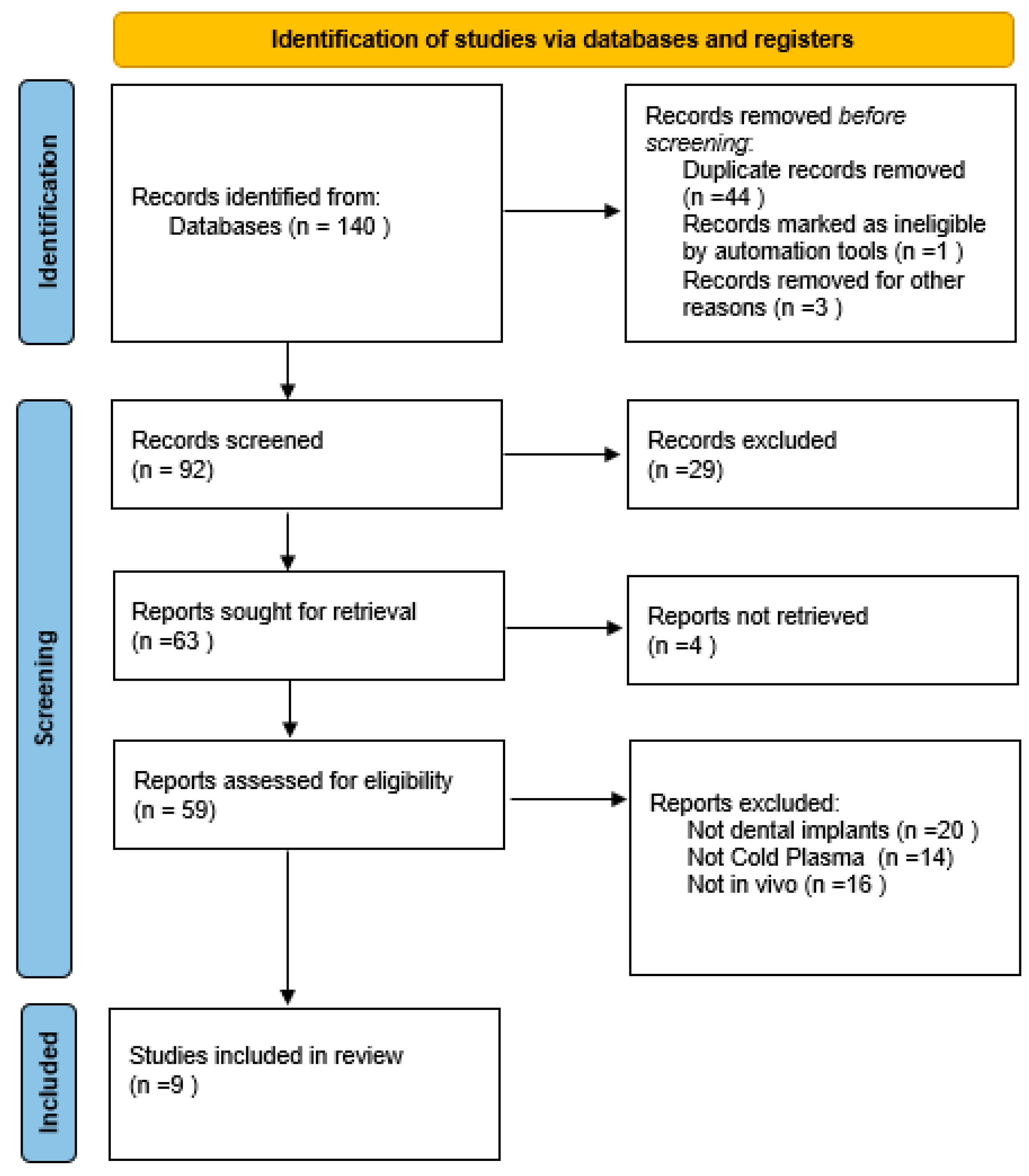
Figure 1. This diagram illustrates the systematic process of identifying, screening and selecting studies for inclusion in the systematic review.
Applsci 15 10302 g001
Figure 2. This table presents the assessment of the risk of bias for each included study, evaluated across the ten domains of the SYRCLE Risk of Bias tool [44,45,46,47,48,49,50,51,52].
Applsci 15 10302 g002
Table 1. This table provides an overview of the studies selected for inclusion in the systematic review. It outlines key information for each study.
TitleAuthors + YearStudy DesignAnimal ModelNumber of ImplantsType of Plasma TreatmentFollow-UpComparisonBIC (%)ISQBAFO %Removal TorquePIBD (Peri Implant Bone Density)%IBD (Interthread Bone Density) %BDWT (Bone-to-Implant Distance Within Threads) %Bone Loss (mm)
Effects of non-thermal plasma on sandblasted titanium dental implants in beagle dogsHung et al., 2018 [44]Preclinical in vivo9 Beagle dogs36 implants (18 treated, 18 untreated), 4 implants per dogArgon NTAPPT0: placement T1: 4 weeks
T2: 8 weeks
T3: 12 weeks
Argon NTAPP treated VS Non treated implantsCONTROL GROUP
(T1): 65.27% ± 3.62
(T2): 82.10% ± 0.23
(T3): 90.20% ± 3.49
STUDY GROUP
(T1): 66.40% ± 3.71
(T2): 80.30% ± 0.66
(T3): 87.38% ± 1.98
(p = non significant)
CONTROL GROUP
(T0): 68.04 ± 3.37
(T1): 66.53 ± 7.40
(T2): 69.20 ± 2.55
(T3): 74.20 ± 2.68
STUDY GROUP
(T0): 67.36 ± 0.52
(T1): 70.17 ± 0.76
(T2): 71.50 ± 1.41
(T3): 77.00 ± 5.87
(p = non significant)
Not reportedNot reportedNot reportedNot reportedNot reportedNot reported
Surface conditioning with cold argon plasma and its effect on the osseointegration of dental implants in miniature pigsNaujokat et al., 2019 [45]Preclinical in vivo4 mini pigs16 implants
(8 treated,8 not treated), 4 implants per pig
Argon NTAPP8 weeksArgon NTAPP treated VS Non treated implantsCONTROL GROUP
86.5 ± 1.23
STUDY GROUP
90.4 ± 1.24
(p = non significant)
Not reportedNot reportedNot reportedCONTROL GROUP 61.14 ± 6.10
STUDY GROUP
60.48 ± 5.03
(p = non significant)
CONTROL GROUP
63.36 ± 6.21
STUDY GROUP
72.47 ± 5.21
(p = 0.002)
Not reportedNot reported
Hard and soft tissue changes around implants activated using plasma of argon: A histomorphometric study in dogCanullo et al., 2018 [46]Preclinical in vivo8 Beagle dogs32 implants (16 treated,16 untreated), 4 implants per dogArgon NTAPPT1:1 month T2: 2 monthsArgon NTAPP treated VS Non treated implantsCONTROL GROUP (T1): 57.2 ± 13.1
(T2): 64.7 ± 17.3
STUDY GROUP
(T1): 60.1 ± 15.6
(T2): 72.5 ± 12.4
p = 0.012 (T2)
Not reportedNot reportedNot reportedNot reportedNot reportedNot reportedNot reported
Assessment of a chair-side argon-based non-thermal plasma treatment on the surface characteristics and integration of dental implants with textured surfacesTeixeira
et al., 2012 [47]
Preclinical in vivo6 Beagle dogs36 implants (24 treated, 12 untreated), 6 per dog (2 plasma 20’, 2 plasma 60’, 2 untreated)Argon NTAPPT1: 2 weeks T2: 4 weeksImplants treated with NTAPP for 20’ VS Implants treated for 60’ VS Non treatedNot reportedNot reportedNot reportedCONTROL(mean rank)
(T1): 8.48 ± 2.08
(T2): 9.50 ± 1.96
PLASMA 20’ (m.r)
9.75 ± 2.40
PLASMA 60’ (m.r)
12.33 ± 2.40
(p = 0–001)
Not reportedNot reportedNot reportedNot reported
Ag-plasma modification enhances bone apposition around titanium dental implants: an animal study in Labrador dogsQiao
et al., 2015 [48]
Preclinical in vivo6 Labrador dogs48 implants (4 types: Control, Ag-PIII 30 min,60 min,90 min)Silver-ion plasma immersion (Ag-PIII)T0: placement
T1:4 weeks
T2: 8 weeks
T3: 12 weeks
Ag-PIII 30 min VS Ag-PIII 60 min VS Ag-PIII 90 min VS Control GroupCONTROL GROUP
(T2): 61.99 ± 4.66
Ag-PIII 30 MIN
(T2): 73.18 ± 5.23
Ag-PIII 60 MIN
(T2): 69.92 ± 4.10
Ag-PIII 90 MIN
(T2): 66.05 ± 3.97
CONTROL GROUP
(T0): >65
(T1): <59
(T2): ~65
(T3): ~70
STUDY GROUPS (T0): >65
(T1): <59
(T2): significant increase (p = 0.01)
(T3):~ 70–72
Not reportedNot reportedNot reportedNot reportedCONTROL GROUP (T2): 66.52 ± 3.46
Ag-PIII 30 MIN
(T2): 77.58 ± 4.3
Ag-PIII 60 MIN
(T2): 77.97 ± 3.34
Ag-PIII 90 MIN
(T2): 69.69 ± 3.68
Not reported
Osseointegration assessment of chairside argon-based nonthermal plasma-treated Ca-P coated dental implants
Giro
et al., 2013 [49]
Preclinical in vivo6 Beagle dogs12 implants (6 treated,6 untreated), 2 implants per dog (1 treated, 1 untreated)NTAPP on CaPT1: 1 week
T2: 3 weeks
CaP coating (control group) VS CaP+ NTP coating (study group)T1: no differences
T3: increase of 100% Vs Control Group
Not reportedT1: no differences
T3: increase of 82% Vs Control Group
Not reportedNot reportedNot reportedNot reportedNot reported
Effect of Plasma Oxidation-Treated TiOx Film on Early OsseointegrationJiang
et al., 2018 [50]
Preclinical in vivo20 rats40 implants (20 treated,20 untreated)Anodic oxidation plasma (PO-SLA)4 weeksSLA surface VS PO-SLA surfaceCONTROL GROUP
39.41 ± 9.00
STUDY GROUP
47.79 ± 9.59
(p < 0.05)
Not reportedCONTROL GROUP
29.01 ± 7.24
STUDY GROUP: 39.10 ± 10.01
(p <0.05)
CONTROL GROUP
9.05 ± 1.42
STUDY GROUP
12.68 ± 1.07
(p <0.05)
Not reportedNot reportedNot reportedNot reported
Osseointegration of titanium implants after surface treatment with ultraviolet light or cold atmospheric plasma in vivoHenningsen et al., 2023 [51]Preclinical in vivo6 pigs54 implants (18 untreated,18 UV treated, 18 plasma treated), 9 implants per pigCAPT0: placement T1: 2 weeks
T2: 4 weeks
T3: 8 weeks
UV treated VS CAP treated VS untreatedCONTROL GROUP
(T1): >69%
(T3): 73.0 ± 2.8
STUDY GROUP
(T1): (p < 0.05)
(T3): 80.6 ± 5.0
CONTROL GROUP
(T0): 90.4 ± 7.2
(T3): 93.1 ± 5.4
STUDY GROUP
(T0): 92.4 ± 5.9
(T3): 89.2 ± 8.3
CONTROL GROUP
(T3): 73.9 ± 12.2
STUDY GROUP
(T3): 80.4 ± 7.0
(p = 0.027)
Not reportedNot reportedNot reportedNot reportedNot reported
Gas Plasma Treatment Improves Titanium Dental Implant Osseointegration—A Preclinical In Vivo Experimental StudyNevins
et al., 2023 [52]
Preclinical in vivo6 Foxhounds dogs36 implants (18 treated,18 untreated)Argon NTAPPT0: placement
T1: 2 weeks
T2: 4 weeks
T3: 6 weeks
Argon NTAPP treated VS non treated implantsCONTROL GROUP
(T2): 88.3 ± 4.8
STUDY GROUP
(T2): 93.7 ±3.3
(p = 0.046)
CONTROL GROUP
(T0): 79.39 ± 2.95
STUDY GROUP
(T0): 79.53 ± 4.05
(p = 0.6, non significant)
Not reportedNot reportedNot reportedNot reportedNot reportedCONTROL GROUP (T3): 0.79 ± 0.20
STUDY GROUP
(T3): 0.56 ± 0.24
(p = 0.016)
Table 2. This table summarizes the plasma parameters used in each study.
TitleAuthorsType of Plasma TreatmentParameters
Effects of non-thermal plasma on sandblasted titanium dental implants in beagle dogsHung
et al., 2018 [44]
Argon NTAPPDevice: Yih Dar Technology, Hsinchu County, Taiwan (ISO 9001 certified)
Electrodes: Aluminum tape electrodes on quartz tube
(4 mm inner diameter, 10 mm spacer)
Argon flow rate: 1.8 L/min
Oxygen flow rate: 0.01 L/min
Voltage: High-voltage mono-polar square pulses
Repetition rate: 0.5–4 kHz
Duration: 60 s per implant
Surface conditioning with cold argon plasma and its effect on the osseointegration of dental implants in miniature pigsNaujokat
et al., 2019 [45]
Argon NTAPPPressure: 2 bar
Flow rate: 4.3–4.4 L/min
Frequency: 20 kHz
Voltage: 115–230 V
Temperature: <40 °C at the point of application
Distance: 7 mm
Duration: 240 s total (60 s per quadrant, from the neck
to the tip of the implant)
Hard and soft tissue changes around implants activated using plasma of argon: A histomorphometric study in dogCanullo
et al., 2018 [46]
Argon NTAPPPower: 75 W
Pressure: −10 mbar
Temperature: room temperature
Duration: 12 min
Assessment of a chair-side argon-based non-thermal plasma treatment on the surface characteristics and integration of dental implants with textured surfacesTeixeira
et al., 2012 [47]
Argon NTAPPPressure: atmospheric
Temperature: room temperature
Duration: 20 or 60 s per quadrant (KinPen™ device, Neoplas tool GmbH, Greifswald, Germany)
Ag-plasma modification enhances bone apposition around titanium dental implants: an animal study in Labrador dogsQiao
et al., 2015 [48]
Silver-ion plasma immersion
(Ag-PIII)
Cathode rod: Pure silver (99.99% purity, 10 mm diameter)
Temperature during silver release test: 37 °C
Duration of silver release test: 3 months (samples in 10 mL water)
Analysis: Silver release quantified by Inductively Coupled
Plasma Optical Emission Spectrometry (ICP-OES)
Osseointegration assessment of chairside argon-based nonthermal plasma-treated Ca-P coated dental implantsGiro
et al., 2013 [49]
NTAPP on CaPPressure: atmospheric
Temperature: Room temperature
Duration: 20 s per quadrant (KinPen™ device, portable, length 155 mm, diameter 20 mm, weight 170 g)
Effect of Plasma Oxidation-Treated TiOx Film on Early OsseointegrationJiang
et al., 2018 [50]
Anodic oxidation plasma (PO-SLA)Plasma oxidation treatment technical parameters not specified in detail
Osseointegration of titanium implants after surface treatment with ultraviolet light or cold atmospheric plasma in vivoHenningsen
et al., 2023 [51]
CAPDevice: Yocto III CAP reactor (Diener electronic GmbH, Ebhausen, Germany)
Power: 24 W
Pressure: vacuum of –0.5 mbar
Duration: 12 min
Gas Plasma Treatment Improves Titanium Dental Implant Osseointegration—A Preclinical In Vivo Experimental Study Nevins
et al., 2023 [52]
Argon NTAPPDevice: ACTILINK Reborn (Plasmapp Co., Ltd., Daejon, Republic of Korea)
Vacuum base pressure: ~5 torr reached within 30 s
Operating pressure: 5–10 torr (optimal range for hydrocarbon removal)
Electrical input: sinusoidal power, frequency 100 kHz, voltage 3 kV
Treatment time: 8 s plasma exposure
Additional cycle: Vacuum generation: 30 s, Plasma treatment: 8 s,
Purification/by-product elimination: 17 s, Venting: 5 s
Total cycle: ~60 s
Gas management: chamber vented via HEPA filter to
prevent reattachment of impurities
Disclaimer/Publisher’s Note: The statements, opinions and data contained in all publications are solely those of the individual author(s) and contributor(s) and not of MDPI and/or the editor(s). MDPI and/or the editor(s) disclaim responsibility for any injury to people or property resulting from any ideas, methods, instructions or products referred to in the content.