Article

Effect of Anti-Pronation Athletic Tape Types: A Randomized Crossover Trial on Ankle Strength, Gait Parameters, and Balance Control Ability in Women with Flexible Flat Feet

1
Department of Physical Therapy, Uiduk University, Gyeongju 38004, Republic of Korea
2
Department of Physical Therapy, College of Health and Life Science, Korea National University of Transportation, Chungbuk 27909, Republic of Korea
*
Author to whom correspondence should be addressed.
Appl. Sci. 2025, 15(12), 6858; https://doi.org/10.3390/app15126858
Submission received: 21 April 2025 / Revised: 16 June 2025 / Accepted: 16 June 2025 / Published: 18 June 2025
(This article belongs to the Special Issue Advanced Physical Therapy for Rehabilitation)

Abstract

:
Athletic Tape is widely used as an immediate and cost-effective intervention for flexible flat feet, offering a practical alternative to orthotic devices and exercise therapies. This study aimed to compare the effects of low-dye and anti-pronation taping (elastic and inelastic) on ankle strength, gait parameters, and balance control in women with flexible flat feet. Thirty women were evaluated under four conditions: no taping, low-dye taping, elastic anti-pronation taping, and inelastic anti-pronation taping. Each condition was tested at 3-day intervals. Outcome measures included ankle muscle strength, step length, stride length, balance control ability assessed using the Romberg and limits of stability tests. Repeated-measures analysis of variance and post hoc least significant difference analyses were used to determine statistical significance. Additionally, effect sizes (η2) were calculated for the primary outcomes. Dorsiflexion strength significantly improved with elastic taping (p < 0.05). Step length increased with both elastic and inelastic taping, whereas stride length improved only with elastic taping. All taping methods significantly reduced the limits of stability compared with the no-taping condition (p < 0.05). Athletic Tape interventions, especially elastic anti-pronation taping, may reduce excessive foot pronation and improve ankle strength and gait performance in women with flexible flat feet.

1. Introduction

The medial and lateral longitudinal arches (MLAs and LLAs) are a key structural component of the foot that plays an essential role in absorbing impacts and providing propulsion during walking and running [1,2]. One of the most common musculoskeletal conditions affecting the MLAs is flexible flatfoot, in which the entire sole of the foot makes contact with the ground during weight-bearing, regardless of the presence of a normal arch during non-weight-bearing [3,4].
Although the exact causes of flexible flatfoot, a condition reported to be more prevalent in women, are unclear, weight bearing is known to cause plantar flexion, medial deviation of the talus, and eversion of the calcaneus, resulting in excessive pronation of the foot [5]. An altered alignment impairs normal foot function and may contribute to fatigue and discomfort during walking.
A key biomechanical mechanism affected by flexible flatfoot is the windlass mechanism, which involves the plantar aponeurosis, a structure that connects the heel to the base of the toes. During toe dorsiflexion, the plantar fascia tightens and elevates the MLA, providing rigidity to the foot and supporting propulsion during gait [6,7]. However, in individuals with flexible flat feet, a decrease in the MLA height compromises the effectiveness of this mechanism and negatively affects gait performance and shock absorption [4].
Flexible flatfeet can also impair postural balance. The soles of the feet contain mechanoreceptors that are sensitive to pressure changes, contributing to proprioceptive feedback and postural control [8]. When the arch collapses during weight bearing, the increased plantar contact area may disrupt sensory input, leading to reduced balance ability. Several studies have reported improved balance in individuals with flat feet following the use of orthotic insoles [9,10].
Athletic Tape has emerged as a popular intervention because of its immediate effects and cost-effectiveness compared with orthotic devices. It is commonly used to support the arch and reduce excessive pronation during movement [11,12,13,14,15]. Among the various taping techniques, low-dye taping has been widely studied and is known to elevate the navicular bone and reinforce the MLA by applying tension across the midfoot [16,17,18]. This technique has demonstrated effectiveness in redistributing plantar pressure during standing and walking [19,20].
Another method, anti-pronation spiral taping, is simpler to apply and more adherent to the foot than traditional methods. It offers enhanced stability owing to its spiraling structure [21]. Despite the growing interest in taping techniques, studies directly comparing different taping techniques, particularly low-dye and anti-pronation taping, are limited [22].
Despite the increasing clinical use of both elastic and inelastic taping techniques for managing flexible flatfoot, few studies have systematically compared their effects under standardized conditions. The lack of comparative evidence limits informed clinical decision-making, particularly in relation to functional outcomes such as gait, balance, and ankle strength. Therefore, this study aimed to investigate the effects of low-dye taping and anti-pronation taping (elastic and inelastic) on ankle strength, gait parameters, and balance control in adult women with flexible flat feet. We hypothesized that elastic anti-pronation taping would yield superior improvements in gait and dorsiflexion strength compared to other taping methods, that is, the effects of low-dye taping and anti-pronation taping (elastic and inelastic) on ankle strength, gait parameters, and balance control in adult women with flexible flat feet.

2. Materials and Methods
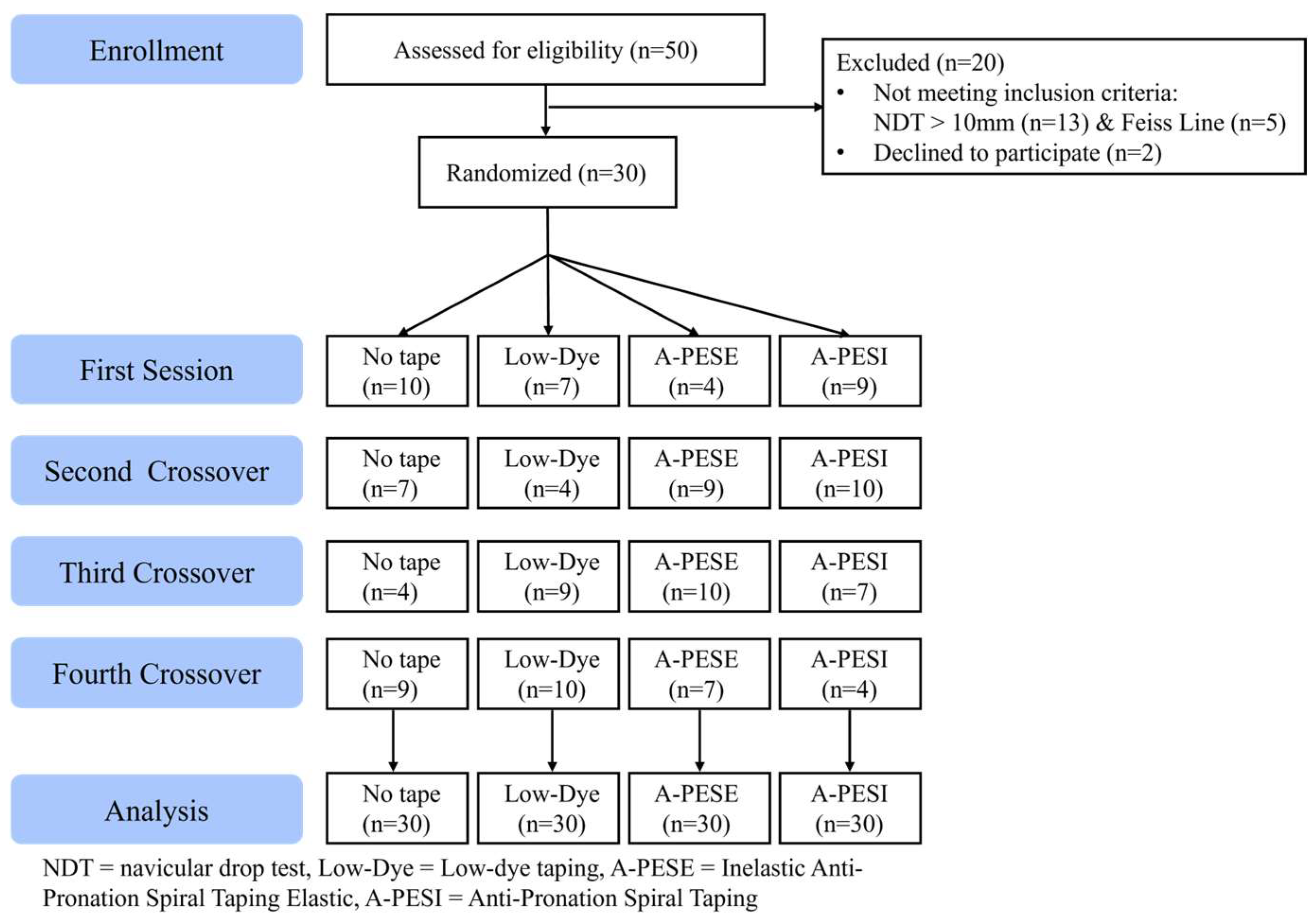
2.1. Participants

Thirty women with flexible flat feet were recruited from U University, Republic of Korea. The inclusion criteria were (i) being a female aged between 20 and 39 years, selected to control for age-related degenerative changes in foot structure; (ii) exhibiting a medial longitudinal arch collapse pattern confirmed using either the navicular drop test (NDT) with a drop greater than 10 mm; or (iii) a positive result on the Feiss line test. The exclusion criteria were (i) any recent (within two years) history of lower extremity musculoskeletal injury; (ii) neurological conditions; or (iii) prior foot or ankle surgery. The general characteristics of the participants, including age, height, and weight, are presented in Table 1.
Individual Navicular Drop Test values for all participants are provided in Supplementary Table S1.
The required sample size was calculated using G*Power software (v3.1.9.7). For a repeated-measures ANOVA, an a priori power analysis indicated that a minimum of 28 participants was needed to achieve 80% power with a medium effect size (f = 0.25) at a significance level of α = 0.05, assuming four measurements with a correlation of 0.4. Anticipating a high exclusion rate during the screening process for flexible flatfoot, we aimed to recruit approximately double the required number. Consequently, 50 individuals were initially assessed for eligibility, from which our final sample of 30 was enrolled, satisfying the power requirement.

2.2. Experimental Procedure

Fifty participants were initially recruited and underwent a one-week screening process, during which the NDT and the Feiss line test were conducted to assess the presence of flexible flatfoot. Of these, 30 met the inclusion criteria and were enrolled. Prior to participation, all participants were informed of the purpose of the study and provided written consent.
Each participant underwent testing under four different taping conditions: no taping, low-dye taping, elastic anti-pronation taping, and inelastic anti-pronation taping. Athletic Tape was applied to the participant’s dominant foot in all conditions. The dominant foot was defined as the foot used to kick a ball. The order of the taping conditions was randomized using a random number table generated in Microsoft Excel to minimize order effects, and there was a 3-day interval between each condition to prevent carryover effects (Figure 1). To control for potential diurnal variations, testing for each participant was consistently conducted within the same period of the day (i.e., all sessions in the morning or all sessions in the afternoon).
Gait, balance, and ankle muscle strength were measured during each testing session. A rest period of 60 s was provided between the trials to prevent fatigue and ensure data reliability.
The study protocol was approved by the Institutional Review Board of SUNMOON University (IRB No. SM-202205-027-3).

2.3. Screening Methods for Flexible Flat Feet

Flexible flatfoot was identified using the NDT. First, the participants were seated with their feet in a neutral, non-weight-bearing position. Vertical height from the floor to the navicular tuberosity was measured using a ruler. The participants were then asked to stand in a relaxed weight-bearing posture, and the same measurement was repeated.
The navicular drop was calculated as the difference in height between the seated and standing positions. A drop greater than 10 mm was used as the threshold for inclusion.
In the Feiss line test, participants stood with their feet approximately 15 cm apart, evenly distributing weight on both legs. A line was drawn from the inferior aspect of the medial malleolus to the first metatarsophalangeal joint, and the position of the navicular tuberosity was observed. If the tuberosity was located below this line, it was considered a positive finding, indicating a collapsed medial longitudinal arch consistent with flexible flatfoot. All measurements were performed by the same trained examiner using calibrated instruments, and the mean of three trials was used for the analysis to ensure reliability. The NDT is recognized as a valid and reliable clinical measure for quantifying navicular bone drop, which is a key indicator of flexible flatfoot [23].

2.4. Interventions

2.4.1. Low-Dye Taping

Low-dye taping was applied using a non-elastic tape (Mueller Sports Medicine, Inc., Prairie du Sac, WI, USA) following standard procedures [16,17]. Participants were seated with their legs extended and feet in a neutral position with the subtalar joint maintained in neutral and the forefoot slightly supinated. Two 35–40 cm anchor strips and five to six 15–20 cm mini stirrups were used, adjusted according to foot size. The tape was first anchored around the heel, beginning at the fifth metatarsal, and wrapped medially across the sole to the first metatarsal to promote forefoot adduction and plantarflexion. Additional mini stirrups were applied vertically from the lateral to the medial side of the plantar surface, overlapping by approximately two-thirds of their width, to support the medial longitudinal arch and the navicular tuberosity. Each strip covered the area from the anterior ankle to the calcaneus. The final fixation strip was applied dorsally across the midfoot, connecting the medial and lateral anchors to secure the tape in place.

2.4.2. Anti-Pronation Spiral Taping

For anti-pronation spiral taping, 5 cm wide, 25 cm long elastic (Nasara, Paju, Republic of Korea) or inelastic tape was used. Participants remained seated with their legs extended and feet in a neutral position. The tape was applied from the lateral aspect of the fifth metatarsal head, pulled diagonally across the plantar surface, and secured to the medial side while the foot was dorsiflexed and slightly inverted. This taping method was intended to support the arch and resist pronation (Figure 2c–f).

2.5. Measurements

2.5.1. Gait Parameters

Gait was evaluated using the Dartfish software (version 9 Pro; Dartfish, Fribourg, Switzerland), which enables video-based motion analysis. Video-based analysis software has been established as a valid tool for measuring spatiotemporal gait parameters [24]. Participants were instructed to walk barefoot at a comfortable, self-selected pace along a straight path, ensuring at least two steps before reaching the measurement zone to capture a natural gait pattern.
The gait video was analyzed to identify specific phases such as swing and heel strike. Step length was defined as the distance from the heel strike of one foot to the heel strike of the opposite foot, whereas stride length was defined as the distance between two consecutive heel strikes of the same foot.
All recordings and measurements were performed by the same trained evaluator to ensure consistency. Measurements were taken over three trials, and the average value was used for the analysis to ensure reliability.

2.5.2. Balance Control Ability

Balance control was assessed using the BioRescue System (RM Ingenierie, Rodez, France), which consists of a pressure-sensitive platform connected to a computer for real-time postural analyses and training. Force platforms, such as the one used in this study, are considered the gold standard for reliably quantifying postural stability and balance control [25]. The system measures the center of pressure path, area (cm2), length (cm), and average velocity (cm/s) during specific balance tasks.
Two tests were conducted: the Romberg test in a standing posture and the limits of stability (LOS) test. During both assessments, participants received verbal instructions and demonstrations. Each test was performed before and after tape application, with a 60 s rest in between the trials. Each test was performed three times, and the average values were used for the analysis to ensure reliability.
In the Romberg test, participants stood quietly on the platform for 30 s with feet shoulder-width apart and eyes closed. For the LOS test, participants stood on the platform with their feet approximately shoulder-width apart and heels 3 cm apart, maintaining a forward gaze. They were positioned 1 to 1.5 m in front of a monitor in a quiet room to minimize distraction. A trained assistant stood nearby during the test to ensure safety, without physical contact unless required.

2.5.3. Ankle Strength

Ankle muscle strength was measured using a handheld dynamometer (HHD) (Commander Muscle Tester; JTech Medical, Midvale, UT, USA), which is a portable device commonly used to assess muscle function and strength in clinical and research settings. The HHD is particularly useful for evaluating foot strength and monitoring progress in patients with musculoskeletal or neurological impairments. The HHD has demonstrated high levels of validity and reliability for measuring isometric strength in lower limb muscles, including those controlling the ankle [26]. The peak force exerted against the dynamometer was recorded in kilograms (kg).
In this study, the isometric strength of the four ankle motions (dorsiflexion, plantarflexion, inversion, and eversion) was assessed. The participants were seated with the ankle in a neutral position, and resistance was applied manually by the examiner using an HHD. For dorsiflexion and plantarflexion, force was applied to the dorsal and plantar surfaces of the foot at the level of the metatarsal heads, respectively. Inversion and eversion were assessed by applying resistance to the medial and lateral aspects of the foot, simultaneously capturing supination and pronation movements of the forefoot, respectively. Only the taped foot was tested. To prevent displacement during testing, the examiner stabilized their posture by leaning them against a wall. Each motion was tested once per condition, aligning with prior studies on immediate taping effects. Isokinetic strength testing was not performed, which was a limitation and is recommended for future studies. All the tests were conducted by the same trained evaluator to ensure reliability.

2.6. Statistical Analysis

All statistical analyses were performed using SPSS software (version 23.0; IBM Corp., Armonk, NY, USA). Descriptive statistics were used to summarize participants’ general characteristics.
A repeated-measures analysis of variance was conducted to examine the differences across the four taping conditions (no taping, low-dye, elastic anti-pronation, and inelastic anti-pronation). When a significant main effect was found, Fisher’s least significant difference post hoc test was applied to determine pairwise differences.
All data are reported as mean ± standard deviation, and statistical significance was set at α = 0.05. To evaluate the magnitude of the differences, effect sizes were calculated using partial eta squared (η2), in accordance with established guidelines [27].

3. Results

3.1. Differences in Ankle Muscle Strength Based on the Type of Taping

Dorsiflexion strength showed a significant increase following the application of elastic anti-pronation taping compared with that in the other taping types (p < 0.05). Although muscle strength increased during other ankle movements (plantar flexion, inversion, and eversion) after taping, the differences were not significant (p > 0.05).
The detailed results for each taping condition are presented in Table 2.

3.2. Analysis of Gait Parameters Based on the Type of Taping

Step length significantly increased after the application of both elastic and inelastic anti-pronation taping compared with that after the other taping types (p < 0.05). However, stride length showed a significant improvement only with elastic taping (p < 0.05), whereas it showed no significant change with low-dye or inelastic taping.
The detailed gait parameter values for each condition are listed in Table 3.

3.3. Analysis of Balance Control Ability Based on the Type of Taping

In the Romberg test, no significant differences were found in sway length or sway velocity across any of the taping conditions compared with the those in the no-taping condition (p > 0.05).
In contrast, the LOS values significantly decreased after each taping-type application (low-dye, elastic, and inelastic) compared with that after the no-taping condition (p < 0.05), indicating a reduced movement range.
A summary of the balance control measurements is provided in Table 4.

4. Discussion

This study examined the effects of low-dye and anti-pronation spiral taping on ankle strength, gait parameters, and balance control ability in young women with flexible flat feet. These findings suggest that taping, particularly elastic anti-pronation taping, may be an effective intervention for improving certain aspects of functional movement in this population.
The most notable improvement in ankle strength was observed in dorsiflexion after elastic taping. This result supports the findings of Ho et al., who found that taping enhances dorsiflexion strength in basketball players with overpronated feet [28]. One possible explanation for this finding is that elastic taping facilitates greater proprioceptive input and muscle activation around the tibialis anterior. Additionally, Bruening et al. (2023) emphasized the importance of the tape tension direction, which supports the methodological rigor of our taping application and its enhancement in neuromuscular control [29]. Furthermore, elastic tape may allow for a degree of dynamic movement in which inelastic or rigid taping methods are restricted, leading to increased functional recruitment of the dorsiflexor muscles [29,30]. Although other directions of ankle movement, such as plantar flexion, inversion, and eversion, showed some increase in strength, the lack of any significant difference suggests that the effect of taping may be more localized or movement-specific.
Concerning gait, both elastic and inelastic taping led to significant increases in step length, whereas only elastic taping led to an improvement in stride length. This difference may be attributed to the mechanical flexibility of the elastic tape, allowing for a more natural joint excursion and muscle engagement during the gait cycle. Previous studies noted that elastic taping can assist in restoring normal foot mechanics, thereby promoting more efficient heel-to-toe progression [20,30]. In contrast, low-dye taping, although effective in arch support, did not significantly influence either step or stride length in this study. This finding corroborates the findings of Radford et al. [16], who suggested that low-dye taping may offer structural stability but lacks the dynamic facilitation required to enhance gait parameters in healthy or mildly symptomatic individuals.
Concerning balance control, Romberg test results revealed no significant changes in sway length or sway velocity following taping. It is also worth noting that the Romberg test was performed with eyes closed, which typically increases the sensitivity of the assessment to proprioceptive or vestibular influences. However, the absence of significant changes suggests that short-term taping may not sufficiently impact static balance under deprived sensory conditions. Future research may benefit from incorporating dynamic balance tasks or perturbation-based protocols to assess functional postural control. This finding is consistent with the findings of a study indicating that short-term taping may not sufficiently alter static postural stability [31]. However, all taping types led to a significant reduction in the LOS, which requires careful interpretation. While decreased LOS may indicate improved control, it may alternatively indicate a reduced range of voluntary postural movement due to mechanical restrictions imposed by taping. Similar interpretations were made by Correia et al. [32] and Calmels et al. [31] who noted that although taping can provide mechanical support, it may concurrently limit the body’s capacity to explore postural boundaries.
The differential effects observed between elastic and inelastic taping further highlight the importance of the mechanical properties in therapeutic taping. Compared with inelastic materials, elastic materials may promote better proprioceptive feedback and allow for a functional range of motion, which are essential for gait and strength gain. Conversely, compared with elastic taping, rigid or inelastic taping may offer greater structural correction at the expense of joint mobility.
Overall, the study results indicate that while all forms of taping may provide arch support and contribute to static alignment, elastic anti-pronation taping demonstrated superior outcomes in dorsiflexion strength and stride length. However, inelastic taping also showed benefits, especially for step length, and should not be overlooked. The lack of improvement in balance measures underscores the complexity of postural control and suggests that sensory or motor training may be needed in addition to mechanical intervention.
This study has several limitations. First, the sample included only young women, limiting the generalizability of the results to males and older adults. Second, the interventions were tested over short time intervals without assessing the long-term effects or adherence. Third, while strength and balance were measured using reliable tools, electromyography and three-dimensional motion analysis can provide more comprehensive insights into neuromuscular and biomechanical changes. Fourth, baseline data on the participants’ physical activity levels were not collected, which is a limitation as regular physical activity could act as a confounding variable influencing ankle strength, gait, and balance outcomes. Finally, although the order of the taping interventions was randomized, neither the participants nor the assessors were blinded to the specific taping method being applied, which could have introduced potential performance or measurement bias. Given the significant improvement observed in dorsiflexion strength following elastic taping, future research could explore its application in populations requiring explosive or stabilizing ankle control, such as female athletes with flexible flat feet or individuals with chronic ankle instability. Investigating the effects of taping on jump mechanics, dynamic balance, and injury risk may provide clinically meaningful insights for sports rehabilitation.

5. Conclusions

This study demonstrated that anti-pronation taping can improve dorsiflexion strength and specific gait parameters in young women with flexible flat feet. Both elastic and inelastic taping resulted in a significant increase in step length, while only elastic taping improved stride length. Furthermore, all taping methods—low-dye, inelastic, and elastic—led to a significant reduction in the limits of stability (LOS) compared to the no-taping condition. However, taping did not enhance static balance control, suggesting that the mechanical restriction may limit dynamic postural adjustment.
These findings suggest that elastic taping may serve as a useful intervention to enhance functional mobility in individuals with flexible flat feet. While elastic taping showed the most comprehensive benefits, inelastic taping also proved to be an effective option for improving step length. Future studies should explore the long-term effects, incorporate diverse populations, and use additional biomechanical assessments.

Supplementary Materials

The following supporting information can be downloaded at: https://www.mdpi.com/article/10.3390/app15126858/s1, Table S1: Individual Navicular Drop Test Values.

Author Contributions

S.-Y.P.: Conceptualization, Investigation, Methodology, Writing—Original Draft (Introduction and Methods). S.-G.K.: Formal Analysis, Writing—Original Draft (Results and Discussion), Writing—Review and Editing. All authors have read and agreed to the published version of the manuscript.

Funding

This work was supported by Uiduk University Foundation Grant, 2025.

Institutional Review Board Statement

The study was conducted in accordance with the Declaration of Helsinki and approved by the Institutional Review Board of SUNMOON university (IRB No. SM-202205-027-3).

Informed Consent Statement

The data presented in this study are available on request from the corresponding author. The data are not publicly available due to privacy and ethical restrictions.

Data Availability Statement

All data generated or analyzed during this study are included in this published article.

Acknowledgments

We acknowledge the financial support from Uiduk University Foundation Grant, 2025.

Conflicts of Interest

The authors declare no conflicts of interest.

Abbreviations

The following abbreviations are used in this manuscript:
MLAmedial longitudinal arch
NDTnavicular drop test
HHDhandheld dynamometer
LOSlimits of stability

References

  1. McKeon, P.O.; Fourchet, F. Freeing the foot: Integrating the foot core system into rehabilitation for lower extremity injuries. Clin. Sports Med. 2015, 34, 347–361. [Google Scholar] [CrossRef] [PubMed]
  2. Welte, L.; Kelly, L.A.; Lichtwark, G.A.; Rainbow, M.J. Influence of the windlass mechanism on arch-spring mechanics during dynamic foot arch deformation. J. R. Soc. 2018, 15, 20180270. [Google Scholar] [CrossRef] [PubMed]
  3. Pourhoseingholi, E.; Pourhoseingholi, M.A. Comparison of arch index of flat foot and healthy foot in pre-school children. Thrita 2013, 2, 15–18. [Google Scholar] [CrossRef]
  4. de Cesar Netto, C.; Schon, L.C.; Thawait, G.K.; da Fonseca, L.F.; Chinanuvathana, A.; Zbijewski, W.B.; Siewerdsen, J.H.; Demehri, S. Flexible adult acquired flatfoot deformity: Comparison between weight-bearing and non-weight-bearing measurements using cone-beam computed tomography. J. Bone Joint Surg. Am. 2017, 99, e98. [Google Scholar] [CrossRef]
  5. Kido, M.; Ikoma, K.; Imai, K.; Maki, M.; Takatori, R.; Tokunaga, D.; Inoue, N.; Kubo, T. Load response of the tarsal bones in patients with flatfoot deformity: In vivo 3D study. Foot Ankle Int. 2011, 32, 1017–1022. [Google Scholar] [CrossRef] [PubMed]
  6. Aydin, M.; Mutlu, R.; Singh, D.; Sariyildiz, E.; Coman, R.; Mayland, E.; Shemmell, J.; Lee, W. Novel soft haptic biofeedback—Pilot study on postural balance and proprioception. Sensors 2022, 22, 3779. [Google Scholar] [CrossRef]
  7. Yu, I.; Ko, M.; Oh, J. The effects of biofeedback training for efficient activation of infraspinatus on proprioception and EMG activity during shoulder external rotation. J. Electromyogr. Kinesiol. 2023, 71, 102798. [Google Scholar] [CrossRef]
  8. Maurer, C.; Mergner, T.; Bolha, B.; Hlavacka, F. Human balance control during cutaneous stimulation of the plantar soles. Neurosci. Lett. 2001, 302, 45–48. [Google Scholar] [CrossRef]
  9. Zijlstra, A.; Mancini, M.; Chiari, L.; Zijlstra, W. Biofeedback for training balance and mobility tasks in older populations: A systematic review. J. Neuroeng. Rehabilit. 2010, 7, 58. [Google Scholar] [CrossRef]
  10. Alghadir, A.H.; Iqbal, Z.A. Effect of deep cervical flexor muscle training using pressure biofeedback on pain and forward head posture in school teachers with neck pain: An observational study. BioMed Res. Int. 2021, 2021, 5588580. [Google Scholar] [CrossRef]
  11. Brijwasi, T.; Borkar, P. A comprehensive exercise program improves foot alignment in people with flexible flat foot: A randomised trial. J. Physiother. 2023, 69, 42–46. [Google Scholar] [CrossRef]
  12. MacKenzie, A.J.; Rome, K.; Evans, A.M. The efficacy of nonsurgical interventions for pediatric flexible flat foot: A critical review. J. Pediatr. Orthop. 2012, 32, 830–834. [Google Scholar] [CrossRef] [PubMed]
  13. Siu, W.; Shih, Y.; Lin, H. Effects of kinesio tape on supporting medial foot arch in runners with functional flatfoot: A preliminary study. Res. Sports Med. 2020, 28, 168–180. [Google Scholar] [CrossRef] [PubMed]
  14. Vicenzino, B.; Franettovich, M.; McPoil, T.; Russell, T.; Skardoon, G.; Bartold, S.J. Initial effects of anti-pronation tape on the medial longitudinal arch during walking and running. Br. J. Sports Med. 2005, 39, 939–943. [Google Scholar] [CrossRef] [PubMed]
  15. Kachanathu, S.J.; Algarni, F.S.; Nuhmani, S.; Alenazi, A.M.; Hafez, A.R.; Algarni, A.D. Functional outcomes of kinesio taping versus standard orthotics in the management of shin splint. J. Sports Med. Phys. Fit. 2018, 58, 1666–1670. [Google Scholar] [CrossRef]
  16. Radford, J.A.; Landorf, K.B.; Buchbinder, R.; Cook, C. Effectiveness of low-dye taping for the short-term treatment of plantar heel pain: A randomised trial. BMC Musculoskelet. Disord. 2006, 7, 64. [Google Scholar] [CrossRef]
  17. Chen, T.L.; Wong, D.W.; Peng, Y.; Zhang, M. Prediction on the plantar fascia strain offload upon fascia taping and low-dye taping during running. J. Orthop. Transl. 2020, 20, 113–121. [Google Scholar] [CrossRef]
  18. Landorf, K.B.; Radford, J.A.; Keenan, A.-M.; Redmond, A.C. Effectiveness of low-dye taping for the short-term management of plantar fasciitis. J. Am. Podiatr. Med. Assoc. 2005, 95, 525–530. [Google Scholar] [CrossRef]
  19. Russo, S.J.; Chipchase, L.S. The effect of low-dye taping on peak plantar pressures of normal feet during gait. Aust. J. Physiother. 2001, 47, 239–244. [Google Scholar] [CrossRef]
  20. O’Sullivan, K.; Kennedy, N.; O’Neill, E.; Ni Mhainin, U. The effect of low-dye taping on rearfoot motion and plantar pressure during the stance phase of gait. BMC Musculoskelet. Disord. 2008, 9, 111. [Google Scholar] [CrossRef]
  21. Prusak, K.M.; Prusak, K.A.; Hunter, I.; Seeley, M.K.; Hopkins, J.T. Comparison of two taping techniques on navicular drop and center-of-pressure measurements during stance. Athl. Train. Sports Health Care 2014, 6, 252–260. [Google Scholar] [CrossRef]
  22. Hoang, N.; Chen, S.; Chou, L. The impact of foot orthoses and exercises on pain and navicular drop for adult flatfoot: A network meta-analysis. Int. J. Environ. Res. Public Health 2021, 18, 8063. [Google Scholar] [CrossRef]
  23. Aenumulapalli, A.; Manoj, M.K.; Achleshwar, R.G. Prevalence of flexible flat foot in adults: A cross-sectional study. J. Clin. Diagn. Res. 2017, 11, AC17–AC20. [Google Scholar] [CrossRef]
  24. Michelini, A.; Arezoo, E.; Jan, A. Two-dimensional video gait analysis: A systematic review of reliability, validity, and best practice considerations. Prosthet. Orthot. Int. 2020, 44, 245–262. [Google Scholar] [CrossRef] [PubMed]
  25. Salavati, M.; Hadian, M.R.; Mazaheri, M. Test-retest reliability of center of pressure measures of postural stability during quiet standing in a group with musculoskeletal disorders. Gait Posture 2009, 29, 382–386. [Google Scholar] [CrossRef]
  26. Stark, T.; Walker, B.; Phillips, J.K. Hand-held dynamometry correlation with the gold standard isokinetic dynamometry: A systematic review. PM&R 2011, 3, 472–479. [Google Scholar]
  27. Lakens, D. Calculating and reporting effect sizes to facilitate cumulative science: A practical primer for t-tests and ANOVAs. Front. Psychol. 2013, 4, 863. [Google Scholar] [CrossRef] [PubMed]
  28. Ho, I.M.; Weldon, A.; Yeung, N.C.Y.; Luk, J.T.C.; Cè, E. Low-dye taping may enhance physical performance and muscle activation in basketball players with overpronated feet. PLoS ONE 2022, 17, e0275178. [Google Scholar] [CrossRef]
  29. Bruening, D.; Messick, C.; Waid, D.; Krupp, T.D.; Stringer, J.R.; Parry, D.J.; Berry, L.J. For plantar taping, direction of elasticity matters. Sci. Rep. 2023, 13, 22811. [Google Scholar] [CrossRef]
  30. Choi, Y.; Nam, C.; Lee, J.; Park, Y.H. The effects of taping prior to PNF treatment on lower extremity proprioception of hemiplegic patients. J. Phys. Ther. Sci. 2013, 25, 1119–1122. [Google Scholar] [CrossRef]
  31. Correia, C.; Lopes, S.; Gonçalves, R.; Torres, R.; Pinho, F.; Gonçalves, P.; Rodrigues, M.; Costa, R.; Lopes, M.; Ribeiro, F. Kinesiology taping does not change fibularis longus latency time and postural sway. J. Bodyw. Mov. Ther. 2016, 20, 132–138. [Google Scholar] [CrossRef] [PubMed]
  32. Calmels, P.; Escafit, M.; Domenach, M.; Minaire, P. Posturographic evaluation of the proprioceptive effect of ankle orthoses in healthy volunteers. Int. Disabil. Stud. 1991, 13, 42–45. [Google Scholar] [CrossRef] [PubMed]
Figure 1. Flow chart.
Applsci 15 06858 g001
Figure 2. Athletic Tape methods. (a) Low-dye taping—sole. (b) Low-dye taping—side. (c) Anti-pronation spiral taping elastic (Lat.). (d) Anti-pronation spiral taping elastic (Med.). (e) Anti-pronation spiral taping inelastic (Lat.). (f) Anti-pronation spiral taping inelastic (Med.).
Applsci 15 06858 g002
Table 1. General participant characteristics (N = 30).
CharacteristicsMean ± SD
Age (years)21.03 ± 1.21
Height (cm)161.51 ± 5.85
Weight (kg)57.87 ± 11.41
Table 2. Difference in ankle strength according to Athletic Tape types.
Taping TypeNo TapeLow-DyeA-PESIA-PESEFpη2 (Effect Size)
Dorsiflexion (kg)19.36 ± 6.0420.06 ± 6.8120.85 ± 8.0121.41 ± 7.244.240.02 *
No Tape < Elastic,
Low Dye < Elastic
0.329
Plantarflexion (kg)17.26 ± 3.6219.58 ± 4.8319.64 ± 6.8319.69 ± 5.692.690.080.237
Inversion (kg)12.38 ± 3.4312.80 ± 2.8013.38 ± 3.8212.28 ± 3.820.320.470.036
Eversion (kg)12.33 ± 2.5413.53 ± 2.3113.09 ± 2.7512.60 ± 2.532.480.070.222
* p < 0.05 (mean ± SD), F-statistic from a repeated-measures ANOVA, A-PESI: anti-pronation spiral taping inelastic, A-PESE: inelastic anti-pronation spiral taping elastic.
Table 3. Difference in gait parameters (step length and stride length) according to Athletic Tape types.
Taping TypeNo TapeLow-DyeA-PESIA-PESEFpη2 (Effect Size)
step length
(cm)
37.82 ± 7.7840.03 ± 8.0440.43 ± 5.6340.77 ± 7.833.870.01 *
No Tape < Inelastic = Elastic
0.309
stride
length (cm)
114.15 ± 18.09117.64 ± 17.84116.99 ± 15.67120.24 ± 17.986.430.01 *
No Tape < Elastic
0.426
* p < 0.05 (mean ± SD), F-statistic from a repeated measures ANOVA, A-PESI: anti-pronation spiral taping inelastic, A-PESE: inelastic anti-pronation spiral taping elastic.
Table 4. Difference in balance control ability parameters (sway length, velocity, and LOS).
Taping TypeNo TapeLow-DyeA-PESIA-PESEFpη2 (Effect Size)
sway length (cm)12.03 ± 3.7713.67 ± 6.4812.82 ± 5.4212.93 ± 5.991.170.340.119
sway velocity (cm/s)0.41 ± 0.130.46 ± 0.210.43 ± 0.180.43 ± 0.201.640.200.159
LOS (mm2)10,096.06 ± 3251.568991.61 ± 3500.219038.29 ± 2884.419245.77 ± 3019.213.840.01 *No Tape < Low Dye = Inelastic = Elastic0.307
* p < 0.05 (mean ± SD), LOS: Areas of limits of stability, F-statistic from a repeated-measures ANOVA, A-PESI: anti-pronation spiral taping inelastic, A-PESE: inelastic anti-pronation spiral taping elastic.
Disclaimer/Publisher’s Note: The statements, opinions and data contained in all publications are solely those of the individual author(s) and contributor(s) and not of MDPI and/or the editor(s). MDPI and/or the editor(s) disclaim responsibility for any injury to people or property resulting from any ideas, methods, instructions or products referred to in the content.

