Article

Evaluation of the Inclusion of the Seaweed Ulva lactuca Produced in an Integrated System with Biofloc in the Diet of Juvenile Tilapia Oreochromis niloticus

1
Estação Marinha de Aquacultura, Instituto de Oceanografia, Universidade Federal do Rio Grande—FURG, Rua do Hotel, no. 2, Cassino, Rio Grande 96210-030, RS, Brazil
2
Instituto de Ciências Biológicas (ICB), Universidade Federal do Rio Grande—FURG, Av. Itália, km 8, Carreiros, Rio Grande 96203-900, RS, Brazil
3
Environmental Science Center, Qatar University, Doha P.O. Box 2713, Qatar
4
Aquaculture Department, Fisheries Faculty, Ege University, Bornova 35100, Turkey
5
Centro de Ciencias Computacionais, Universidade Federal do Rio Grande—FURG, Av. Itália, km 8, Carreiros, Rio Grande 96203-900, RS, Brazil
*
Author to whom correspondence should be addressed.
Appl. Sci. 2025, 15(12), 6410; https://doi.org/10.3390/app15126410
Submission received: 20 March 2025 / Revised: 30 May 2025 / Accepted: 4 June 2025 / Published: 6 June 2025
(This article belongs to the Special Issue Advances in Aquatic Animal Nutrition and Aquaculture)

Abstract

Macroalgae biomass produced in integrated biofloc systems can become a high-quality nutritional product to replace ingredients in the diet of tilapia. This study aimed to evaluate the effects of different concentrations of Ulva lactuca on the performance and antioxidant capacity of Nile tilapia. There were four isoprotein and isolipid diets with 5%, 10%, and 15% macroalgae meal, and a control treatment without macroalgae inclusion. The experiment lasted for 42 days in a recirculating system, with animal performance, blood sampling, and proximal composition being carried out. To assess the potential benefits of including algal biomass, a salinity stress test was carried out on the fish, and samples were collected for biochemical analysis. There were no significant differences in carcass performance and composition between the treatments. The results showed that the inclusion of 10% macroalgae resulted in a higher granulocyte count, while the antioxidant capacity obtained better results in the 5 and 10% macroalgae inclusions, followed by the modulation of the antioxidant system, as evidenced by an increase in glutathione-S-transferase activity and reduced glutathione levels. However, protein and lipid oxidation did not occur only in the 5% macroalgae inclusion compared with the treatments with higher algae inclusion. Therefore, the inclusion of 5% macroalgae in the tilapia diet is indicated to improve antioxidant capacity in the face of stress.

1. Introduction

Macroalgae production in an integrated system has shown significant results, such as increased yields and nutrient removal from cultivation [1,2,3]. This association of organisms from different trophic levels is defined as Integrated Multitrophic Aquaculture (IMTA), which involves choosing organisms that will take advantage of organic and/or inorganic compounds in the system with the aim of reusing waste produced by the species with the highest trophic level [4]. As a result, the availability of nutrients during cultivation allows macroalgae to be cultivated in integrated systems, which can show a higher growth rate than open sea cultivation, as well as removing approximately 90% of the ammonia available in the system [5]. Resende et al. [3] reported an increase in tissue nitrogen and capacity to absorb ammonia, nitrate, and phosphate by the macroalga Ulva spp. when cultivated in an integrated system on a pilot scale with sea bream (Sparus aurata) and sea bass (Dicentrarchus labrax).
Thus, the insertion of macroalgae into intensive systems with low water exchange can contribute to the reuse of inorganic wastes to form biomass. Biofloc technology (BFT) is an intensive system that allows an increase in the density of organisms and the maintenance of water quality through the growth of microbial communities, such as bacteria, microalgae, and protozoa [6]. The bacteria present can oxidize ammonia to nitrite and subsequently to nitrate or transform ammonia into bacterial biomass [7]. In this system, nitrate and phosphate represent more than 80% of the inorganic compounds produced and accumulated in the system after 42 days of cultivation, according to Silva et al. [8], which is why macroalgae should be included in the system to reuse nutrients.
The high availability of nutrients and high organic load present in the biofloc system can alter the nutritional composition of macroalgae. Through of the low light penetration caused by the accumulation of suspended solids [9], macroalgae cultivated in biofloc systems tend to increase chlorophyll a and b levels to maximize photosynthesis [10,11]. In addition to the increase in chlorophyll, U. lactuca macroalgae produced in the biofloc system have a higher protein concentration than those produced in a laboratory solution [12]. This is due to high nutrient availability. Thus, different environments can affect the nutritional value of macroalgae, such as salinities below 25 ppt, which influence the concentrations of amino acids and fatty acids in macroalgae [13].
As a result, the production of macroalga biomass in an integrated system with biofloc, having a high nutritional content, can provide a high-quality product that can be returned to the production system as an additive in the feed of aquatic organisms [14]. Unlike the linear economy, which consists of the production and sale of a product, the circular economy proposal has a wider concept and aims to obtain products or by-products and insert them back into the cultivation system [15]. Therefore, in this practice, the resources produced in the system are reused several times in a closed circuit [16]. The term circular economy reflects the adoption of practices related to the better utilization of the nutrients produced, waste management, and the use of new ingredients to replace feed [17]. This concept has been promoted by the European Commission as a green economy, made applicable to maritime and coastal sectors as a stimulus to new management practices due to limited resources and major environmental impacts, with a focus on providing sustainable aquaculture [18].
According to Mwendwa et al. [19], the production of macroalgae for commercial purposes has a high financial return with increased infrastructure in local communities, especially when associated with integrated production systems. When applied to diets, this production can reduce the commercial feed costs associated with improved performance, as observed by Saleh et al. [20]. Marinho et al. [21], testing inclusions of 10, 15, and 20% of Chlorophyta macroalgae meal produced in the IMTA system in tilapia diets, found that diets with up to 10% macroalgae inclusion are possible without affecting the species’ performance. One advantage of using macroalgae in aquaculture diets is their phenolic compound content, which is linked to increased feed palatability [22]. In addition, phenolic compounds have antioxidant properties [23], which can help remove excess reactive oxygen species (ROS) and recover the organism’s antioxidant system in high-density cultivation. Green macroalgae also have immunostimulant characteristics due to the presence of a sulfated polysaccharide (Ulvana), which may be linked to a reduction in the incidence of chronic diseases and antiviral and antioxidant properties [24].
The use of O. niloticus in ingredient substitution studies is due to the omnivorous feeding habits of the species, as well as the ease of obtaining juveniles, rapid growth, and the possibility of cultivation in various locations [25]. Although tilapia’s omnivorous habits allow them to eat diverse diets, the use of foods with low digestibility can influence animal performance, the cost of the feed, and the water quality of cultivation [26]. Because they are composed of plant cells with a cell wall, macroalgae can have a high fiber content [27], which, when added to the feed of aquatic organisms, can act as an anti-nutritional factor [28]. Therefore, the aim of this study was to evaluate the effects of different levels of inclusion of the macroalgae Ulva lactuca from cultivation in an integrated biofloc system on the performance and antioxidant responses of tilapia Oreochromis niloticus.

2. Materials and Methods

2.1. Location and Origin of the Animals and Macroalgae

The experiment was carried out at the Marine Aquaculture Station (EMA) for 42 days, under CEUA (Animal Ethics Committee) process number 000476.0013263/2024. The fish (0.51 ± 0.02 g) were obtained from a commercial farm and were fed a commercial GuabiTech Initial Larvae feed with 55% crude protein powder (Guabi Nutrition and Animal Health S.A., Campinas, São Paulo, SP, Brazil) for an acclimatization period of three weeks. The salinity was gradually adjusted from 5 to 15 at the start of the experiment.
The macroalgae came from integrated cultivation with shrimp Penaeus vannamei and Oreochromis niloticus in a biofloc system, with a cultivation time of 70 days, and average nitrate and phosphate values of 63.06 ± 26.00 mg L−1 nitrate and 5.71 ± 3.97 mg L−1 phosphate. Macroalga biomass was collected and washed with freshwater. The samples were then placed in an oven to dry at 60 °C for 24 h, ground into a powder, and taken for proximal composition in triplicate (Table 1).

2.2. Experimental Design

To carry out the experiment, four feeds (40% crude protein, 8% ether extract) were produced at the Aquatic Organism Nutrition Laboratory (LANOA) at FURG: control = control feed produced in the laboratory without the addition of macroalgae; T5 = 5% inclusion of macroalgae in the feed; T10 = 10% inclusion of macroalgae; T15 = 15% inclusion of macroalgae; all experimental diets were tested in quadruplicate (Table 2). To make the diets, the dry ingredients were mixed, from the smallest to the largest, finishing with the liquid ingredients. The mixture was then pelletized in a meat grinder (Metalúrgica 9000, PC- 22, São Paulo, Brazil) and the feed was dried in an oven at 60 °C for 24 h. The pellets were broken down to the desired diameter.
The experiment was carried out in tanks with recirculating water that contained a macrocosm of 800 L of useful volume, with the presence of artificial substrate (0.60 m2), and, using a submerged pump (SPA 4000 L/h, BOYU©, Chaozhou, China), the water was recirculated to 16 units with 50 L of useful volume at a speed of 2.60 L min−1. The recirculation system was operated at a temperature of 26.20 °C, dissolved oxygen at 6.95 mg L−1, pH 7.92, salinity at 14.82 ppt, and alkalinity at 150 mg CaCO3 L−1. Nutrients were maintained at 0.29, 0.18, 12.00, and 0.42 mg L−1, for total ammoniacal nitrogen, nitrite, nitrate, and phosphate, respectively. The tanks were siphoned three times a week to remove organic matter from the bottom.
Ten fish were used per tank with an initial weight of 0.94 ± 0.01 g. The fish were fed at a fixed rate of 6% of the total biomass per day, divided into three feedings during the day (8:00 a.m., 12:00 p.m., and 5:00 p.m.) [29]. Initial and final weightings were performed, and the feed was adjusted. At the end of the experiment, the fish were anesthetized with benzocaine [30] and weighed to determine performance variables. Nine samples for treatment (three fish per tank) for proximal composition and nine samples for treatment for hematology were collected at the end of the experiment after the animals were anesthetized and euthanized with benzocaine at a concentration of 400 ppm [30].
After collecting samples and weighing the animals, three fish were selected from each tank of the control, T5, T10, and T15 treatments to carry out a salinity stress test. The fish were transferred and maintained for 24 h in new experimental units composed of polyethylene tanks with a useful volume of 30 L, and the salinity was reduced from 15 to 0 ppt without acclimatization. As a control for saline stress, three fish were collected from the control treatment and kept at the salinity of the experiment (15 ppt). Fifteen experimental units and five treatments were used for saline stress: Sal15 (control without algae at a salinity of 15 ppt), Sal0C0 (control without algae at a salinity of 0 ppt), Sal0T5 (diet with 5% macroalgae at a salinity of 0 ppt), Sal0T10 (diet with 10% macroalgae at a salinity of 0 ppt), and Sal0T15 (diet with 15% macroalgae at a salinity of 0 ppt). After 24 h of exposure, three fish from each tank (nine from each treatment group) were euthanized by freezing and dissected. The gills, liver, and muscle of each fish were collected and preserved at −80 °C for future biochemical analysis.

2.3. Water Quality and Fish Performance

The temperature (°C) and dissolved oxygen (DO, mg L−1) were measured daily using a multiparameter probe (YSI, model Pro-20, Yellow Springs, OH, USA). Salinity (ppt) was measured twice a week using a multiparameter probe (YSI, model Pro-20, USA). The pH was measured daily using a bench pH meter (Mettler Toledo, FEP20, Barueri, Brazil). The total alkalinity (mg CaCO3 L−1) was monitored according to the methodology presented by APHA [31] and was measured twice a week. Sodium bicarbonate was used to maintain alkalinity above 150 mg L−1. Total ammoniacal nitrogen (TAN, mg L−1) and nitrite (mg L−1) were analyzed according to the methodology of Unesco [32] and Bendschneider and Robinson [33] on a daily basis until the system stabilized and then twice a week. Nitrate (mg L−1) and phosphate (mg L−1) were analyzed weekly using the methodology described by Aminot and Chaussepied [34].
The following formulae were used to evaluate animal performance:
1.
Initial body weight (g): initial biomass of live animals (g)/total number of animals;
2.
Average final weight (g): final biomass of live animals (g)/total number of animals;
3.
Weight gain (g): final weight (g) − initial weight (g);
4.
Specific growth rate (% d−1): 100 × [ln (final weight (g) − initial weight (g))/(cultivation time)];
5.
Feed conversion rate (FCR) = ∑ration offered (g)/(final biomass (g) − initial biomass (g));
6.
Survival (%) = (final number of animals/initial number of animals) × 100.

2.4. Proximal Composition and Hematology

Nine fish muscle samples (3 fish per tank) were collected from each experimental unit at the beginning and end of the experiment. The samples were weighed to determine the wet weight and then placed in an oven at 60 °C for 24 h, after which they were weighed again to obtain the dry weight. The nitrogen content was determined using the Kjeldahl titration method [35] at the Laboratory of Aquatic Organism Nutrition-LANOA (EMA; FURG, Rio Grande, Brazil). The lipid content was determined by cold extraction using the Bligh–Dyer method, and the ash content was obtained by the gravimetric method in a muffle furnace at 600 °C, as described by AOAC [35]. Crude fibers were obtained by washing in acidic and basic media. The same proximal composition methodology was applied to the macroalgae and diets. To obtain the protein content in the macroalgae, a conversion factor of 5.45 was used in the formula [36].
For haematological analyses, three fish were sampled per tank, resulting in a total of nine fish per treatment. They were anaesthetized in clean water with 50 mg L−1 of benzocaine hydrochloride, and a caudal puncture was performed to remove blood, which was then smeared and stained with May–Grünwald–Giemsa [37] for cell counting.

2.5. Biochemical Analysis

At the end of the stress test, nine fish from each treatment group were euthanized by freezing, and three tissues were collected: gills, liver, and muscle from each fish, and preserved in an ultrafreezer at −80 °C until homogenization and analysis. Tissues were homogenized (1:5, w/v) in Tris-HCl buffer (100 mM, pH 7.75) with EDTA (2 mM) and Mg2+ (5 mM). The homogenate was centrifuged at 20,000× g (14,010 rpm) for 10 min at 4.0 °C and used for the following analyses. The total protein content was determined using the Biuret method.
The total antioxidant capacity against peroxyl radicals (ACAP) was determined according to the methodology of Amado et al. [38], with the detection of reactive oxygen species. The reading was carried out on a microplate containing 10 μL of tissue extract, 7.5 μL of ABAP solution (2,2-azobis-2-methylpropionamidine dihydrochloride) at a final concentration of 20 μM, and finally 10 μL H2DCF-DA (2′, 7′ dichlorofluorescein diacetate) at a concentration of 40 μM. The plate was read using a fluorimeter (Victor 2, Perkin Elmer) at a wavelength of 485/520 nm and maintained at 37 °C. The result was interpreted as follows: the smaller the area, the greater the antioxidant capacity.
Lipid peroxidation levels were determined by quantifying thiobarbituric acid reactive substances (TBARS), as proposed by Oakes and Van Der Kraak [39]. For this, 10 μL of homogenate was used, and 20 μL of butylated hydroxytoluene (BHT, 67 μM) solution, 150 μL of 20% acetic acid, 150 μL of 0.8% TBA solution, 50 μL of milli Q water, and 20 μL of 8.1% sodium dodecyl sulfate were added. The samples were then placed in a water bath at 95 °C for 30 min, followed by a 10 min rest at room temperature. Milli-Q water (100 μL of milli Q water) and n-butanol (500 μL of n-butanol) were added, and the samples were vortexed and centrifuged at 3000 rpm for 10 min at 15 °C to separate the phases. Then, 150 μL of the supernatant was removed and placed in a microplate, and fluorescence was measured at 553 nm for emission and 515 nm for excitation using a fluorimeter (Victor 2, Perkin Elmer). The results are expressed as MDA per milligram of tissue.
Glutathione-S-transferase (GST) activity was determined following the methodology described by Habig et al. [40]. Following the procedure of incorporating 15 μL of homogenized sample, 10 μL of GSH 25 Mm, 80 μL of CDNB, and a potassium phosphate reaction medium heated to 25 °C in a water bath. The absorbance of the samples was read using a spectrophotometer (BiotelELx 800, DeMorellis, Osasco, SP, Brazil) at 340 nm. The concentration of reduced glutathione (GSH) was measured according to the methodology proposed by Sedlak and Lindsay [41]. After incubating 240 μL of sample extract with 28 μL of trichloroacetic acid at a final concentration of 5% (TCA), centrifugation was performed for 10 min at 20,000 g at 4 °C. After centrifugation, 100 μL of the supernatant was removed to mount the plate, 200 µL of 0.4 M Tris-Base pH 8.9, and 10 μL of 5, 5′-dithiobis (2-nitrobenzoic acid) (DTNB) were added. The plate was incubated at room temperature for 15 min and then read using a spectrophotometer (BiotelELx 800) at an absorbance of 405 nm. The results are expressed as μmol of GSH per mg of protein.
To measure the sulfhydryl group (P-SH) [41], the pellet formed in the previous analysis was resuspended in 240 µL of homogenization buffer. Then, 20 µL of the extract obtained was added to a microplate along with 160 µL of 0.2 M Tris-Base (pH 8.2) and 10 µL of DTNB. The plate was read using a spectrophotometer (BiotelELx 800) at an absorbance of 405 nm after an incubation time of 15 min.

2.6. Statistical Analysis

The normality and homoscedasticity of the data were verified using the Shapiro–Wilk and Levene tests, respectively. When the ANOVA assumptions were satisfied, one-way ANOVA was performed, followed by Tukey’s post hoc test to verify the difference between the treatments. When ANOVA assumptions were not met, the non-parametric Kruskal–Wallis test was used. A significance level of 5% (p < 0.05) was applied to all the analyses.

3. Results

3.1. Water Quality and Fish Performance

Water quality parameters were the same for all treatments. Water temperature was maintained at 27.57 ± 1.42 °C, dissolved oxygen at 6.26 ± 2.18 mg L−1, pH 8.18 ± 0.24, salinity at 15.61 ± 0.62 ppt, and alkalinity at 163.49 ± 16.43 mg CaCO3 L−1. Nutrients were maintained at an average of 0.12 ± 0.07, 0.10 ± 0.15, 18.69 ± 7.29, and 0.85 ± 0.54 mg L−1, for total ammoniacal nitrogen, nitrite, and nitrate and phosphate, respectively. The performance of the animals is shown in Table 3. There were no significant differences (p > 0.05, Tukey’s test) between the treatments.

3.2. Fish Proximal Composition and Hematology

The proximal composition of the fish showed no significant differences (p > 0.05, Tukey’s test) between treatments. For the hematological parameters, there was a difference in the concentration of granulocytes between the treatments, with higher concentrations in the treatments with the inclusion of macroalgae in the feed than in the control (Table 4 and Table 5).

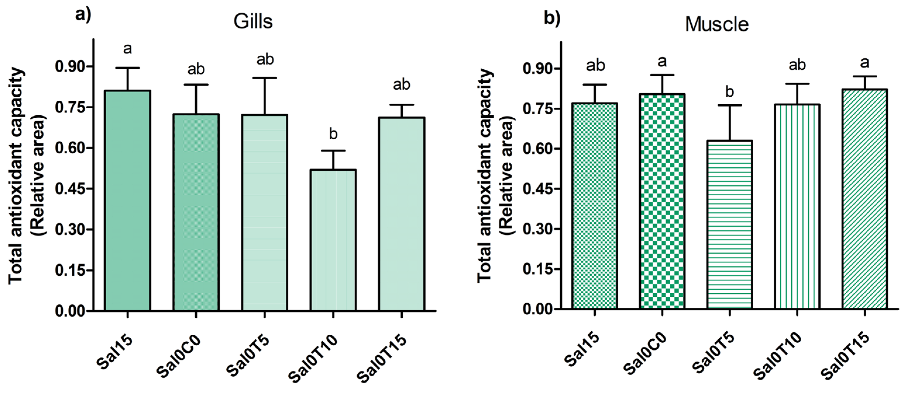
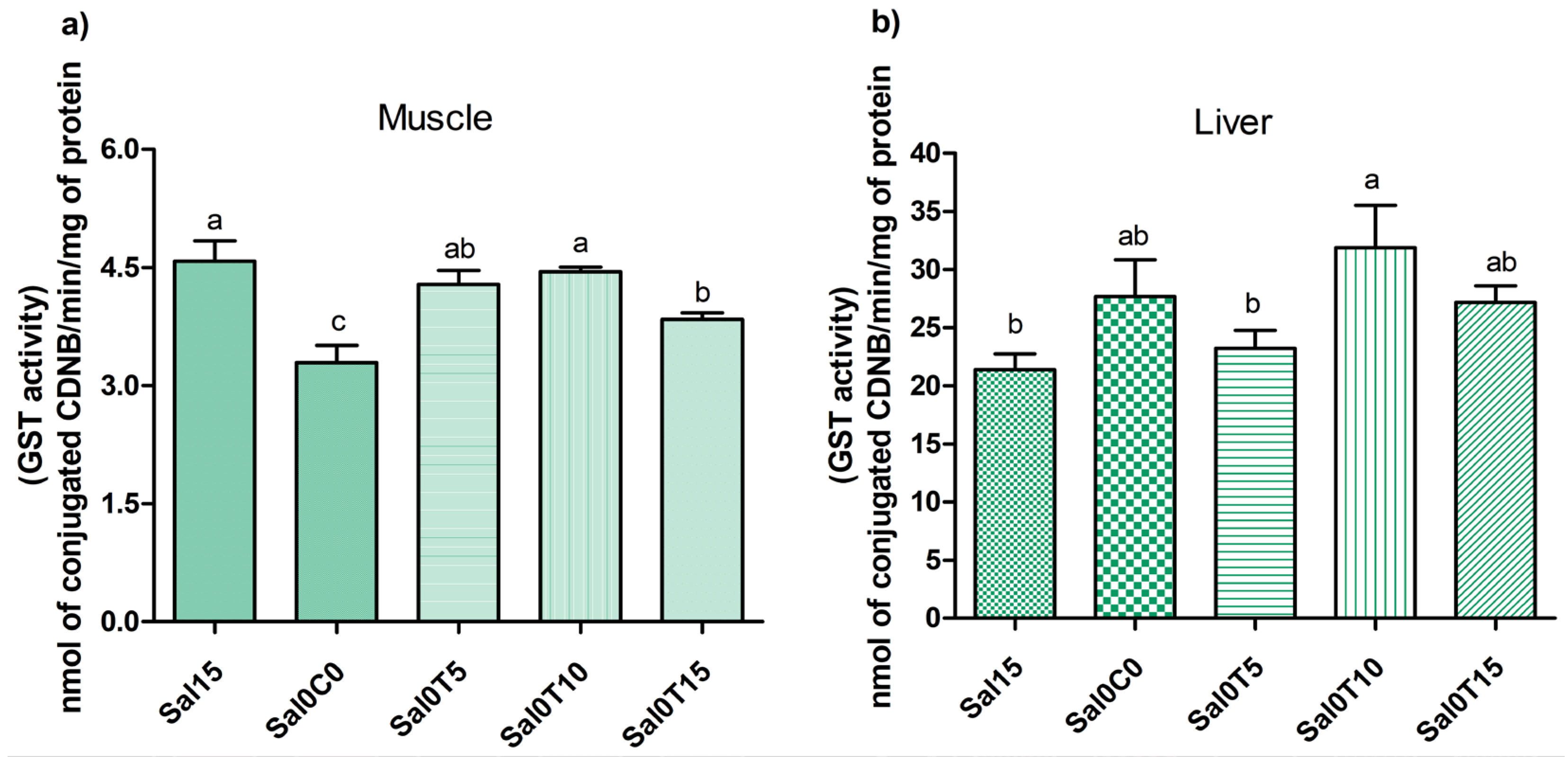
3.3. Biochemical Analysis

After the salinity stress test, there was no mortality in the tanks, showing only differences in the biochemical results of the gills, muscle, and liver. The results of the total antioxidant capacity (ACAP) presented in Figure 1a,b show that in the gills, the treatment with 10% macroalgae inclusion after stress had a higher antioxidant capacity than the treatment without stress and without macroalgae inclusion. In contrast, muscle showed a higher antioxidant capacity in the treatment with 5% macroalgae after stress compared with the control treatment without algae after stress. Both results showed the influence of macroalgae inclusion on the increase in antioxidant capacity compared with the control.
In the gills, it was observed that all levels of inclusion gradually increased GSH levels, whereas saline stress did not differ from the control (salinity 15) (Figure 2a). In the muscles, only the treatment with 10% macroalgae in the diet showed a higher concentration of antioxidants compared with the control treatment without stress (Figure 2b).
In the muscle, salt stress was shown to reduce detoxification capacity (evidenced by a decrease in GST activity), and the animals that received the inclusion feed were shown to have restored enzyme activity compared with the animals subjected to salt stress. In the liver, salt stress did not induce changes in GST activity; however, the group that received 10% salt stress showed an increase in GST activity compared with the control group without salt stress (Figure 3a,b).
In the muscle, treatment with 15% macroalgae in the diet after stress showed an increase in P-SH levels, indicating an increase in the reducing state. In the gills, this result was observed in the 5% inclusion group (Figure 4a,b).
In the muscle, the inclusion of 10 and 15% was shown to increase lipid damage compared with the other treatments (Figure 5).

4. Discussion

The nutritional composition of macroalgae depends on the environmental variables in which they are cultivated. In this study, the protein content of macroalgae cultivated in an integrated system with biofloc was 19%, which was higher than that reported by Limiñana et al. [42], who obtained 14% protein from macroalgae collected in the environment, and by Angell et al. [43], working with Ulva ohnoi, who collected from the environment with a protein content of 13.6% in the macroalgae. The protein increase observed in the present study may be associated with the high concentrations of nitrogen available for absorption in the biofloc system, as well as a high load of solid production, causing an increase in chlorophyll-a as a way of maximizing photosynthesis, as reported by Carvalho et al. [10]. Legarda et al. [44], working in an integrated system with shrimp, mullet, and macroalgae, showed an increase in nitrogen in the macroalgae, from 1% nitrogen at the beginning of the experiment to 2.9% at the end of the experiment, associated with an increase in chlorophyll-a and carotenoids, showing an increase in the nutritional value of the macroalgae when cultivated in association with other organisms.
This increase in protein and biocompounds in macroalgal tissues makes it a promising and sustainable activity to be included in the feed processes for aquatic organisms. Marinho et al. [21] used a 10, 15, and 20% inclusion of macroalgae in the diet and the replacement of fishmeal for tilapia, with the result that, up to 10% inclusion, the animal’s performance was not affected, with digestibility, apparent retention of protein, and energy being essential factors for the success or failure of the substitution [45]. A similar result was found by Ergün et al. [46] for tilapia, showing that small percentages of Ulva macroalgae inclusion (5 and 10%) did not negatively affect animal performance compared with the control diet. For carnivorous species such as sea bass fry Dicentrarchus labrax, it was found that the inclusion of up to 5% macroalgae meal promoted improvements in growth and survival rates, related to the protein content, minerals, and essential amino acid profile present in the macroalgae [47].
In the present study, the inclusion of macroalgae in the diet resulted in the replacement of ingredients from terrestrial plant sources such as soybean meal and wheat bran. Although macroalgae meal presented lower energy and protein digestibility, as presented by Qiu et al. [45], it did not interfere with the growth performance of tilapia at any level of inclusion in this experiment. Suryaningrum and Samsudin [48] evaluated the digestibility indices of U. lactuca for tilapia and obtained values of 82% for protein, 92% for lipids, 63% for ash, and 74% for energy, showing good indices and feasibility of inclusion in the diet. The results of the present study were similar to those found by Silva et al. [49], who tested the inclusion of 10% Gracilaria from an integrated cultivation system in the diet of tilapia, and showed that there were no differences in the performance or body composition of tilapia between the treatment with U. lactuca and the control. Thus, the application of macroalgal biomass produced in the integrated biofloc system in tilapia feed reflects the circular economy, which consists of adopting sustainable practices related to the better use of the nutrients produced and waste management [17].
Several studies have pointed to different situations regarding the effects of including macroalgae on the proximal composition of fish carcasses. Saleh et al. [20] tested the replacement of wheat flour with macroalgae flour and showed that higher inclusions (75 and 100 g kg−1) resulted in greater growth and changes in the tilapia carcass, with higher protein and lower lipid values as the inclusion of macroalgae in the feed increased. Azaza et al. [50] also found a decrease in lipid values with an increase in the inclusion of macroalgae, but protein values were not altered. According to Ortiz et al. [51], the profiles of vitamins and minerals in macroalgae can directly affect the body composition of organisms. For example, the presence of vitamin C in macroalgae can promote an increase in lipid metabolism in animals and change their body composition [52]. The tilapia fed with macroalgae did not show any differences in the proximal composition of the animals between the treatments; therefore, the inclusion of up to 15% U. lactuca flour in the feed did not interfere with the body composition of the tilapia. The results were similar to those found by Ergün et al. [46], who tested the same concentrations of macroalgae inclusion in the diet.
In addition to performance and bromatological parameters, blood parameters such as cell counts can characterize fish health and physiological effects associated with cultivation and feeding systems [53]. The lymphocyte and granulocyte counts in this study were in the same range as those reported by Jerônimo et al. [54] and Charlie-Silva et al. [55], indicating that the cultivation environment did not present high-stress factors or conditions of low health. Lymphocytes are indicators of inflammation in stressful situations [54], and the results indicated that these processes were not identified in the organisms sampled. However, the number of granulocytes may be associated with water quality parameters. Jerônimo et al. [54] associated the increase in granulocytes with a defense mechanism when temperatures were lower than those recommended for tilapia cultivation. The water quality in this study was maintained under ideal conditions and there was no difference between the treatments. Therefore, the increase in granulocyte concentrations in the 10% treatment compared with the control may be associated with the inclusion of macroalgae in the diet. Amar et al. [56] reported that including ingredients rich in carotenoids can stimulate the immune system of fish, which could explain the increase in granulocyte production in this experiment, stimulated by the presence of carotenoids in the macroalgae. Contrary to these results, Mendonça et al. [57] found that the inclusion of the macroalga Gracilaria dominguensis affected the growth of mullets when it was added to the diet at an inclusion rate of up to 15%, but had no effect on granulocyte and lymphocyte counts. In addition to carotenoids, according to Abdelrhman et al. [58], factors such as the presence of polysaccharides and sulfated carbohydrates in marine macroalgae can stimulate the immunity of animals.
During the cultivation of aquatic organisms, stress related to water quality, management, and biosecurity can occur frequently. Stress can be characterized as an alteration in homeostasis caused by a stressful agent [59] and directly affects the growth and physiological performance of animals. Stressful conditions in cultivation, such as pH changes, temperature, and salinity variations, can induce a pro-oxidative situation in different animal tissues and try to minimize this situation by recruiting antioxidant responses, such as ROS interception or the modulation of antioxidant systems (enzymatic or non-enzymatic), which in turn demand energy from other physiological processes such as growth and reproduction [60]. Tissues such as the gill, liver, and muscle can provide physiological responses to stress in animals, such as osmoregulatory functions, biotransformation processes, and the detoxification of xenobiotics [61]. An alternative to conserving animal energy to deal with pro-oxidant situations is to provide a diet with good antioxidant capacity, such as the use of macroalgae that provide bioactive substances such as carotenoids, which can have higher concentrations if produced in BFT systems compared with algae collected from the environment [62].
In tilapia, despite being an organism that can withstand large variations in salinity between 0 and 16 [63], an abrupt change can cause a pro-oxidant situation with the formation of reactive oxygen species and alterations in the antioxidant system [64]. Antioxidant defense systems are activated in stressful situations, whether due to changes in water quality or an increase in cultivation density; therefore, diet can increase resistance [26]. In this study, gill data showed that increasing the levels of macroalgae inclusion in the diet led to increased levels of reduced glutathione (GSH) and increased antioxidant capacity (ACAP), as well as increases in the P-SH groups. GSH is one of the first defense mechanisms against the action of free radicals [65]. Jiang et al. [66] indicated that a curcumin-inclusive diet may modulate GSH biosynthesis by regulating gene expression. In situations of stress, the gill is the most affected tissue because it is in direct contact with possible contaminants dissolved in water, and it is important to recruit antioxidant molecules to maintain homeostasis [61]. Therefore, the use of macroalgae in diets represents an alternative to using the biomass produced and exogenous input of antioxidant enzymes. According to Tziveleka et al. [67], because macroalgae are able to develop in stressful environments, they have defense mechanisms such as an increase in antioxidant enzymes, such as polyphenols and carotenoids, which are important characteristics in the application of the biomass produced.
Muscle, which is the most common part of fish, is interesting for observing the absence of damage and improvement in antioxidant capacity under stressful conditions. In this study, even when the animals were subjected to salt stress, there was an improvement in the detoxification capacity and absence of lipid damage in animals with 5 and 10% macroalgae inclusion. According to Srikanth et al. [68], antioxidants, such as glutathione S-transferase (GST), glutathione (GSH), and superoxide dismutase (SOD), have been reported in the body. This antioxidant effect of algae may be due to the fact that macroalgae of the Ulva genus have several important metabolites in their composition, such as phenolic compounds, carotenoids, chlorophylls, and minerals [67]. According to Pérez-gálvez et al. [69], carotenoids and chlorophylls can be characterized as antioxidants that seek to prevent/intercept ROS. Therefore, the inclusion of macroalgae in the diet can increase the preparation of animals to prevent oxidative damage. However, the inclusion of 10 and 15% macroalgae in the diet caused lipid peroxidation compared with the control treatments. According to Poljsak et al. [62], an inappropriate amount of antioxidants can convert bioactive compounds into pro-oxidants, caused by the neutralization of physiologically beneficial radicals, which may have occurred at higher levels of macroalgae inclusion. Some anti-nutritional characteristics, such as high ash and fiber content, can also affect animals physiologically, given that the nitrogen digestibility coefficient of macroalgae is lower than that of soybean meal [70].
GST activity also increased in the liver at a 10% macroalgae inclusion concentration compared with that in the unstressed control. GST plays a role in the biotransformation and elimination of toxic compounds in the cell [71], and in this process, GSH is used as a co-substrate in detoxification to maintain homeostasis, so its levels can be altered by GST activity [72]. However, in our study, unlike the accumulation of GSH and GST activity that occurred in the gills, GSH levels in the liver did not increase, which probably indicates that they were used for the detoxification of some compounds accumulated in the body at inclusions of 10 and 15%, which may be related to salinity stress or possible anti-nutritional characteristics of the macroalgae [48]. In addition to the presence of a high fiber and carbohydrate content, which according to the requirements of the species can negatively interfere in the growth of organisms, some factors such as phenolic compounds, which are bioactive compounds present in macroalgae, can be anti-nutritional factors and can cause oxidative stress in the organism [73]. These issues can be improved by using different extraction processes and macroalgae fermentation processes as alternatives to increase nutrient availability and minimize possible physiological effects on animals [74]. Therefore, obtaining a by-product of integrated aquaculture with better nutritional value that is rich in antioxidants is environmentally and economically advantageous and can be used in the system as a bioremediator and returned to the system to replace ingredients of terrestrial plant origin, applying the concept of circular economy [75]. This concept represents a green economy for sustainable aquaculture and waste recycling [18].

5. Conclusions

The use of macroalgae meal produced in an integrated system with biofloc at an inclusion level of 5% in tilapia feed resulted in a growth performance similar to that observed in the control without macroalgae, with the partial replacement of soy and wheat bran with an alternative ingredient. The proximal composition and hematological analysis were not influenced by the inclusion of macroalgae in the diet. The results of the oxidative stress after the salinity change showed a higher antioxidant capacity in the 5% inclusion treatment, associated with an increase in reduced glutathione (GSH) and sulfhydryl groups (P-SH), as well as an improvement in detoxification capacity and the absence of lipid peroxidation compared with the control treatments with and without salinity stress.

Author Contributions

Conceptualization, A.C., V.R., M.B.T., J.V.-L. and L.H.P.; methodology, A.C., L.M., V.R., M.B.T., J.V.-L. and L.H.P.; validation, A.C., L.M. and V.R.; formal analysis, A.C. and L.M.; investigation, A.C., L.M., V.R., M.B.T. and L.H.P.; resources, G.T., M.P., J.V.-L. and L.H.P.; data curation, A.C., L.M., V.R. and M.B.T.; writing—original draft preparation, A.C., V.R. and L.M.; writing—review and editing, A.C., L.M., V.R., M.B.T., J.V.-L., G.T., M.P. and L.H.P.; supervision, M.B.T., G.T. and L.H.P.; project administration, J.V.-L. and L.H.P.; funding acquisition, G.T., M.P., M.B.T., J.V.-L. and L.H.P. All authors have read and agreed to the published version of the manuscript.

Funding

This research was funded by the ASTRAL Project—H2020 grant agreement 863034.

Institutional Review Board Statement

All applicable international, national, and/or institutional guidelines for the care and use of animals were followed by the authors. The experiment was approved by the Ethics and Animal Welfare Committee of FURG (Case number 000476.0013263/2024).

Informed Consent Statement

Informed consent was obtained from all subjects involved in the study.

Data Availability Statement

The data presented in this study are available in the article.

Acknowledgments

Special thanks to the Brazilian Council of Research (CNPq), the Coordination for the Improvement of Higher Level or Education Personnel (CAPES), and the Rio Grande do Sul State Government. Luís H. Poersch and Marcelo B. Tesser receive a productivity research fellowship from CNPq.

Conflicts of Interest

The authors declare no conflicts of interest.

References

  1. Ben-Ari, T.; Neori, A.; Ben-Ezra, D.; Shauli, L.; Odintsov, V.; Shpigel, M. Management of Ulva lactuca as a Biofilter of Mariculture Effluents in IMTA System. Aquaculture 2014, 434, 493–498. [Google Scholar] [CrossRef]
  2. Queirós, A.S.; Circuncisão, A.R.; Pereira, E.; Válega, M.; Abreu, M.H.; Silva, A.M.S.; Cardoso, S.M. Valuable Nutrients from Ulva rigida: Modulation by Seasonal and Cultivation Factors. Appl. Sci. 2021, 11, 6137. [Google Scholar] [CrossRef]
  3. Resende, L.; Flores, J.; Moreira, C.; Pacheco, D.; Baeta, A.; Garcia, A.C.; Rocha, A.C.S. Effective and Low-Maintenance IMTA System as Effluent Treatment Unit for Promoting Sustainability in Coastal Aquaculture. Appl. Sci. 2022, 12, 398. [Google Scholar] [CrossRef]
  4. Chopin, T. Marine Aquaculture in Canada: Well-Established Monocultures of Finfish and Shellfish and an Emerging Integrated Multi-Trophic Aquaculture (IMTA) Approach Including Seaweeds, Other Invertebrates, and Microbial Communities. Fisheries 2015, 40, 28–31. [Google Scholar] [CrossRef]
  5. De Alencar, J.R.; Junior, P.A.H.; Celino, J.J. Cultivo de Camarão Branco Litopenaeus Vannamei (Boone, 1931) Com a Macro- Alga Ulva lactuca Linneaus (Chlorophyta) No Tratamento de Efluentes Em Sistema Fechado de Recirculação. Rev. Biol. Ciências Terra 2010, 10, 117–137. [Google Scholar]
  6. Khanjani, M.H.; Mozanzadeh, M.T.; Sharifinia, M.; Emerenciano, M.G.C. Biofloc: A Sustainable Dietary Supplement, Nutritional Value and Functional Properties. Aquaculture 2023, 562, 738757. [Google Scholar] [CrossRef]
  7. Ebeling, J.M.; Timmons, M.B.; Bisogni, J.J. Engineering Analysis of the Stoichiometry of Photoautotrophic, Autotrophic, and Heterotrophic Removal of Ammonia-Nitrogen in Aquaculture Systems. Aquaculture 2006, 257, 346–358. [Google Scholar] [CrossRef]
  8. Da Silva, K.R.; Wasielesky, W.; Abreu, P.C. Nitrogen and Phosphorus Dynamics in the Biofloc Production of the Pacific White Shrimp, Litopenaeus vannamei. J. World Aquac. Soc. 2013, 44, 30–41. [Google Scholar] [CrossRef]
  9. Reis, W.G.; Wasielesky, W.; Abreu, P.C.; Brandão, H.; Krummenauer, D. Rearing of the Pacific White Shrimp Litopenaeus vannamei (Boone, 1931)in BFT System with Different Photoperiods: Effects on the Microbial Community, Water Quality and Zootechnical Performance. Aquaculture 2019, 508, 19–29. [Google Scholar] [CrossRef]
  10. Carvalho, A.; Braga, Í.; Chaar, F.; Cardozo, A.P.; Monserrat, J.M.; Ramírez, J.R.B.; Wasielesky, W.; Poersch, L.H. Production of the Macroalgae Ulva lactuca Integrated with the Shrimp Penaeus Vannamei in a Biofloc System: Effect of Total Suspended Solids and Nutrient Concentrations. Phycology 2024, 4, 37–52. [Google Scholar] [CrossRef]
  11. Levavasseur, G. Analyse Comparée Des Complexes Pigment-Protéines de Chlorophycophytes Marines Benthiques. Phycologia 1989, 28, 1–14. [Google Scholar] [CrossRef]
  12. Carvalho, A.; Costa, L.C.d.O.; Holanda, M.; Poersch, L.H.; Turan, G. Influence of Total Suspended Solids on the Growth of the Sea Lettuce Ulva lactuca Integrated with the Pacific White Shrimp Litopenaeus vannamei in a Biofloc System. Fishes 2023, 8, 163. [Google Scholar] [CrossRef]
  13. Xu, P.; Tan, H.; Jin, W.; Li, Y.; Santhoshkumar, C.; Li, P.; Liu, W. Antioxidative and Antimicrobial Activities of Intertidal Seaweeds and Possible Effects of Abiotic Factors on These Bioactivities. J. Oceanol. Limnol. 2018, 36, 2243–2256. [Google Scholar] [CrossRef]
  14. Montgomery, W.L.; Gerking, S.D. Marine Macroalgae as Foods for Fishes: An Evaluation of Potential Food Quality. Environ. Biol. Fishes 1980, 5, 143–153. [Google Scholar] [CrossRef]
  15. Kirchherr, J.; Reike, D.; Hekkert, M. Conceptualizing the Circular Economy: An Analysis of 114 Definitions. Resour. Conserv. Recycl. 2017, 127, 221–232. [Google Scholar] [CrossRef]
  16. Cornejo-Ponce, L.; Vilca-Salinas, P.; Lienqueo-Aburto, H.; Arenas, M.J.; Pepe-Victoriano, R.; Carpio, E.; Rodríguez, J. Integrated Aquaculture Recirculation System (Iars) Supported by Solar Energy as a Circular Economy Alternative for Resilient Communities in Arid/Semi-Arid Zones in Southern South America: A Case Study in the Camarones Town. Water 2020, 12, 3469. [Google Scholar] [CrossRef]
  17. Cooney, R.; de Sousa, D.B.; Fernández-Ríos, A.; Mellett, S.; Rowan, N.; Morse, A.P.; Hayes, M.; Laso, J.; Regueiro, L.; Wan, A.H.; et al. A Circular Economy Framework for Seafood Waste Valorisation to Meet Challenges and Opportunities for Intensive Production and Sustainability. J. Clean. Prod. 2023, 392, 136283. [Google Scholar] [CrossRef]
  18. Campanati, C.; Willer, D.; Schubert, J.; Aldridge, D.C. Sustainable Intensification of Aquaculture through Nutrient Recycling and Circular Economies: More Fish, Less Waste, Blue Growth. Rev. Fish. Sci. Aquac. 2021, 30, 143–169. [Google Scholar] [CrossRef]
  19. Mwendwa, R.; Wawire, M.; Kahenya, P. Potential for Use of Seaweed as a Fish Feed Ingredient: A Review. J. Agric. Sci. 2023, 15, 96. [Google Scholar] [CrossRef]
  20. Saleh, N.E.; Shalaby, S.M.; Sakr, E.M.; Elmonem, A.I.A.; Michael, F.R. Effect of Dietary Inclusion of Ulva fasciata on Red Hybrid Tilapia Growth and Carcass Composition. J. Appl. Aquac. 2014, 26, 197–207. [Google Scholar] [CrossRef]
  21. Marinho, G.; Nunes, C.; Sousa-Pinto, I.; Pereira, R.; Rema, P.; Valente, L.M.P. The IMTA-Cultivated Chlorophyta Ulva Spp. as a Sustainable Ingredient in Nile Tilapia (Oreochromis niloticus) Diets. J. Appl. Phycol. 2013, 25, 1359–1367. [Google Scholar] [CrossRef]
  22. Yildiz, G.; Celikler, S.; Vatan, O.; Dere, S. Determination of the Anti-Oxidative Capacity and Bioactive Compounds in Green Seaweed Ulva rigida C. Agardh. Int. J. Food Prop. 2012, 15, 1182–1189. [Google Scholar] [CrossRef]
  23. He, J.; Xu, Y.; Chen, H.; Sun, P. Extraction, Structural Characterization, and Potential Antioxidant Activity of the Polysaccharides from Four Seaweeds. Int. J. Mol. Sci. 2016, 17, 1988. [Google Scholar] [CrossRef] [PubMed]
  24. Kidgell, J.T.; Magnusson, M.; de Nys, R.; Glasson, C.R.K. Ulvan: A Systematic Review of Extraction, Composition and Function. Algal Res. 2019, 39, 101422. [Google Scholar] [CrossRef]
  25. Santos, I.L.M.; Melatti, E.; de Souza Correia, E.; Ferreira, D.A.; da Silva, L.O.B. Economic Viability of Tilapia Cultivation (Oreochromis niloticus) in Net Tanks Located in the Municipality of Glória-BA. Custos Agronegócios Online 2020, 16, 900. [Google Scholar]
  26. Oliva-Teles, A. Nutrition and Health of Aquaculture Fish. J. Fish Dis. 2012, 35, 83–108. [Google Scholar] [CrossRef]
  27. Ju, J.; Yang, J.; Zhang, W.; Wei, Y.; Yuan, H.; Tan, Y. Seaweed Polysaccharide Fibers: Solution Properties, Processing and Applications. J. Mater. Sci. Technol. 2023, 140, 1–18. [Google Scholar] [CrossRef]
  28. Mhlongo, G.; Mnisi, C.M. Effect of Seaweed (Ecklonia maxima) on Apparent Nutrient Digestibility, Growth Performance, and Physiological and Meat Quality Parameters in Boschveld Cockerels. Poult. Sci. 2023, 102, 102361. [Google Scholar] [CrossRef]
  29. Huang, Q.; Huang, K.; Ma, Y.; Qin, X.; Wen, Y.; Sun, L.; Tang, L. Feeding Frequency and Rate Effects on Growth and Physiology of Juvenile Genetically Improved Farmed Nile Tilapia. N. Am. J. Aquac. 2015, 77, 503–512. [Google Scholar] [CrossRef]
  30. Gontijo, Á.M.D.M.C.; Barreto, R.E.; Speit, G.; Valenzuela Reyes, V.A.; Volpato, G.L.; Favero Salvadori, D.M. Anesthesia of Fish with Benzocaine Does Not Interfere with Comet Assay Results. Mutat. Res.-Genet. Toxicol. Environ. Mutagen. 2003, 534, 165–172. [Google Scholar] [CrossRef]
  31. APHA. Standard Methods for the Examination of Water and Wastewater, 21st ed.; American Public Health Association: Washington, DC, USA, 2005. [Google Scholar]
  32. UNESCO. Chemical Methods for Use in Marine Environmental Monitoring; Intergovernmental Oceanographic Commission: Paris, France, 1983. [Google Scholar]
  33. Bendschneider, K.; Robinson, R.J. New Spectrophotometric Method for the Determination of Nitrite in Water. Fresenius Environ. Bull. 1952, 10, 781–784. [Google Scholar]
  34. Aminot, A.; Chaussepied, M. Manuel des Analyses Chimiques en Milieu Marin; Centre National Pour L’exploitation des Océans: Brest, France, 1983. [Google Scholar]
  35. Association of Official Analytical Chemists (A.O.A.C). Official Methods of Analysis of the AOAC; AOAC: New Orleans, LA, USA, 2007. [Google Scholar]
  36. Baethgen, W.E.; Alley, M. A Manual Colorimetric Procedure for Measuring Ammonium Nitrogen in Soil and Plant Kjeldahl Digests. Commun. Soil Sci. Plant Anal. 1989, 20, 961–969. [Google Scholar] [CrossRef]
  37. Rosenfeld, G. Corante Pancrômico Para Hematologia e Citologia Clínica, 20th ed.; Inst. Butantan: São Paulo, Brazil, 1947. [Google Scholar]
  38. Amado, L.L.; Garcia, M.L.; Ramos, P.B.; Freitas, R.F.; Zafalon, B.; Ferreira, J.L.R.; Yunes, J.S.; Monserrat, J.M. A Method to Measure Total Antioxidant Capacity against Peroxyl Radicals in Aquatic Organisms: Application to Evaluate Microcystins Toxicity. Sci. Total Environ. 2009, 407, 2115–2123. [Google Scholar] [CrossRef]
  39. Oakes, K.D.; Van Der Kraak, G.J. Utility of the TBARS Assay in Detecting Oxidative Stress in White Sucker (Catostomus commersoni) Populations Exposed to Pulp Mill Effluent. Aquat. Toxicol. 2003, 63, 447–463. [Google Scholar] [CrossRef]
  40. Ryther, J.H.; DeBusk, T.A.; Peterson, J.E. Studies of Marine Macroalgae: Saline Desert Water Cultivation and Effects of Environmental Stress on Proximate Composition. Sol. Energy 1985, 6456763. [Google Scholar]
  41. Sedlak, J.; Lindsay, R.H. Estimation of Total, Protein-Bound, and Nonprotein Sulfhydryl Groups in Tissue with Ellman’s Reagent. Anal. Biochem. 1968, 25, 192–205. [Google Scholar] [CrossRef]
  42. Arcos Limiñana, V.; Benoist, T.; Anton Sempere, S.; Maestre Pérez, S.E.; Prats Moya, M.S. Chemical Composition of Sustainable Mediterranean Macroalgae Obtained from Land-Based and Sea-Based Aquaculture Systems. Food Biosci. 2023, 54, 102902. [Google Scholar] [CrossRef]
  43. Angell, A.R.; Pirozzi, I.; de Nys, R.; Paul, N.A. Feeding Preferences and the Nutritional Value of Tropical Algae for the Abalone Haliotis asinina. PLoS ONE 2012, 7, e38857. [Google Scholar] [CrossRef]
  44. Legarda, E.C.; da Silva, D.; Miranda, C.S.; Pereira, P.K.M.; Martins, M.A.; Machado, C.; de Lorenzo, M.A.; Hayashi, L.; do Nascimento Vieira, F. Sea Lettuce Integrated with Pacific White Shrimp and Mullet Cultivation in Biofloc Impact System Performance and the Sea Lettuce Nutritional Composition. Aquaculture 2021, 534, 736265. [Google Scholar] [CrossRef]
  45. Qiu, X.; Neori, A.; Kim, J.K.; Yarish, C.; Shpigel, M.; Guttman, L.; Ben Ezra, D.; Odintsov, V.; Davis, D.A. Evaluation of Green Seaweed Ulva Sp. as a Replacement of Fish Meal in Plant-Based Practical Diets for Pacific White Shrimp, Litopenaeus vannamei. J. Appl. Phycol. 2018, 30, 1305–1316. [Google Scholar] [CrossRef]
  46. Ergün, S.; Soyutürk, M.; Güroy, B.; Güroy, D.; Merrifield, D. Influence of Ulva Meal on Growth, Feed Utilization, and Body Composition of Juvenile Nile Tilapia (Oreochromis niloticus) at Two Levels of Dietary Lipid. Aquac. Int. 2009, 17, 355–361. [Google Scholar] [CrossRef]
  47. Wassef, E.A.; El-Sayed, A.F.M.; Sakr, E.M. Pterocladia (Rhodophyta) and Ulva (Chlorophyta) as Feed Supplements for European Seabass, Dicentrarchus labrax L.; Fry. J. Appl. Phycol. 2013, 25, 1369–1376. [Google Scholar] [CrossRef]
  48. Suryaningrum, L.H.; Samsudin, R. Nutrient Digestibility of Green Seaweed Ulva Meal and the Influence on Growth Performance of Nile Tilapia (Oreochromis niloticus). Emir. J. Food Agric. 2020, 32, 488–494. [Google Scholar] [CrossRef]
  49. Silva, D.M.; Valente, L.M.P.; Pereira, R.; Pires, M.A.; Seixas, F.; Rema, P. Evaluation of IMTA-Produced Seaweeds (Gracilaria, Porphyra, and Ulva) as Dietary Ingredients in Nile Tilapia, Oreochromis niloticus L., Juveniles. Effects on Growth Performance and Gut Histology. J. Appl. Phycol. 2015, 27, 1671–1680. [Google Scholar] [CrossRef]
  50. Azaza, B.M.S.; Mensi, F.; Ksouri, J.; Dhraief, M.N.; Brini, B.; Abdelmouleh, A.; Kraı, M.M. Growth of Nile Tilapia (Oreochromis niloticus L.) Fed with Diets Containing Graded Levels of Green Algae Ulva Meal (Ulva rigida) Reared in Geothermal Waters of Southern Tunisia. J. Appl. Ichthyol. 2008, 24, 202–207. [Google Scholar] [CrossRef]
  51. Ortiz, J.; Romero, N.; Robert, P.; Araya, J.; Lopez-Hernández, J.; Bozzo, C.; Navarrete, E.; Osorio, A.; Rios, A. Dietary Fiber, Amino Acid, Fatty Acid and Tocopherol Contents of the Edible Seaweeds Ulva lactuca and Durvillaea antarctica. Food Chem. 2006, 99, 98–104. [Google Scholar] [CrossRef]
  52. Nakagawa, H. Effect of Dietary Algae on Improvement of Lipid Metabolism in Fish. Biomed. Pharmacother. 1997, 51, 345–348. [Google Scholar] [CrossRef]
  53. Satheeshkumar, P.; Ananthan, G.; Kumar, D.S.; Jagadeesan, L. Haematology and Biochemical Parameters of Different Feeding Behaviour of Teleost Fishes from Vellar Estuary, India. Comp. Clin. Path. 2012, 21, 1187–1191. [Google Scholar] [CrossRef]
  54. Jerônimo, G.; Laffitte, L.; Speck, G.; Martins, M. Seasonal Influence on the Hematological Parameters in Cultured Nile Tilapia from Southern Brazil. Braz. J. Biol. 2011, 71, 719–725. [Google Scholar] [CrossRef]
  55. Charlie-Silva, I.; Klein, A.; Gomes, J.M.M.; Prado, E.J.R.; Moraes, A.C.; Eto, S.F.; Fernandes, D.C.; Fagliari, J.J.; Junior, J.D.C.; Lima, C.; et al. Acute-Phase Proteins during Inflammatory Reaction by Bacterial Infection: Fish-Model. Sci. Rep. 2019, 9, 4776. [Google Scholar] [CrossRef]
  56. Amar, E.C.; Kiron, V.; Satoh, S.; Watanabe, T. Enhancement of Innate Immunity in Rainbow Trout (Oncorhynchus mykiss Walbaum) Associated with Dietary Intake of Carotenoids from Natural Products. Fish Shellfish Immunol. 2004, 16, 527–537. [Google Scholar] [CrossRef] [PubMed]
  57. Mendonça, A.J.C.; Rosas, V.T.; Monserrat, J.M.; Romano, L.A.; Tesser, M.B. The Inclusion of Algae Gracilaria domingensis in the Diet of Mullet Juveniles (Mugil liza) Improves the Immune Response. J. Appl. Aquac. 2019, 31, 210–223. [Google Scholar] [CrossRef]
  58. Abdelrhman, A.M.; Ashour, M.; Al-Zahaby, M.A.; Sharawy, Z.Z.; Nazmi, H.; Zaki, M.A.A.; Ahmed, N.H.; Ahmed, S.R.; El-Haroun, E.; Van Doan, H.; et al. Effect of Polysaccharides Derived from Brown Macroalgae Sargassum dentifolium on Growth Performance, Serum Biochemical, Digestive Histology and Enzyme Activity of Hybrid Red Tilapia. Aquac. Rep. 2022, 25, 101212. [Google Scholar] [CrossRef]
  59. Barton, B.A.; Iwama, G.K. Physiological Changes in Fish from Stress in Aquaculture with Emphasis on the Response and Effects of Corticosteroids. Annu. Rev. Fish Dis. 1991, 1, 3–26. [Google Scholar] [CrossRef]
  60. Halliwell, B.; Gutteridge, J.M.C. Fee Radicals in Biology and Medicine Carcinogenesis. J. Free Radic. Biol. Med. 1985, 1, 331–334. [Google Scholar] [CrossRef]
  61. Bernet, D.; Schmidt, H.; Meier, W.; Burkhardt-Holm, P.; Wahli, T. Histopathology in Fish: Proposal for a Protocol to Assess Aquatic Pollution. J. Fish Dis. 1999, 22, 25–34. [Google Scholar] [CrossRef]
  62. Poljsak, B.; Šuput, D.; Milisav, I. Achieving the Balance between ROS and Antioxidants: When to Use the Synthetic Antioxidants. Oxid. Med. Cell. Longev. 2013, 2013, 956792. [Google Scholar] [CrossRef]
  63. de Souza, R.L.; de Lima, E.C.R.; de Melo, F.P.; Ferreira, M.G.P.; de Souza Correia, E. The Culture of Nile Tilapia at Different Salinities Using a Biofloc System. Rev. Cienc. Agron. 2019, 50, 267–275. [Google Scholar] [CrossRef]
  64. Caxico Vieira, C.A.S.; Vieira, J.S.; Bastos, M.S.; Zancanela, V.; Barbosa, L.T.; Gasparino, E.; Del Vesco, A.P. Expression of Genes Related to Antioxidant Activity in Nile Tilapia Kept under Salinity Stress and Fed Diets Containing Different Levels of Vitamin C. J. Toxicol. Environ. Health-Part A Curr. Issues 2018, 81, 20–30. [Google Scholar] [CrossRef]
  65. Ventura-Lima, J.; de Castro, M.R.; Acosta, D.; Fattorini, D.; Regoli, F.; de Carvalho, L.M.; Bohrer, D.; Geracitano, L.A.; Barros, D.M.; Marins, L.F.F.; et al. Effects of Arsenic (As) Exposure on the Antioxidant Status of Gills of the Zebrafish Danio rerio (Cyprinidae). Comp. Biochem. Physiol.-Part C Toxicol. Pharmacol. 2009, 149, 538–543. [Google Scholar] [CrossRef]
  66. Jiang, J.; Wu, X.Y.; Zhou, X.Q.; Feng, L.; Liu, Y.; Jiang, W.D.; Wu, P.; Zhao, Y. Effects of Dietary Curcumin Supplementation on Growth Performance, Intestinal Digestive Enzyme Activities and Antioxidant Capacity of Crucian Carp Carassius auratus. Aquaculture 2016, 463, 174–180. [Google Scholar] [CrossRef]
  67. Tziveleka, L.A.; Tammam, M.A.; Tzakou, O.; Roussis, V.; Ioannou, E. Metabolites with Antioxidant Activity from Marine Macroalgae. Antioxidants 2021, 10, 1431. [Google Scholar] [CrossRef] [PubMed]
  68. Srikanth, K.; Pereira, E.; Duarte, A.C.; Ahmad, I. Glutathione and Its Dependent Enzymes’ Modulatory Responses to Toxic Metals and Metalloids in Fish-a Review. Environ. Sci. Pollut. Res. 2013, 20, 2133–2149. [Google Scholar] [CrossRef] [PubMed]
  69. Pérez-gálvez, A.; Viera, I.; Roca, M. Carotenoids and Chlorophylls as Antioxidants. Antioxidants 2020, 9, 505. [Google Scholar] [CrossRef]
  70. Bikker, P.; Stokvis, L.; van Krimpen, M.M.; van Wikselaar, P.G.; Cone, J.W. Evaluation of Seaweeds from Marine Waters in Northwestern Europe for Application in Animal Nutrition. Anim. Feed Sci. Technol. 2020, 263, 114460. [Google Scholar] [CrossRef]
  71. Goto, S.; Kawakatsu, M.; Izumi, S.i.; Urata, Y.; Kageyama, K.; Ihara, Y.; Koji, T.; Kondo, T. Glutathione S-Transferase π Localizes in Mitochondria and Protects against Oxidative Stress. Free Radic. Biol. Med. 2009, 46, 1392–1403. [Google Scholar] [CrossRef]
  72. Liu, J.D.; Liu, W.B.; Zhang, C.Y.; Xu, C.Y.; Zheng, X.C.; Zhang, D.D.; Chi, C. Dietary Glutathione Supplementation Enhances Antioxidant Activity and Protects against Lipopolysaccharide-Induced Acute Hepatopancreatic Injury and Cell Apoptosis in Chinese Mitten Crab, Eriocheir Sinensis. Fish Shellfish Immunol. 2020, 97, 440–454. [Google Scholar] [CrossRef]
  73. Wan, A.H.L.; Davies, S.J.; Soler-Vila, A.; Fitzgerald, R.; Johnson, M.P. Macroalgae as a Sustainable Aquafeed Ingredient. Rev. Aquac. 2019, 11, 458–492. [Google Scholar] [CrossRef]
  74. Hardjani, D.K.; Suantika, G.; Aditiawati, P. Nutritional Profile of Red Seaweed Kappaphycus alvarezii after Fermentation Using Saccharomyces Cerevisiae as a Feed Supplement for White Shrimp Litopenaeus vannamei Nutritional Profile of Fermented Red Seaweed. J. Pure Appl. Microbiol. 2017, 11, 1637–1645. [Google Scholar] [CrossRef]
  75. Colombo, S.M.; Roy, K.; Mraz, J.; Wan, A.H.L.; Davies, S.J.; Tibbetts, S.M.; Overland, M.; Francis, D.S.; Rocker, M.M.; Gasco, L.; et al. Towards Achieving Circularity and Sustainability in Feeds for Farmed Blue Foods. Rev. Aquac. 2023, 15, 1115–1141. [Google Scholar] [CrossRef]
Figure 1. Total antioxidant capacity (ACAP) (mean ± standard deviation) in the gills (a) and muscle (b) at the end of saline stress, in treatments Sal15 (control without algae and at salinity 15 of cultivation), Sal0C0 (control without algae after saline stress), Sal0T5 (diet with 5% macroalgae after saline stress), Sal0T10 (diet with 10% macroalgae after saline stress), and Sal0T15 (diet with 15% macroalgae after saline stress). Different letters represent a significant difference (p < 0.05, Tukey’s test) between treatments.
Applsci 15 06410 g001
Figure 2. Concentration of reduced glutathione (GSH) (mean ± standard deviation) in gills (a) and muscle (b) at the end of salt stress, in treatments Sal15 (control without algae and at salinity 15 of cultivation), Sal0C0 (control without algae after salt stress), Sal0T5 (diet with 5% macroalgae after salt stress), Sal0T10 (diet with 10% macroalgae after salt stress), and Sal0T15 (diet with 15% macroalgae after salt stress). Different letters represent a significant difference (p < 0.05, Tukey’s test) between treatments.
Applsci 15 06410 g002
Figure 3. Glutathione S-transferase activity (GST) (mean ± standard deviation) in the gills (a) and muscle (b) at the end of saline stress, in treatments Sal15 (control without algae and at salinity 15 of cultivation), Sal0C0 (control without algae after saline stress), Sal0T5 (diet with 5% macroalgae after saline stress), Sal0T10 (diet with 10% macroalgae after saline stress), and Sal0T15 (diet with 15% macroalgae after saline stress). Different letters represent a significant difference (p < 0.05, Tukey’s test) between treatments.
Applsci 15 06410 g003
Figure 4. Concentration of sulfhydryl groups associated with protein (P-SH) (mean ± standard deviation) in gills (a) and muscle (b) at the end of saline stress, in treatments Sal15 (control without algae and at salinity 15 of cultivation), Sal0C0 (control without algae after saline stress), Sal0T5 (diet with 5% macroalgae after saline stress), Sal0T10 (diet with 10% macroalgae after saline stress), and Sal0T15 (diet with 15% macroalgae after saline stress). Different letters represent a significant difference (p < 0.05, Tukey’s test) between treatments.
Applsci 15 06410 g004
Figure 5. Concentration of thiobarbituric acid reactive substances (TBARS) (mean ± standard deviation) in the muscle at the end of saline stress, in treatments Sal15 (control without algae and at salinity 15 of cultivation), Sal0C0 (control without algae after saline stress), Sal0T5 (diet with 5% macroalgae after saline stress), Sal0T10 (diet with 10% macroalgae after saline stress), and Sal0T15 (diet with 15% macroalgae after saline stress). Different letters represent a significant difference (p < 0.05, Tukey’s test) between treatments.
Applsci 15 06410 g005
Table 1. Proximal composition of the macroalgae U. lactuca (% of dry matter) cultivated in an integrated biofloc system.
Protein (%)Lipid (%)Fiber (%)Ash (%)
Ulva meal19.31 ± 0.300.46 ± 0.04 10.34 ± 0.72 29.05 ± 0.63
Table 2. Ingredient composition and proximal composition of experimental diets (% of dry matter) containing U. lactuca meal at different levels.
Ingredients Experimental Diets
Control T5 T10 T15
Fish meal 40.00 40.00 40.00 40.00
Soybean meal 33.00 32.00 31.00 30.00
Wheat bran 12.00 8.00 4.00 0.00
Gelatin 2.00 2.00 2.00 2.00
Soy oil 2.00 2.00 2.00 2.00
Fish oil 2.00 2.00 2.00 2.00
Cellulose 4.00 4.00 4.00 4.00
Vitamin/mineral mix 5.00 5.00 5.00 5.00
Ulva meal 0.00 5.00 10.00 15.00
Proximal composition (%)
Protein42.44 41.82 41.37 39.98
Lipids8.23 8.07 8.05 8.64
Ash16.28 17.45 19.01 20.02
Gross energy 17.63 17.34 17.32 17.42
Gross energy: Expressed in MJ/kg wet weight of feed.
Table 3. Tilapia performance in the treatments: control (no macroalgae included), T5 (5% macroalgae included in the diet), T10 (10% macroalgae included in the diet), and T15 (15% macroalgae included in the diet) during 42 days of cultivation.
Treatments
ParametersControlT5T10T15
Average initial weight (g)0.93 ± 0.010.93 ± 0.010.94 ± 0.010.94 ± 0.00
Average final weight (g)10.78 ± 0.5111.44 ± 0.8410.35 ± 0.9210.33 ± 0.15
Weight gain (g)9.85 ± 05110.50 ± 0.839.41 ± 0.929.39 ± 0.15
SGR (% day−1)5.82 ± 0.115.95 ± 0.155.71 ± 0.205.70 ± 0.40
FCR1.00 ± 0.070.96 ± 0.090.99 ± 0.061.02 ± 0.02
Survival (%)87.50 ± 15.0085.00 ± 5.7795.00 ± 5.7790.0 ± 0.00
SGR specific growth rate; FCR feed conversion rate.
Table 4. Proximal composition of O. niloticus at the end of the 42-day experiment in treatments: control (no macroalgae included), T5 (5% macroalgae included in the diet), T10 (10% macroalgae included in the diet), and T15 (15% macroalgae included in the diet).
Treatments
Parameters (%)ControlT5T10T15
Moisture77.31 ± 1.2377.95 ± 0.3077.68 ± 0.1877.58 ± 0.27
Protein 14.23 ± 0.63 13.73 ± 0.74 13.67 ± 0.79 13.81 ± 0.96
Lipid 3.72 ± 0.53 3.33 ± 0.30 3.74 ± 0.35 3.55 ± 0.28
Ash 4.04 ± 0.17 4.08 ± 0.21 4.01 ± 0.11 4.09 ± 0.18
Table 5. Hematological composition of O. niloticus at the end of the 42-day experiment in the treatments: control (no macroalgae included), T5 (5% macroalgae included in the diet), T10 (10% macroalgae included in the diet), and T15 (15% macroalgae included in the diet).
Treatments
Parameters (×10−4 cel mL−1)ControlT5T10T15
Lymphocytes 273.50 ± 97.65240.00 ± 78.50209.60 ± 78.54278.50 ± 186.76
Granulocytes 10.50 ± 9.20 b26.17 ± 20.73 ab36.60 ± 36.84 a23.17 ± 13.98 ab
Lowercase letters on the same line represent a significant difference (p < 0.05, Tukey’s test) between treatments after a one-way ANOVA.
Disclaimer/Publisher’s Note: The statements, opinions and data contained in all publications are solely those of the individual author(s) and contributor(s) and not of MDPI and/or the editor(s). MDPI and/or the editor(s) disclaim responsibility for any injury to people or property resulting from any ideas, methods, instructions or products referred to in the content.

