Article

Fermentative Liberation and Transformation of Bioactive Compounds: Ellagic Acid from Nut Press Cake Ellagitannins

by
Wolfram Manuel Brück
*,
Yannick Erismann
and
Wilfried Andlauer
*
Institute of Life Technologies, University of Applied Sciences and Arts Western Switzerland Valais, Rue de l’Industrie 19, 1950 Sion, Switzerland
*
Authors to whom correspondence should be addressed.
Appl. Sci. 2024, 14(3), 1141; https://doi.org/10.3390/app14031141
Submission received: 8 December 2023 / Revised: 24 January 2024 / Accepted: 26 January 2024 / Published: 29 January 2024
(This article belongs to the Special Issue Bioactive Compounds: From Extraction to Application)

Abstract

:
Oil extracted from walnuts leaves behind large amounts of defatted press cake that still retains valuable nutrients. Solid state fermentation (SSF) is a promising method to liberate bioactive compounds from food by-products. Aspergillus oryzae, Rhizopus oligosporus, and Streptococcus thermophilus possess the necessary enzymes to utilize these nutrients from the walnut press cake (WPC) and convert walnut ellagitannins into ellagic acid and urolithins. This study conducted SSF with WPC to release ellagic acid from ellagitannins and form urolithins. The growth of the two filamentous fungi could be observed visually and the growth of Streptococcus thermophilus was confirmed by plate count technique. Extracts from fermented products were subjected to analysis using HPLC–DAD to measure the release of ellagic acid from ellagitannins. Additionally, a more sensitive UHPLC–MS method was employed to screen fermented samples for urolithin A. The ellagic acid content exhibited no perceptible change but was already present in the press cake before and after all fermentations. Urolithin A was undetectable, even with the more sensitive MS method. All studies showing urolithin A formation were conducted under anaerobic conditions. This might be a basic prerequisite for the transformation of ellagic acid to urolithins.

1. Introduction

Over the past decade, the food and cosmetic industries have shown significant interest in walnut oil derived from Junglas regia L., recognizing its richness in bioactive compounds such as polyunsaturated triglycerides and fat-soluble vitamins. However, the oil extraction process results in substantial by-products, specifically walnut press cake (WPC), which retains valuable components including oil (20–36%), protein (30–42%), dietary fiber, phenolic compounds (ellagitannins), and minerals [1,2,3,4].
Given the compelling composition of WPC, there is an increasing interest in using it as a nutritional and functional component in food items or as a base for fermentation [1,5]. Historically, fermentation played a crucial role in the processing of food for human consumption. Fermentation was used primarily for preservation; However, it not only preserves the food but also improves its quality and functionality [5]. Nowadays, fermentation is often used with the aim to improve the bioavailability of health promoting compounds or to increase their biological activity by microbial transformations. The bioavailability of natural polyphenols in human beings is very low due to their low absorption in the gastrointestinal tract. In a recent publication, Ran et al. showed a significant increase of the total polyphenol content and the antioxidant activity of apple pulp due to fermentation with Lactobacillus acidophilus [6]. They also observed an increase in the bioavailability of apple polyphenols. The authors explained their findings by suggesting a transformation of larger molecules into smaller ones, which can be easily digested and absorbed.
In contemporary times, advancements in fermentation techniques have significantly improved the capabilities, productivity, and efficiency of industrial fermentation. Presently, two primary processing methods are employed: (I) submerged fermentation and (II) solid-state fermentation (SSF) [2]. The present study employs SSF, involving the use of a solid substrate without liquid water, creating an environment where microorganisms can thrive. This approach maximizes nutrient concentration in the solid substrate [7,8]. The benefits of SSF encompass high yields at low costs, increased microorganism activity, minimal water consumption, reduced waste production, and heightened resistance to contamination [9]. Nevertheless, industrial applications of SSF pose challenges due to issues such as scalability, heat accumulation, and difficulties in regulating growth parameters [8,10].
Filamentous fungi are the predominant microorganisms employed in SSF. The fermentation temperature emerges as a critical parameter influencing microorganism growth in SSF. Furthermore, the microbial development is affected by variables including the moisture level, chemical composition, and particle dimensions of the substrate, the height of the substrate layer, accessibility of oxygen, initial spore concentration, distribution of microorganisms in the substrate, and the age of the microorganisms [9,11].
Ellagic acid (EA) is a natural phenolic compound found in various fruits (especially in raspberries, strawberries and pomegranate), vegetables, and nuts (e.g., pecan nuts and walnuts) and has gained notable interest as one of the prevalent antioxidants found abundantly in the human diet [12]. In plants, ellagic acid is mainly present in the precursor form of ellagitannins. Ellagitannins are part of the hydrolysable tannins, compounds composed of hexahydroxydiphenoyl units, bound to sugar moieties. These tannins are astringent compounds, protecting the plant from predators. Ellagic acid has to be liberated from these precursor compounds. The industrial liberation of EA from ellagitannins traditionally involves acidic or alkaline hydrolysis, but these methods, besides their considerable environmental impact, are associated with drawbacks such as high production costs and low yields [13]. Aligned with green biotechnology and the objectives of the United Nations Sustainable Development Goals, the fermentative release of EA presents itself as a favorable alternative to chemical liberation [8,9,12,14].
In addition to serving as an antioxidant, EA provides numerous health advantages, such as antimutagenic, antitumor, anti-inflammatory, and antimicrobial properties [15,16,17]. These effects are attributed not only to EA, but they are also linked to EA metabolites known as urolithins. Urolithins are microbial cleavage products of ellagic acid, which are produced by colonic microbiota in anaerobic conditions [18,19]. Urolithin A seems to be the most promising urolithin with significant health benefits, making it a key focus from a health perspective. Obviously, the formation of urolithins depends on the composition of the microbiota. Thomas-Barberan observed both producers and nonproducers of specific urolithins, leading him to introduce the concept of metabotypes. A metabotype is a metabolic phenotype capable of producing selected urolithins [20]. And obviously, these metabotypes are closely linked to a particular gut microbiota composition. In essence, three metabotypes have been identified so far: the metabotypes A, B, and 0. Metabotype A individuals exclusively produce urolithin A as the final urolithin, while metabotype B individuals generate, besides urolithin A, also isourolithin A and urolithin B. On the other hand, metabotype 0 individuals are unable to produce any of the aforementioned urolithins. Therefore, urolithin A, which has been demonstrated to have associations with the most beneficial health effects, remains elusive for metabotype 0 persons.
There are currently efforts to form urolithin A from EA in the human body. Only a few microorganisms have been described that transform EA to urolithin A. Yang et al. used in their study Gordonibacter urolithfaciens [21]. They used laborious techniques to form microorganism-loaded alginate hydrogels, or to encapsulate the microorganisms in alginate coated beds, to protect Gordonibacter from gastrointestinal digestion. The so-protected microorganisms have been simultaneously applied with EA in mice. The authors observed, in fact, an enhanced formation, systemic circulation, and tissue accumulation of various urolithins, including urolithin A.
Another approach to provide urolithin A to nonproducers would be to form urolithin A directly in the food during fermentation. The aim to create a functional food based on urolithin A in one-step fermentation becomes intriguing.
For the liberation of ellagic acid from filamentous fungi using SSF, the organisms need to be able to cleave ester and depside bonds to liberate ellagic acid from ellagitannins. Many microorganisms produce tannin acyl hydrolases (EC 3.1.1.20). Commercial enzymes could also be used, but this approach has not been considered commercially viable in previous studies [22]. Aspergillus oryzae, producing a higher level of ellagitannin acyl hydrolase, has been shown to yield a cost-effective approach to ellagic acid production from forestry by-products, yielding 17.7% ellagic acid in 84 h of fermentation. Results from that research suggested that the physiological parameters influence ellagitannin acyl hydrolase activity during fermentation, and subsequently altered the ellagic acid yield. Specifically, it was determined that optimal yields were obtained using a 3% substrate concentration, an incubation time of 72 h, and an initial pH of 5.5 [22]. Similar results were observed in SSF of pomegranate husk using Aspergillus niger [23]. Here, however, the optimal pH for the best enzyme activity in the fermentation was determined to be pH 5.0, with no activity detected at pH 6–7.
A recent study explored the application of SSF on WPC using the fungi A. oryzae and R. oligosporus, with a particular focus on the liberation of ellagitannins to EA, but showed limited success in the transformation of free EA to urolithins [24]. Yet, recent findings have demonstrated the formation of urolithin A from EA by Streptococcus thermophilus with a high conversion efficacy of EA to urolithin A [25].
This study’s objective is to employ SSF on WPC using the GRAS strains S. thermophilus, A. oryzae, and R. oligosporus, with a keen interest in the liberation of EA from ellagitannins and its possible transformation into urolithin A.

2. Materials and Methods

2.1. Microorganisms

Cultures of Aspergillus oryzae DSM 1863 and Rhizopus oligosporus DSM 1862 were obtained from DSMZ (Braunschweig, Germany). In soybean tempeh, R. oligosporus had lipase, endoglucanase, endoxylanase, and aminopeptidase activities [26]. Similarly, A. oryzae DSM 1863 also secretes numerous hydrolytic enzymes and organic acids, while having an increased tolerance to liquid pyrolysis [25]. Streptococcus thermophilus FAM2932 was obtained from the Swiss Dairy Research Station (Liebefeld, Bern, Switzerland) and is used in dairy fermentation. The choice of these microorganisms was based on their widespread availability in the market, their resilience, and their established role in the food industry for tempeh production. A. oryzae DSM 1863 and R. oligosporus DSM 1862 were grown from conidia on potato dextrose agar (Biolife, Milan, Italy) for 7 d at 25 °C [26,27]. S. thermophilus FAM2932 was grown at 37 °C overnight in M17 broth (Biolife, Milan, Italy). Before WPC inoculation, S. thermophilus culture cell densities as well as spore concentrations of A. oryzae DSM 1863 and R. oligosporus DSM 1862 were assessed using a Neubauer improved counting chamber (chamber dept = 0.02 mm, Assistent, Glaswarenfabrik Karl Hecht GmbH & Co KG, Sondheim vor der Rhön, Germany). Spore suspensions (3.6 × 10⁷ spores/mL) were prepared using maximum recovery diluent containing of 5.0 g/L of peptic digest of animal tissue, and 42.5 g/L of NaCl (Biolife, Milan, Italy). S. thermophilus cultures were adjusted to 3.6 × 10⁷ cells/mL using M17 broth.

2.2. Walnut Press Cake (WPC)

5.5 kg of walnut (Juglans regia) press cake (WPC), a waste product from the edible oils industry, was kindly supplied in dry powder form by Huilerie Pré-Girard (Pompaples, Switzerland). The WPC underwent homogenization, and 500 g portions were separated and vacuum-sealed at room temperature until employed for fermentation experiments.
The characterization of WPC before fermentation revealed a dry matter content of 95.45% (Halogen Balance, Mettler-Toledo GmbH, Columbus, OH, USA). In terms of dry matter, the WPC exhibited a protein content of 29.7% (Kjeldahl), a fat content of 37.6% (Soxhlet), a mineral content of 4.1% (ash), and a carbohydrate and dietary fiber content of 28.6% (calculated). The predominant particle size was 60 μm, with 50% of particles measuring less than 340 μm and 90% measuring less than 1050 μm (Camsizer® XT, Retsch Technologie, Haan, Germany).

2.3. Preparation of WPC for Fermentation

A Hydration solution containing the following elements was prepared: 2.5 g/L NaNO3 (Fluka, Aesch, Switzerland), 1 g/L KH2PO4 (Brunschwig, Basel, Switzerland), 0.5 g/L KCl (Sigma-Aldrich, Buchs, Switzerland) and 0.5 g/L MgSO4 (Fluka, Aesch, Switzerland). This solution was autoclaved for 15 min at 121 °C and was used to hydrate the WPC. To each 100 g of WPC, 80 mL was added. The WPC underwent acidification using an aqueous acetic acid solution (Carl Roth, Arlesheim, Switzerland) with a concentration of 45 g/L (pH 2.18). Five mL of this solution was added to every 100 g of WPC. NaCl (ITW Reagents, Monza, Italy) was incorporated into the hydrated and acidified WPC at a concentration of 1% (w/w).

2.4. WPC Inoculation

The prepared and well mixed WPC was inoculated to contain 3.6 × 10⁷ spores or colonies for each 100 g of WPC. After inoculation, mixing was carried out in the same way as in the previous steps. For cocultures of S. thermophilus FAM2932 and R. oligosporus DSM 1862, 1.8 × 10⁷ spores or colonies of each strain per 100 g of prepared WPC were inoculated.

2.5. Fermentation

Once the WPC was hydrated, acidified, salted, and inoculated, small disks were formed by molding the homogenized paste with small 60 × 15 mm sterile Petri dishes. The mass of a disc was approximately 30 g. These were arranged in a glass tray covered with perforated cling film. The incubation temperature depended on the strain inoculated: 25 °C for A. oryzae DSM 1863 and R. oligosporus DSM 1862, 37 °C for S. thermophilus FAM2932, and 30 °C for the coculture of S. thermophilus FAM2932 and R. oligosporus DSM 1862. CO2 values and relative humidity in the three incubators were measured using a climate analyzer and a Bluetooth probe® (Testo, Lenzkirch, Germany; Table 1). All fermentations and subsequent analysis were performed in triplicate.

2.6. Sampling, Measuring pH, and Water Activity (aw)

Two g of fermented WPC was collected with a spatula from each of the discs formed immediately after inoculation and after 24, 48, and 72 h in a 15 mL Falcon tube. Samples were stored in a freezer at −18 °C. Two g of the fermented WPC was also added to 20 mL of demineralized water for pH measurement (Metrohm, Herisau, Switzerland) in a 50 mL Falcon tube. For S. thermophilus, 1:10 dilutions in a peptone salt solution (Maximum Recovery Diluent, Merck, Schaffhausen, Switzerland) were prepared from 2 g of fermented WPC and spread on M17 agar (Biolife, Milan, Italy). Countable dilutions containing 10–300 colonies per plate were used for enumeration at 24, 48, and 72 h to monitor changes in the number of CFU per gram of WPC. Quantification was not possible for A. oryzae and R. oligosporus strains, as these are filamentous fungi.
Water activity was measured using an aw-meter (AW SPRINT TH500, Novasina, Lachen, Switzerland) on one of the discs for each of the three strains at the inoculation and at the end of the experiment after 72 h of incubation.

2.7. Extraction of EA

About 200 mg of frozen WPC samples was extracted in an ultrasonic bath (TPC-120, TELSONIC, Bronschhofen, Switzerland) for 15 min at room temperature with 5 mL of a methanol (Sigma-Aldrich, Buchs, Switzerland)/water (80/20, v/v) solution. Following centrifugation at 3000 RPM for 10 min at room temperature, 500 μL of the supernatant was retrieved, and the residual pellet underwent a repeated extraction using an identical procedure. Subsequently, another 500 μL was obtained and combined with the initial 500 μL. This volume was filtered (CHROMAFIL® Xtra PTFE, 0.45 μm, 13 mm; Machery-Nagel, Düren, Germany) and used for HPLC analysis.

2.8. HPLC–DAD Analysis

Quantification of EA and urolithin A was performed using an HPLC–DAD 1220 Infinity LC system (Agilent Technologies, Waldbronn, Germany) with a Kinetex® 2.6 μm C18 100 Å column 150 × 4.6 mm (Phenomenex, Basel, Switzerland). Column temperature was maintained at 40 °C, flow rate at 0.5 mL/min, and high-pressure limit at 600 bar. The injection volume was set at 2 μL. Eluent A consisted of H2O + 0.1% formic acid (v/v) (Sigma-Aldrich, Buchs, Switzerland), eluent B of CH3CN (Sigma-Aldrich, Buchs, Switzerland) + 0.1% formic acid (v/v). The gradient applied was 0–10 min, 5–40% B; 10–18 min, 40–90% B; 18–24 min, 90% B; 20–20.1 min, 90–5% B. The quantification wavelength for EA was 255 nm and for urolithin A 280 nm. Quantification was conducted by external calibration with EA (Fluka, Aesch, Switzerland) in DMSO (Sigma-Aldrich, Buchs, Switzerland). Urolithin A (Sigma-Aldrich, Buchs, Switzerland) was solubilized in 1 mL DMSO, diluted methanol—water 80:20 (v/v). The limits of detection (LOD) were 8.1 mg/L for EA and 3.6 mg/L for urolithin A. The LOD for WPC was 4.0 μg/g for EA and 1.8 μg/g for urolithin A. Analyses were conducted in triplicate, and all values were calculated on a fresh matter basis.

2.9. HPLC–MS Analysis

For the verification of the presence of urolithin A, this more sensitive system was applied. The MS-Analyses were made on a UHPLC 1290 Agilent QQQ 6460 Infinity II system (Agilent, Basel, Switzerland) with the same column and the same conditions as for the DAD analyses. The injection volume was set at 20 μL. The ion source used was an AJS ESI with a capillary voltage of (+) 3500 (−) 3500. The scan segments for urolithin A were 227/198, 227/182, and 227/154, and for EA were 301/228.8, 301/217.2, and 301/201.3. The LOD for urolithin A was 50 μg/L, which corresponds to 25 ng/g of WPC.

2.10. Satistical Analysis

All results were analyzed using Student’s t-tests with the Student factor t = 4.303 at a probability P = 95% with a degree of freedom ϕ = 2. The measure of dispersion was obtained by calculating the coefficient of variation (CV). In addition, a first Dixon quotient test (Q-test) was carried out to determine whether the maximum or minimum value was aberrant for a sample size of 3 at a significance level α = 0.05. A David, Hartley and Pearson (DHP) test was performed to determine whether a gross error was contained in a sample size n = 3 at a significance level α = 0.05. If P was less than pn, α = 1.999, the sample did not contain any outliers.

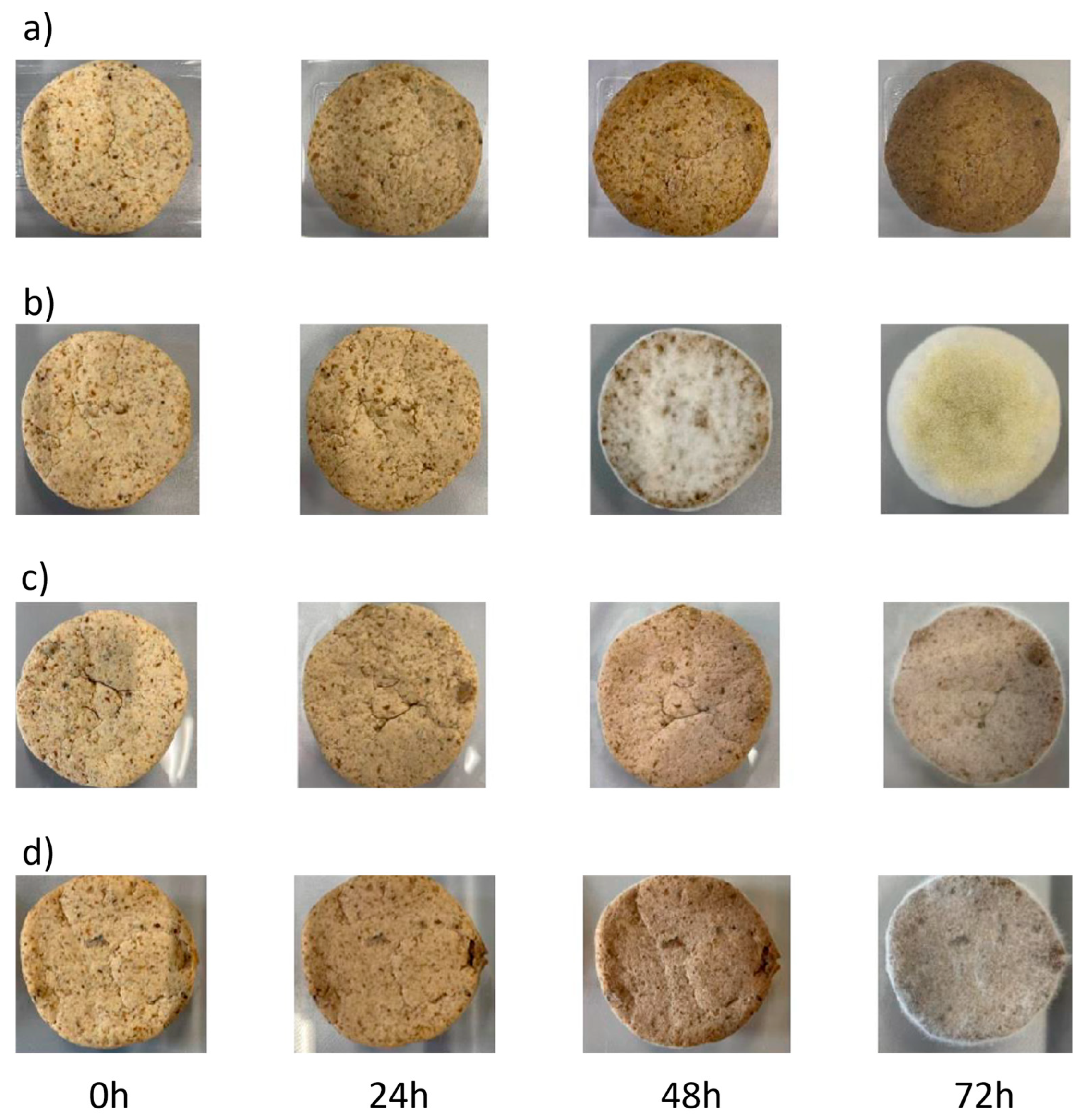
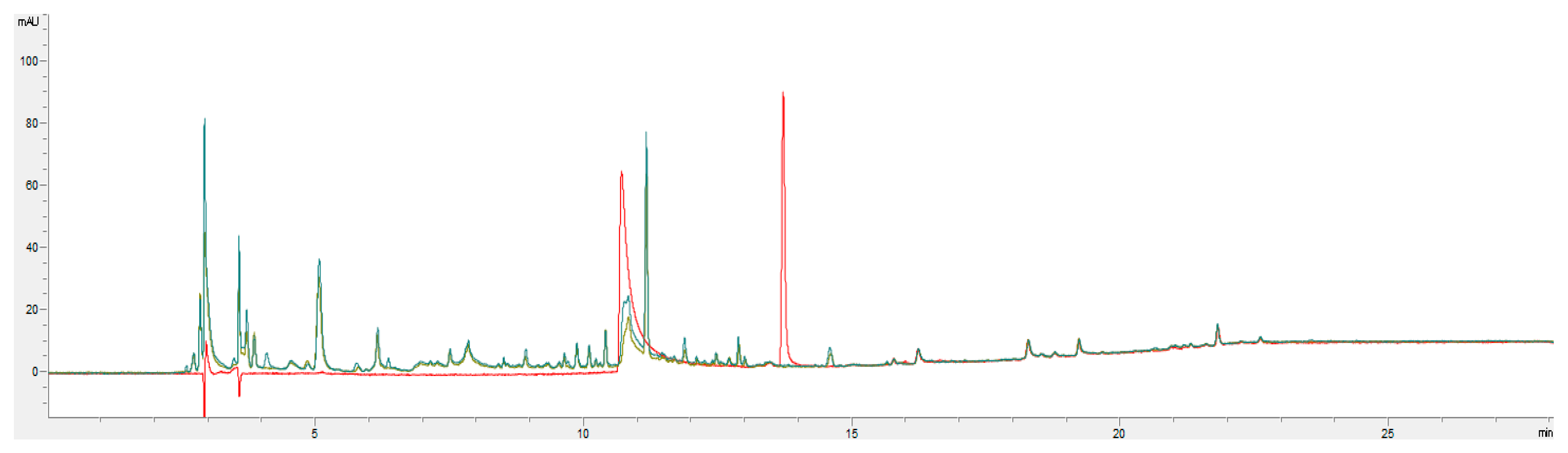
3. Results and Discussion

In this study, we adapted the fermentation conditions presented by Huang et al. (2007) and performed SSF of 100 g portions of walnut press cake using inocula of either S. thermophilus, A. ozyzae, R. oligosporus, or S. thermophilus and R. oligosporus in coculture. The WPC was examined visually after 0, 24, 48, and 72 h (Figure 1) [21]. As the optimal fermentation time for ellagic acid liberation was after 72 h of fermentation, we also stopped our assays at that time [22].
For fermentations with A. ozyzae, R. oligosporus visible mycelium formation was observed after 48 h. Similarly, in cocultures with R. oligosporus and S. thermophilus, visible mycelium formation by R. oligosporus was also observed after 48 h. The densest mycelium formation appeared in fermentation with A. oyzae after 72 h. The pH of the fermentations was monitored every 24 h. The water activity (aw) of the WPC was evaluated at the beginning and the end of the fermentation (Table 2).
Despite visible mycelium growth and a more than 300-fold increase in S. thermophilus concentration within the first 48 h of incubation, neither the pH, water content, nor the aw showed any significant changes throughout the incubation time.
The presence of EA was confirmed through HPLC–DAD analysis (Figure 2). Nonetheless, concentrations of EA in all fermentations remained constant, with only marginal variations observed from the initial concentrations, which were attributed to raw material heterogeneity (Table 3). The absence of EA liberation is unexpected, considering the results reported in a recent article involving SSF under comparable conditions [24].
The liberation of EA from WPC could potentially be influenced by insufficient moisture content. Based on the WPC preparation and the original humidity of the WPC, an approximate 48% water content was estimated in the fermentation setups at the beginning. For SSF using fungi, a moisture content of 60–70% is optimal to allow for adequate interaction of the mycelium with the substrate [11,24]. Due to adequate levels of water activity (aw) the growth of neither the two fungi nor the S. thermophilus was hindered.
The results obtained contrast with previous data using the raw material source and using identical methods, where the liberation of EA from ellagitannins in WPC was achieved using either A. oryzae or R. oligosporus [24]. However, as the studies where more than a year apart, the nut press cake used in this study did not originate from the same batch of production as before and may have been processed differently. Previous studies concluded that while ellagitannins are stable in acidic conditions, they are rapidly degraded in neutral and basic environments at elevated temperatures (60–80 °C). Ellagitannins may also oxidize to form compounds with a dehydrohexahydroxydiphenoyl (DHHDP) group such as mallotusinic acid [27]. Unfortunately, the authors of this study did not have control over any parameters involved in the production or storage of the walnut press cake before its procurement. Hence, even though the press cake was purchased at the same oil mill, variations in handling and processing parameters may be expected. Though unlikely, the differences in the results obtained previously and those reported here may be due to these factors. In addition, the effect of immobilization systems in improving productivity and efficiency was also previously documented using other ellagitannin-rich substrates. SSF using A. niger GH1 showed that fungal ellagitannase and ellagitannin acyl hydrolases (EAH) were able convert ellagitannins to EA within 18 h of culture using a polyurethane foam support. The conversion corresponded to fungal biomass production [16]. Similarly, Aspergillus oryzae, under optimum conditions, was able to convert ellagitannins from oak acorn fringe, yielding 17.7% EA. Yields were dependant on pH and incubation time [22]. Bioconversions using R. oligosporus and cranberry pomace provided a maximum EA production of 400 mg/100 g of pomace. However, fungal fermentation lasted 12 d [28]. Cocultures of A. niger and R. oryzae produced 6.9 mg/g of pomegranate husk [29]. SSF from powdered pomegranate husks using Saccharomyces cerevisiae yielded five-fold higher concentrations of EA than those obtained by A. niger fermentations [30]. Comparison of the fermentations suggests that punicalagins (α and β) are the targets of the microorganisms’ hydrolase activity during formation of EA [30]. To further convert EA to urolithin A during fermentation, S. thermophilus FAM2932 was used in the present study. A related strain, S. thermophilus FUA329, isolated from human breast milk was recently shown to convert EA to urolithin A in vitro [25] S. thermophilus FUA329 degraded EA during the exponential phase, with urolithin A being produced in the stationary phase. A maximum concentration of 7.3 µmol/L of urolithin A was produced after 50 h of anaerobic culture in an ABB medium supplemented with 20 µmol/L EA and 0.005% L-cysteine. Other bacteria, such as Enterococcus faecium FUA029 and Lactococcus garviae FUA009, were also implicated in the production of urolithin A under similar growth conditions [31,32,33]. Urolithin A and B from EA were produced by Bifidobacterium pseudocatenulatum INIA P815 [34]. The organism was incubated anaerobically for 5 d at 37 °C in BHI containing 0.5 g/L L-cysteine and 20 mg/L EA. Unfortunately, in this study, even with the more sensitive HPLC–MS analysis, no urolithin A, with a limit of detection of 25 ng/g WPC, was detected in the fermented products. However, the existence of EA has been unequivocally confirmed in all WPC samples. While human gut bacteria have been previously implicated in urolithin production, the use of microorganisms used in food production would provide a new pathway for accessing this bioactive compound for people without the capacity to form urolithin from their microbiota [35]. In all studies mentioned before, incubation under anaerobic conditions was crucial for the production of urolithins from EA. However, the fermentation of food waste substrates containing ellagitannins to produce urolithins remains to be demonstrated.

4. Conclusions

Walnut press cake, a relevant by-product from oil production, can be utilized as a substrate for solid-state fermentation with Aspergillus oryzae, Rhizopus oligosporus, and Streptococcus thermophilus. These microorganisms possess the necessary enzymes to release ellagic acid from ellagitannins and convert ellagic acid into urolithin A. The growth of microorganisms on the substrate was confirmed. However, the liberation of ellagic acid was not observed in detectable amounts. Additionally, even with a sensitive MS-method, urolithin A was not detectable in the fermented walnut press cake. It appears that anaerobic conditions, as well as immobilization systems for laboratory scale fermentation, are fundamental prerequisites for the transformation of ellagic acid into urolithins. In addition, variations of raw materials have to be scrutinized to eliminate ellagitannin degradation before their use. Further investigations into EA and urolithin A production under anaerobic conditions are currently in progress.

Author Contributions

Conceptualization, W.A. and W.M.B.; methodology, W.M.B.; formal analysis, Y.E.; investigation, Y.E.; writing—original draft preparation, and review and editing, W.A. and W.M.B.; supervision, W.M.B.; project administration, W.M.B. All authors have read and agreed to the published version of the manuscript.

Funding

This research received no external funding.

Data Availability Statement

The data presented in this study are available on request from the corresponding author.

Acknowledgments

We acknowledge the support of this study by the Analytical and Bioanalytical Platform of the Institute of Life Technologies, University of Applied Sciences and Arts of Western Switzerland (HES-SO Valais Wallis).

Conflicts of Interest

The authors declare no conflicts of interest.

References

  1. Bakkalbasi, E.; Meral, R.; Dogan, I.S. Bioactive Compounds, Physical and Sensory Properties of Cake Made with Walnut Press-Cake. J. Food Qual. 2015, 38, 422–430. [Google Scholar] [CrossRef]
  2. Bakkalbaşı, E. Oxidative stability of enriched walnut oil with phenolic extracts from walnut press-cake under accelerated oxidation conditions and the effect of ultrasound treatment. J. Food Meas. Charact. 2019, 13, 43–50. [Google Scholar] [CrossRef]
  3. Day, L.; Cakebread, J.A.; Loveday, S.M. Food proteins from animals and plants: Differences in the nutritional and functional properties. Trends Food Sci. Technol. 2022, 119, 428–442. [Google Scholar] [CrossRef]
  4. Pop, A.; Păucean, A.; Socaci, S.A.; Alexa, E.; Man, S.M.; Mureșan, V.; Chiş, M.S.; Salanță, L.; Popescu, I.; Berbecea, A.; et al. Quality Characteristics and Volatile Profile of Macarons Modified with Walnut Oilcake By-Product. Molecules 2020, 25, 2214. [Google Scholar] [CrossRef] [PubMed]
  5. Garcia-Mendoza, M.d.P.; Espinosa-Pardo, F.A.; Savoire, R.; Etchegoyen, C.; Harscoat-Schiavo, C.; Subra-Paternault, P. Recovery and antioxidant activity of phenolic compounds extracted from walnut press-cake using various methods and conditions. Ind. Crops Prod. 2021, 167, 113546. [Google Scholar] [CrossRef]
  6. Ran, J.; Su, Y.; Wang, P.; Yang, W.; Li, R.; Jiao, L.; Zhao, R. Effect of Lactobacillus acidophilus fermentation on bioaccessibility: The relationship between biotransformation and antioxidant activity of apple polyphenols based on metabolomics. LWT 2023, 190, 115360. [Google Scholar] [CrossRef]
  7. Martí-Quijal, F.J.; Khubber, S.; Remize, F.; Tomasevic, I.; Roselló-Soto, E.; Barba, F.J. Obtaining Antioxidants and Natural Preservatives from Food By-Products through Fermentation: A Review. Fermentation 2021, 7, 106. [Google Scholar] [CrossRef]
  8. Tamang, J.P.; Cotter, P.D.; Endo, A.; Han, N.S.; Kort, R.; Liu, S.Q.; Mayo, B.; Westerik, N.; Hutkins, R. Fermented foods in a global age: East meets West. Compr. Rev. Food Sci. Food Saf. 2020, 19, 184–217. [Google Scholar] [CrossRef] [PubMed]
  9. Lizardi-Jiménez, M.A.; Hernández-Martínez, R. Solid state fermentation (SSF): Diversity of applications to valorize waste and biomass. 3 Biotech 2017, 7, 44. [Google Scholar] [CrossRef] [PubMed]
  10. Šelo, G.; Planinić, M.; Tišma, M.; Tomas, S.; Koceva Komlenić, D.; Bucić-Kojić, A. A Comprehensive Review on Valorization of Agro-Food Industrial Residues by Solid-State Fermentation. Foods 2021, 10, 927. [Google Scholar] [CrossRef]
  11. Doriya, K.; Jose, N.; Gowda, M.; Kumar, D.S. Chapter Six—Solid-State Fermentation vs Submerged Fermentation for the Production of l-Asparaginase. In Advances in Food and Nutrition Research; Kim, S.-K., Toldrá, F., Eds.; Academic Press: Cambridge, MA, USA, 2016; Volume 78, pp. 115–135. [Google Scholar]
  12. Srivastava, N.; Srivastava, M.; Ramteke, P.W.; Mishra, P.K. Chapter 23—Solid-State Fermentation Strategy for Microbial Metabolites Production: An Overview. In New and Future Developments in Microbial Biotechnology and Bioengineering; Gupta, V.K., Pandey, A., Eds.; Elsevier: Amsterdam, The Netherlands, 2019; pp. 345–354. [Google Scholar] [CrossRef]
  13. Landete, J.M. Ellagitannins, ellagic acid and their derived metabolites: A review about source, metabolism, functions and health. Food Res. Int. 2011, 44, 1150–1160. [Google Scholar] [CrossRef]
  14. Devanthi, P.V.P.; Gkatzionis, K. Soy sauce fermentation: Microorganisms, aroma formation, and process modification. Food Res. Int. 2019, 120, 364–374. [Google Scholar] [CrossRef]
  15. Anwar, F.; Qadir, R.; Abbas, A. Chapter 44—Cold pressed walnut (Juglans regia L.) oil. In Cold Pressed Oils; Ramadan, M.F., Ed.; Academic Press: Cambridge, MA, USA, 2020; pp. 491–495. [Google Scholar] [CrossRef]
  16. Buenrostro-Figueroa, J.; Gutierrez-Sánchez, G.; Prado-Barragán, L.A.; Rodríguez-Herrera, R.; Aguilar-Zárate, P.; Sepúlveda, L.; Ascacio-Valdés, J.A.; Tafolla-Arellano, J.C.; Aguilar, C.N. Influence of culture conditions on ellagitannase expression and fungal ellagitannin degradation. Bioresour. Technol. 2021, 337, 125462. [Google Scholar] [CrossRef]
  17. Nevara, G.A.; Giwa Ibrahim, S.; Syed Muhammad, S.K.; Zawawi, N.; Mustapha, N.A.; Karim, R. Oilseed meals into foods: An approach for the valorization of oilseed by-products. Crit. Rev. Food Sci. Nutr. 2023, 63, 6330–6343. [Google Scholar] [CrossRef]
  18. Evtyugin, D.D.; Magina, S.; Evtuguin, D.V. Recent Advances in the Production and Applications of Ellagic Acid and Its Derivatives. A Review. Molecules 2020, 25, 2745. [Google Scholar] [CrossRef] [PubMed]
  19. Nasser, H.A.; Mahmoud, M.; Tolba, M.M.; Radwan, R.A.; Gabr, N.M.; ElShamy, A.A.; Yehya, M.S.; Ziemke, A.; Hashem, M.Y. Pros and cons of using green biotechnology to solve food insecurity and achieve sustainable development goals. Euro-Mediterr. J. Environ. Integr. 2021, 6, 29. [Google Scholar] [CrossRef]
  20. Tomás-Barberán, F.A.; González-Sarrías, A.; García-Villalba, R.; Núñez-Sánchez, M.A.; Selma, M.V.; García-Conesa, M.T.; Espín, J.C. Urolithins, the rescue of “old” metabolites to understand a “new” concept: Metabotypes as a nexus among phenolic metabolism, microbiota dysbiosis, and host health status. Mol. Nutr. Food Res. 2017, 61, 1500901. [Google Scholar] [CrossRef]
  21. Yang, Y.; Lee, P.-K.; Wong, H.-C.; Zhao, D. Oral supplementation of Gordonibacter urolithinfaciens promotes ellagic acid metabolism and urolithin bioavailability in mice. Food Chem. 2024, 437, 137953. [Google Scholar] [CrossRef]
  22. Huang, W.; Niu, H.; Gong, G.H.; Lu, Y.R.; Li, Z.S.; Li, H. Individual and combined effects of physicochemical parameters on ellagitannin acyl hydrolase and ellagic acid production from ellagitannin by Aspergillus oryzae. Bioprocess Biosyst. Eng. 2007, 30, 281–288. [Google Scholar] [CrossRef] [PubMed]
  23. Buenrostro Figueroa, J.; Mireles, M.; Ascacio-Valdés, J.; Aguilera-Carbo, A.; Sepúlveda, L.; Contreras-Esquivel, J.; Rodriguez, R.; Aguilar, C. Enzymatic Biotransformation of Pomegranate Ellagitannins: Initial Approach to Reaction Conditions. Iran. J. Biotechnol. 2020, 18, 30–36. [Google Scholar] [CrossRef]
  24. Brück, W.M.; Díaz Escobar, V.D.; Droz-dit-Busset, L.; Baudin, M.; Nicolet, N.; Andlauer, W. Fermentative Liberation of Ellagic Acid from Walnut Press Cake Ellagitannins. Foods 2022, 11, 3102. [Google Scholar] [CrossRef] [PubMed]
  25. Liu, Q.; Liu, S.; Ye, Q.; Hou, X.; Yang, G.; Lu, J.; Hai, Y.; Shen, J.; Fang, Y. A Novel Streptococcus thermophilus FUA329 Isolated from Human Breast Milk Capable of Producing Urolithin A from Ellagic Acid. Foods 2022, 11, 3280. [Google Scholar] [CrossRef]
  26. Varzakas, T. Rhizopus oligosporus mycelial penetration and enzyme diffusion in soya bean tempe. Process Biochem. 1998, 33, 741–747. [Google Scholar] [CrossRef]
  27. Sójka, M.; Janowski, M.; Grzelak-Błaszczyk, K. Stability and transformations of raspberry (Rubus idaeus L.) ellagitannins in aqueous solutions. Eur. Food Res. Technol. 2019, 245, 1113–1122. [Google Scholar] [CrossRef]
  28. Vattem, D.A.; Shetty, K. Solid-State Production of Phenolic Antioxidants from Cranberry Pomace by Rhizopus oligosporus. Food Biotechnol. 2002, 16, 189–210. [Google Scholar] [CrossRef]
  29. Hu, H.L.; van den Brink, J.; Gruben, B.S.; Wösten, H.A.B.; Gu, J.D.; de Vries, R.P. Improved enzyme production by co-cultivation of Aspergillus niger and Aspergillus oryzae and with other fungi. Int. Biodeterior. Biodegrad. 2011, 65, 248–252. [Google Scholar] [CrossRef]
  30. Moccia, F.; Flores-Gallegos, A.C.; Chávez-González, M.L.; Sepúlveda, L.; Marzorati, S.; Verotta, L.; Panzella, L.; Ascacio-Valdes, J.A.; Aguilar, C.N.; Napolitano, A. Ellagic Acid Recovery by Solid State Fermentation of Pomegranate Wastes by Aspergillus niger and Saccharomyces cerevisiae: A Comparison. Molecules 2019, 24, 3689. [Google Scholar] [CrossRef] [PubMed]
  31. Mi, H.; Liu, S.; Hai, Y.; Yang, G.; Lu, J.; He, F.; Zhao, Y.; Xia, M.; Hou, X.; Fang, Y. Lactococcus garvieae FUA009, a Novel Intestinal Bacterium Capable of Producing the Bioactive Metabolite Urolithin A from Ellagic Acid. Foods 2022, 11, 2621. [Google Scholar] [CrossRef]
  32. Xia, M.; Mu, S.; Fang, Y.; Zhang, X.; Yang, G.; Hou, X.; He, F.; Zhao, Y.; Huang, Y.; Zhang, W.; et al. Genetic and Probiotic Characteristics of Urolithin A Producing Enterococcus faecium FUA027. Foods 2023, 12, 1021. [Google Scholar] [CrossRef]
  33. Zhang, X.; Fang, Y.; Yang, G.; Hou, X.; Hai, Y.; Xia, M.; He, F.; Zhao, Y.; Liu, S. Isolation and characterization of a novel human intestinal Enterococcus faecium FUA027 capable of producing urolithin A from ellagic acid. Front. Nutr. 2022, 9, 1039697. [Google Scholar] [CrossRef]
  34. Gaya, P.; Peirotén, Á.; Medina, M.; Álvarez, I.; Landete, J.M. Bifidobacterium pseudocatenulatum INIA P815: The first bacterium able to produce urolithins A and B from ellagic acid. J. Funct. Foods 2018, 45, 95–99. [Google Scholar] [CrossRef]
  35. Iglesias-Aguirre, C.E.; García-Villalba, R.; Beltrán, D.; Frutos-Lisón, M.D.; Espín, J.C.; Tomás-Barberán, F.A.; Selma, M.V. Gut Bacteria Involved in Ellagic Acid Metabolism To Yield Human Urolithin Metabotypes Revealed. J. Agric. Food Chem. 2023, 71, 4029–4035. [Google Scholar] [CrossRef] [PubMed]
Figure 1. WPC fermentation after 0 h, 24 h, 48 h, and 72 h using (a) S. thermophilus, (b) A. ozyzae, (c) R. oligosporus, and (d) S. thermophilus and R. oligosporus coculture.
Applsci 14 01141 g001
Figure 2. Chromatograms of a standard solution with 50 mg/L of ellagic acid (Rt = 10.7 min) and urolithin A (Rt = 13.7 min) in red. Samples of the fermentation with R. oligosporus at t = 0 h (green) and t = 72 h (blue).
Applsci 14 01141 g002
Table 1. Measured CO2 and relative humidity for the fermentation incubators.
Temperature
[°C]
CO2 [ppm]Humidity [%]
2561157.2
3061146.1
3749933.9
Table 2. pH and aw of WPC fermentation after 0 h, 24 h, 48 h, and 72 h using either S. thermophilus, A. ozyzae, R. oligosporus, or S. thermophilus and R. oligosporus coculture.
Time (h)S. thermophilusA. oryzaeR. oligosporusS. thermophilus and R. oligosporus Co-Culture
pHawpHawpHawpHaw
05.95 ± 0.020.955.90 ± 0.020.965.88 ± 0.020.965.99 ± 0.020.96
245.76 ± 0.01 5.83 ± 0.01 5.77 ± 0.03 5.80 ± 0.02
485.70 ± 0.03 5.87 ± 0.01 5.67 ± 0.06 5.81 ± 0.03
725.61 ± 0.020.945.85 ± 0.030.955.61 ± 0.070.935.79 ± 0.020.96
Table 3. EA concentrations of WPC (mg/g fresh matter) for the four fermentations as determined by HPLC–DAD analysis (mean ± SD, n = 3).
Fermentation0 h24 h48 h72 h
S. thermophilus0.14 ± 0.010.13 ± 0.000.14 ± 0.000.17 ± 0.03
A. oryzae0.15 ± 0.040.18 ± 0.030.14 ± 0.040.13 ± 0.02
R. oligosporus0.26 ± 0.010.21 ± 0.130.22 ± 0.140.28 ± 0.07
S. thermophilus and R. oligosporus co-culture0.19 ± 0.030.21 ± 0.060.20 ± 0.020.16 ± 0.02
Disclaimer/Publisher’s Note: The statements, opinions and data contained in all publications are solely those of the individual author(s) and contributor(s) and not of MDPI and/or the editor(s). MDPI and/or the editor(s) disclaim responsibility for any injury to people or property resulting from any ideas, methods, instructions or products referred to in the content.

