Article

A Comparative Study of Oleuropein Extraction from Wild Olive Leaves (Olea europea subsp. oleaster, Hoffmanns. & Link), Its Gastrointestinal Stability, and Biological Potential

1
Faculty of Sciences, University of Split, R. Boškovića 33, 21000 Split, Croatia
2
Faculty of Chemistry and Technology, University of Split, R. Boškovića 35, 21000 Split, Croatia
3
Department of Biotechnology, University of Rijeka, R. Matejčić 2, 51000 Rijeka, Croatia
4
School of Medicine, University of Split, Šoltanska 2, 21000 Split, Croatia
5
Department of Gastroenterology, Clinic for Internal Medicine, University Hospital of Split, Spinčićeva 1, 21000 Split, Croatia
6
Faculty of Science, University of Zagreb, Horvatovac 102a, 10000 Zagreb, Croatia
7
Institute for Medical Research and Occupational Health, Ksaverska Cesta 2, 10000 Zagreb, Croatia
*
Author to whom correspondence should be addressed.
Appl. Sci. 2024, 14(2), 869; https://doi.org/10.3390/app14020869
Submission received: 12 December 2023 / Revised: 11 January 2024 / Accepted: 17 January 2024 / Published: 19 January 2024
(This article belongs to the Special Issue Bioactive Compounds: From Extraction to Application)

Abstract

:
Olive leaves are the richest source of phenolic compounds, particularly oleuropein, which has many beneficial effects on human health. This study compares the effect of three extraction techniques: ultrasonic-solvent extraction, microwave-assisted extraction, and supercritical CO2 extraction of freeze-dried wild olive leaves (Olea europea subps. oleaster, Hoffmanns. & Link) on oleuropein concentration in obtained extracts. The extract with the highest concentration of oleuropein was obtained after low-frequency ultrasound solvent extraction (WOLE-S) (71.97 µg/mL). Oleuropein from that extract showed low stability after simulated gastric digestion with human gastric juices (35.66%) and high stability after 60 min of intestinal phase (65.97%). The highest antiproliferative activity of WOLE-S and pure oleuropein was detected against T24 cancer cells. The antiproliferative activity of WOLE-S against MD-MBA-123, A549, and A1235 cells was moderate, while the activity of oleuropein was high. WOLE-S significantly down-regulated the expression of IL-6 in A549 cells, indicating a possible anti-inflammatory effect in lung cancer cells. The expression of IL-8 was slightly reduced by treating the cells with WOLE-S, while the expression of IL-10 was not altered by the treatment with WOLE-S or oleuropein.

1. Introduction

Oleuropein is a phenolic secoiridoid glycoside, derived from ligstroside, and found in all parts of the olive tree, mainly in the fruit and especially in the leaf. It was discovered in 1908 by Bourquelot and Vintilesco [1] who described it as a non-crystalline, intensely bitter substance that is easily soluble in alcohol, and fairly soluble in water [2]. Its concentration in the young olive fruit (up to 140 mg/g of dry matter) and in the leaves (60–80 mg/g dry matter) caused a very strong perception of bitterness [3,4]. Degradation products of oleuropein are hydroxytyrosol, elenolic acid, and glucose. The olive leaf has been used in Mediterranean folk medicine since ancient times to treat diabetes, hypertension, and hypercholesterolemia [5]. More recently, olive leaf extract (OLE) has become one of the most valuable by-products of the olive oil industry due to its diverse biological potential (antimicrobial, antioxidant, antidiabetic, anticarcinogenic, immunomodulatory) [6,7,8,9]. Valuable biological potential is associated with the presence of phenolic compounds, especially phenolic secoiridoids such as oleuropein, hydroxytyrosol, verbascoside, oleuropein aglycone, ligstroside aglycone [10].
The conditions for the extraction of oleuropein from olive leaves have been demonstrated in many studies [1,11,12]. The choice of the optimal extraction method, solvent, and extraction time are decisive factors. Oleuropein from olive leaves can be extracted by various methods such as Soxhlet extraction, cold solvent extraction, ultrasound-assisted extraction (UAE), supercritical fluid extraction (SFE), microwave-assisted extraction (MAE), pressurized liquid extraction, and novel techniques using membranes [1]. In this study, low-frequency ultrasound was used for extraction. The effects of cavitation of ultrasound extraction are described in the literature [1], leading to mechanical stress on the cells and rupture of the cells. Ruptured cells release water, which is necessary for the hydration of the plant material and the mass transfer between the plant material and the solvent. The frequency of the device is a decisive factor in ultrasound extraction. It is assumed that low frequencies of around 20 kHz are more effective for plant materials such as olive leaves, as the bubbles generated by cavitation can implode more easily than those generated at high frequencies [1]. The highest yield of oleuropein (812.9 mg/g) extracted in methanol:water (80:20) with an ultrasonic bath of 25 kHz was reported by Yasemi et al. [11]. MAE extraction is generally considered an environmentally friendly technique characterized by a lower solvent requirement and a short extraction time. Da Rosa et al. [13] compared three extraction methods for total phenols from olive leaves (MAE, maceration, and ultrasound-assisted extraction). They found that MAE with water as solvent was the most efficient (82% recovery of total phenols). Di Meo et al. [12] investigated the total phenolic content in olive leaves of five Italian cultivars extracted with MAE under different extraction conditions and found that the cultivar and harvest period had a greater influence than the MAE conditions. Supercritical fluid extraction is also considered an environmentally friendly technique due to the use of non-toxic solvents [14]. The extraction of oleuropein from olive leaves with supercritical fluid has been described in several studies [15,16,17]. Some authors reported very low amounts of OLE after SC-CO2 extraction [18]. The highest yield of OLE was obtained by SC-CO2 at 300 bar, 100 °C and using methanol 20% v/v [1].
It has already been mentioned that oleuropein is known for its various pharmacological and biological properties (antioxidant, antimicrobial, antiviral, anticancer, hypolipidemic, cardioprotective, anti-inflammatory), which puts oleuropein in the focus of numerous in vivo and in vitro studies [19,20,21]. Gastrointestinal digestion can influence the stability, bioavailability, and biological activity of oleuropein. The use of in vitro digestion models is relatively simple and fast compared to time-consuming and costly in vivo studies [22]. In our study, an in vitro model with human digestive juices was used to simulate physiological digestion, as described by Ulleberg et al. [23]. The available data on the gastrointestinal stability of oleuropein from OLE show that pH affects its stability and bioavailability only in an acidic medium (about 2) [24,25,26,27], while after the intestinal phase, its stability was higher [26]. This study also includes the investigation of the antiproliferative and anti-inflammatory activities of wild olive leaf extract obtained after low-frequency ultrasound solvent extraction (WOLE-S) and pure oleuropein. Their antiproliferative effect has been proven in many studies against different cancer cell lines [28,29,30,31,32]. OLE showed a significant anti-inflammatory effect in rats by reducing TNF, IL-1, COX-2 and NO [20] and the mRNA level of proinflammatory cytokines in the brain of diabetic rats [33].
The aim of this study was to investigate the influence of three extraction techniques (ultrasound-solvent, microwave-assisted, and supercritical CO2) on the oleuropein content of freeze-dried wild olive leaf. The extract with the highest concentration of oleuropein was used to investigate the gastrointestinal stability of oleuropein after simulated two-phase digestion with human digestive enzymes. The analysis of the composition of phenolic compounds in wild olive leaf extract was performed using the HPLC-UV/VIS technique, while the analysis of the gastrointestinal stability of oleuropein from WOLE-S was performed using the UHPLC-DAD technique. In addition, the antiproliferative activity of WOLE-S vs. pure oleuropein was analysed against four human cancer cell lines: T24, MDA-MB-231, A549, and A1235. As far as we know, this is the first report on the antiproliferative activity of oleuropein and olive leaf extract against A1235 cells. The effect of WOLE-S and pure oleuropein on the expression of inflammation-linked genes (IL-6, IL-8, IL-10) in the A549 cell line was detected using the quantitative Real-Time PCR method.

2. Material and Methods

2.1. Plant Material

Fresh green olive leaves from wild olive trees (Olea europea L. subsp. oleaster) from the island of Lastovo (Dalmatia region, Croatia). The fresh plant material (leaves) was weighed (300 g) and frozen with liquid nitrogen (−196 °C) immediately before freeze-drying (Labconco, Kansas City, MO, USA). The conditions for freeze-drying were as follows: Pressure: 0.122 mbar; drying time: 24 h; condenser temperature: −55 °C. The sample was placed as a thin single layer on the drying tray. The dried plant material was packed in aluminium barrier bags and stored at room temperature and a relative humidity of 35% until analysis.

2.2. Extractions of Wild Olive Leaves

2.2.1. Solvent Extraction

Thirty grams of freeze-dried and ground wild olive leaves were extracted three times with 150 mL methanol: water, 80:20 (v/v) in an ultrasonic bath (15 min, room temperature, 40 kHz frequency). The solvent was evaporated on a rotary evaporator (Büchi, R200, Allschwil, Switzerland), and the dry residue was dissolved in 25 mL of distilled water. The extract obtained was stored at 20 °C and sealed with an inert septum until analysis.

2.2.2. Microwave Assisted-Extraction (MAE)

Microwave extraction (Milestone, Sorisole, Italy) of freeze-dried ground wild olive leaves used ethanol as solvent. The temperature of extraction was 70 °C, the time of extraction was 15 min, and the irradiation power was 500 watts. The extract obtained was filtered through a 0.45 µm cellulose membrane filter and stored at the temperature of −20 °C until analysis.

2.2.3. Supercritical CO2 Extraction

The dried plant material was ground in a coffee blender, and the mean particle size of the raw material (0.355 mm) was determined using sieve sets (CISA Cedaceria Industrial, Barcelona, Spain). Supercritical fluid extraction (SFE) was performed on a laboratory-scale, high-pressure extraction plant (HPEP, NOVA, Swiss, Efferikon, Switzerland) described in Pekić et al. [34]. The prepared sample (50.0 g) was placed in an extraction vessel and extracted for 4 h. The extract was obtained at 150 bar, a temperature of 40 °C, and a CO2 flow rate of 0.2 kg/h.

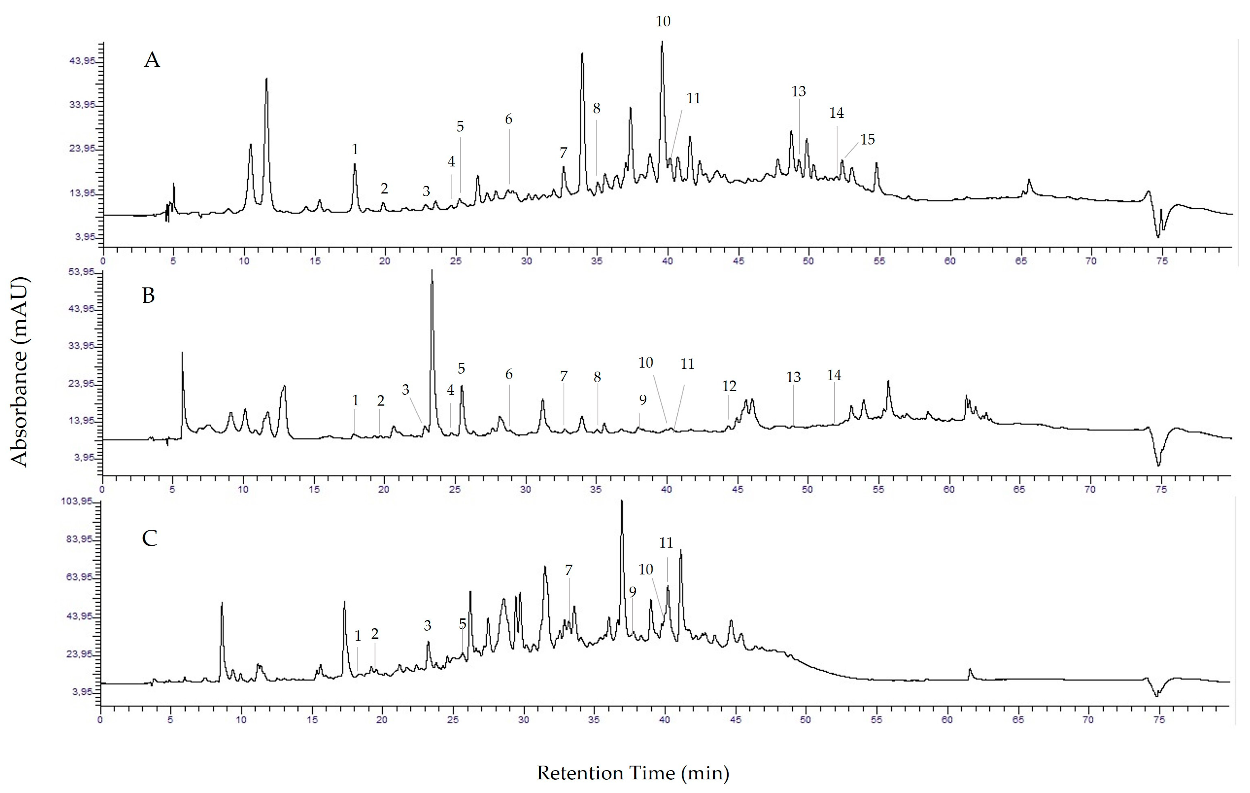
2.3. HPLC Analysis of Phenolic Compounds Extracted from Wild Olive Leaves

The phenolic compounds from wild olive leaf extract were separated by high-performance liquid chromatography on an HPLC system (Perkin Elmer, Waltham, MA, USA) using COI/T method 20/Doc. No. 29 [35]. The system is labelled Series 200 and consists of units for the automatic sample application, a binary pump, a system for removing gases from the mobile phase using a vacuum, a column heater, and a UV/VIS detector. The separation was performed on an Ultra Aqueous C18 column (250 × 4.60 mm) with a stationary phase diameter of 5 µm (Restek, Bellefonte, PA, USA). The mobile phase consisted of 0.2% phosphoric acid (solvent A) and a 1:1 mixture of methanol and acetonitrile (solvent B). Gradient elution was achieved by a pump programme that initially provided 4% solvent B, and increased the proportion of solvent B to 50% over a period of 40 min. The increase in solvent B to 60% continued over the next 5 min, with the proportion of solvent B increasing to 100% from 45 to 60 min. In the next 8 min, solvent B flowed through the column. A steep linear decrease of solvent B to 4% occurred from 68 to 70 min, and the achieved solvent ratio was maintained for the last 10 min to ensure stabilisation of the column to the initial conditions. The column was thermostatted at 25 °C, the flow rate was 0.8 mL/min, and the injection volume was 20 µL.
A UV detector with a wavelength of 280 nm was used for the detection of phenol. The quantification of oleuropein aglycone is expressed in µg/mL oleuropein, while separate calibration lines were established for the other 14 standards (Supplementary Materials Figure S1a–m). Oleuropein was identified by the retention time of the standard and quantified using the calibration curve of the standard in the range of 5.16 to 201.60 µg/mL (R2 = 0.9997). All results represent the average of two measurements.
Standards and solvents were of analytical grade and were purchased from Sigma-Aldrich (Steineheim, Germany). Deionised water (Milli Q) was used to prepare the solvents.

2.4. Collection of Human Digestive Enzymes and Determination of the Enzymatic Activity of Digestive Juices

The collection of human digestive juices from the stomach and duodenum was done according to the method described by Blažević et al. [36], while the determination of the enzymatic activity of collected juices was performed according to the method described by Almaas et al. [37]. Briefly, human digestive juices from the stomach and duodenum were collected from four healthy volunteers using an endoscope. Digestive juices were collected separately from the stomach and duodenum, collected in a sterile tube and centrifuge at 10,000 rpm at room temperature (my Spin Minicentrifuges, Thermo Fisher Scientific, Waltham, MA, USA) to remove mucosa and cell debris. Juices were divided into aliquots, treated with inert gas (argon), and stored at −20 °C until further analysis. The enzymatic activity of collected human digestive juices was determined using bovine haemoglobin solution for gastric juices and casein solution for duodenal juices. One unit of enzyme activity (U) represents the amount of enzyme that causes the absorbance change of 1 between the blank and the sample at the wavelength of 280 nm (Microplate Reader, Tecan, Mannedorf/Zurich, Switzerland).

2.5. In Vitro Digestion

The method used to simulate the digestion process was a static, two-phase model of digestion. The method is described in detail by Furlund et al. [38]. A horizontal thermo-shaker (BioSan, Berlin, Germany) was used for the digestion simulation. The shaking speed was 1200 rpm at a temperature of 37 °C. For 1 U of gastric and intestinal juice, 20 and 25 µL were required, respectively (the calculation of 1U is explained in the previous paragraph). The pH value of the simulated gastric phase was 2.5, while the pH value of the simulated intestinal phase was 7.5. For the simulated stomach phase, 20 U per g sample and for the simulated intestinal phase, 62.4 U samples were used. After the simulated digestion process, the digested samples were centrifuged for 10 min at room temperature at 9800× g using the mySpin 12 microcentrifuge (Thermo Scientific, Walthman, MA, USA).
The supernatant was filtered through a 0.45 µm cellulose membrane filter stored at −20° until analyses. The formula for the gastrointestinal stability rate (%) was as follows:
g a s t r o i n t e   s t i n a l s t a b i l i t y   r a t e % = s a m p l e   c o n c e n t r a t i o n   a f t e r   d i g e s t i o n s a m p l e   c o n c e n t r a t i o n   b e f o r e   d i g e s t i o n × 100  

2.6. UHPLC Analysis of In Vitro Digesta Samples

The samples for gastrointestinal stability of pure oleuropein and WOLE were filtered through a filter with a pore size of 0.22 µm before analysis. Qualitative and quantitative analysis was performed using an Agilent UHPLC 1290 Infinity with a DAD detector, (Agilent Technologies, Palo Alto, CA, USA). The oleuropein standard was purchased from Extrasynthesis (Genay, France). The phenolic compounds were separated on an Agilent ZORBAX RRHD SB-C18 (2.1 mm × 50 mm, 1.8 µm) column. The mobile phase consisted of 0.1% formic acid (solvent A) and a mixture of acetonitrile and 0.1% formic acid (solvent B). The gradient elution was achieved using the pump program, which ensured an increase in the proportion of solvent B from 0 to 5% from 0 to 1.8 min. In the period from 1.8 to 3.6 min, the proportion of solvent B increased to 15%, and in the following period, from 3.6 to 13 min to 40%. A steep increase in the proportion of solvent B to 100% was achieved from 13 to 14.5 min. A steep linear decrease of solvent B to 0% occurred in the period from 14.5 to 15 min, and solvent A flowed through the column during the last 2.5 min. The column was thermostatted at 30 °C, the flow rate was 0.3 mL/min, and the volume injected was 3 µL. Detection was performed with a UV-DAD detector at 280 nm and 325 nm. The calibration curve of the external standard oleuropein dissolved in water was established in the concentration range of 133 to 1200 µg/mL, y = 2.0536x − 11,591 (R2 = 0.9998). All results represent the average of two measurements.

2.7. Antiproliferative Activity

The antiproliferative activity of WOLE-S and pure oleuropein was investigated against four cancer cell lines: glioblastoma cancer cells (A1235), breast cancer cells (MDA-MB-231), lung cancer cells (A549), and bladder cancer cells (T24). The stock solutions of the samples were 10 mg/mL (pure oleuropein was dissolved in DMSO, dimethyl sulfoxide). The cancer cells were grown in Dulbecco’s modified Eagle’s medium (DMEM, Euroclone, Milan, Italy), supplemented by 10% foetal bovine serum (FBS, Euroclone, Milan, Italy), and 1% antibiotics (penicillin and streptomycin, Euroclone, Milan, Italy), in a humidified incubator at 37 °C and 5% CO2. The equal numbers of cells (1 × 10⁴) were transferred to 96 wells and allowed to adhere overnight. The cells were then treated with samples (WOLE-S and oleuropein) at concentrations of 0.5 and 1 µg/L for a period of 4, 24, 48, and 72 h, followed by the addition of the yellow tetrazoline MTT (3-(4,5-dimethylthiazolid-2)-2,5-diphenyltetrazoline bromide), which is reduced to purple formazan in metabolically active cells. After 2 h, the medium with MTT was removed, and DMSO was added. The plates were incubated for 10 min at 37 °C with shaking. The absorbance was measured by the microplate photometer (HiPo MPP-96, Biosan, Riga, Latvia) at 570 nm. All experiments were performed in triplicate.

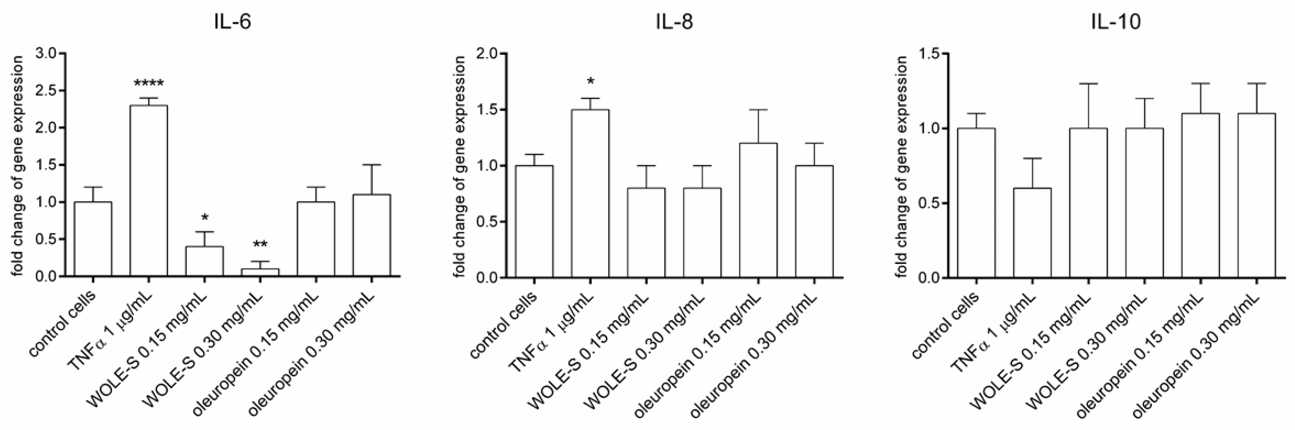
2.8. Anti-Inflammatory Activity

2.8.1. Cell Culture

A549 cells were purchased from ATCC (ATCC, Manassas, VA, USA) and cultured in T75 flasks (Eppendorf, Hamburg, Germany) in Dulbecco’s Modified Eagle Medium (DMEM, Sigma Aldrich, St. Louis, MO, USA) supplemented with 10% Fetal Bovine Serum (FBS, Sigma Aldrich, St. Louis, MO, USA) and 1% Antibiotic/Antimycotic (Thermo Fisher Scientific, Waltham, MA, USA) at 37° C and 5% CO2. Cells were grown until reaching 80% confluence, after which they were washed with sterile phosphate-buffered saline (PBS). Cells were then detached by adding 1.5 mL of Trypsin-EDTA solution (Sigma Aldrich, St. Louis, MO, USA) to the flask, followed by 5 min incubation at 37 °C and 5% CO2. Detached cells were collected, transferred to fresh cell culture medium, and seeded in 12-well plates (Eppendorf, Hamburg, Germany) in 1000 μL/well of cell culture medium (100,000 cells/well). Plates were incubated for 24 h at 37 °C and 5% CO2 prior to cell treatment.

2.8.2. Gene Expression Analysis–qPCR

Cell culture medium was removed from 12-well plates and fresh medium was added. WOLE-S and oleuropein suspensions were prepared in ultra-pure H2O and added to wells. Final concentrations were 0.15 mg/mL and 0.33 mg/mL for both WOLE-S and oleuropein. TNFα (Sigma Aldrich, St. Louis, MO, USA) was dissolved in ultra-pure H2O and added to wells to a final concentration of 1 μg/mL. Plates were incubated for 120 min at 37 °C and 5% CO2, after which cell culture medium was removed and total RNA from samples was extracted using Aurum Total RNA Mini Kit (BioRad Hercules, CA, USA) according to the manufacturer’s instructions. The quantitative real-time PCR method (qPCR) was used to assess inflammatory response and induction of oxidative stress following cell treatment with WOLE-S and oleuropein. Extracted sample RNA was used as a template for cDNA synthesis (5 µL of sample RNA was used for each reaction) by iScript cDNA synthesis kit (BioRad, Hercules, CA, USA). cDNA synthesis was performed following kit instructions on Eppendorf ThermoMixer C (Eppendorf, Germany). PCR was performed with iTaq Universal SYBR Green PCR Supermix (BioRad, USA) on Applied Biosystems 7500 Real-Time PCR System (Applied Biosystems, Waltham, MA, USA). The following program was used for cDNA amplification: 95 °C for 10 min, followed by 40 cycles of 95 °C for 15 s and 60 °C for 60 s. Glyceraldehyde-3-phosphate dehydrogenase (GAPDH) was used as a reference gene. Primers used to amplify targeted genes are listed in Table 1. Relative quantification of interleukin expression was calculated using the 2−ΔΔCq method [39].

2.9. Statistical Analysis

HPLC results were compared using a one-way analysis of variance (ANOVA) followed by a post-hoc test of multiple comparisons (Duncan’s New Multiple Range Test, DNMRT). Values that differ at the level p ≤ 0.05 were considered statistically significant. For antiproliferative activity, statistical significance was determined using the Student’s t-test for two independent environments and one-way ANOVA followed by the post-hoc Duncan’s new multiple range test, with p ≤ 0.05. Statistical analysis for gene expression experiments was performed in GraphPad Prism 6 (GraphPad Software, v8.0.2., San Diego, CA, USA). Statistical significance was determined with one-way ANOVA, followed by Dunnett’s Multiple Comparison Test where all treatment values for all assays were compared to control cell values. The threshold for statistical significance was set at p < 0.05. Statistically significant results were denoted with asterisks (* for p < 0.05, ** for p < 0.01, *** for p < 0.001, **** for p < 0.0001).

3. Results and Discussion

3.1. Extraction of Wild Olive Leaves

The results of the HPLC analysis of wild olive leaves after the use of three extraction methods: low-frequency ultrasound solvent extraction, supercritical CO2 extraction, and MAE extraction, are shown in Table 2 and Figure 1.
The focus of this study was on the effects of the extraction methods on oleuropein content. The results showed that the highest concentration of oleuropein was obtained after low-frequeny ultrasound solvent extraction (71.97 µg/mL) (Table 2, Figure 1A), compared to supercritical CO2 extraction (2.05 µg/mL) (Table 2, Figure 1B), and MAE extraction (13.62 µg/mL) (Table 2, Figure 1C). The results obtained are consistent with those of other studies that found very low amounts of oleuropein in olive leaf extract after supercritical CO2 extraction [18] on the one hand and an optimal effect of the combination of low-frequency ultrasound (20–40 kHz) and solvent (methanol:water, 80:20) for the extraction process on the other [11]. Baldino et al. [17] concluded that when supercritical CO2 extraction alone was used, only small amounts of oleuropein were extracted. According to the available literature, MAE extraction is a better choice than supercritical CO2 extraction to obtain a higher concentration of oleuropein in olive leaf extract, but not the best [40]. Extraction with low-frequency ultrasound has the best effect due to the cavitation phenomenon already explained in the introduction.
If we look at the effect of extraction methods on the proportion of all identified compounds, ultrasound solvent extraction resulted in significantly the highest yield for all the compounds, except 4-hydroxybenzoic acid, o-coumaric acid, tyrosol, and pinoresinol (Table 2). 4-hydroxybenzoic acid and pinoresinol were best extracted using supercritical CO2, while o-coumaric acid and tyrosol by applying microwave-assisted extraction.

3.2. Gastrointestinal Stability of Oleuropein from WOLE-S

The extract of wild olive leaves obtained after low-frequency ultrasonic solvent extraction (WOLE-S) was used to analyse the gastrointestinal stability of oleuropein after a simulated biphasic digestion process (stomach and duodenum) with human digestive juices, as this extract contains the highest concentration of oleuropein, as shown in Table 2 and explained previously. The oleuropein from the extract showed a gastrointestinal stability of 35.66% after a 30-min gastric digestion period. After the simulated 60-min duodenal phase, we observed higher stability of oleuropein from WOLE-S (65.97%). The results represent an average of two independent analyses. The acidic environment and digestive enzymes of the human stomach affected the decrease of oleuropein concentration in WOLE-S. In contrast, almost twice the amount of oleuropein was retained in the duodenal phase, even after a period twice as long. These results are consistent with the results of other studies [24,25,26]. Duque-Soto et al. [26] reported low stability of oleuropein after gastric digestion and higher stability after the intestinal phase, while Cedola et al. [27] documented low oleuropein stability after the intestinal phase. Gonzalez et al. [41] used microencapsulation of oleuropein with sodium alginate to enhance its gastric stability. Corona et al. [42] showed low gastric stability of oleuropein after in vivo digestion as a consequence of rapid hydrolysis in acidic gastric conditions. The combined effect of enzymatic activity and low pH resulted in the degradation of oleuropein and other secoiridoids from WOLE-S in the first phase of digestion. Reboredo-Rodriquez et al. [43] used the INFOGEST standardised method to assess the gastrointestinal stability of phenolic compounds from olive oil, and they proved extensive hydrolysis of secoiridoids, such as oleuropein, after gastric digestion.

3.3. Biological Potential of WOLE-S vs. Pure Oleuropein

The investigation of the biological potential of WOLE-S vs. pure oleuropein focussed on the antiproliferative activity against four human cancer cell lines: MD-MBA-231, T24, A1235, and A549 cells (Table 3a–d) and its anti-inflammatory activity by regulating the expression of genes involved in the inflammatory process (IL-6, IL-8, and IL-10) in A549 cells (Figure 2).
In general, the highest inhibitory effect of WOLE-S and pure oleuropein on cell proliferation was achieved against T24 cells (Table 3b). Two concentrations of WOLE-S and pure oleuropein (0.5 and 1 g/L) were tested after different incubation times (4, 24, 48, and 72 h) on all cancer cell lines. The antiproliferative effect of both samples was dose-dependent in all tested cells. Both concentrations of the samples showed high inhibitory activity against T24 cancer cells, already after 24 h of incubation (83.61% WOLE-S and 81.12% pure oleuropein). The antiproliferative effect of both samples remained high even after 48 and 72 h of incubation (Table 3b). Table 3a,c,d shows that in other cancer cell lines tested (MD-MBA-231, A1235, and A549), the antiproliferative effect of pure oleuropein was significantly higher compared to WOLE-S at both concentrations and after all incubation times tested. Compared to T24 cells, the antiproliferative effect of WOLE-S against A1235, A549, and MD-MBA 231 cells was generally moderate (the average rate was 35.48% at a concentration of 1 g/L after 72 h of incubation). In contrast, the antiproliferative activity of pure oleuropein against A1235, A549, and MD-MBA 231 cells was high (the average rate was 79.57% at a concentration of 1 g/L after 72 h of incubation). Except T24 at a concentration of 1.00 g/L after 24 h and 48 h of incubation, A1235 at 1.00 g/L after 4 h of incubation, and A549 at 1.00 g/L and 0.50 g/L after 24 h of incubation, in all other tested cell line/concentration/incubation time combinations oleuropein showed significantly higher inhibition of cell proliferation than WOLE-S. Regarding the incubation time, in the majority of the combinations, the highest inhibition by oleuropein was achieved after 72 h. However, in T24 cells, that was after 48 h of incubation with oleuropein 0.50 g/L, and in the A549 line after 48 h of incubation with oleuropein 1.00 g/L. Moreover, in A549 cells, with oleuropein 1.00 g/L, we got significantly better results after 48 h and 24 h compared to 72 h. Incubation for 4 h resulted in the weakest effect in all combinations. The results obtained are interesting because, on the one hand, 1 g/L of WOLE-S contains only 0.072 g/L of oleuropein incorporated into the plant matrix on one hand and on the other hand, the average rate of antiproliferative activity of pure oleuropein is only 2–3 times higher than that of WOLE-S against all cancer cell lines tested (for T24 cells, their activity was very similar at a concentration of 1 g/L).
Considering that the amount of 1 g/L pure oleuropein is approximately 14 times higher than the concentration of oleuropein in 1 g/L WOLE-S, one might expect a much higher antiproliferative activity of pure oleuropein compared to WOLE-S. Moreover, their antiproliferative activity against T24 cells at a concentration of 1 g/L was similar after 24, 48, and 72 h. This could be due to the fact that both samples were very effective against T24 cells and reached a maximum of their activity after 24 h of incubation. Dugue-Soto et al. [26] identified 74 phenolic compounds in olive leaf extract whose synergistic effect is expected, but it is impossible to characterise the exact mechanisms between so many biologically active compounds in the olive leaf plant matrix. Also, the effect of the plant matrix could cause a change in the stability of antioxidants from wild olive leaves in MTT media after 72 h of incubation.
To the best of our knowledge, this is the first report on the antiproliferative activity of WOLE-S and pure oleuropein against A1235 cancer cells. The antiproliferative activity of oleuropein and olive leaf extract is documented in many studies [28,29,30,31,32]. Najibulah et al. [44] showed the antiproliferative and pro-apoptotic activity of olive leaf extract in the A549 cell line, while other authors observed that oleuropein decreases the cell viability in T24 cancer cells [45,46]. Bal et al. [47] showed that the microincapsulation of olive leaf extract with calcium alginate and chitosan enhanced its antiproliferative activity against A549 cells. Figure 2 shows the expression of genes encoding the inflammatory biomarkers, such as interleukin 1 (IL-6), IL-8, and IL-10 involved in the inflammatory process in A549 cells.
The results show that WOLE-S significantly downregulates the expression of the anti-inflammatory cytokine IL-6 in A549 cells, indicating a possible anti-inflammatory effect in lung cancer cells. Given the importance of IL-6 in initiating and sustaining inflammatory response [48], reduced levels of IL-6 mRNA expression after WOLE-S treatment of A549 cells is evidence of WOLE-S anti-inflammatory potential. The expression of IL-8 was slightly reduced by treating the cancer cell line with WOLE-S, but the reduction was not statistically significant. Considering that TNFα treatment did not increase IL-8 expression as much as IL-6 expression in A549 cells, it can be concluded that IL-6 expression is a more sensitive indicator of the inflammatory process at a 2-h timepoint. The expression of IL-10 was not altered by treatment with WOLE-S or oleuropein. According to other in vitro studies, oleuropein from olive oil and olive leaf extract modulates the inflammatory process in the tissue [49,50]. Lockyer et al. [51] demonstrated a change in the pattern of cytokine expression (IL-1β, IL-6, IL-10, TNF-α, and IL-8) of secoiridoids from olive leaf extract in an in vivo study. Interestingly, our results have shown that the anti-inflammatory activity of WOLE-S was not caused exclusively by oleuropein. Considering the different anti-inflammatory, as well as anti-proliferative, activities of WOLE-S and pure oleuropein demonstrated by these results, it is apparent that oleuropein is not the only biologically active component of the extract. Specifically, hydroxytyrosol has previously shown anti-inflammatory activity [52], and its presence in WOLE-S could be the cause for different biological activity compared to oleuropein alone. However, the downregulation of inflammatory cytokines might not be a desirable property for potential anticarcinogens, considering the importance of the immune system in cancer suppression. Future studies should explore both antiproliferative and anti-inflammatory properties of WOLE-S to determine which holds greater potential.

4. Conclusions

The use of the low-frequency, ultrasound-solvent extraction method yielded the highest concentration of oleuropein in wild olive leaf extract (WOLE-S) compared to extraction with microwaves and supercritical CO2. This study proved the gastrointestinal instability of oleuropein from WOLE-S, especially after the gastric phase. In this sense, the use of microencapsulation techniques is necessary to protect the stability of oleuropein under digestive conditions and, thus, its biological potential. Pure oleuropein showed high antiproliferative activity against all tested cancer cell lines (T24, A549, A1235, and MD-MBA-123), while the antiproliferative activity of olive leaf extract was high against T24 cells and moderate against the other tested cell lines. WOLE-S caused a down-regulation of the expression of the inflammatory cytokine IL-6 in A549 cells. The results show the optimal effect of low-frequency ultrasound in combination with a suitable solvent extraction for a high yield of oleuropein in olive leaf extract. In addition, the gastrointestinal instability of oleuropein was demonstrated under simulated gastrointestinal conditions with human digestive enzymes, and a strong antiproliferative and anti-inflammatory effect of pure oleuropein and olive leaf extract was shown. However, this biological potential of oleuropein as a dietary supplement or olive leaf extract or infusate should be protected by the use of microencapsulation techniques due to the gastrointestinal instability of oleuropein.

Supplementary Materials

The following supporting information can be downloaded at: https://www.mdpi.com/article/10.3390/app14020869/s1, Figure S1: Calibration curve each of the 14 phenolic standards used in HPLC analysis, (a) hydroxytyrosol, (b) 3,4-hydroxybenzoic acid, (c) tyrosol, (d) catechin, (e) 4-hydroxybenzoic acid, (f) coffee acid, (g) p-coumaric acid, (h) t-ferulic acid, (i) o-coumaric acid, (j) oleuropein, (k) pinoresinol, (l) luteolin, (m) apigenin and (n) kaempferol. The amount of the standard is expressed in µg/mL.

Author Contributions

Conceptualization, B.S. and T.B.; methodology, B.S., T.B., J.G., I.L., V.Č.Č., A.B., P.B., I.Š. and K.I.; formal analysis, B.S., T.B., J.G., I.L., V.Č.Č., P.B., I.Š. and K.I.; investigation, B.S., T.B., J.G., I.L., V.Č.Č., A.B., P.B. and K.I.; resources, B.S., T.B., J.G., I.L., V.Č.Č., A.B., P.B., I.Š. and K.I.; data curation, B.S. and T.B.; writing-original draft preparation, B.S.; visualization, B.S., T.B. and I.Š.; writing—review and editing, B.S.,T.B., J.G., I.L., V.Č.Č., A.B., P.B., I.Š. and K.I.; supervision, B.S. All authors have read and agreed to the published version of the manuscript.

Funding

This research received no external funding.

Institutional Review Board Statement

The study was conducted in accordance with the Declaration of Helsinki, and approved by the Ethics Committee of University of Hospital Centre Split (protocol code: 2181-147-01/06J.B-16.2 and date of approval 19 May 2016).

Informed Consent Statement

Informed consent was obtained from all subjects involved in the study of gastrointestinal stability of oleuropein from WOLE-S. The approval for the collection of digestive juices was obtained from the Ethics Committee of the University of Hospital Centre Split.

Data Availability Statement

The data supporting our findings and analyses are contained in the article itself. Readers can access this data by referring to the article.

Conflicts of Interest

The authors declare no conflicts of interest.

References

  1. Coppa, C.; Gonçalves, B.; Lee, S.; Nunes, V. Extraction of Oleuropein from Olive Leaves and Applicability in Foods. Qual. Assur. Saf. Crops Foods 2020, 12, 50–62. [Google Scholar] [CrossRef]
  2. Cruess, W.V.; Alsberg, C.L. The bitter glucoside of the olive. J. Am. Chem. Soc. 1934, 56, 2115–2117. [Google Scholar] [CrossRef]
  3. Kranz, P.; Braun, N.; Schulze, N.; Kunz, B. Sensory Quality of Functional Beverages: Bitterness Perception and Bitter Masking of Olive Leaf Extract Fortified Fruit Smoothies. J. Food Sci. 2010, 75, S308–S311. [Google Scholar] [CrossRef] [PubMed]
  4. Omar, S.H. Oleuropein in Olive and its Pharmacological Effects. Sci. Pharm. 2010, 78, 133–154. [Google Scholar] [CrossRef] [PubMed]
  5. Rios, J.L.; Gonzalez Arbelaez, L.F.; Schinella, G.; Andújar, I. Olive Leaf: A Traditional Phytomedicine for Diabetes and Hypertension. In Phytotherapy in the Management of Diabetes and Hypertension; Bentham Science Publishers: Sharjah, United Arab Emirates, 2020; Volume 4, pp. 79–99. [Google Scholar] [CrossRef]
  6. Boss, A.; Bishop, K.S.; Marlow, G.; Barnett, M.P.; Ferguson, L.R. Evidence to Support the Anti-Cancer Effect of Olive Leaf Extract and Future Directions. Nutrients 2016, 8, 513. [Google Scholar] [CrossRef]
  7. Magrone, T.; Spagnoletta, A.; Salvatore, R.; Magrone, M.; Dentamaro, F.; Russo, M.A.; Difonzo, G.; Summo, C.; Caponio, F.; Jirillo, E. Olive Leaf Extracts Act as Modulators of the Human Immune Response. Endocr. Metab. Immune Disord. Drug Targets 2018, 18, 85–93. [Google Scholar] [CrossRef]
  8. Selim, S.; Albqmi, M.; Al-Sanea, M.M.; Alnusaire, T.S.; Almuhayawi, M.S.; AbdElgawad, H.; Al Jaouni, S.K.; Elkelish, A.; Hussein, S.; Warrad, M.; et al. Valorizing the usage of olive leaves, bioactive compounds, biological activities, and food applications: A comprehensive review. Front. Nutr. 2022, 9, 1008349. [Google Scholar] [CrossRef]
  9. Mansour, H.M.M.; Zeitoun, A.A.; Abd-Rabou, H.S.; El Enshasy, H.A.; Dailin, D.J.; Zeitoun, M.A.A.; El-Sohaimy, S.A. Antioxidant and Anti-Diabetic Properties of Olive (Olea europaea) Leaf Extracts: In Vitro and In Vivo Evaluation. Antioxidants 2023, 12, 1275. [Google Scholar] [CrossRef]
  10. Talhaoui, N.; Gómez-Caravaca, A.M.; León, L.; de la Rosa, R.; Segura-Carretero, A.; Fernández-Gutiérrez, A. Determination of Phenolic Compounds of “Sikitita” Olive Leaves by HPLC-DAD-TOF-MS. Comparison with its Parents “Arbequina” and “Picual” Olive Leaves. LWT—Food Sci. Technol. 2014, 58, 28–34. [Google Scholar] [CrossRef]
  11. Yasemi, M.; Heydarinasab, A.; Rahimi, M.; Ardjmand, M. Microchannels Effective Method for the Extraction of Oleuropein Compared with Conventional Methods. J. Chem. 2017, 2017, 6594156. [Google Scholar] [CrossRef]
  12. Di Meo, M.C.; Izzo, F.; Rocco, M.; Zarrelli, A.; Mercurio, M.; Varricchio, E. Mid-Infrared Spectroscopic Characterization: New Insights on Bioactive Molecules of Olea europea L. Leaves from Selected Cultivars. Infrared Phys. Technol. 2022, 127, 104439. [Google Scholar] [CrossRef]
  13. Silveira da Rosa, G.; Martiny, T.R.; Dotto, G.L.; Kranthi Vanga, S.; Parrine, D.; Gariepy, Y.; Lefsrud, M.; Raghavan, V. Eco-friendly extraction for the recovery of bioactive compounds from Brazilian olive leaves. Sustain. Mater. Technol. 2021, 28, e00276. [Google Scholar] [CrossRef]
  14. Uwineza, P.A.; Waśkiewicz, A. Recent Advances in Supercritical Fluid Extraction of Natural Bioactive Compounds from Natural Plant Materials. Molecules 2020, 25, 3847. [Google Scholar] [CrossRef] [PubMed]
  15. Xynos, N.; Papaefstathiou, G.; Psychis, M.; Argyropoulou, A.; Aligiannis, N.; Skaltsounis, A.L. Development of a Green Extraction Procedur with Super/Subcritical Fluids to Produce Extracts Enriched in Oleuropein from Olive Leaves. J. Supercrit. Fluids 2012, 67, 89–93. [Google Scholar] [CrossRef]
  16. Bastante, C.C.; Casas Cardoso, L.; Fernández Ponce, M.T.; Mantell Serrano, C.; Martínez de la Ossa-Fernández, E.J. Characterization of Olive Leaf Extract Polyphenols Loaded by Supercritical Solvent Impregnation into PET/PP Food Packaging Films. J. Supercrit. Fluids 2018, 140, 196–206. [Google Scholar] [CrossRef]
  17. Baldino, L.; Della Porta, G.; Sesti Osseo, L.; Reverchon, E.; Adami, R. Concentrated Oleuropein Powder from Olive Leaves Using Alcoholic Extraction and Supercritical CO2 Assisted Extraction. J. Supercrit. Fluids 2018, 133, 65–69. [Google Scholar] [CrossRef]
  18. Le Floch, F.; Tena, M.T.; Ríos, V.; Valcárcel, M. Supercritical Fluid Extraction of Phenol Compounds from Olive Leaves. Talanta 1998, 46, 1123–1130. [Google Scholar] [CrossRef]
  19. Nediani, C.; Ruzzolini, J.; Romani, A.; Calorini, L. Oleuropein, a Bioactive Compound from Olea europaea L., as a Potential Preventive and Therapeutic Agent in Non-Communicable Diseases. Antioxidants 2019, 8, 578. [Google Scholar] [CrossRef]
  20. Fayez, N.; Khalil, W.; Abdel-Sattar, E.; Abdel-Fattah, A.M. In Vitro and In vivo Assessment of the Anti-Inflammatory Activity of Olive Leaf Extract in Rats. Inflammopharmacology 2023, 3, 1529–1538. [Google Scholar] [CrossRef]
  21. Emma, M.R.; Augello, G.; Di Stefano, V.; Azzolina, A.; Giannitrapani, L.; Montalto, G.; Cervello, M.; Cusimano, A. Potential Uses of Olive Oil Secoiridoids for the Prevention and Treatment of Cancer: A Narrative Review of Preclinical Studies. Int. J. Mol. Sci. 2021, 22, 1234. [Google Scholar] [CrossRef]
  22. Sensoy, I.A. Review on the Food Digestion in the Digestive Tract and the Used In Vitro Models. Curr. Res. Food Sci. 2021, 4, 308–319. [Google Scholar] [CrossRef] [PubMed]
  23. Ulleberg, E.K.; Comi, I.; Holm, H.; Herud, E.B.; Jacobsen, M.; Vegarud, G.E. Human Gastrointestinal Juices Intended for Use in In Vitro Digestion Models. Food Dig. 2011, 2, 52–61. [Google Scholar] [CrossRef] [PubMed]
  24. Markopoulos, K.; Vertzoni, M.; Agalias, A.; Magiatis, P. Stability of Oleuropein in the Human Proximal Gut. J. Pharm. Pharmacol. 2009, 61, 143–149. [Google Scholar] [CrossRef] [PubMed]
  25. Villalva, M.; Silvan, J.M.; Guerrero-Hurtado, E.; Gutierrez-Docio, A.; Navarro Del Hierro, J.; Alarcón-Cavero, T.; Prodanov, M.; Martin, D.; Martinez-Rodriguez, A.J. Influence of In Vitro Gastric Digestion of Olive Leaf Extracts on Their Bioactive Properties against H. pylori. Foods 2022, 11, 1832. [Google Scholar] [CrossRef] [PubMed]
  26. Duque-Soto, C.; Quirantes-Piné, R.; Borrás-Linares, I.; Segura-Carretero, A.; Lozano-Sánchez, J. Characterization and Influence of Static In Vitro Digestion on Bioaccessibility of Bioactive Polyphenols from an Olive Leaf Extract. Foods 2022, 11, 743. [Google Scholar] [CrossRef]
  27. Cedola, A.; Palermo, C.; Centoze, D.; Del Nobile, M.A.; Conte, A. Characterization and Bio-Accessibility Evaluation of Olive Leaf Extract-Enriched “Taralli”. Foods 2020, 9, 1268. [Google Scholar] [CrossRef]
  28. Elamin, M.H.; Daghestani, M.H.; Omer, S.A.; Elobeid, M.A.; Virk, P.; Al-Olayan, E.M.; Hassan, Z.K.; Mohammed, O.B.; Aboussekhra, A. Olive Oil Oleuropein has Anti-Breast Cancer Properties with Higher Efficiency on ER-Negative Cells. Food Chem. Toxicol. 2013, 53, 310–316. [Google Scholar] [CrossRef]
  29. Asgharzade, S.; Sheikhshabani, S.H.; Ghasempour, E.; Heidari, R.; Rahmati, S.; Mohammadi, M.; Jazaeri, A.; Amini-Farsani, Z. The effect of oleuropein on apoptotic pathway regulators in breast cancer cells. Eur. J. Pharmacol. 2020, 886, 173509. [Google Scholar] [CrossRef]
  30. Antoniou, C.; Hull, J. The Anti-Cancer Effect of Olea europaea L. Products: A Review. Curr. Nutr. Rep. 2021, 10, 99–124. [Google Scholar] [CrossRef]
  31. Morandi, F.; Bensa, V.; Calarco, E.; Pastorino, F.; Perri, P.; Corrias, M.V.; Ponzoni, M.; Brignole, C. The Olive Leaves Extract has Anti-Tumor Effects against Neuroblastoma through Inhibition of Cell Proliferation and Induction of Apoptosis. Nutrients 2021, 13, 2178. [Google Scholar] [CrossRef]
  32. Rishmawi, S.; Haddad, F.; Dokmak, G.; Karaman, R. A Comprehensive Review on the Anti-Cancer Effects of Oleuropein. Life 2022, 12, 1140. [Google Scholar] [CrossRef] [PubMed]
  33. Berkoz, M.; Kahraman, T.; Shamsulddin, Z.N.; Krośniak, M. Antioxidant and Anti-inflammatory Effect of Olive Leaf Extract Treatment in Diabetic Rat Brain. J. Basic Clin. Physiol. Pharmacol. 2021, 34, 187–196. [Google Scholar] [CrossRef]
  34. Pekić, B.; Zeković, Z.; Petrović, L.; Aleksandar Tolić, A. Behavior of ()-α-Bisabolol and ()-α-Bisabololoxides A and B in Camomile Flower Extraction with Supercritical Carbon Dioxide. Sep. Sci. Technol. 1995, 18, 3567–3576. [Google Scholar] [CrossRef]
  35. IOC. Determination of Biophenols in Olive Oil by HPLC; COI/T.20/Doc No. 29; International Olive Council: Madrid, Spain, 2009. [Google Scholar]
  36. Blažević, I.; Đulović, A.; Burčul, F.; Popović, M.; Montaut, S.; Bilušić, T.; Vrca, I.; Markić, J.; Ljubenkov, I.; Ruščić, M.; et al. Stability and Bioaccessibility during Ex Vivo Digestion of Glucoraphenin and Glucoraphasatin from Mathiola incana (L.). R. Br. J. Food Comp. Anal. 2020, 90, 103483. [Google Scholar] [CrossRef]
  37. Almaas, H.; Cases, A.L.; Devold, T.G.; Holm, H.; Langsrud, T.; Aabakken, L.; Aadnoey, T.; Vegarud, G.E. In Vitro Digestion of Bovine and Caprine Milk by Human Gastric and Duodenal Enzymes. Int. Dairy J. 2006, 16, 961–968. [Google Scholar] [CrossRef]
  38. Furlund, C.B.; Ulleberg, E.K.; Devold, T.G.; Flengsrud, R.; Jacobsen, M.; Sekse, C.; Holm, H.; Vegarud, G.E. Identification of Lactoferrin Peptides Generated by Digestion with Human Gastrointestinal Enzymes. J. Dairy Sci. 2013, 96, 75–88. [Google Scholar] [CrossRef]
  39. Livak, K.J.; Schmittgen, T.D. Analysis of Relative Gene Expression Data Using Real-Time Quantitative PCR and the 2−ΔΔCT Method. Methods 2001, 25, 402–408. [Google Scholar] [CrossRef]
  40. Otero, D.M.; Oliveira, F.M.; Lorini, A.; da Fonseca Antunes, B. Oleuropein: Methods for Extraction, Purifying and Applying. Rev. Ceres 2020, 67, 315–329. [Google Scholar] [CrossRef]
  41. Gonzalez, E.; Gomez-Caravaca, A.M.; Giménez, B.; Cebrián, R.; Maqueda, M.; Martínez- Férez, A.; Segura-Carretero, A.; Robert, P. Evolution of the Phenolic Compounds Profile of Olive Leaf Extract Encapsulated by Spray-Drying during In Vitro Gastrointestinal Digestion. Food Chem. 2019, 279, 40–48. [Google Scholar] [CrossRef]
  42. Corona, G.; Tzounisa, X.; Dessìb, M.A.; Deianab, M.; Debnamc, E.S.; Visiolid, F.; Spencer, J.P.E. The Fate of Olive Oil Polyphenols in the Gastrointestinal Tract: Implications of Gastric and Colonic Microflora-Dependent Biotransformation. Free Radic. Res. 2009, 40, 647–658. [Google Scholar] [CrossRef]
  43. Reboredo-Rodríguez, P.; Olmo-García, L.; Figueiredo-González, M.; González-Barreiro, C.; Carrasco-Pancorbo, A.; Cancho-Grande, B. Application of the INFOGEST Standardized Method to Assess the Digestive Stability and Bioaccessibility of Phenolic Compounds from Galician Extra-Virgin Olive Oil. J. Agric. Food Chem. 2021, 69, 11592–11605. [Google Scholar] [CrossRef] [PubMed]
  44. Najibullah, S.N.M.; Ahamad, J.; Sultana, S.; Uthirapathy, S. Potential Anticancer Activity of Chemically Characterized Extract of Olea europea (Olive) Leaves. Emir. J. Food Agric. 2023, 35, 890–896. [Google Scholar] [CrossRef]
  45. Goulas, V.; Exarchou, V.; Troganis, A.N.; Psomiadou, E.; Fotsis, T.; Briasoulis, E.; Gerothanassi, J.P. Phytochemicals in Olive-Leaf Extracts and Their Antiproliferative Activity against Cancer and Endothelial Cells. Mol. Nutr. Food Res. 2009, 53, 600–6008. [Google Scholar] [CrossRef] [PubMed]
  46. Imran, M.; Nadeem, M.; Gilani, S.A.; Khan, S.; Sajid, M.W.; Amir, R.M. Antitumor Perspectives of Oleuropein and Its Metabolite Hydroxytyrosol: Recent Updates. J. Food Sci. 2018, 83, 1781–1791. [Google Scholar] [CrossRef] [PubMed]
  47. Bal, Y.; Sürmeli, Y.; Şanlı-Mohamed, G. Antiproliferative and Apoptotic Effects of Olive Leaf Extract Microcapsules on MCF-7 and A549 Cancer Cells. ACS Omega 2023, 8, 28984–28993. [Google Scholar] [CrossRef]
  48. Tanaka, T.; Narazaki, M.; Kishimoto, T. IL-6 in Inflammation, Immunity, and Disease. Cold Spring Harb. Perspect. Biol. 2014, 6, 016295. [Google Scholar] [CrossRef]
  49. Pojero, F.; Aiello, A.; Gervasi, F.; Caruso, C.; Ligotti, M.E.; Calabrò, A.; Procopio, A.; Candore, G.; Accardi, G.; Allegra, M. Effects of Oleuropein and Hydroxytyrosol on Inflammatory Mediators: Consequences on Inflammaging. Int. J. Mol. Sci. 2022, 24, 380. [Google Scholar] [CrossRef]
  50. Silvestrini, A.; Giordani, C.; Bonacci, S.; Giuliani, A.; Ramini, D.; Matacchione, G.; Sabbatinelli, J.; Di Valerio, S.; Pacetti, D.; Procopio, A.D.; et al. Anti-Inflammatory Effects of Olive Leaf Extract and Its Bioactive Compounds Oleacin and Oleuropein-Aglycone on Senescent Endothelial and Small Airway Epithelial Cells. Antioxidants 2023, 12, 1509. [Google Scholar] [CrossRef]
  51. Lockyer, S.; Corona, G.; Yaqoob, P.; Spences, J.P.E.; Rowland, I. Secoiridoids Delivered as Olive Leaf Extract induce Acute Improvements in Human Vascular Function and Reduction of an Inflammatory Cytokine: A Randomised, Double-blind, Placebo-controlled, Cross-over Trial. Br. J. Nutr. 2015, 114, 75–83. [Google Scholar] [CrossRef]
  52. Wu, H.; Jiang, K.; Zhang, T.; Zhao, G.; Deng, G. Hydroxytyrosol Exerts an Anti-inflammatory Effect by Suppressing Toll-like Receptor 2 and TLR 2 Downstream Pathways in Staphylococcus aureus-Induced Mastitis in Mice. J. Funct. Foods 2017, 35, 595–604. [Google Scholar] [CrossRef]
Figure 1. HPLC chromatograms of three extraction methods; (A) low-frequency ultrasound solvent extraction; (B) supercritical CO2 extraction, (C) microwave-assisted extraction. Peaks: (1) hydroxytyrosol, (2) 3,4-hydroxybenzoic acid, (3) tyrosol, (4) catechin, (5) 4-hydroxybenzoic acid, (6) coffee acid, (7) p-coumaric acid, (8) t-ferulic acid, (9) o-coumaric acid, (10) oleuropein, (11) oleuropein aglycone, (12) pinoresinol, (13) luteolin, (14) apigenin, (15) kaempferol.
Applsci 14 00869 g001
Figure 2. Analysis of IL-6, IL-8, and IL-10 expression in A549 cells treated for 2 h with WOLE-S and oleuropein. Statistically significant results were denoted with asterisks (* for p < 0.05, ** for p < 0.01, **** for p < 0.0001).
Applsci 14 00869 g002
Table 1. Primers used in qPCR for quantification of selected genes. F—forward primers, R—reverse primers. GAPDH—glyceraldehyde-3-phosphate dehydrogenase, IL-6—interleukin 6, IL-8—interleukin 8, IL-10—interleukin 10.
GAPDHForwardACCCACTCCTCCACCTTTGAC
ReverseCATACCAGGAAATGAGCTTGACAA
IL-6ForwardCCCCCAGGAGAAGATTCCA
ReverseTCAATTCGTTCTGAAGAGGTGAGT
IL-8ForwardCTGGCCGTGGCTCTCTTG
ReverseCCTTGGCAAAACTGCACCTT
IL-10ForwardTGAGAACAGCTGCACCCACTT
ReverseGCTGAAGGCATCTCGGAGAT
Table 2. HPLC analysis of wild olive leaf extracts.
Solvent ExtractionSupercritical CO2 ExtractionMAE
Extraction
Retention Time
CompoundConcentration (µg/mL)(min)
hydroxytyrosol9.72 ± 0.48 a1.41 ± 0.02 b0.16 ± 0.01 c18.10
3,4-hydroxybenzoic acid1.70 ± 0.09 a0.20 ± 0.01 c0.89 ± 0.05 b19.86
tyrosol3.04 ± 0.34 b3.51 ± 0.38 b17.76 ± 1.05 a22.42
catechin1.91 ± 0.15 a0.74 ± 0.05 bn.d.24.86
4-hydroxybenzoic acid1.24 ± 0.09 c4.84 ± 0.16 a2.96 ± 0.11 b25.27
coffee acid0.93 ± 0.11 a0.47 ± 0.03 abn.d.28.66
p-coumaric acid8.60 ± 1.17 a1.78 ± 0.13 b6.49 ± 0.61 a33.46
t-ferulic acid1.56 ± 0.18 a0.72 ± 0.08 bn.d.35.04
o-coumaric acidn.d.0.53 ± 0.08 b1.91 ± 0.15 a37.73
oleuropein71.97 ± 3.77 a2.05 ± 0.13 c13.62 ± 0.77 b39.57
oleuropein aglycone20.62 ± 1.89 a0.97 ± 0.08 b20.60 ± 1.47 a40.38
pinoresinoln.d.3.34 ± 0.58 an.d.44.57
luteoline4.40 ± 0.62 a0.37 ± 0.04 bn.d.48.40
apigenine2.70 ± 0.10 a0.33 ± 0.03 bn.d.51.77
kaempferol0.84 ± 0.07 a0.18 ± 0.20 bn.d.52.14
Values represent the mean ± standard deviation of two measurements. Different letters in a row indicate significant differences (one-way ANOVA and the post-hoc Duncan’s new multiple range test, with p ≤ 0.05). n.d.—not detectable compound.
Table 3. In vitro antiproliferative activity (inhibition of cell proliferation (%)) of WOLE-S and pure oleuropein against (a) MD-MBA-231, (b) T24, (c) A1235, (d) A549 cancer cells. Different small letters in a row, for each concentration separately, indicate significant differences between the WOLE-S and oleuropein (Student’s t-test, p ≤ 0.05). Different capital letters in a column, for each concentration and sample separately, indicate significant differences (one-way ANOVA and post-hoc Duncan’s new multiple range test, with p ≤ 0.05).
Inhibition of Cell Proliferation /%
Sample ConcentrationIncubation Period
4 h
Incubation Period
24 h
Incubation Period
48 h
Incubation Period
72 h
(a)
1.00 g/L
WOLE-S8.77 ± 0.03 b D34.22 ± 0.02 b B26.12 ± 0.02 b C37.87 ± 0.04 b A
oleuropein52.59 ± 0.01 a D63.12 ± 0.03 a C77.94 ± 0.01 a B89.12 ± 0.03 a A
0.50 g/L
WOLE-S2.70 ± 0.02 b D18.14 ± 0.03 b C25.55 ± 0.01 b B31.12 ± 0.04 b A
oleuropein23.60 ± 0.02 a D44.12 ± 0.03 a C60.14 ± 0.01 a B77.52 ± 0.03 a A
Positive control (taxol 0.047 g/L)16.25 ± 0.0116.15 ± 0.0750.65 ± 0.0533.16± 0.02
(b)
1.00 g/L
WOLE-S26.88 ± 0.01 b D83.61 ± 0.04 a C88.07 ± 0.03 a A86.67 ± 0.04 b B
oleuropein56.25 ± 0.03 a D81.12 ± 0.02 b C87.61 ± 0.02 b B89.12 ± 0.01 a A
0.50 g/L
WOLE-S0.00 ± 0.00 b D28.83 ± 0.04 b C45.16 ± 0.03 b B50.12 ± 0.02 b A
oleuropein2.10 ± 0.03 a D79.51 ± 0.01 a C89.67 ± 0.02 a A88.24 ± 0.04 a B
Positive control (cisplatin 0.05 g/L)8.44 ± 0.1213.40 ± 0.0843.80 ± 0.947.60 ± 0.04
(c)
1.00 g/L
WOLE-S16.91 ± 0.03 a D19.20 ± 0.02 b C32.45 ± 0.02 b B36.14 ± 0.04 b A
oleuropein15.12 ± 0.01 b D74.74 ± 0.03 a C83.77 ± 0.01 a B87.66 ± 0.03 a A
0.50 g/L
WOLE-S5.40 ± 0.02 b D14.04 ± 0.03 b C25.30 ± 0.01 b B29.88 ± 0.03 b A
oleuropein8.93 ± 0.03 a D55.30 ± 0.01 a C61.12 ± 0.02 a B73.09 ± 0.04 a A
(d)
1.00 g/L
WOLE-S18.94 ± 0.01 a D19.20 ± 0.01 b C29.14 ± 0.07 b B32.44 ± 0.04 b A
oleuropein15.59 ± 0.04 b D74.74 ± 0.02 a B79.73 ± 0.02 a A61.95 ± 0.03 a C
0.50 g/L
WOLE-S10.25 ± 0.02 a D14.04 ± 0.02 b C17.10 ± 0.05 b B28.05 ± 0.02 b A
oleuropein10.07 ± 0.02 b D55.30 ± 0.01 a C60.14 ± 0.01 a B65.29 ± 0.01 a A
Positive control (cisplatin 0.050 g/L)0.00 ± 0.007.55 ± 0.0818.43 ± 0.0616.00 ± 0.02
Values represent the mean ± standard deviation of tree replicates.
Disclaimer/Publisher’s Note: The statements, opinions and data contained in all publications are solely those of the individual author(s) and contributor(s) and not of MDPI and/or the editor(s). MDPI and/or the editor(s) disclaim responsibility for any injury to people or property resulting from any ideas, methods, instructions or products referred to in the content.

