

Numerical and Experimental Demonstration of a Silicon Nitride-Based Ring Resonator Structure for Refractive Index Sensing

 ,
,  , , ,
, , ,  and
and
Abstract
1. Introduction
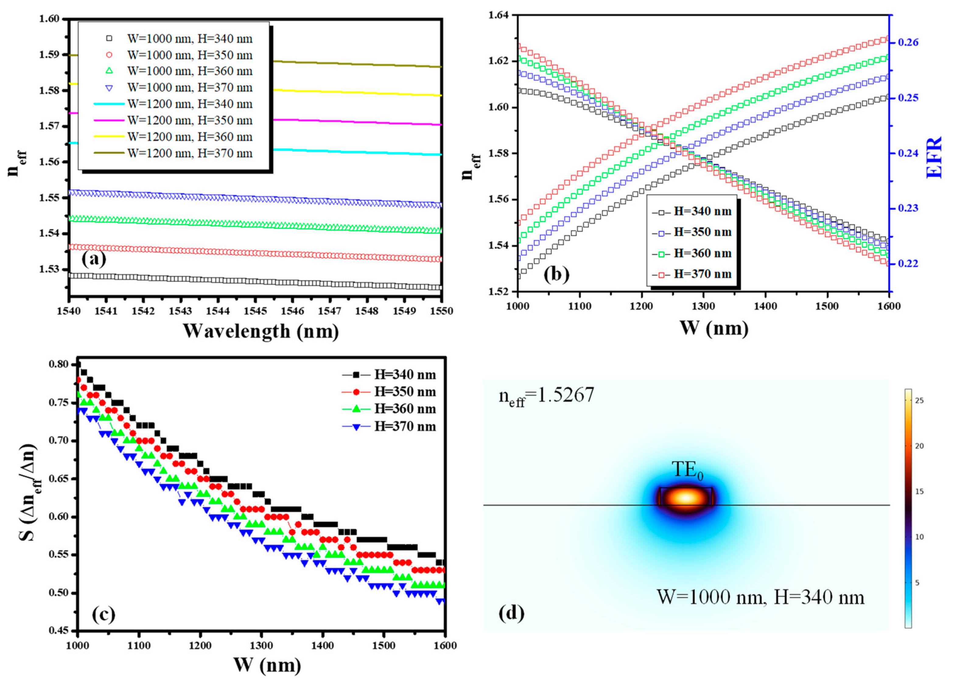
2. Results
3. Discussion
4. Numerical Simulations
5. The Fabrication Process
6. The Measurement Procedure
7. Future Directions for Ring Resonator Structure-Based Sensors
Author Contributions
Funding
Institutional Review Board Statement
Informed Consent Statement
Data Availability Statement
Acknowledgments
Conflicts of Interest
References
- Romero-García, S.; Merget, F.; Zhong, F.; Finkelstein, H.; Witzens, J. Silicon nitride CMOS-compatible platform for integrated photonics applications at visible wavelengths. Opt. Express 2013, 21, 14036–14046. [Google Scholar] [CrossRef] [PubMed]
- Klamkin, J.; Zhao, H.; Song, B.; Liu, Y.; Isaac, B.; Pinna, S.; Sang, F.; Coldren, L. Indium Phosphide Photonic Integrated Circuits: Technology and Applications. In Proceedings of the 2018 IEEE BiCMOS and Compound Semiconductor Integrated Circuits and Technology Symposium (BCICTS), San Diego, CA, USA, 15–17 October 2018; pp. 8–13. [Google Scholar] [CrossRef]
- Siew, S.Y.; Li, B.; Gao, F.; Zheng, H.Y.; Zhang, W.; Guo, P.; Xie, S.W.; Song, A.; Dong, B.; Luo, L.W.; et al. Review of Silicon Photonics Technology and Platform Development. J. Light. Technol. 2021, 39, 4374–4389. Available online: https://ieeexplore.ieee.org/document/9380443 (accessed on 4 July 2024).
- Kazanskiy, N.L.; Khonina, S.N.; Butt, M.A. A Review of Photonic Sensors Based on Ring Resonator Structures: Three Widely Used Platforms and Implications of Sensing Applications. Micromachines 2023, 14, 1080. [Google Scholar] [CrossRef] [PubMed]
- Xiang, C.; Jin, W.; Bowers, J.E. Silicon nitride passive and active photonic integrated circuits: Trends and prospects. Photonics Res. 2022, 10, A82–A96. [Google Scholar] [CrossRef]
- Blumenthal, D.J.; Heideman, R.; Geuzebroek, D.; Leinse, A.; Roeloffzen, C. Silicon Nitride in Silicon Photonics. Proc. IEEE 2018, 106, 2209–2231. [Google Scholar] [CrossRef]
- Xiang, C.; Guo, J.; Jin, W.; Wu, L.; Peters, J.; Xie, W.; Chang, L.; Shen, B.; Wang, H.; Yang, Q.-F.; et al. High-performance lasers for fully integrated silicon nitride photonics. Nat. Commun. 2021, 12, 6650. [Google Scholar] [CrossRef] [PubMed]
- Ji, X.; Okawachi, Y.; Gil-Molina, A.; Corato-Zanarella, M.; Roberts, S.; Gaeta, A.L.; Lipson, M. Ultra-Low-Loss Silicon Nitride Photonics Based on Deposited Films Compatible with Foundries. Laser Photonics Rev. 2023, 17, 2200544. [Google Scholar] [CrossRef]
- Urs, K.M.B.; Sahoo, K.; Bhat, N.; Kamble, V. Complementary Metal Oxide Semiconductor-Compatible Top-Down Fabrication of a Ni/NiO Nanobeam Room Temperature Hydrogen Sensor Device. ACS Appl. Electron. Mater. 2022, 4, 87–91. Available online: https://pubs.acs.org/doi/abs/10.1021/acsaelm.1c00912 (accessed on 4 July 2024).
- Butt, M.A. Integrated Optics: Platforms and Fabrication Methods. Encyclopedia 2023, 3, 824–838. [Google Scholar] [CrossRef]
- Barrios, C.A.; Bañuls, M.J.; González-Pedro, V.; Gylfason, K.B.; Sánchez, B.; Griol, A.; Maquieira, A.; Sohlström, H.; Holgado, M.; Casquel, R. Label-free optical biosensing with slot-waveguides. Opt. Lett. 2008, 33, 708–710. [Google Scholar] [CrossRef] [PubMed]
- Uchida, Y.; Arakawa, T.; Higo, A.; Ishizaka, Y. Silicon Microring Resonator Biosensor for Detection of Nucleocapsid Protein of SARS-CoV-2. Sensors 2024, 24, 3250. Available online: https://www.mdpi.com/1424-8220/24/10/3250 (accessed on 4 July 2024).
- Butt, M.A. Loop-Terminated Mach-Zehnder Interferometer Integrated with Functional Polymer for CO2 Gas Sensing. Appl. Sci. 2024, 14, 4714. [Google Scholar] [CrossRef]
- Yuan, D.; Dong, Y.; Liu, Y.; Li, T. Mach-Zehnder Interferometer Biochemical Sensor Based on Silicon-on-Insulator Rib Waveguide with Large Cross Section. Sensors 2015, 15, 21500–21517. [Google Scholar] [CrossRef]
- Claes, T.; Girones Molera, J.; De Vos, K.; Schacht, E.; Baets, R.; Bienstman, P. Label-Free Biosensing With a Slot-Waveguide-Based Ring Resonator in Silicon on Insulator. IEEE Photonics J. 2009, 1, 197–204. [Google Scholar] [CrossRef]
- Muñoz, P.; Micó, G.; Bru, L.A.; Pastor, D.; Pérez, D.; Doménech, J.D.; Fernández, J.; Baños, R.; Gargallo, B.; Alemany, R.; et al. Silicon Nitride Photonic Integration Platforms for Visible, Near-Infrared and Mid-Infrared Applications. Sensors 2017, 17, 2088. [Google Scholar] [CrossRef] [PubMed]
- Ciminelli, C.; Dell’Olio, F.; Armenise, M.N.; Soares, F.M.; Passenberg, W. High performance InP ring resonator for new generation monolithically integrated optical gyroscopes. Opt. Express 2013, 21, 556–564. [Google Scholar] [CrossRef] [PubMed]
- Haas, J.; Artmann, P.; Mizaikoff, B. Mid-infrared GaAs/AlGaAs micro-ring resonators characterized via thermal tuning. RSC Adv. 2019, 9, 8594–8599. [Google Scholar] [CrossRef] [PubMed]
- Girault, P.; Lorrain, N.; Poffo, L.; Guendouz, M.; Lemaitre, J.; Carré, C.; Gadonna, M.; Bosc, D.; Vignaud, G. Integrated polymer micro-ring resonators for optical sensing applications. J. Appl. Phys. 2015, 117, 104504. [Google Scholar] [CrossRef]
- Butt, M.A. Racetrack Ring Resonator-Based on Hybrid Plasmonic Waveguide for Refractive Index Sensing. Micromachines 2024, 15, 610. [Google Scholar] [CrossRef] [PubMed]
- Bryan, M.R.; Butt, J.N.; Bucukovski, J.; Miller, B.L. Biosensing with Silicon Nitride Microring Resonators Integrated with an On-Chip Filter Bank Spectrometer. ACS Sens. 2023, 8, 739–747. Available online: https://pubs.acs.org/doi/10.1021/acssensors.2c02276 (accessed on 3 December 2023).
- Zubkova, E.; An, P.; Kovalyuk, V.; Korneev, A.; Ferrari, S.; Pernice, W.; Goltsman, G. Integrated Bragg Waveguides as an Efficient Optical Notch Filter on Silicon Nitride Platform. J. Phys. Conf. Ser. 2017, 917, 062042. Available online: https://iopscience.iop.org/article/10.1088/1742-6596/917/6/062042 (accessed on 3 December 2023).
- Hermans, A.; Daele, M.V.; Dendooven, J.; Clemmen, S.; Detavernier, C.; Baets, R. Integrated silicon nitride electro-optic modulators with atomic layer deposited overlays. Opt. Lett. 2019, 44, 1112–1115. [Google Scholar] [CrossRef] [PubMed]
- Paik, H.; Osborn, K.D. Reducing Quantum-Regime Dielectric Loss of Silicon Nitride for Superconducting Quantum Circuits. Appl. Phys. Lett. 2010, 96, 072505. Available online: https://pubs.aip.org/aip/apl/article-abstract/96/7/072505/121275/Reducing-quantum-regime-dielectric-loss-of-silicon?redirectedFrom=fulltext (accessed on 3 December 2023).
- Kim, H.T.; Yu, M. Cascaded Ring Resonator-Based Temperature Sensor with Simultaneously Enhanced Sensitivity and Range. Opt. Express 2016, 24, 9501–9510. Available online: https://opg.optica.org/oe/fulltext.cfm?uri=oe-24-9-9501&id=340158 (accessed on 30 November 2023).
- de Oliveira, B.N.M.; Daniel, W.G.; Herbster, A.F. Multimode Photonic Biosensor Based on Cascaded Ring Resonator with Mach-Zehnder Interferometer for Vernier-Effect Refractive Index Sensing. In Proceedings of the 2019 SBFoton International Optics and Photonics Conference (SBFoton IOPC), Sao Paulo, Brazil, 7–9 October 2019; pp. 1–5. [Google Scholar] [CrossRef]
- Butt, M.A.; Shahbaz, M.; Piramidowicz, R. Racetrack Ring Resonator Integrated with Multimode Interferometer Structure Based on Low-Cost Silica–Titania Platform for Refractive Index Sensing Application. Photonics 2023, 10, 978. [Google Scholar] [CrossRef]
- Mudumba, S.; de Alba, S.; Romero, R.; Cherwien, C.; Wu, A.; Wang, J.; Gleeson, M.A.; Iqbal, M.; Burlingame, R.W. Photonic ring resonance is a versatile platform for performing multiplex immunoassays in real time. J. Immunol. Methods 2017, 448, 34–43. [Google Scholar] [CrossRef]
- Xu, Y.; Bai, P.; Zhou, X.; Akimov, Y.; Png, C.E.; Ang, L.K.; Knoll, W.; Wu, L. Optical Refractive Index Sensors with Plasmonic and Photonic Structures: Promising and Inconvenient Truth. Adv. Opt. Mater. 2019, 7, 1801433. Available online: https://onlinelibrary.wiley.com/doi/abs/10.1002/adom.201801433 (accessed on 24 December 2023).
- Wang, F.; Wei, Y.; Han, Y. High Sensitivity and Wide Range Refractive Index Sensor Based on Surface Plasmon Resonance Photonic Crystal Fiber. Sensors 2023, 23, 6617. [Google Scholar] [CrossRef]
- Butt, M.A.; Kazanskiy, N.L.; Khonina, S.N.; Voronkov, G.S.; Grakhova, E.P.; Kutluyarov, R.V. A Review on Photonic Sensing Technologies: Status and Outlook. Biosensors 2023, 13, 568. [Google Scholar] [CrossRef] [PubMed]
- Gupta, B.D.; Dodeja, H.; Tomar, A.K. Fibre-optic evanescent field absorption sensor based on a U-shaped probe. Opt. Quantum Electron. 1996, 28, 1629–1639. [Google Scholar] [CrossRef]
- Khonina, S.N.; Kazanskiy, N.L.; Butt, M.A. Evanescent Field Ratio Enhancement of a Modified Ridge Waveguide Structure for Methane Gas Sensing Application. IEEE Sens. J. 2020, 20, 8469–8476. [Google Scholar] [CrossRef]
- Zhang, Y.; Zeng, C.; Li, D.; Gao, G.; Huang, Z.; Yu, J.; Xia, J. High-quality-factor photonic crystal ring resonator. Opt. Lett. 2014, 39, 1282–1285. [Google Scholar] [CrossRef] [PubMed]
- Ali, L.; Khan, M.; Mohammed, M.U.; Yousuf, A.H.B.; Chaudhry, M.H. High Quality Silicon Photonics Optical Ring Resonator Biosensor Design. In Proceedings of the 2018 IEEE Nanotechnology Symposium (ANTS), Albany, NY, USA, 14–15 November 2018; Available online: https://ieeexplore.ieee.org/document/8653557 (accessed on 26 December 2023).
- Zhang, X.; Zhou, C.; Luo, Y.; Yang, Z.; Zhang, W.; Li, L.; Xu, P.; Zhang, P.; Xu, T. High Q-factor, ultrasensitivity slot microring resonator sensor based on chalcogenide glasses. Opt. Express 2022, 30, 3866–3875. [Google Scholar] [CrossRef] [PubMed]
- Sub-Wavelength Grating for Enhanced Ring Resonator Biosensor. Available online: https://opg.optica.org/oe/fulltext.cfm?uri=oe-24-14-15672&id=345275 (accessed on 1 December 2023).
- Kundal, S.; Khandelwal, A. Improving the Performance of Ring Resonator Refractive Index Sensor through Structural Modifications. Optik 2024, 296, 171555. Available online: https://www.sciencedirect.com/science/article/pii/S0030402623010537 (accessed on 25 December 2023).
- Tu, Z.; Gao, D.; Zhang, M.; Zhang, D. High-sensitivity complex refractive index sensing based on Fano resonance in the subwavelength grating waveguide micro-ring resonator. Opt. Express 2017, 25, 20911–20922. [Google Scholar] [CrossRef] [PubMed]
- Liu, N.; Wang, S.; Lv, J.; Lu, Y.; Zhang, J. Refractive Index Biosensor Based on Topological Ring Resonator. Opt. Commun. 2023, 541, 129542. Available online: https://www.sciencedirect.com/science/article/pii/S0030401823002894 (accessed on 1 December 2023).
- Butt, M.A.; Khonina, S.N.; Kazanskiy, N.L. Highly sensitive refractive index sensor based on hybrid plasmonic waveguide microring resonator. Waves Random Complex. Media 2020, 30, 292–299. [Google Scholar] [CrossRef]
- Tazawa, H.; Steier, W.H. Linearity of ring resonator-based electro-optic polymer modulator. Electron. Lett. 2005, 41, 1297–1298. [Google Scholar] [CrossRef]
- Asuero, A.G.; Sayago, A.; González, A.G. The Correlation Coefficient: An Overview. Crit. Rev. Anal. Chem. 2006, 36, 41–59. [Google Scholar] [CrossRef]
- Castelló-Pedrero, L.; Gómez-Gómez, M.I.; García-Rupérez, J.; Griol, A.; Martínez, A. Performance improvement of a silicon nitride ring resonator biosensor operated in the TM mode at 1310 nm. Biomed. Opt. Express 2021, 12, 7244–7260. [Google Scholar] [CrossRef]
- Huang, W.; Luo, Y.; Zhang, W.; Li, C.; Li, L.; Yang, Z.; Xu, P. High-sensitivity refractive index sensor based on Ge–Sb–Se chalcogenide microring resonator. Infrared Phys. Technol. 2021, 116, 103792. [Google Scholar] [CrossRef]
- Ou, X.; Yang, Y.; Sun, F.; Zhang, P.; Tang, B.; Li, B.; Liu, R.; Liu, D.; Li, Z. Wide-range, ultra-compact, and high-sensitivity ring resonator biochemical sensor with CMOS-compatible hybrid plasmonic waveguide. Opt. Express 2021, 29, 19058–19067. [Google Scholar] [CrossRef] [PubMed]
- Sahraeibelverdi, T.; Guo, L.J.; Veladi, H.; Malekshahi, M.R. Polymer Ring Resonator with a Partially Tapered Waveguide for Biomedical Sensing: Computational Study. Sensors 2021, 21, 5017. [Google Scholar] [CrossRef] [PubMed]
- Lu, K.; Huang, B.; Lv, X.; Zhang, Z.; Ma, Z. Ultrasensitive Silicon Photonic Refractive Index Sensor Based on Hybrid Double Slot Subwavelength Grating Microring Resonator. Sensors 2024, 24, 1929. [Google Scholar] [CrossRef] [PubMed]
- COMSOL: Multiphysics Software for Optimizing Designs. Available online: https://www.comsol.com/ (accessed on 3 December 2023).
- Kern, W. Evolution of silicon wafer cleaning technology. Proc.—Electrochem. Soc. 1990, 90, 3–19. [Google Scholar] [CrossRef]
- Lelit, M.; Słowikowski, M.; Filipiak, M.; Juchniewicz, M.; Stonio, B.; Michalak, B.; Pavłov, K.; Myśliwiec, M.; Wiśniewski, P.; Kaźmierczak, A.; et al. Passive Photonic Integrated Circuits Elements Fabricated on a Silicon Nitride Platform. Materials 2022, 15, 1398. [Google Scholar] [CrossRef] [PubMed]
- Wang, Z.; Xu, X.; Fan, D.; Wang, Y.; Chen, R.T. High quality factor subwavelength grating waveguide micro-ring resonator based on trapezoidal silicon pillars. Opt. Lett. 2016, 41, 3375–3378. [Google Scholar] [CrossRef]
- Butt, M.A.; Kozlowski, L.; Piramidowicz, R. Optimized hybrid plasmonic waveguide-based ring resonator for advanced refractive index sensing. J. Opt. 2024, 26, 075802. [Google Scholar] [CrossRef]
- Chrostowski, L.; Grist, S.; Flueckiger, J.; Shi, W.; Wang, X.; Ouellet, E.; Yun, H.; Webb, M.; Nie, B.; Liang, Z.; et al. Silicon photonic resonator sensors and devices. In Proceedings of the Laser Resonators, Microresonators, and Beam Control XIV, San Francisco, CA, USA, 22–25 January 2012; Volume 8236, pp. 387–402. [Google Scholar] [CrossRef]
- Juan-Colás, J.; Parkin, A.; Dunn, K.E.; Scullion, M.G.; Krauss, T.F.; Johnson, S.D. The electrophotonic silicon biosensor. Nat. Commun. 2016, 7, 12769. [Google Scholar] [CrossRef]


| FSR (nm) | Sensitivity (nm/RIU) | Q Factor | LOD (RIU) | |||
|---|---|---|---|---|---|---|
| Radius (µm) | 50 ± 0.02 | 75 ± 0.02 | 100 ± 0.02 | |||
| Numerical calculations | ~4.2 | ~2.8 | ~2.1 | ~110 | - | - |
| Experimental data | ~3.64 | ~2.42 | ~1.82 | ~112.5 | 1.7154 × 10⁴ | 7.99 × 10⁻⁴ |
| Relative percent error (%) | ~15.4 | ~15.7 | ~15.3 | ~2.2 | - | - |
| Platform | Numerical/Experimental | Waveguide Configuration | Sensitivity (nm/RIU) | Q Factor | LOD | Ref. |
|---|---|---|---|---|---|---|
| Silicon nitride | Numerical and experimental | Ridge waveguide | 164.8 | 2575 | 3.65 × 10⁻³ | [44] |
| Ge-As-Se chalcogenide | Experimental | Ridge waveguide | 123 | 7.74 × 10⁴ | 3.24 × 10⁻⁴ | [45] |
| Silicon | Numerical | Suspended slot hybrid plasmonic waveguide | 458.1 | - | 3.7 × 10⁻⁵ | [46] |
| Polymer | Numerical | Tapered ridge waveguide | 84.6 to 101.74 | 4.6 × 10³ | - | [47] |
| Silicon | Numerical | Hybrid double-slot subwavelength grating | 1005 | 22,429 | 6.86 × 10⁻⁵ | [48] |
| Silicon nitride | Numerical and experimental | Ridge waveguide | 112.5 | 1.7154 ×10⁴ | 7.99 × 10⁻⁴ | This work |
Disclaimer/Publisher’s Note: The statements, opinions and data contained in all publications are solely those of the individual author(s) and contributor(s) and not of MDPI and/or the editor(s). MDPI and/or the editor(s) disclaim responsibility for any injury to people or property resulting from any ideas, methods, instructions or products referred to in the content. |
© 2024 by the authors. Licensee MDPI, Basel, Switzerland. This article is an open access article distributed under the terms and conditions of the Creative Commons Attribution (CC BY) license (https://creativecommons.org/licenses/by/4.0/).
Share and Cite
Butt, M.A.; Kozłowski, Ł.; Golas, M.; Slowikowski, M.; Filipiak, M.; Juchniewicz, M.; Bieniek-Kaczorek, A.; Dudek, M.; Piramidowicz, R. Numerical and Experimental Demonstration of a Silicon Nitride-Based Ring Resonator Structure for Refractive Index Sensing. Appl. Sci. 2024, 14, 6082. https://doi.org/10.3390/app14146082
Butt MA, Kozłowski Ł, Golas M, Slowikowski M, Filipiak M, Juchniewicz M, Bieniek-Kaczorek A, Dudek M, Piramidowicz R. Numerical and Experimental Demonstration of a Silicon Nitride-Based Ring Resonator Structure for Refractive Index Sensing. Applied Sciences. 2024; 14(14):6082. https://doi.org/10.3390/app14146082
Chicago/Turabian StyleButt, Muhammad A., Łukasz Kozłowski, Michał Golas, Mateusz Slowikowski, Maciej Filipiak, Marcin Juchniewicz, Aleksandra Bieniek-Kaczorek, Michał Dudek, and Ryszard Piramidowicz. 2024. "Numerical and Experimental Demonstration of a Silicon Nitride-Based Ring Resonator Structure for Refractive Index Sensing" Applied Sciences 14, no. 14: 6082. https://doi.org/10.3390/app14146082
APA StyleButt, M. A., Kozłowski, Ł., Golas, M., Slowikowski, M., Filipiak, M., Juchniewicz, M., Bieniek-Kaczorek, A., Dudek, M., & Piramidowicz, R. (2024). Numerical and Experimental Demonstration of a Silicon Nitride-Based Ring Resonator Structure for Refractive Index Sensing. Applied Sciences, 14(14), 6082. https://doi.org/10.3390/app14146082