Article

A Self-Supervised Detail-Sensitive ViT-Based Model for COVID-19 X-ray Image Diagnosis: SDViT

1
Qianjinag College, Hangzhou Normal University, Hangzhou 311121, China
2
Department of Computer Science, Gonzaga University, 502 E Boone Ave, Spokane, WA 99258, USA
*
Author to whom correspondence should be addressed.
Appl. Sci. 2023, 13(1), 454; https://doi.org/10.3390/app13010454
Submission received: 4 November 2022 / Revised: 23 December 2022 / Accepted: 24 December 2022 / Published: 29 December 2022
(This article belongs to the Special Issue Applications of Artificial Intelligence in Medical Imaging)

Abstract

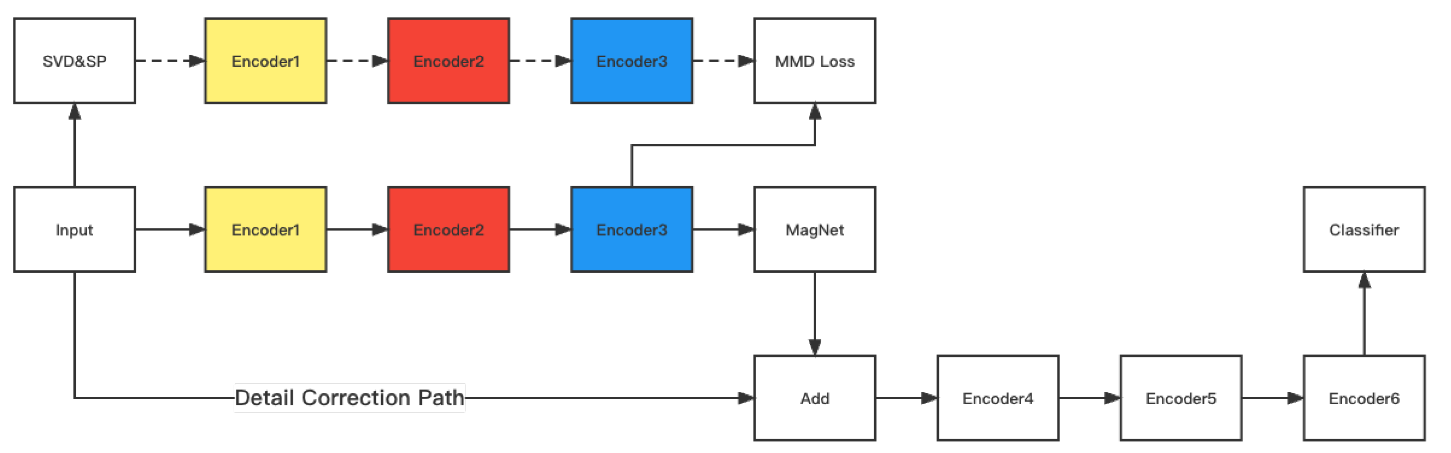
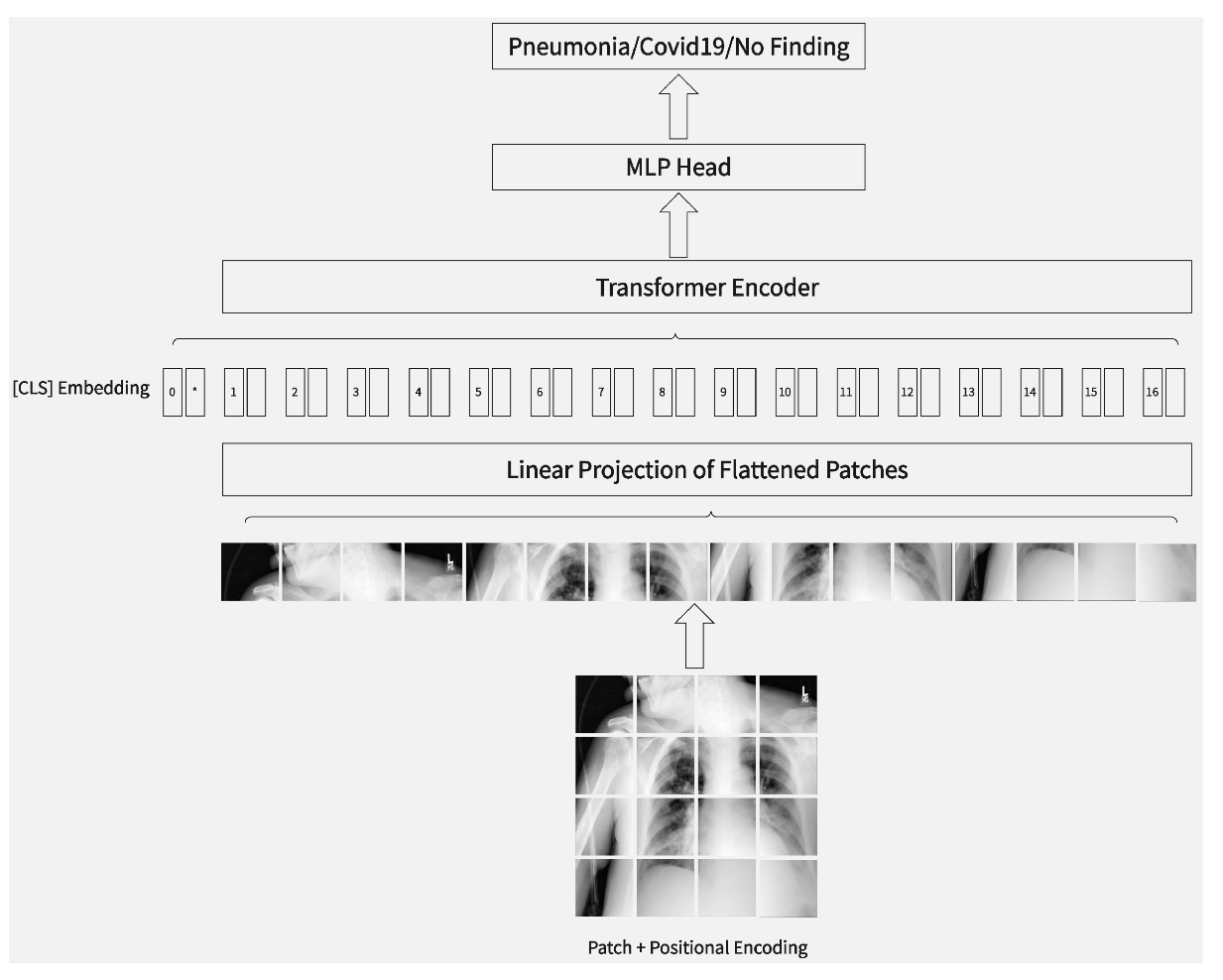
COVID-19 has led to a severe impact on the society and healthcare system, with early diagnosis and effective treatment becoming critical. The Chest X-ray (CXR) is the most time-saving and cost-effective tool for diagnosing COVID-19. However, manual diagnosis through human eyes is time-consuming and tends to introduce errors. With the challenge of a large number of infections and a shortage of medical resources, a fast and accurate diagnosis technique is required. Manual detection is time-consuming, depends on individual experience, and tends to easily introduce errors. Deep learning methods can be used to develop automated detection and computer-aided diagnosis. However, they require a large amount of data, which is not practical due to the limited annotated CXR images. In this research, SDViT, an approach based on transformers, is proposed for COVID-19 diagnosis through image classification. We propose three innovations, namely, self-supervised learning, detail correction path (DCP), and domain transfer, then add them to the ViT Transformer architecture. Based on experimental results, our proposed method achieves an accuracy of 95.2381%, which is better performance compared to well-established methods on the X-ray Image dataset, along with the highest precision (0.952310), recall (0.963964), and F1-score (0.958102). Extensive experiments show that our model achieves the best performance on the synthetic-covid-cxr dataset as well. The experimental results demonstrate the advantages of our design for the classification task of COVID-19 X-ray images.
Keywords: COVID-19; vision transformer; chest X-ray image; image classification COVID-19; vision transformer; chest X-ray image; image classification