Article

The Effects of Periodontal Laser Therapy on Pain in Adult Patients with Orthodontic Treatment: A Randomized Clinical Trial

1
Department of Occlusology and Oral Rehabilitation, George Emil Palade University of Medicine, Pharmacy, Science, and Technology of Târgu Mureș, 38 Ghe. Marinescu Street, 540139 Târgu Mureș, Romania
2
Department of Odontology and Oral Pathology, George Emil Palade University of Medicine, Pharmacy, Science, and Technology of Târgu Mureș, 38 Ghe. Marinescu Street, 540139 Târgu Mureș, Romania
3
Department of Pedodontics, George Emil Palade University of Medicine, Pharmacy, Science, and Technology of Târgu Mureș, 38 Ghe. Marinescu Street, 540139 Târgu Mureș, Romania
4
Department of Periodontology, George Emil Palade University of Medicine, Pharmacy, Science, and Technology of Târgu Mureș, 38 Ghe. Marinescu Street, 540139 Târgu Mureș, Romania
5
Department of Oral and Maxillofacial Surgery, George Emil Palade University of Medicine, Pharmacy, Science, and Technology of Târgu Mureș, 38 Ghe. Marinescu Street, 540139 Târgu Mureș, Romania
6
Department of Periodontology, Grigore T. Popa University of Medicine and Pharmacy, 16 Universității Street, 700115 Iași, Romania
7
Department of Orthodontics, George Emil Palade University of Medicine, Pharmacy, Science, and Technology of Târgu Mureș, 38 Ghe. Marinescu Street, 540139 Târgu Mureș, Romania
*
Author to whom correspondence should be addressed.
These authors contributed equally to this work.
Appl. Sci. 2022, 12(7), 3601; https://doi.org/10.3390/app12073601
Submission received: 10 March 2022 / Revised: 29 March 2022 / Accepted: 30 March 2022 / Published: 1 April 2022
(This article belongs to the Special Issue Diagnosis and Treatment of Periodontal Diseases)

Abstract

:
(1) Background: Pain is the most common complaint of patients with fixed orthodontic treatment, especially when applying and activating the orthodontic device. The effectiveness of low-level laser therapy (LLLT) in reducing pain caused by the orthodontic treatment has been frequently studied over the last few years. In this study, we aim to evaluate the influence of laser therapy on pain caused by orthodontic forces acting on periodontal tissues during fixed orthodontic treatment. (2) Methods: A total of 36 patients who met the inclusion criteria were submitted to laser therapy after beginning a fixed orthodontic treatment. For each patient, one half-arch was treated with laser (HL), the opposite one being the control half-arch (HC). The evaluation of pain was conducted by asking the patients to rate their pain on a VAS scale from 0 to 10. (3) Results: Analyzing the VAS score values reported after applying the orthodontic device, we noticed that the pain increased in the first few hours, it was the highest 24 h after application, and it decreased towards the 7th day. The pain reported by most patients was significantly reduced in the HL compared to the HC, in all three moments that were evaluated (p < 0.0001). (4) Conclusions: Repeated LLLT in patients with orthodontic treatment leads to a significant decrease in pain levels, as the comparison between the laser half-arch and the control half-arch showed.

1. Introduction

Pain is the most common complaint of patients with fixed orthodontic treatment, especially when applying and activating the orthodontic device, as a large number of systematic reviews have shown [1,2]. Clinical studies have shown that almost 90% of patients felt pain at some point during their treatment caused by either tooth movement, mucosal ulcers, and activation or debonding of brackets [3]. Pain related to debonding of brackets is materialized through sensitivity to thermal stimuli mostly immediately after accidental or intentional debonding, as a recent clinical trial showed [4].
Pain related to dental treatments is in most cases strongly influenced by certain factors such as personality, sex, and especially prior experience in a dentistry practice [5].
The aim of the orthodontic treatment is to align the teeth and to obtain a correct occlusion, by modifying the position of the teeth using mechanical forces. Orthodontic tooth movement is a process in which the appliance of a force causes bone resorption on the pressure side and bone apposition on the tension side. Thus, regular tooth movement is associated with biological cascades of resorption and apposition caused by the mechanical forces. The alveolar bone of dental sockets contains many fenestrations that allow compressed fluid in the extracellular matrix to be expressed into the marrow spaces. The marrow spaces decrease some of the force values that the cells must endure during mechanical perturbation [6]. The “holes” serve as a pathway for peripheral hematopoietic cells from marrow spaces to communicate with and migrate to an area of hyalinization. The next notable factor is that in the anatomy of the periodontal ligament, the blood vessels, in general, are located in the structure in an asymmetrical way. The blood vessels are commonly located about one-third of the distance closer to the bone than to the tooth. This means that cells extruded from a dilated vessel have less of a distance to migrate to the bone surface than to the root surface. These cells build or remodel the connective tissue infrastructure of the bone or cementum [7]. Therefore, due to all these biological modifications, tooth movement causes pain for the patient [8]. Pain can be one of the main reasons why patients stop orthodontic treatments [9,10].
The management of orthodontic-related periodontal pain consists of two broad categories: pharmacological methods and nonpharmacological methods. The pharmacological approach includes nonsteroidal anti-inflammatory drugs (NSAIDs) which, due to multiple side effects, tend to lose ground to nonpharmacological methods of pain relief such as ozone and photobiomodulation therapy, laser therapy, and transcutaneous electrical nerve stimulation. [11,12,13]
Low-level laser therapy (LLLT) has attracted interest during the last few decades due to its advantages as a noninvasive pain management therapy without significant side-effects [14]. The effectiveness of LLLT in reducing pain caused by the orthodontic treatment has been frequently studied over the last few years [15]. It has been demonstrated that LLLT has analgesic and anti-inflammatory properties due to the fact that it increases local blood flow, reduces levels of prostaglandin E2, and suppresses collagenase [16,17]. As recent studies have shown, LLLT therapy uses visible light (generally red) or almost infrared light generated by a laser or a diode (LED-light emitting diode). The light spectrum is narrow with a wavelength of 600 to 1000 nm. The intensity is approximately 1 to 20 J/cm2 [14]. The anti-inflammatory effect of LLLT functions through nonthermal photobiological mechanisms in which the laser light is absorbed by chromophores (functional groups graphed in a molecule’s structure responsible for its color), thus modulating physiopathological processes. Pain arises as a result of the activation of harmful nerve terminations in the epidermis or mucosa. At the level of these somatosensitive nerves, harmful stimuli such as heat, mechanical forces, and also chemical stimuli related to the inflammatory process are turned into action potential. The latter are then transmitted to the central nervous system. By blocking these nerves, and this transmission by default, the painful sensation will be reduced. The specific action of the LLLT is either to suppress harmful receptors or to block the neuronal response to harmful stimuli and the generated action potential [18,19].
In order to analyze the degree of pain that patients report during an orthodontic treatment, a series of studies have been conducted (including questionnaires and telephone interviews). However, due to the small number of patients included in the studies and different results and methodology, there is still no clear proof of the positive effects of LLLT [20].
As pain is the most common complaint and reason for quitting the orthodontic treatment, in this study we aim to evaluate the influence of laser therapy on pain during the orthodontic therapy. The null hypothesis states that there is no statistically significant difference between the VAS score values of HC and HL immediately after bonding the orthodontic appliance, after the first, second, and third activation.

2. Materials and Methods

2.1. Study Design

This study was conducted as a randomized trial, placebo-control, and double blind.

2.2. Subject Selection

From all the adult patients with orthodontic treatment that came to CMI Dr. Lazăr Luminița Târgu Mureș, 36 patients (20 females and 16 males), who met the following inclusion criteria were selected:
-
Age between 18 and 55 years;
-
Presence of dentoalveolar disharmony (DAD) with mild crowding (3–7 mm);
-
No prior orthodontic treatment;
-
No prior anti-inflammatory treatment (NSAIDs).
The exclusion criteria were as follows:
-
Presence of carious lesions;
-
Signs of active periodontal disease;
-
Lesions of the oral mucosa;
-
Systemic conditions that have an impact on periodontal tissues (diabetes, immunological conditions, acute articular rheumatism, tuberculosis, etc.).

2.3. Periodontal Status

A complex oral examination by an experienced periodontist was performed for periodontal status assessment prior to applying the orthodontic device. The following parameters were determined: gingival index (GI), plaque index (PqI), and papillary bleeding index (PBI). The values we assessed were within normal limits, with a few exceptions.

2.4. Orthodontic Protocol

Patients presenting dentoalveolar disharmony with crowding were examined and received orthodontic treatment using standard edgewise brackets with the same slot size for all subjects (0.22) (American Orthodontics, Sheboygan, WI, USA). The following sequence of archwires was used: NiTi size 0.12 when applying the orthodontic device, and sizes 0.16 for the first activation, 0.16 × 0.16 for the second, and 0.16 × 0.22 for the third (BioForce, Dentsply Sirona, Charlotte, NC, USA).

2.5. LLLT Protocol

One member of the research team performed the laser therapy on a half-arch (HL), left or right, randomly for each patient (split mouth study design). The patients were informed that only one half-arch would receive laser therapy, without specifying which one.
The laser therapy was performed with a dental laser (Prime, Litemedics, Lambda SpA, Milano, Italy), with a power of 12 watts, in a pulsating system and operating wave of 980 nm, using therapy and biostimulation mode. The optic fiber of 320 μm was held at a distance of 10 mm from the gingiva, perpendicular on the root surface, at a level that corresponded to the center of the root length, for 20 s.
Laser therapy was applied on one half-arch on the incisor and the molar at six points (mesio-buccal, centro-buccal, distobuccal, mesio-oral, centro-oral, and disto-oral) for each tooth (HL). On the other side, the control half-arch (HC) was submitted to the same protocol but without active light. During the irradiation process, both the doctor and the patient wore protection glasses.
The laser therapy was initiated immediately after applying the orthodontic device (T00), 24 h later (T01), and after 7 days (T02). This protocol was repeated monthly, after activating the orthodontic appliance, with the same intervals of applying laser therapy. Therefore, for the first month: T10 (activation moment), T11 (24 h later), and T12 (after seven days); for the second month: T20 (activation moment), T21 (24 h later), and T22 (after seven days); for the third month: T30 (activation moment), T31 (24 h later), and T32 (after seven days).
The evaluation of pain was conducted by asking the patients to rate their pain from 0 to 10 on a VAS scale, 0 being “no pain”, 1–3 “mild pain”, 4–6 “moderate pain”, 7–9 “severe pain”, and 10 “the worst pain imaginable”. Every patient filled in the score for each half-arch in a questionnaire 3 h (T00, T10, T20, and T30), 24 h (T01, T11, T21, and T31) and 7 days (T02, T12, T22, and T32) after applying the device and for each activation session.

2.6. Statistical Analysis
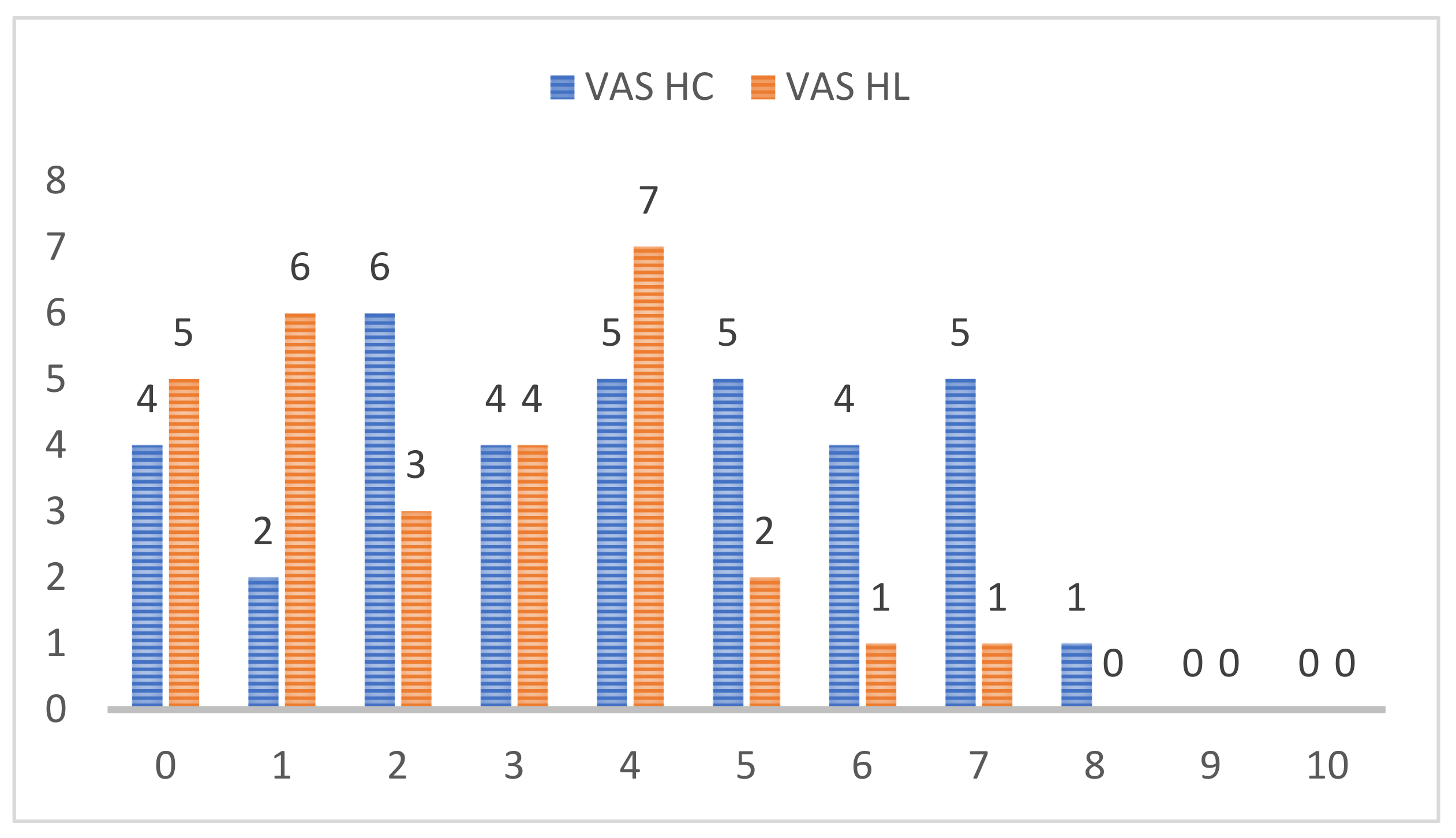
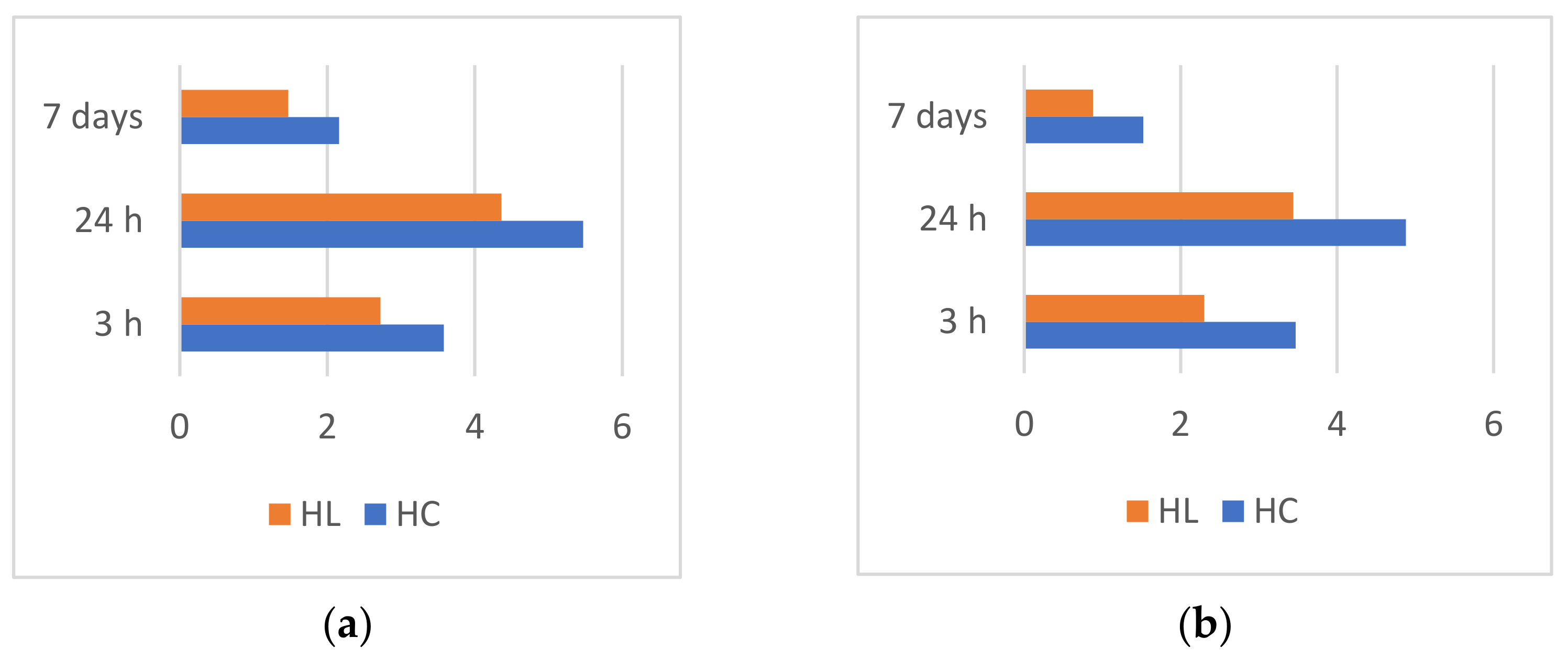
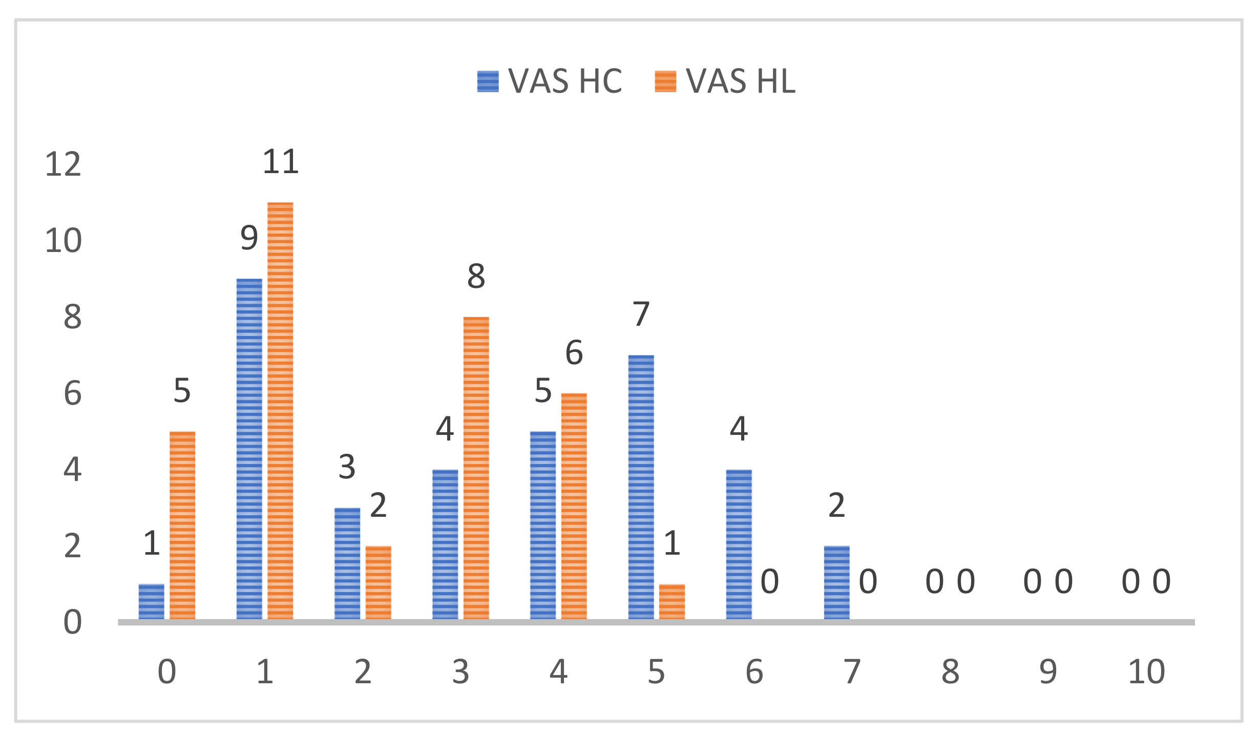
All data were collected in Microsoft Excel work sheets (Microsoft Corporation, 2018, Redmond, WA, USA). The statistical analysis was carried out in GraphPad Prism version 8.0.0 for Windows (GraphPad Software, San Diego, CA, USA). For each group of data, descriptive statistics such as mean, standard deviation, and median, minimum, and maximum value were assessed. Data normality was determined using the Kolmogorov–Smirnov test. Pain evaluation comparison between the HC and HL measured at different time intervals after applying the orthodontic device and activation were determined using the Wilcoxon test. The chosen significance level was set at 0.05.

3. Results

After analyzing the VAS score values reported subsequent to applying the orthodontic device, we noticed that the pain level increased in the first few hours, it was the highest 24 h after, and it decreased towards the 7th day of activation. (Figure 1) The comparison between the VAS score values in the HS and the HL were statistically significant (T00—p < 0.0001, T01—p < 0.0001, T02—p = 0.0002).
The levels of pain reported by patients one month after starting the treatment, when the first activation took place are represented in Figure 2. By comparing the VAS values of the HC and HL in T10, T11, and T12, statistically significant results were found (p < 0.0001 for T10, T11, and T12).
VAS score values given by the patients for the two half-arches, HL and HC, 3 h (T0), 24 h (T01), and 7 days (T02) after applying the orthodontic device are represented in Figure 3a, and those for 1 month after, at the first activation, in Figure 3b.

4. Discussion

In our study we analyzed the effects of repeated LLLT on pain management in patients with orthodontic treatment. We evaluated the intensity of the pain consecutive to mechanical forces and compared the values for each patient in the laser half-arch (HL) and the control half-arch (HC).
Analyzing the VAS score values reported after applying the orthodontic device, we noticed that the pain increased in the first few hours, it was the highest 24 h after, and it decreased towards the 7th day. The highest value of the VAS score at T00, in HC was 7, reported by three patients, whilst in HL it was reported by one patient. For T01 (24 h after) severe pain was reported, in HC, the VAS score value was nine in four patients and eight in three patients. At the same time in HL, the VAS score value was nine in one patient and eight in one patient. On the 7th day, in HC most patients reported mild pain, four patients moderate pain, and six patients had no more pain, whereas in HL most patients had mild pain, and 11 had no more pain. None of the patients reported a VAS score value of 10, which represents the worst pain imaginable.
One month after applying the orthodontic device, when the first activation was performed, the pain reported by the patients had similar characteristics to the one reported at the beginning of the treatment, the values reported 24 h after being higher than those registered 3 h after the procedure, then decreasing towards the 7th day.
Analyzing VAS score values for the two half-arches, the pain reported by most patients was significantly reduced in HL compared to HC, both at the beginning of the treatment, when the orthodontic device was placed and one month after, when the first activation was performed. However, four patients reported same VAS score values for HL and HC, probably due to an increased sensitivity to pain, given the high VAS score values registered by these patients.
For the activations performed 2 months and 3 months after beginning the treatment, the characteristics of the pain reported by the patients were not significantly different compared to the pain reported in the beginning and one month after applying the orthodontic device at the first activation.
Significant decreases in pain reported by patients treated with LLLT after applying orthodontics wires were demonstrated by a double-blind study [21]. Similar conclusions were reported by other studies that showed immediate lowering of pain values after using LLLT [22].
Similar to our study, another research showed maximum values of pain 24 h after activating the orthodontic device, both for the patients treated with LLLT and for the placebo group. Afterwards, the pain decreased gradually and disappeared after the 7th day [23]. These results are in accordance with other studies that also illustrate a maximum value of pain 24 h after applying orthodontic forces [24]. Furthermore, there are numerous studies that report pain lasting for less time in patients who received laser therapy [25,26,27,28].
The exact mechanisms that lead to the analgesic effects of LLLT are unclear. Certain studies assume that the LLLT has antinociceptive properties as a result of biologic mechanisms that stimulate cellular differentiation and proliferation [29,30,31]. A study on experimental animals showed that the anti-inflammatory effects of LLLT can be similar to those of NSAIDs (nonsteroidal anti-inflammatory drugs) [32].
The literature shows that the inflammatory infiltrate located in the lamina propria is the critical morphological trait for controlling the healing process. Laser therapy can help diminish the number of lymphocytes and macrophages, which will implicitly determine a lower production of chemical mediators interfering with the sequence of the healing process. Other studies demonstrated that LLLT improves local vascularization and has a significant role in the healing processes of soft tissues [32,33,34].
Other factors that can contribute to the analgesic effect of laser therapy could be the activation of certain enzymes, the inhibition of nervous depolarization, the production of ATP, and the reduction in prostaglandin levels [18,33,35].
When it comes to the pain level reported by patients, the placebo effects cannot be totally excluded. However, clinical observations support the results that show the superiority of adjuvant laser therapy in pain management caused by orthodontic forces.

Limitations of This Study

An element of limitation comes from the fact that the patients included in the study were both male and female. It is known that between the two sexes there are significant differences in regard to the sensitivity to pain [36]. On the other hand, having a mixed group is a positive element, because the results can be generalized.
Another limitation is related to patient subjectivity in describing pain. However, in our study we used the VAS score in order to counteract this limitation.
Even though a wide age range (18–55) can be considered a weak point, we think that it is an advantage in order to be able to generally apply our results to further treatment guidelines.

5. Conclusions

Pain is the most common complaint of patients when applying and activating the fixed orthodontic device (approximately 90% of patients experience pain during the orthodontic treatment). The values of the VAS score showed that, when applying the orthodontic device, pain increased during the first few hours, was the highest after 24 h, and decreased towards the 7th day.
One month after applying the orthodontic device, the pain had similar characteristics to that reported at the beginning of the treatment. In the 2nd and 3rd month, the characteristics of the pain reported by the patients were not significantly different compared to the pain reported previously.
Repeated LLLT in patients with orthodontic treatment leads to the significant reduction in pain levels, comparing the laser half-arch with the control half-arch.

Author Contributions

Conceptualization, A.-P.L. and L.L.; methodology, A.-P.L. and M.P.; software, T.D.; validation, A.-P.L., L.L. and M.P.; formal analysis, A.B., T.D. and M.-A.M.; investigation, L.L. and A.O.; resources, T.D., M.P. and M.-A.M.; data curation, A.B.; writing—original draft preparation, A.V. and M.P.; writing—review and editing, T.D. and M.-A.M.; visualization, A.-P.L. and A.O.; supervision, L.L.; project administration, M.P. and A.O.; funding acquisition, L.L. All authors have read and agreed to the published version of the manuscript.

Funding

This research was funded by the George Emil Palade University of Medicine, Pharmacy, Science, and Technology of Târgu Mureș, grant Determination of the levels of osteocalcin and N-telopeptide from the crevicular fluid, number 511/4/17.01.2022.

Institutional Review Board Statement

The study was conducted in accordance with the Declaration of Helsinki and approved by the Ethics Committee of our university (Comisia de Etică a Cercetării Științifice a UMFST George Emil Palade din Tg. Mureș), number 1203/03.12.2020.

Informed Consent Statement

Informed consent was obtained from all subjects involved in the study and to publish this paper. Patients received information about the treatment protocol and about the fact that they could leave the study at any time.

Data Availability Statement

The data presented in this study are available on request from the corresponding author. The data are not publicly available due to personal protection.

Conflicts of Interest

The authors declare no conflict of interest.

References

  1. Bergius, M.; Berggren, U.; Kiliaridi, S. Experience of pain during an orthodontic procedure. Eur. J. Oral Sci. 2002, 110, 92–98. [Google Scholar] [CrossRef] [PubMed]
  2. Erdinc, A.M.; Dincer, B. Perception of pain during orthodontic treatment with fixed appliances. Eur. J. Orthod. 2004, 26, 79–85. [Google Scholar] [CrossRef] [PubMed]
  3. Sonesson, M.; de Geer, E.; Subraian, J.; Petren, S. Efficacy of low-level laser therapy in accelerationg tooth movement, preventing relapse and managing acute pain during othodontic treatment in humans: A systematic review. BMC Oral Health 2017, 17, 11. [Google Scholar] [CrossRef] [PubMed] [Green Version]
  4. Scribante, A.; Gallo, S.; Celmare, R.L.; D’Anto, V.; Grippaudo, C.; Gandini, P.; Sfondrini, M.F. Orthodontic debonding and tooth sensitivity of anterior and posterior teeth: A prospective clinical trial. Angle Orthod. 2020, 90, 766–773. [Google Scholar] [CrossRef]
  5. Tecco, S.; D’Attilio, M.; Tetè, S.; Festa, F. Prevalence and type of pain during conventional and self-ligating orthodontic treatment. Eur. J. Orthod. 2009, 31, 380–384. [Google Scholar] [CrossRef]
  6. Li, Y.; Jacox, L.A.; Little, S.; Ko, C.C. Orthodontic tooth movement: The biology and clinical implications. Kaoshiung J. Med. Sci. 2018, 34, 207–2014. [Google Scholar] [CrossRef] [Green Version]
  7. Feller, L.; Khammissa, R.A.G.; Schechter, I.; Moodley, A.; Thamadakis, G.; Lemmer, J. Periodontal biological events associated with orthodontic tooth movement: The biomechanics of the cytoskeleton and the extracellular matrix. Sci. World J. 2015, 8, 11–17. [Google Scholar] [CrossRef] [Green Version]
  8. Vandevska-Radunovic, V. Neural modulation of inflammatory reactions in dental tissues incident to orthodontic tooth movement. A review of the literature. Eur. J. Orthod. 1999, 21, 231–247. [Google Scholar] [CrossRef]
  9. Popa, C.G.; Luchian, I.; Ioanid, N.; Goriuc, A.; Martu, I.; Bosinceanu, D.; Martu, M.A.; Tirca, T.; Martu, S. ELISA Evaluation of RANKL Levels in Gingival Fluid in Patients with Periodontitis and Occlusal Trauma. Rev. Chim. 2018, 69, 1578–1580. [Google Scholar] [CrossRef]
  10. Wilson, S.; Ngan, P.; Kess, B. Time course of the discomfort in young patients undergoing orthodontic treatment. Pediatr. Dent. 1989, 11, 107–110. [Google Scholar]
  11. Matys, J.; Jaszsczak, E.; Flieger, R.; Kostrzewska-Kaminiarz, L. Effect of ozone and diode laser (635 nm) in reducing orthodontic pain in the maxillary arch—A randomized clinical controlled trial. Lasers Med. Sci. 2020, 35, 487–496. [Google Scholar] [CrossRef] [Green Version]
  12. Scribante, A.; Gallo, S.; Pascadopoli, M.; Soleo, R.; di Fonso, F.; Politi, L.; Venugopal, A.; Marya, A.; Butera, A. Management of periodontal disease with adjuvant therapy with ozone and photobiomodulation (PBM): A randomized clinical trail. Photonics 2022, 9, 138. [Google Scholar] [CrossRef]
  13. Scribante, A.; Butera, A.; Alovisi, M. Customized minimally invasive protocols for the clinical and microbiological management of the oral microbiota. Microorganisms 2022, 10, 675. [Google Scholar] [CrossRef]
  14. Xiaoting, L.; Yin, T.; Yangxi, C. Interventions for pain during fixed orthodontic appliance therapy. A systematic review. Angle Orthod. 2010, 80, 925–932. [Google Scholar] [CrossRef]
  15. Li, F.J.; Zhang, J.Y.; Zeng, X.T.; Guo, Y. Low-level laser therapy for orthodontic pain: A systematic review. Laser Med. Sci. 2015, 30, 1789–1803. [Google Scholar] [CrossRef]
  16. Sakurai, Y.; Yamaguchi, M.; Abiko, Y. Inhibitory effect of low-level laser irradiationon LPS-stimulated prostaglandin E2 production and cyclooxygenase-2 in human gingival fibroblasts. Eur. J. Oral Sci. 2000, 108, 29–34. [Google Scholar] [CrossRef]
  17. Ren, C.; McGrath, C.; Yang, Y. The effectiveness of low-level diode laser therapy on orthodontic pain management: A systematic review and meta-analysis. Laser Med. Sci. 2015, 30, 1881–1893. [Google Scholar] [CrossRef] [Green Version]
  18. Song, W.; Yinan, C.J.Z.; Wenjing, C.; Sheng, S.; Huijie, S.; Ling, Z.; Ping, Y.; Svensson, P.; Wang, K. Effect of low-level laser therapy on tooth-related pain and somatosensory function evoked by orthodontic treatment. Int. J. Oral Sci. 2018, 10, 56–60. [Google Scholar]
  19. Isola, G.; Matarese, M.; Briguglio, F.; Grassia, V.; Picciolo, G.; Fiorillo, L.; Matarese, G. Effectiveness of low-level laser therapy during tooth movement: A randomized clinica trial. Materials 2019, 12, 2187. [Google Scholar] [CrossRef] [Green Version]
  20. Deana, N.F.; Zaror, C.; Sandoval, P.; Alves, N. Effectiveness of low-level laser therapy in reducing orthodontic pain. A systematic review and meta-analysis. Pain Res. Manag. 2017, 2017, 8560652. [Google Scholar] [CrossRef]
  21. Silveira, P.C.; Silva, L.A.; Freitas, T.P.; Latini, A.; Pinho, L.A. Effects of low-power laser irradiation (LPLI) at different wave lengths and doses on oxidative stress and fibrogenesis parameters in ananimal model of wound healing. Laser Med. Sci. 2011, 26, 125–131. [Google Scholar] [CrossRef]
  22. Lim, H.M.; Lew, K.K.; Tay, D.K. A clinical investigation of the efficacy of low level laser therapy in reducing orthodontic postadjustment pain. Am. J. Orthod. Dentofac. Orthop. 1995, 108, 614–622. [Google Scholar] [CrossRef]
  23. Abtahi, S.M.; Mousavi, S.A.; Shafaee, H.; Tanbakuchi, B. Effect of low-level laser therapy on dental pain induced byseparator force in orthodontic treatment. Dent. Res. J. 2013, 10, 647–651. [Google Scholar]
  24. Guram, G.; Reddy, R.K.; Dharamsi, A.M.; Ismail, P.M.S.; Mishra, T.; Prakashkumar, M.D. Evaluation of low-level laser therapy on orthodontic tooth movement: A randomized control study. Contemp. Clin. Dent. 2018, 9, 105–109. [Google Scholar]
  25. Kim, W.T.; Bayome, M.; Park, J.B.; Park, J.H.; Baek, S.H.; Kook, Y.A. Effect of frequent laser irradiation on orthodontic pain. A single blind randomized clinical trial. Angle Orthod. 2013, 83, 611–616. [Google Scholar] [CrossRef]
  26. Tortamano, A. Low-level laser therapy for pain caused by placement of the first orthodontic archwire: A randomized clinical trial. Am. J. Orthod. Dentofac. Orthop. 2009, 136, 662–667. [Google Scholar] [CrossRef]
  27. Turhani, D.; Scheriau, M.; Kapral, D.; Benesch, T. Pain relief by single low-level laser irradiation in orthodontic patients undergoing fixed appliance therapy. Am. J. Orthod. Dentofac. Orthop. 2006, 130, 371–377. [Google Scholar] [CrossRef]
  28. Martu, A.; Agop-Forna, D.; Luchian, I.; Toma, V.; Picus, M.; Solomon, S. Influence of laser therapy as an adjunct to periodontaly optimizing orthodontic tooth movement. Rom. J. Oral Rehabil. 2019, 11, 161–169. [Google Scholar]
  29. Sobouti, F.; Rakhshan, V.; Chiniforush, N.; Khatami, M. Effects of laser-assisted cosmetic smile lift gingivectomy on postoperative bleeding and pain in fixed orthodontic patients: A controlled clinical trial. Prog. Orthod. 2014, 15, 66. [Google Scholar] [CrossRef] [PubMed] [Green Version]
  30. Abi-Ramia, L.B.; Stuani, A.S.; Stuani, A.S.; Stuani, M.B.S.; Moares Mendes, A. Effects of low-level laser therapy and orthodontic tooth movement on dental pulps in rats. Angle Orthod. 2010, 80, 116–122. [Google Scholar] [CrossRef] [PubMed] [Green Version]
  31. Borzabadi-Farahani, A. The adjunctive soft-tissue diode laser in orthodontics. Compend. Contin. Educ. Dent. 2017, 38, e18–e31. [Google Scholar] [PubMed]
  32. Mârțu, S.; Amălinei, C.; Tatarciuc, M.; Rotaru, M.; Potârniche, O.; Liliac, L.; Căruntu, I.D. Healing process and laser therapy in the superficial periodontium: A histological study. Rom. J. Morophol. Embryol. 2012, 53, 111–116. [Google Scholar]
  33. Mizutani, K.; Musya, Y.; Wakae, K. A clinical study on serum prostaglandin E2 with low-level lasertherapy. Photomed. Laser Surg. 2004, 22, 537–539. [Google Scholar] [CrossRef] [PubMed]
  34. Sioustis, I.A.; Martu, M.A.; Aminov, L.; Pavel, M.; Cianga, P.; Kappenberg-Nitescu, D.C.; Luchian, I.; Solomon, S.M.; Martu, S. Salivary metalloproteinase-8 and metalloproteinase-9 evaluation in patients undergoing fixed orthodontic treatment before and after periodontal Therapy. Int. J. Environ. Res. Public Health 2021, 18, 1583. [Google Scholar] [CrossRef]
  35. Luchian, I.; Martu, M.A.; Tatarciuc, M.; Scutariu, M.M.; Ioanid, N.; Pasarin, L.; Kappenberg-Nitescu, D.C.; Sioustis, I.A.; Solomon, S.M. Using fem to assess the effect of orthodontic forces on affected periodontium. Appl. Sci. 2021, 11, 7183. [Google Scholar] [CrossRef]
  36. Ronen, A.; Shochat, T.; Eidelman, L. Sex differences in physiological responses to pain throughout surgical procedures. Harefuah 2017, 1, 342–344. [Google Scholar]
Figure 1. VAS score value reported by the subjects 24 h after applying the orthodontic device.
Applsci 12 03601 g001
Figure 2. VAS score value reported by the subjects 24 h after the first activation.
Applsci 12 03601 g002
Figure 3. (a) VAS score value after applying the orthodontic device; (b) VAS score value after the first activation.
Applsci 12 03601 g003
Publisher’s Note: MDPI stays neutral with regard to jurisdictional claims in published maps and institutional affiliations.

