Article

Pre-Disinfection of Poly-Methyl-Methacrylate (PMMA) Reduces Volatile Sulfides Compounds (VSC) Production in Experimental Biofilm In Vitro

Department of Prosthodontics, Goldschleger School of Dental Medicine, Tel-Aviv University, Ramat-Aviv 69978, Israel
*
Author to whom correspondence should be addressed.
These authors contributed equally to this work.
Appl. Sci. 2022, 12(4), 1947; https://doi.org/10.3390/app12041947
Submission received: 9 December 2021 / Revised: 30 January 2022 / Accepted: 7 February 2022 / Published: 13 February 2022
(This article belongs to the Collection State-of-the-Art Dentistry and Oral Health)

Abstract

:
Temporary dental crowns and bridges are commonly made of poly-methylmethacrylate (PMMA), a porous material attracting the microbial biofilm associated with malodor production. The purpose of the present study was to test pre-disinfection of PMMA on malodor-related parameters in an experimental oral biofilm. PMMA discs were pre-soaked in anti-malodor disinfecting solutions and controls: (i) Saline, (ii) essential oils (EO), (iii) herbal extracts (HE), and (iv) chlorhexidine (CHX). Following, discs were subjected to a salivary incubation assay and monitored for malodor-producing bacteria within the biofilm using confocal microscopy (CLSM), malodor production (organoleptic scale 0–5), volatile sulfide levels (Halimeter), and salivary protein degradation (SDS-PAGE). Results showed that disinfection solutions were significantly effective in reducing malodor-related parameters (CHX > HE > EO > Saline). Taken together, these results suggest that pre-disinfection may help to reduce malodor production in PMMA temporary dental restorations.

1. Introduction

Oral malodor is considered a quite common and disturbing state affecting some 25% of the adult population [1]. In most cases, this condition derives from the proteolysis of anaerobic Gram-negative oral bacteria situated in the deep layers of the mature oral biofilm [2]. Those bacteria deconstruct oral proteins and glycoproteins to their amino acids building blocks. These are further metabolized, resulting in malodorous by-products such as volatile sulfide compounds (VSC) that are felt during exhalation and speech.
Temporary dental restorations (e.g., temporary crowns and bridges) most regularly comprise poly-methylmethacrylate (PMMA). This porous material encourages microbial biofilm accumulation [3]. The microbial biofilm may harbor VSC-producing bacteria such as Fusobacterium nucleatum, Porphyromonas gingivalis, and Prevotella intermedia that have been related with oral malodor production [4]. Furthermore, a recent study concluded that dental crowns, especially faulty ones, are associated with increased readings of oral VSC levels [5].
We hypothesized that the pre-disinfection of PMMA would not reduce the risk for malodor-producing biofilm formation. Therefore, the purpose of the present study is to test the influence of solutions containing anti-malodor agents on the levels of VSC-producing bacteria in the biofilm adhered to PMMA discs.

2. Materials and Methods

2.1. Tested Material

Self-cured acrylic resin (Unifast Trad, GC America Inc. Allsip, IL, USA) discs were prepared using a prefabricated Teflon mold (9 mm diameter and 2 mm thickness). The discs were made according to the manufacturer’s instructions. Finishing and polishing were carried out by a single operator (G.M.) on one side of the discs. Finishing was performed by an electric motor handpiece using a tungsten carbide burr and applied for 10 sec. Polishing was performed using a hard brush and pumice paste, followed by a soft brush and universal polishing paste (Ivoclar, VivaDent).

2.2. Predisinfection Protocol

Discs were pre-disinfected by soaking for 10 min in commercially available mouth rinsing formulations and controls as follows: (i) Saline as negative control, (ii) essential oil formulation (EO; Listerine®, Johnson & Johnson, Pomezia, Italy), (iii) herbal extracts formulation (HE; Nova Breath®, Tree of Life, Lod, Israel), and (iv) chlorhexidine formulation (CHX; Corsodyl®, GlaxoSmithKline, Herrenberg, Germany) as positive control. Following pre-disinfection, discs were rinsed with water and tested as described below.

2.3. Experimental Protocol

Fresh stimulated whole saliva was taken from the same donor positive for oral malodor (E.D.). Pre-disinfected discs were placed at the bottom of test tubes, and saliva (40 µL) was placed on top of the discs and allowed to rest for three minutes at room temperature. Next, test tubes were gently added with 2 mL of decarboxylase medium and 1 mL filtered saliva (Stericup, Millipore). Test tubes were incubated anaerobically at 37 °C for 72 h. After incubation, discs were tested for malodor presence, VSC levels, salivary protein degradation, and VSC-producing bacteria in biofilm as described below.

2.4. Malodor Scores

Following incubation and randomization, malodor production levels were blindly scored by a trained odor judge (NS). Malodor levels were measured organoleptically immediately after opening each test tube. The scores were recorded according to a semi-integer scale of 0 to 5, as follows: 0, no appreciable odor; 1, barely noticeable odor; 2, slight, but clearly noticeable odor; 3, moderate odor; 4, strong odor; 5, extremely strong odor.

2.5. VSC Levels

Using a sulfide monitor (HalimeterTM, Interscan Corp. Simi Valley, CA, USA), volatile sulfide compounds (VSC) levels were measured in the test tubes’ headspace. Monitor was zeroed on ambient air, and a quarter inch diameter disposable plastic straw was attached to the air inlet of the monitor. VSC levels were measured by inserting the other end of the straw 2 cm into the gas headspace of each test tube immediately after removing the cap and recording the maximal reading in ppb sulfide equivalents.

2.6. Salivary Proteins Degradation

Salivary protein degradation was determined using SDS-PAGE densitometry. Samples (40 µL) were prepared according to Laemmli [6] and applied to a 12% polyacrylamide gel in Tris-glycine-SDS buffer (0.025 M Tris, 0.192 M glycine, 0.1% SDS, pH 8.6) followed by electrophoresis (80 mV) using a Mini-PROTEAN 3 electrophoresis minigel cell system (BIO-RAD, Hercules, CA, USA). The gels were stained with Coomassie brilliant blue (BIO-RAD, Hercules, CA, USA). Protein levels were determined densitometrically (B.I.S. 202 D Bio imaging system, Jerusalem, Israel) by analyzing the digital images of the stained gels (ImageJ, NIH, Bethesda, MD, USA).

2.7. VSC-Producing Bacteria in Biofilm

With special care not to disrupt the formed biofilms, discs were carefully removed from the test tubes and placed at the bottom of a 48 wells microplate and gently added with decarboxylase medium (600 µL) supplemented with sodium thiosulfate (5% w/v) and ferrous sulfate (2% w/v) and incubated over-night under anaerobic conditions at 37 °C. Following second incubation, the discs were analyzed using a Zeiss LSM 510 META confocal laser scanning microscope (CLSM). An argon laser at 458 nm was used, with a pin-hole diameter equivalent to 1 Airy unit. The fluorescence emanating from the cell-associated ferric sulfide precipitate on the VSC-producing bacteria [2] was read at a wavelength of 565–615 nm.

2.8. Statistical Analysis

To compare the effect of the tested predisinfection solutions on malodor-related parameters ANOVA was performed with post-hoc pairwise comparisons according to Dunnet and Scheffe. Tests applied were two-tailed and p ≤ 0.05 was considered statistically significant. Experiments were conducted in six replicates.

3. Results

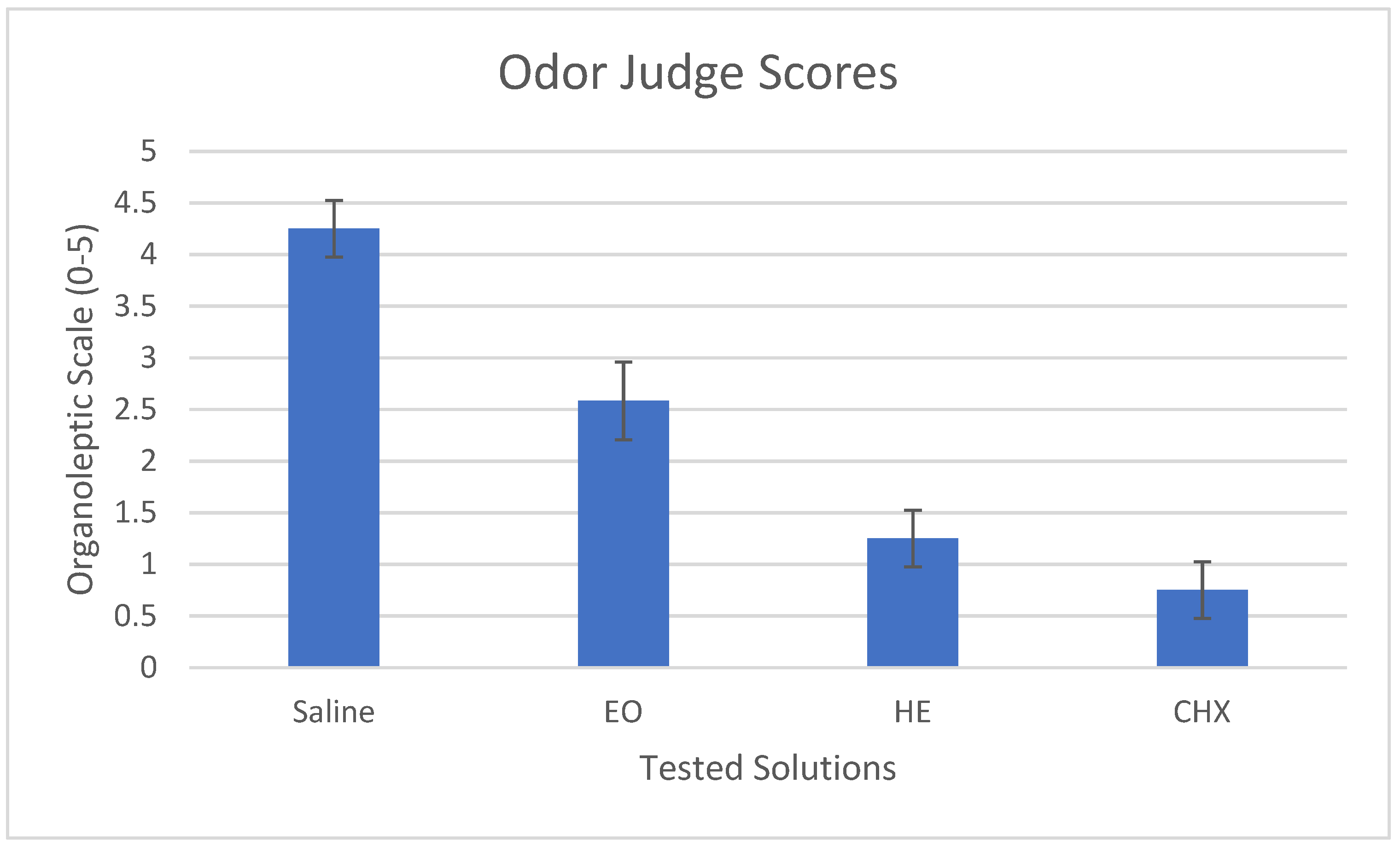
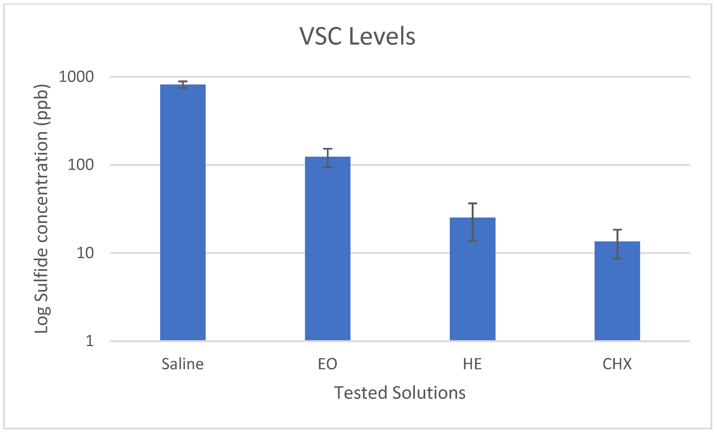
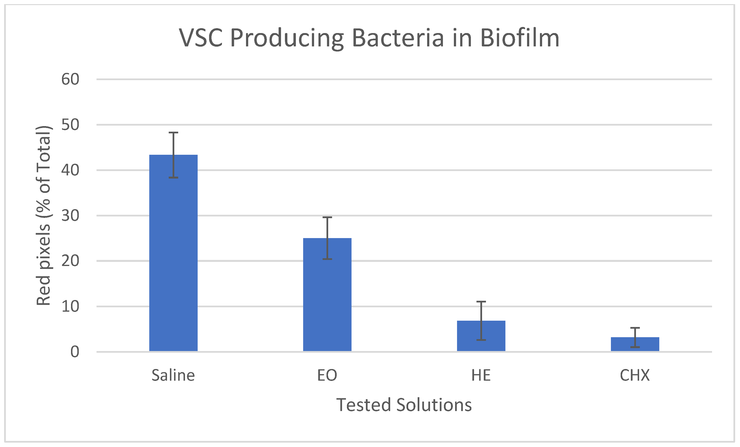
3.1. Malodor and VSC levels

The influence of several predisinfection solutions on malodor scores and VSC-production is shown in Figure 1 and Figure 2. Both chlorhexidine (CHX) and herbal extract (HE) formulations effectively reduced malodor and VSC levels below detection threshold (OJ scores < 2 and VSC < 75 ppb), with only borderline statistically significant difference between them (p = 0.037 and 0.045, respectively).

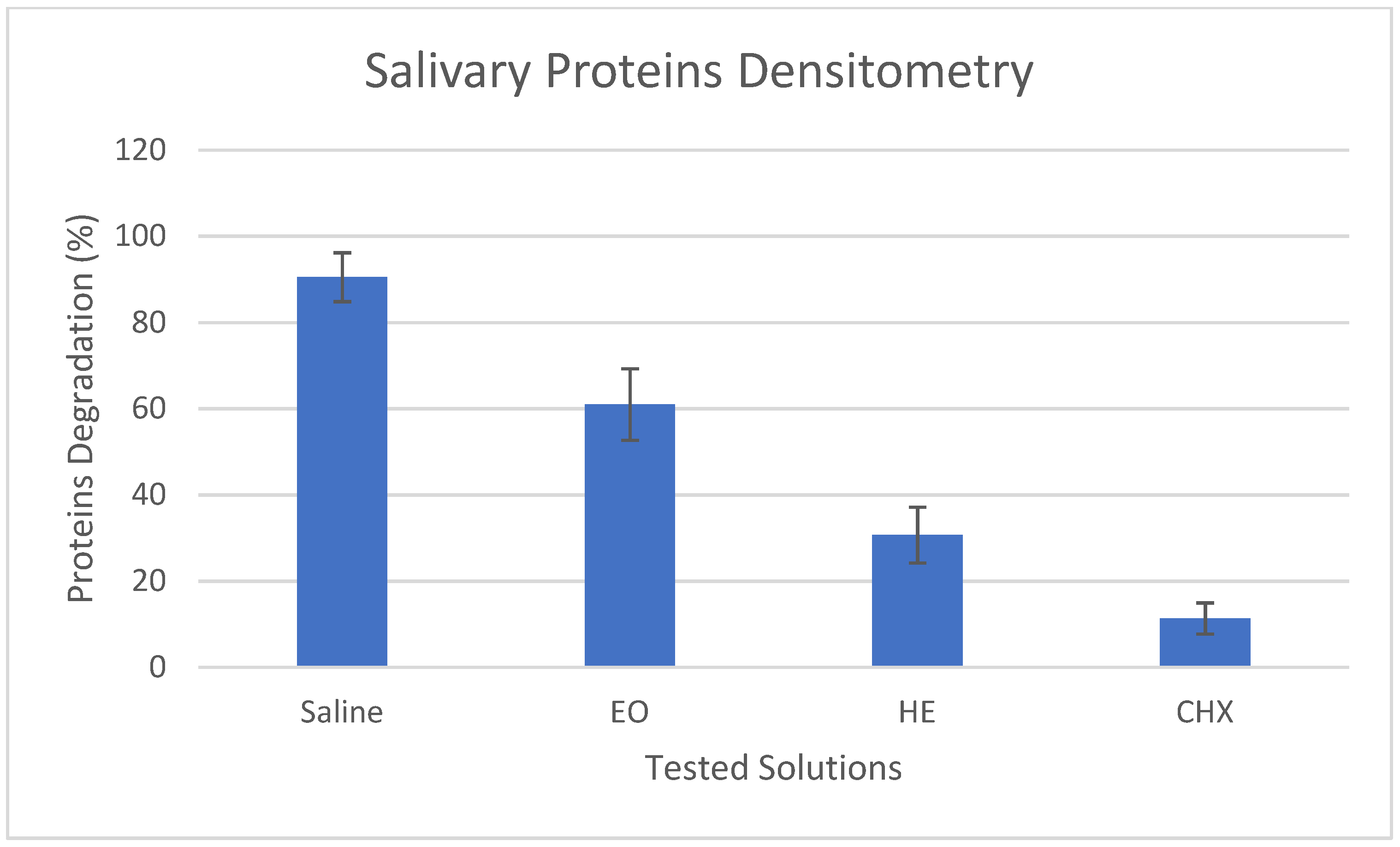
3.2. Salivary Proteins Degradation

The effects of the tested solutions on salivary protein degradation are presented in Figure 3 and Figure 4. Chlorhexidine (CHX) was most effective showing 88% reduction in proteolysis followed by the herbal extract (HE) formulation with 67% and essential oils (EO) with 32% reduction compared with the saline (negative) control. These differences were statistically significant (p < 0.001).

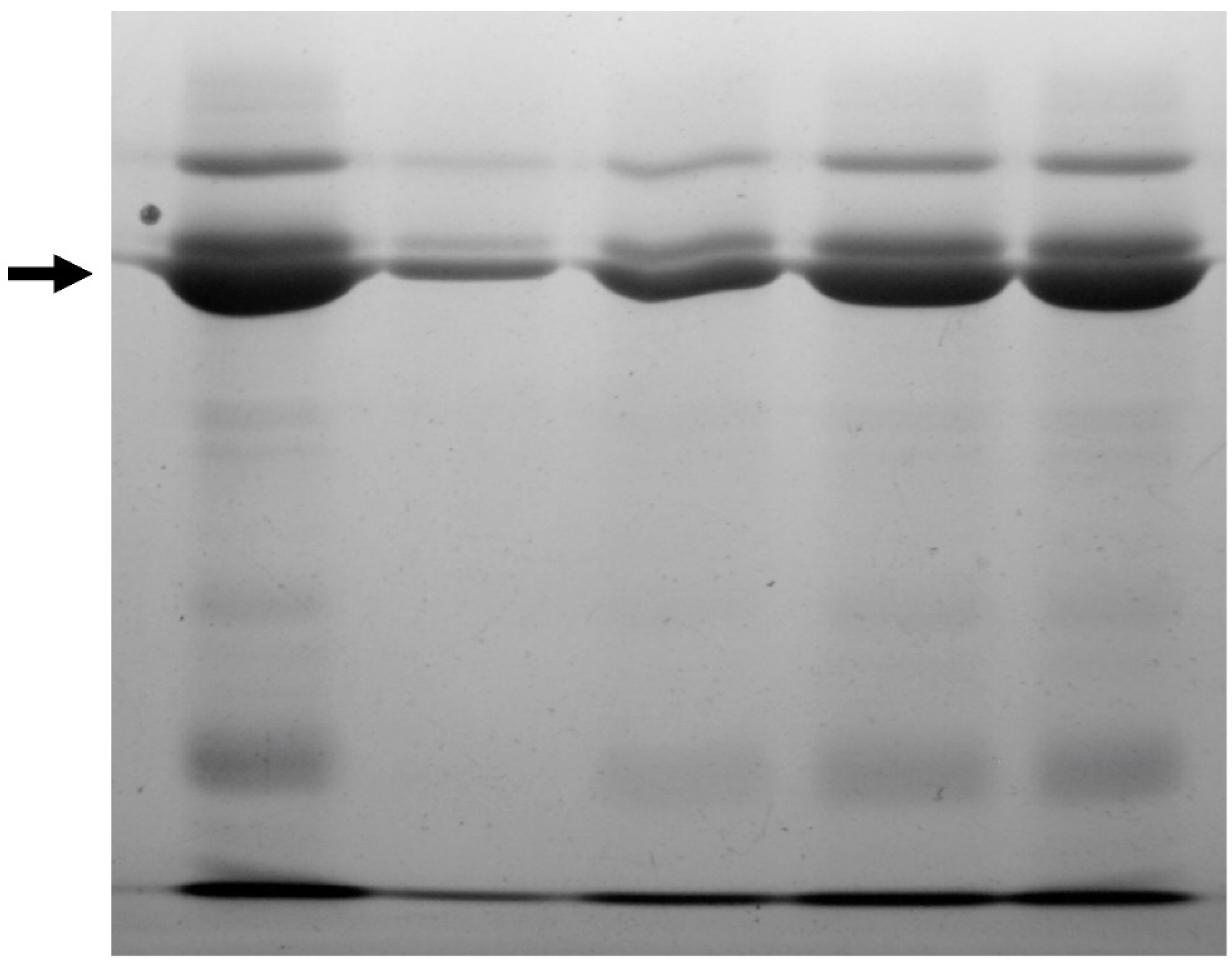
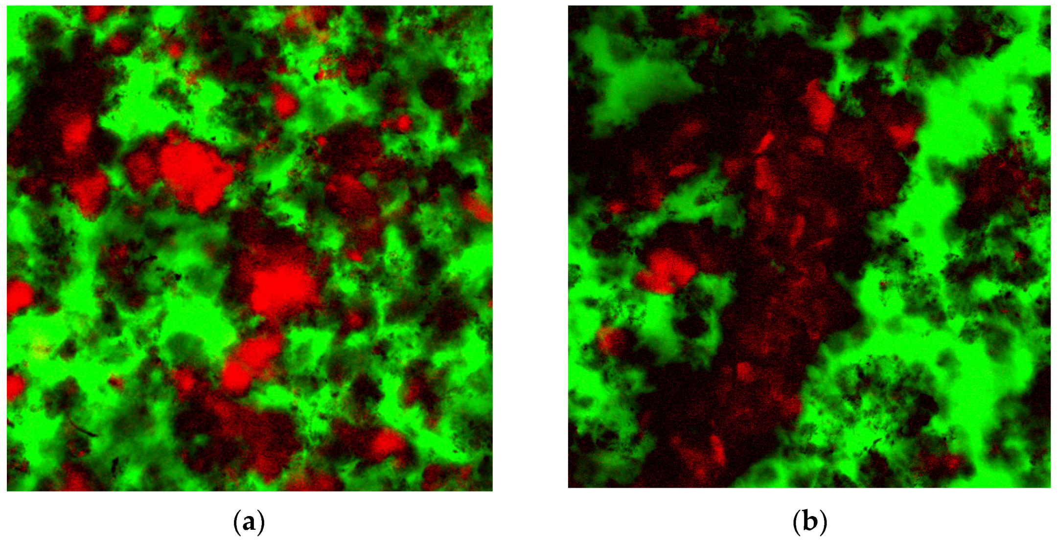
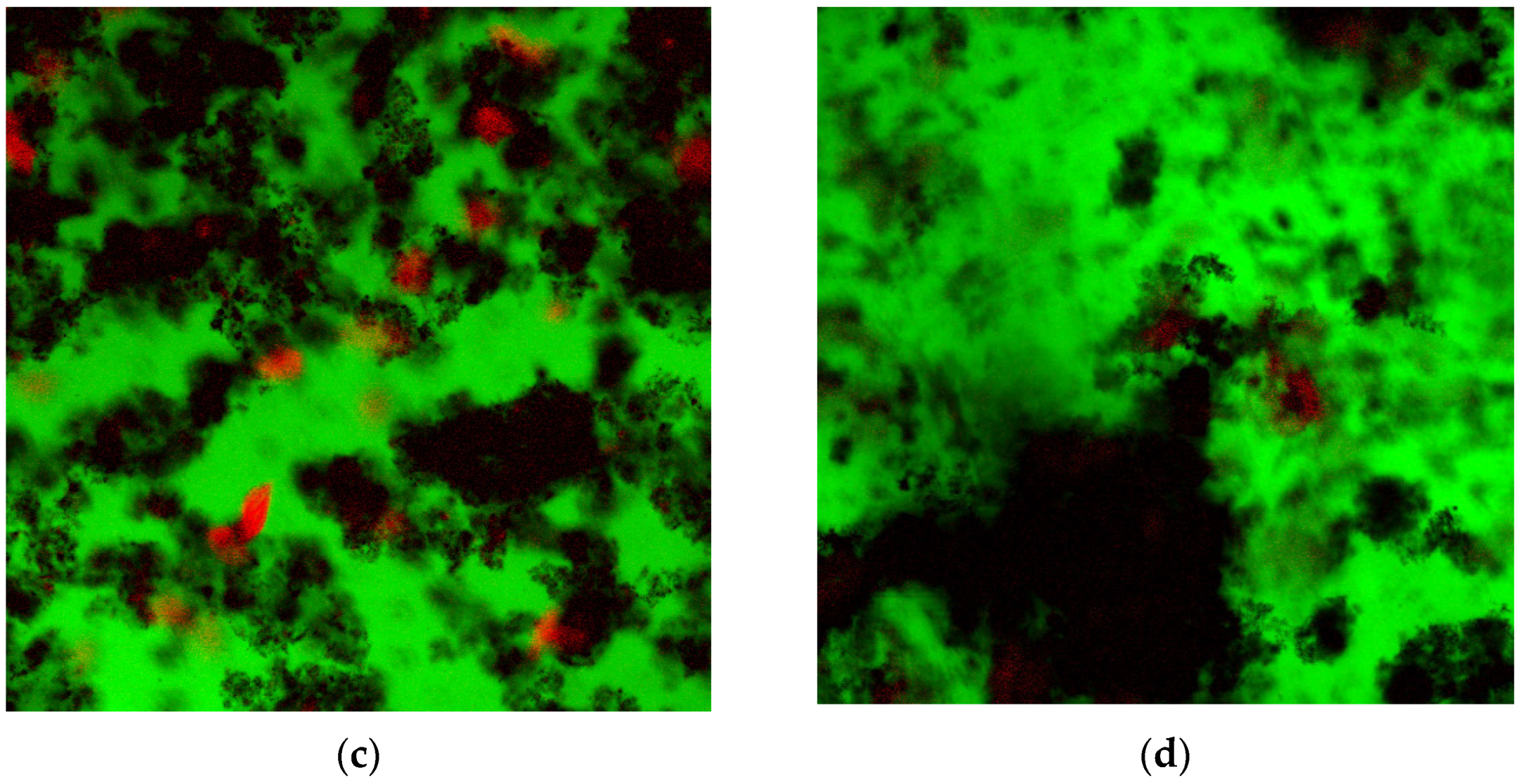
3.3. Confocal Laser Scanning Microscopy (CLSM)

The effects of the different tested solutions on VSC-producing bacteria in the biofilm formed on the pre-treated PMMA discs are shown in Figure 5 and Figure 6. Chlorhexidine (CHX) was most effective, showing 93% reduction in VSC-producing bacteria, whereas the herbal extract (HE) formulation showed 86% reduction compared with the saline (negative) control. However, the differences between those two formulations were statistically insignificant (p = 0.08).

4. Discussion

Our study hypothesis was rejected, as the results of the present research show that the pre-disinfection of PMMA discs by soaking in a disinfecting solution for 10 min prior to exposing them to incubation with whole saliva significantly reduces the presence of malodor-producing bacteria in the biofilm formed on these discs concomitant with the reduction in proteolytic activity and resulting VSC and malodor production. Salivary incubation assays are routinely used in oral malodor investigations and were shown to be a useful tool in screening for anti-malodor agents [7].
The disinfecting solutions used in this study are commercially available mouthwash formulations with claims of oral malodor reduction. The chlorhexidine formulation was the most effective pre-disinfecting solution in reducing malodor-producing bacteria in the biofilm and resulting malodor-related parameters. This is in agreement with other malodor studies [8,9]. However, the use of 0.2% chlorhexidine is not without the risk of side effects, the most common of which is staining and discoloration [10] that may pose serious aesthetic challenges in temporary crowns. Fortunately, in this study, a 10 min duration exposure was used, which is the equivalent of a 5-day rinsing in routine life. Such an extent of exposure is unlikely to yield a significant color change [11]. Furthermore, the results of the present study imply that natural-based products may also offer an esthetically safe and effective alternative.
Although routine removal of temporary crowns is often accompanied by the emission of malodor it was not clear to what extent this contributes to the overall problem of oral malodor in everyday life. A recent study comparing VSC levels in the oral cavities of patients with or without fixed partial dentures (i.e., dental crowns and bridges) found that crowns and bridges, especially faulty ones, were a significant risk factor for elevated VSC levels [5]. Temporary crowns and bridges are more than likely to exacerbate this problem, mainly due to the porosity of PMMA [3].
Taken together, the results of the present study suggest that pre-disinfection of temporary crowns may be beneficial in reducing the crowns’ contribution to oral malodor production. However, as this is an in vitro study, further clinical trials are warranted. The mechanism responsible for such results remains to be also investigated under in vitro conditions. It may well be discovered that the porous PMMA harbored a substantial reservoir of chlorhexidine, which influenced the malodor production. The experiment should be repeated with the samples thoroughly washed after exposure to the disinfection solutions. This will reveal the effect of each solution due to the actual 10 min exposure with less impact of the remnant disinfectant material.

Author Contributions

Conceptualization, O.R. and G.M.; methodology, N.S.; software, E.D.; validation, S.L.; U.J. and H.H.; formal analysis, G.B.I.; writing—original draft preparation, N.S.; writing—review and editing, U.J., O.R., G.M., E.D. and H.H. visualization, S.L.; supervision, O.R. and G.B.I.; project administration, N.S. All authors have read and agreed to the published version of the manuscript.

Funding

This research received no external funding.

Institutional Review Board Statement

The Research protocol was approved by Tel Aviv university ethics committee.

Informed Consent Statement

Not applicable.

Data Availability Statement

Not applicable.

Conflicts of Interest

Author (NS) declare ownership of patent EP2066304.

References

  1. Seemann, R.; Conceicao, M.D.; Filippi, A.; Greenman, J.; Lenton, P.; Nachnani, S.; Quirynen, M.; Roldan, S.; Schulze, H.; Sterer, N.; et al. Halitosis management by the general dental practitioner--results of an international consensus workshop. J. Breath Res. 2014, 8, 017101. [Google Scholar] [CrossRef] [PubMed] [Green Version]
  2. Sterer, N.; Shaharabany, M.; Rosenberg, M. β-Galactosidase activity and H(2)S production in an experimental oral biofilm. J. Breath Res. 2009, 3, 016006. [Google Scholar] [CrossRef] [PubMed]
  3. Gad, M.M.; Al-Thobity, A.M.; Shahin, S.Y.; Alsaqer, B.T.; Ali, A.A. Inhibitory effect of zirconium oxide nanoparticles on Candida albicans adhesion to repaired polymethyl methacrylate denture bases and interim removable prostheses: A new approach for denture stomatitis prevention. Int. J. Nanomed. 2017, 12, 5409–5419. [Google Scholar] [CrossRef] [PubMed] [Green Version]
  4. Takahashi, N. Oral Microbiome Metabolism: From “Who Are They?” to “What Are They Doing?”. J. Dent. Res. 2015, 94, 1628–1637. [Google Scholar] [CrossRef] [PubMed]
  5. Alzoman, H.; Rashid Habib, S.; Alghamdi, S.; Al-Juhani, H.; Daabash, R.; Al-Khalid, W.; Al-Askar, M.; Al-Johany, S. Relationship between Fixed Dental Crowns and Volatile Sulphur Compounds. Int. J. Environ. Res. Public Health 2021, 18, 1283. [Google Scholar] [CrossRef] [PubMed]
  6. Laemmli, U.K. Cleavage of structural proteins during the assembly of the head of bacteriophage T4. Nature 1970, 227, 680–685. [Google Scholar] [CrossRef] [PubMed]
  7. Sterer, N.; Rubinstein, Y. Effect of various natural medicinals on salivary protein putrefaction and malodor production. Quintessence Int. 2006, 37, 653–658. [Google Scholar]
  8. Carvalho, M.D.; Tabchoury, C.M.; Cury, J.A.; Toledo, S.; Nogueira-Filho, G.R. Impact of mouthrinses on morning bad breath in healthy subjects. J. Clin. Periodontol. 2004, 31, 85–90. [Google Scholar] [CrossRef] [PubMed]
  9. Sharma, K.; Acharya, S.; Verma, E.; Singhal, D.; Singla, N. Efficacy of chlorhexidine, hydrogen peroxide and tulsi extract mouthwash in reducing halitosis using spectrophotometric analysis: A randomized controlled trial. J. Clin. Exp. Dent. 2019, 11, e457–e463. [Google Scholar] [CrossRef] [PubMed]
  10. Addy, M.; Mahdavi, S.A.; Loyn, T. Dietary staining in vitro by mouthrinses as a comparative measure of antiseptic activity and predictor of staining in vivo. J. Dent. 1995, 23, 95–99. [Google Scholar] [CrossRef]
  11. Van Strydonck, D.A.C.; Slot, D.E.; Van der Velden, U.; Van der Weijden, F. Effect of a chlorhexidine mouthrinse on plaque, gingival inflammation and staining in gingivitis patients: A systematic review. J. Clin. Periodontol. 2012, 39, 1042–1055. [Google Scholar] [CrossRef] [PubMed]
Figure 1. The mean results (±SD) of the odor judge scores given using an organoleptic scale of 0–5, for the various tested solutions: Saline, essential oils (EO), herbal extracts (HE), and chlorhexidine (CHX) formulations as indicated.
Applsci 12 01947 g001
Figure 2. The mean results (±SD) of the Volatile Sulfide Compounds (VSC) levels measured using a sulfide monitor (Halimeter), for the various tested solutions: Saline, essential oils (EO), herbal extracts (HE), and chlorhexidine (CHX) formulations as indicated.
Applsci 12 01947 g002
Figure 3. SDS-PAGE analysis of the salivary proteins (arrow, alpha amylase, 55 KD) in the incubation mixture following pre-disinfection of the PMMA discs using the various treatment solutions with lane designations as follows: (1) Filtered saliva control, (2) saline control, (3) essential oils treatment, (4) herbal extracts treatment, and (5) chlorhexidine treatment.
Applsci 12 01947 g003
Figure 4. The mean results (±SD) of the salivary proteins degradation measured using SDS PAGE densitometry, for the various tested solutions: Saline, essential oils (EO), herbal extracts (HE), and chlorhexidine (CHX) formulations as indicated.
Applsci 12 01947 g004
Figure 5. Confocal Laser Scanning Microscopy (CLSM) images showing VSC-producing bacteria (stained red) within the biofilm (black) formed on the PMMA discs (green) following pre-disinfection using: (a) Saline (negative) control, (b) essential oil formulation, (c) herbal extract formulation, and (d) chlorhexidine formulation.
Applsci 12 01947 g005aApplsci 12 01947 g005b
Figure 6. The mean results (±SD) of the quantification of VSC-producing bacteria within the biofilm formed on the PMMA discs measured using confocal microscopy for the various tested solutions: Saline, essential oils (EO), herbal extracts (HE), and chlorhexidine (CHX) formulations as indicated.
Applsci 12 01947 g006
Publisher’s Note: MDPI stays neutral with regard to jurisdictional claims in published maps and institutional affiliations.

