Article

Long-Term Prosthetic Aftercare of Mandibular Implant-Supported Overdenture

1
Department of Prosthodontics, The Maurice and Gabriela Goldschleger School of Dental Medicine, Tel-Aviv University, Ramat-Aviv 69978, Israel
2
Department of Pediatric Dentistry, The Maurice and Gabriela Goldschleger School of Dental Medicine, Tel-Aviv University, Ramat-Aviv 69978, Israel
3
Department of Periodontics and Implant Dentistry, The Maurice and Gabriela Goldschleger School of Dental Medicine, Tel-Aviv University, Ramat-Aviv 69978, Israel
*
Author to whom correspondence should be addressed.
These authors contributed equally to this work.
Appl. Sci. 2022, 12(17), 8673; https://doi.org/10.3390/app12178673
Submission received: 7 August 2022 / Revised: 28 August 2022 / Accepted: 28 August 2022 / Published: 29 August 2022
(This article belongs to the Collection State-of-the-Art Dentistry and Oral Health)

Abstract

:
Background: Two of the most popular resilient attachment systems for mandibular implant-supported overdenture (MISOD) are locator and ball attachments. The purpose of the present retrospective cohort study was to assess the long-term prosthetic aftercare and oral hygiene status in edentulous patients rehabilitated with MISOD. Materials and Methods: Forty-five consecutive patients were included (22, group A- ball vs. 23, group B- locator attachments). Attachment incorporation into the MISOD was conducted in a direct (chair-side) intraoral technique at the time of denture insertion. Routine follow-up included yearly visits. The number of visits requiring prosthetic aftercare, either during the follow-up or during the additional visit, was recorded. Outcome parameters included—prosthetic aftercare—the number of aftercare (primary outcome parameter) visits, and dental treatment received (pressure sores relief, liner changes due to loss of retention, loss of retention due to debris accumulation, denture repair—secondary outcome parameters); oral hygiene—gingival index (primary outcome parameter). Results: The mean follow-up of the entire study population was 84 ± 21 months, range 39–120 months. Statistical analysis revealed a lower need for prosthetic aftercare interventions in group A (p < 0.001). The mean number of visits dedicated to pressure sores relief (6.09 ± 1.04 vs. 3.03 ± 0.77, p < 0.001) and liner exchange due to loss of retention (5.6 ± 1.03 vs. 2.09 ± 1.04, p < 0.001), were significantly lower in group A. Debris (food/calculus) accumulation inside the attachment was noted only for the locator’s group (p < 0.001). No statistically significant differences between the groups were noted for denture repair (p = 0.318). Oral hygiene also exhibited statistically significant differences in favor of group A (2.3 ± 0.3 vs. 1.03 ± 0.2, p < 0.001). Conclusions: It can be concluded that using ball attachments for MISOD yields less need for aftercare treatments and improved oral hygiene status over the years.

1. Introduction

Complete edentulism remains a substantial problem inflicting the quality of life of millions of dental patients worldwide. Its prevalence may reach values as high as 14.4% in young age groups and 32.3% in older age groups. Unfortunately, it is also associated with depression and poor self—related health [1]. The obsolete treatment modality of a conventional mandibular complete denture has lost its position as the primary standard of care for that predicament. The use of two endosseous dental implants placed in the anterior mandible as an aid to support and retain complete mandibular dentures proved itself an effective and predictable treatment option [2]. It has been favored as the primary standard of care for treating mandibular edentulism for almost twenty years [3]. The therapy of a maxillary complete denture with mandibular implant-supported overdenture (MISOD) among totally edentulous patients is a well-documented and successful treatment modality with proven patient satisfaction and chewing performance [4,5].
One of the research foci concerns the attachment systems that convey the load to the implants and the supporting tissues. A large systematic review concerning prosthetic aftercare with MISOD summarized in one of its conclusions that the implant attachment system has no clear effect on prosthetic aftercare requirements [6]. A more recent Cochrane review investigating prosthetic and patient-related outcomes of different attachment systems for maxillary and mandibular overdentures revealed deficient data to compose any meta-analyses due to the non-uniformity of included studies. Accordingly, that review concluded that there was insufficient evidence to support improved performance and outcomes of any specific attachment system [7]. A popular and simplified treatment philosophy encourages using two separate implants to support/retain a mandibular overdenture due to anatomical restraints, chair time, cost, and oral hygiene limitations [8,9].
Marginal bone loss (MBL) around implants is another important issue. With time, there seem to be slightly shallower probing depths around separate implants supporting an overdenture in comparison to splinted implants after 10 years of function. In addition, healthy mucosal conditions and stable marginal bone levels (determined radiographically) were found [10]. Another study comparing implant manufacturers found significantly less MBL around Straumann implants in comparison to Branemark implants over a five-year observation period [11]. These two studies [10,11] refer to MISOD on two separate implants. A meta-analysis showed that splinting implants through any specific attachment system does not offer any advantage concerning implant survival, marginal bone loss, and prosthetic complications [12]. However, the findings of the present systematic review should be analyzed with caution due to other influencing factors such as implant number/position, connection type, length/diameter, loading protocol, and follow-up period [12].
Two of the most popular resilient attachment systems available today are locator and ball attachments. Their comfortable ease of repair and installation on both implants and the supported overdenture made them highly acceptable among practitioners. In a recent meta-analysis, mean marginal bone loss was reported comparable between the locator attachment and the ball anchor [13] after a minimum of 1 year follow-up period. The same was true for an immediate and delayed loading protocol.
Peri-implant clinical parameters, as well as patient satisfaction, yielded no significant difference between the attachment systems in a clinical study [14]. Another study repeated the non-significant difference in aftercare requirements between the groups [15]. On the other hand, Matthys et al. found more aftercare requirements and less retention with the locator system versus ball attachments. A significantly higher plaque score was noted in the locator group [16]. Other studies also showed a preference for the locator attachment system with regard to the quality-of-life parameters and prosthetic aftercare [17,18].
Several quoted studies lacked data regarding the state of the opposing jaw and its edentulousness, which may influence the biting force and the attachment system’s long-lasting durability [13,17,18]. Others failed to substantiate the superiority of specific attachment systems due to heterogeneity and inconsistency in background parameters [6,9,12]
The somewhat confusing or even contradictory results listed above call for further research, which will find the parameter that can improve the quality of life among edentulous patients, in our case—the attachment system.
The alternative is bone grafting. A comparison between pre-operative and post-operative CT cross-sections showed statistically significant differences both in maxillary and mandibular posterior regions subjected to vertical onlay grafts. However, such procedures are complicated and not always predictable [19].
To the best of our knowledge, no comparable evidence exists regarding the long-term prosthetic aftercare impact of those two popular attachment systems regarding treatment with implant-supported mandibular overdenture. This is a medical and prosthetic topic that seems to be critical for patients’ comfort, function, and quality of life.
The purpose of the present long-term retrospective study was to compare ball vs. locator attachment systems regarding prosthetic aftercare requirements and oral hygiene outcomes between edentulous patients rehabilitated using two implants MISOD.

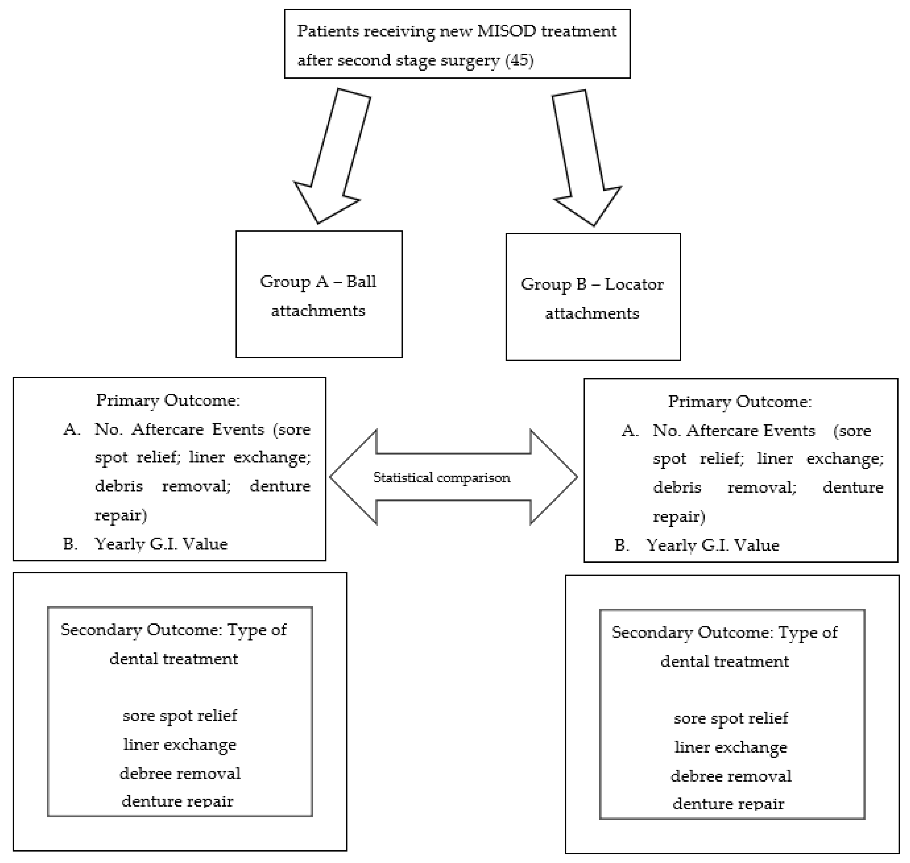
2. Materials and Methods

2.1. Inclusion Criteria

  • Addresses the Oral Rehabilitation Department, School of Dentistry, Tel-Aviv University;
  • MISOD treatment;
  • Reduced stability and insufficient retention of their former traditional mandibular complete denture;
  • Edentulous period concurrent with standard mandibular complete dentures of at least 12 months;
  • Classes III–VI resorption of the mandible according to Cawood and Howell (1988, [20]);
  • Presence of keratinized mucosa at the future implantation sites.

2.2. Exclusion Criteria

  • Stable mandibular denture without patient complaint;
  • Lack of available data;
  • Edentulous period <12 months;
  • Classes I–II resorption of the mandible according to Cawood and Howell (1988, [20]);
  • Absence of keratinized mucosa at the future implantation sites.
All patients were selected after meticulous medical histories, clinical dental examinations, and radiological evaluation that included panoramic and dental computerized tomography (CT) scans. All procedures were fully explained to the patients (informed consent was signed), and the Ethics Committee of Tel Aviv University approved the study (No. 7102008, October 2008). The attachment type alternative was chosen at the discretion of the operating prosthodontist. After collecting data from all consecutive individuals corresponding with the inclusion and exclusion criteria, the cohort resulted in two groups where 22 patients constituted group A to be treated with ball attachment and 23 patients in group B to be treated with locator attachment.

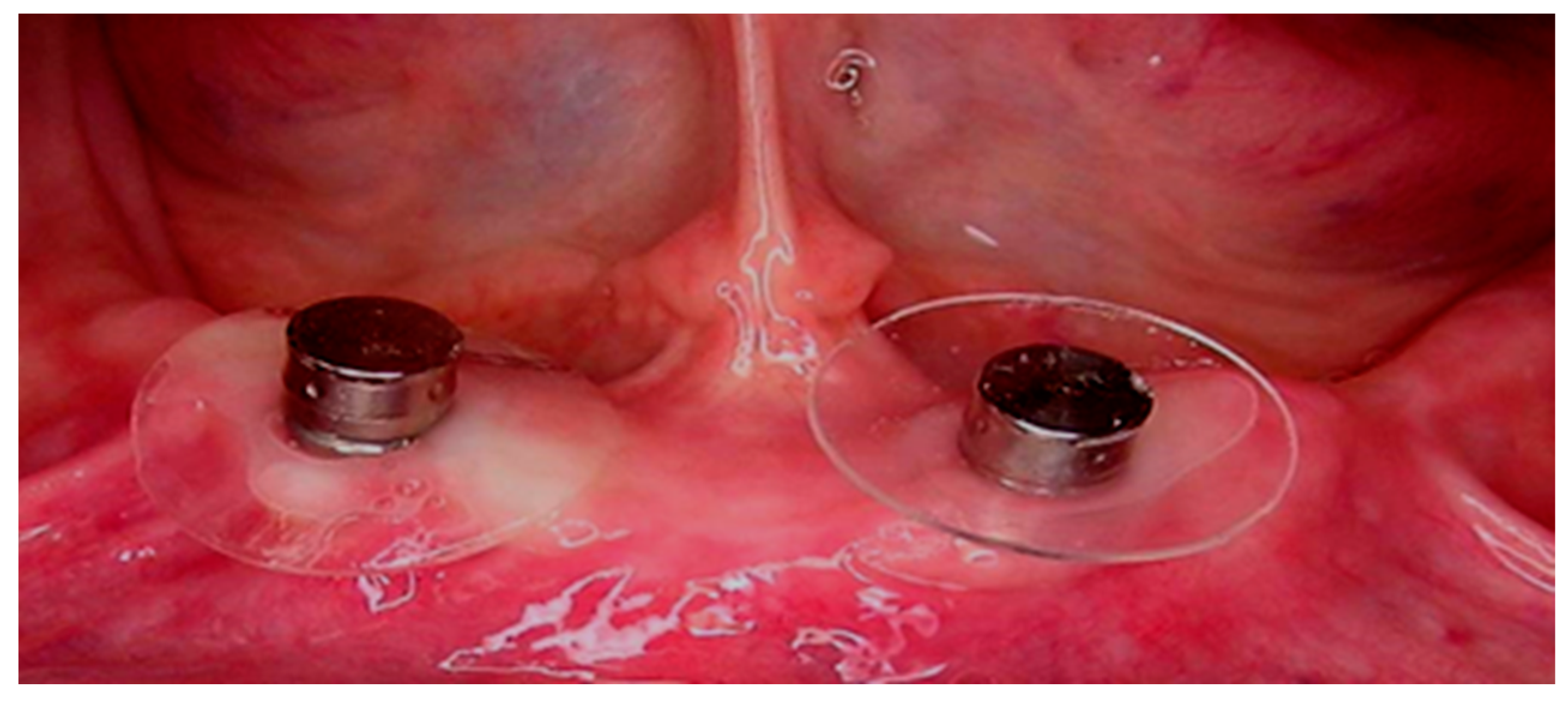
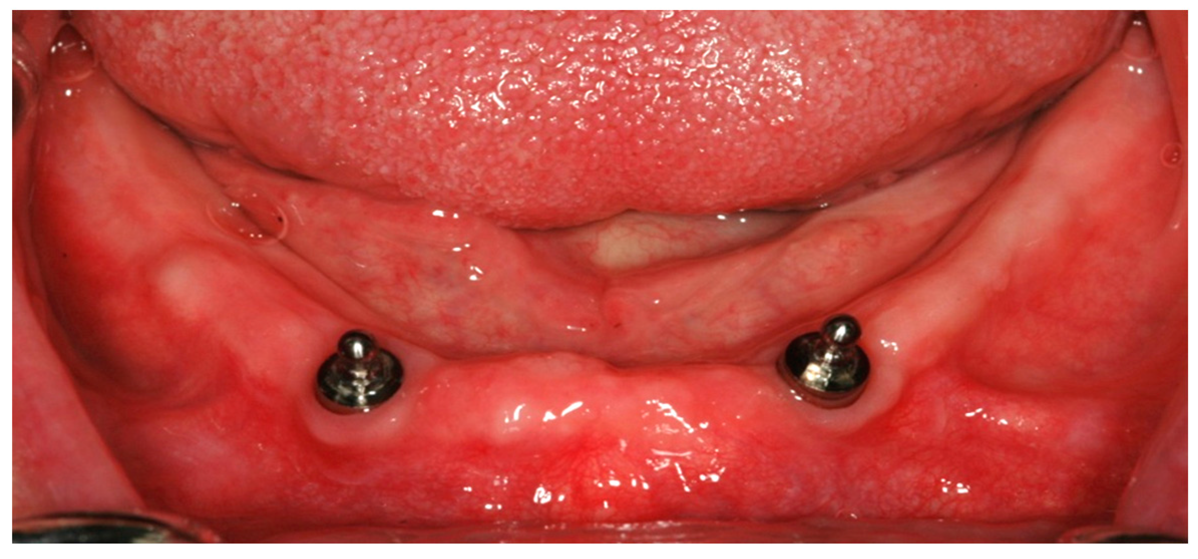
2.3. Surgical Treatment

Standard pre-operative treatment composed of chlorhexidine 0.2% mouth rinses, analgesics, and antibiotics. The implants were inserted under local anesthesia into the interforaminal region. A midcrestal incision was used, followed by implant osteotomy, bone level implant insertion, and primary soft tissue closure. All implants were placed using a prefabricated surgical stent as a two-stage procedure. Standard post-operative treatment composed of chlorhexidine 0.2% mouth rinses, analgesics, and antibiotics.
Post-operative panoramic and orthoradial periapical radiographs were taken prior to implant uncovering. Three months after implant placement, second-stage surgery was performed. After 4 weeks, standard prosthetic treatment was carried out—a new MISOD supported by ball or locator attachments. Attachment incorporation into the implants occurred depending on the height of the keratinized mucosa adjacent to them. In both experimental groups, attachments emerged 1 mm. supragingivally (range: 1–3 mm).

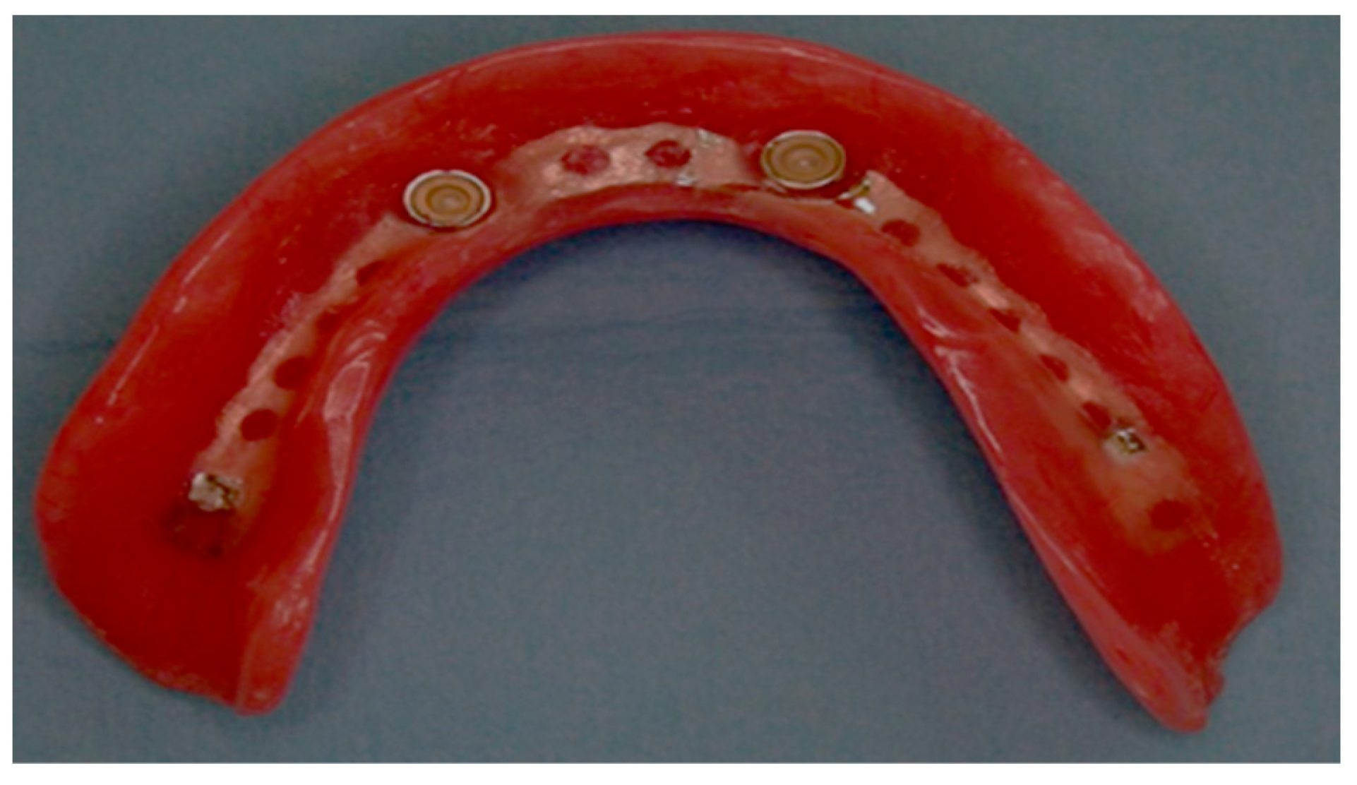
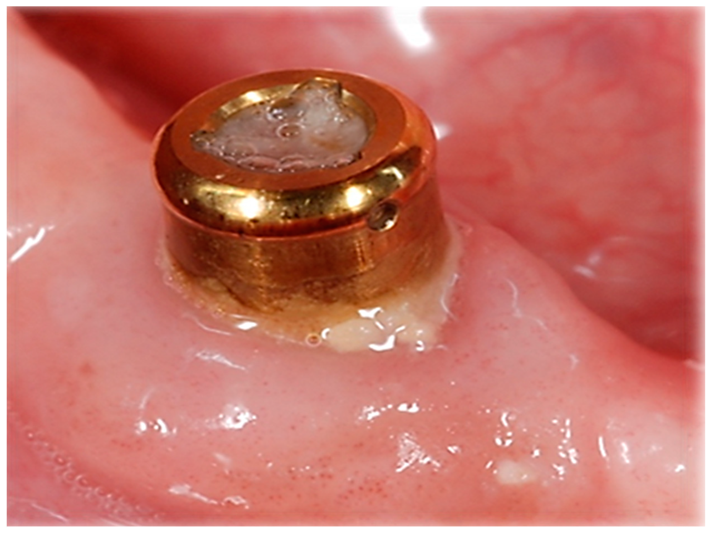
2.4. Prosthetic Treatment

Attachment incorporation into the MISOD was conducted in a direct (chair-side) intraoral technique at the time of denture insertion, and for both systems, we used the strongest silicone liner offered, also for both MISOD groups (Figure 1, Figure 2 and Figure 3).
In all patients, a balanced occlusion and monoplane acrylic teeth were used. The same dental laboratory constructed all of the overdentures as well as repairs and revisions during the aftercare period. All patients were treated by well-experienced oral-maxillofacial surgeons/periodontists and well-experienced specialists in prosthodontics/residents in oral rehabilitation.

2.5. Follow-Up and Data Collection

From the first day the patients visited the clinic and up to 10 years post initial examination, all surgical or prosthetic appointments and interventions were recorded. Prosthetic aftercare included routine recall visits every year. At a routine recall visit, the quality of opposing maxillary dentures (retention, support, and stability), implants, attachments, keratinized mucosa, prostheses, and oral hygiene (plaque accumulation was checked all over the attachment surface and also near peri-implant mucosa for each system) were evaluated. If needed, there were additional procedures for plaque debridement, calculus removal, oral hygiene instructions and support (instructions and removal of food debris), and adjustment or repair of the MISOD (teeth and/or base). Any reparative treatment was recorded. Consequently, patients used the same prosthesis for the entire study length.
In order to properly isolate the variable of the attachment system, our study group eliminated several masking factors. Hence, the entire patient cohort presented the same treated arch (mandible), the opposing arch (complete maxillary dentures), number of interforaminal implants (two), and mandibular bone quality (type III and IV according to Cawood and Howell [20]). All the data for the study were collected from the patients’ records.

2.6. Outcome Parameters

  • Number of aftercare visits—primary outcome parameter;
  • Dental treatment received— secondary outcome parameters:
    • Pressure sores relief;
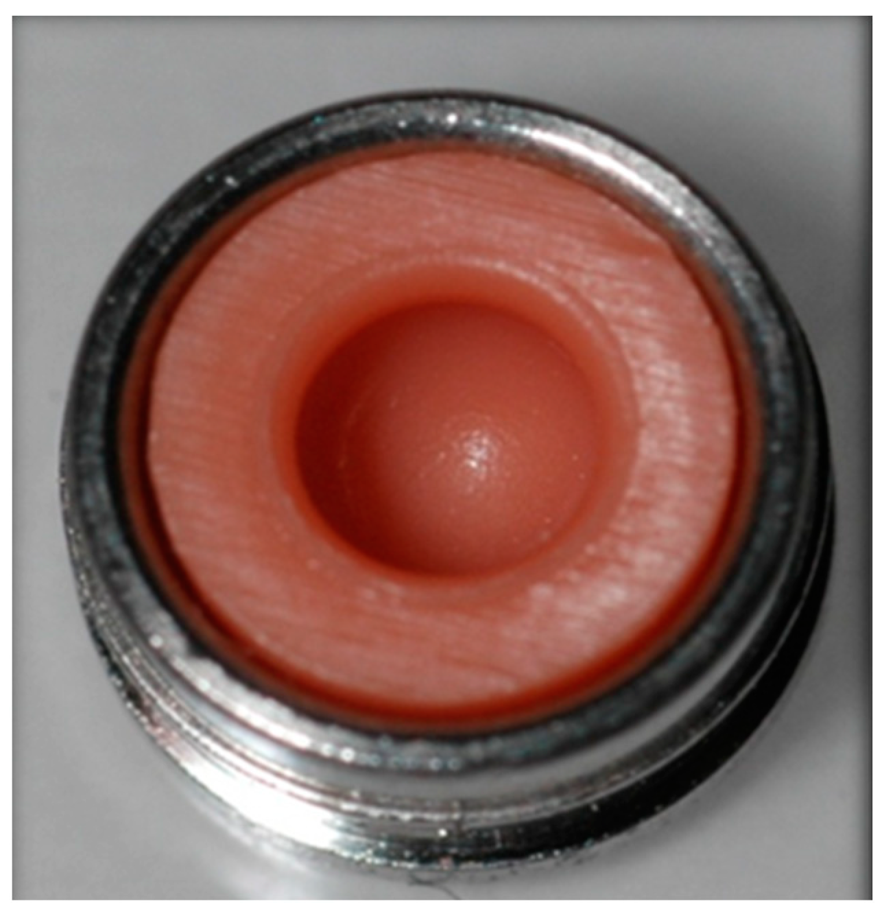
    • Liner (silicone insert) exchanges due to loss of retention;
    • Cleaning debris accumulation after loss of retention;
    • Denture repair;
  • Oral hygiene (gingival index 0–3 according to Loe and Silness [21])—primary outcome parameter.
The gingival index was used for the assessment of prevalence and severity of gingivitis. Score 0 = Normal gingiva; Score 1 = Mild inflammation—slight change in color, slight edema, no bleeding on probing; Score 2 = Moderate inflammation—redness, edema, glazing, bleeding on probing; Score 3 = Severe inflammation—marked redness and edema, ulceration, tendency toward spontaneous bleeding.

2.7. Statistical Analysis
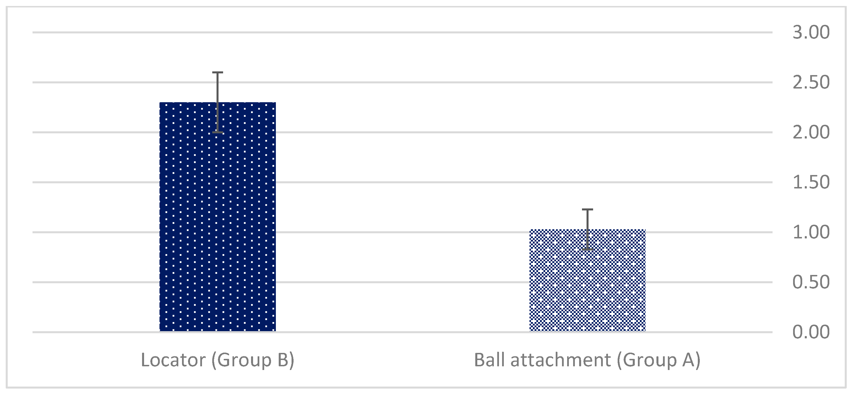
The statistical analysis was performed using SPSS software version 24.0 (SPSS Inc., Chicago, IL, USA; STATA 15.1, StataCorp LLC, College Station, TX, USA). A p-value < 0.05 was considered statistically significant. The Student’s t-test and Fisher’s exact test were used for statistical analysis of the data. Independent variables included age, gender, implant length, and type.
A scheme depicting the course of our study is illustrated below (Scheme 1).

3. Results

Forty-five consecutive patients (23 women and 22 men; mean age 65 ± 8 years, range 47–80 years) were included. The demographic characteristics showed insignificant differences between groups (mean age—63 vs. 65 years, p = 0.573; gender (male/female)—10/12 vs. 12/11).
All individuals were healthy or with mild controlled systemic disease. Individuals with severe systemic uncontrolled disease (e.g., smokers, diabetics, obese, on medications) were excluded.
A total of 90 (44 group A vs. 46 group B) implants were inserted. Two implant types were used: MIS Seven ™ implants (Implant Technologies, Bar Lev Industry, Israel)—23 patients; Zimmer TSV implants (Zimmer Dental, Carlsbad, CA, USA)—22 patients. The implant lengths used were 10, 11.5, or 13 mm, depending on the height of the mandible. Implant diameter was standard platform—3.75 mm. All abutments used were MIS (Implant Technologies, Bar Lev Industry, and Israel) due to university financial policy.
The mean follow-up of the entire study population was 84 ± 21 months, range of 39–120 months; group A mean follow-up was 88 ± 25, range of 39–120 months; group B mean follow-up was 80 ± 19, range of 45–110 months.
Statistical analysis revealed a greater need for prosthetic and oral hygiene interventions in group B (locator) vs. group A (ball). The mean number of aftercare visits was 11.69 ± 1.04 vs. 5.12 ± 0.4, p < 0.001. The mean number of visits due to pressure sores relief was 6.09 ± 1.04 vs. 3.03 ± 0.77, p < 0.001 (Figure 4), also for group B vs. group A.
Liner (silicone insert) exchange due to loss of retention (5.6 ± 1.03 vs. 2.09 ± 1.04, p < 0.001), was significantly higher in group B (Figure 5).
Debris (food/calculus) accumulation inside the attachment was noted only for the locator’s group in 75% of the MISODs (p < 0.001). Debris was checked only at the intraoral attachment level, not in the denture part of the attachment for both systems. No statistically significant differences between the groups were noted for the denture repair aftercare treatments (8/23 vs. 7/22) nor for dentures in the opposing arch (p = 0.318). The mean Gingival index also exhibited statistically significant differences (2.3 ± 0.3 vs. 1.03 ± 0.2, p < 0.001) (Figure 6).
The independent variables including age, gender, implant length, and type did not significantly affect the need for prosthetic aftercare.

4. Discussion

The present study demonstrated a significantly higher aftercare demand with locator vs. ball-borne MISOD wearers. Similar findings were also noted in other studies [22,23], where significantly more aftercare events and liner exchange took place among the locator group. It can be concluded that such need is related to the food accumulation in the systems’ matrix due to its coronal concave shape, surrounded by elevated edges, which increase retention, for creating a double retention system. Accordingly, the opposite topography has food impacting niches as well (Figure 7, Figure 8 and Figure 9). Although defined as a “nuisance” in one study [15], it appears to be much more meaningful. The tight intimate contact of the two attachment components is disrupted due to the food debris squeezed between them each time the denture is worn in the mouth or loaded under function. Gradually, permanent distortion—especially on the matrix level—occurs in addition to its normal wear and worsens the denture’s retention properties. Another retrospective study described the clinical outcome of 23 MISOD patients treated at the prosthodontic residency program and reported the same observation [24], stating that the main finding regarding the abutments was the debris (food/calculus) impacted inside the locator attachments. The authors further noted that 8 out of 23 prostheses had distorted or missing matrices [24]. Oral hygiene measures should eliminate such phenomenon, especially among older age groups wearing two implant MISODs, that due to less manual dexterity, exhibit more plaque around locator abutments.
Another factor that may facilitate the loss of retention with the locator system due to rapid wear is the technique used to incorporate the metal housing into the intaglio surface of the overdenture. In order to overcome it, attachment incorporation into the MISOD was conducted in a direct (chair-side) intraoral technique at the time of denture insertion for both systems. A previous study reported higher accuracy between the attachment components when the direct incorporation technique was used [25]. Minute misfits that may have occurred had a smaller effect on the ball vs. the locator group due to the spatial uniform round shape of the ball matrices. The locator abutment, on the other hand, has a stud cylindrical shape (axial parallel walls with a concave coronal edge justify increasing retention) may lead to spatial inaccuracies, which are more pronounced under loading conditions [26].
Implant parallelism may also influence the mechanical wear with the locator attachment, as shown in in vitro [23] as well as in clinical studies [27]. When comparing the two attachment systems through the same conditions of parallelism and near parallelism, the ball attachment showed superior results compared to the locator after an in vitro fatigue simulation period of 5.5 years [23]. This should lead to the conclusion that in surgical–clinical situations, where complete parallelism is rarely achieved, the ball attachment will show less compromised retention with time and require less liner exchange. The superiority should be related to the round uniform geometry with the ball anchors, which compensates for the non-parallel position between the two implants. It is worth mentioning that comparable values of liner exchange re-occurred in another retrospective cohort study, investigating ball anchored MISOD wearers opposing maxillary complete dentures as well but with an average 20-year follow-up period [27].
Finally, an in vitro study supporting our long-term clinical results compared stress distribution beneath a MISOD anchored by four popular attachment systems [28] and also found that the MISOD using ball attachment presented the least pressure exerted on the oral mucosa.
Bearing in mind that the locator system was also examined in that study and under the same conditions, the advantageous stress dissipation of the ball attachment is noteworthy. This was true under unilateral as well as bilateral loading protocols indicating that this type of attachment ball contributes the greatest reduction in oral mucosal pressure, rendering less traumatic injuries to the supporting tissues, as the present clinical study demonstrated. From a financial point of view, it is way cheaper to replace the liner (silicone insert) of a ball abutment than to replace the liner of a locator. Furthermore, the locator system is more expensive than the ball system. Age is also an issue that might be considered when choosing the attachment system (an older patient does not have the same expectation and manual abilities as a younger one).
Limitations of the present study include its retrospective nature, limited extent to only one treatment center, which leads to relatively small sample size, and two types of attachment. Another limitation of the study is the exclusion of unhealthy patients. For example, hyperlipidemia and obesity act as contributory factors in periodontitis development. Consequently, it was impossible to assess the effect of systemic conditions on peri-implant tissue health conditions [29,30].
This research contributes to the potential development of a protocol to rehabilitate edentulous patients not only through restoring their form and function but at a later stage—to decrease their dependence on off-schedule aftercare visits and improve long-term quality of life. A mechanism explaining the superiority of the ball attachment was elucidated, relating the difficulty of maintaining meticulous oral hygiene to higher incidence of liner exchange with the locator system, especially among older age groups. The known different metal compositions of the two attachment systems might also play a role in attracting plaque and thus influencing the gingival index of each one. Future research may allow us a pre-operative evaluation of “personalized” therapeutic options as in other fields of medicine [31]. For instance, In the field of orthopedics, the use of hard-on-soft bearings, such as metal-on-polyethylene, is common through hip replacement surgery. Unfortunately, the wear particles of the polyethylene material in their articulation with the hard material can result in a reduced life span of the artificial hip joint and mechanical loosening of implant components [32,33,34].

5. Conclusions

It can be concluded that using ball attachments for MISOD yields less need for aftercare treatments dedicated to sore spot relief, liner exchange, and oral hygiene compared with the locator attachment system. It can be suggested that using ball attachments may decrease the need for aftercare. Further studies are recommended to alleviate the anguish of MISOD wearers regarding soreness under the denture base and reduce the frequency of aftercare visits. Developing a product that will facilitate adequate cleaning and rinsing of the locator attachment may enhance the wellbeing of patients and practitioners using this highly popular prosthetic anchor.

Author Contributions

Conceptualization, E.Z., J.N., O.R. and S.N.; methodology, L.C., S.B., H.S., I.M., G.B.-I. and O.G.; formal analysis, L.C., S.B., J.N. and S.N.; investigation, O.R., E.Z., O.G. and J.N.; resources, I.M.; data curation, G.B.-I., O.G., H.S., I.M., S.B. and S.N.; writing—original draft preparation, G.B.-I., O.R., S.N. and E.Z.; writing—review and editing, S.N., L.C. and O.R. All authors have read and agreed to the published version of the manuscript.

Funding

This research received no external funding.

Institutional Review Board Statement

All procedures were fully explained to the patients and the Ethics Committee of Tel Aviv University approved the study (No. 7102008).

Informed Consent Statement

Informed consent was obtained from all subjects involved in the study.

Data Availability Statement

Data supporting reported results can be found in the table/graph.

Conflicts of Interest

The authors declare no conflict of interest. All authors have read and agreed to the published version of the manuscript.

References

  1. Tyrovolas, S.; Koyanagi, A.; Panagiotakos, D.B.; Haro, J.M.; Kassebaum, N.J.; Chrepa, V.; Kotsakis, G.A. Population prevalence of edentulism and its association with depression and self-rated health. Sci. Rep. 2016, 6, 37083. [Google Scholar] [PubMed]
  2. Allen, P.F.; McMillian, A. Food selections and perceptions of chewing ability following provision of implant and conventional prostheses to complete denture wearers. Clin. Oral Implant. Res. 2002, 12, 320–326. [Google Scholar]
  3. Feine, J.S.; Carlsson, G.E.; Awad, M.A.; Chehade, A.; Duncan, W.J.; Gizani, S.; Head, T.; Lund, J.P.; MacEntee, M.; Mericske-Stern, R.; et al. The McGill consensus statement on overdentures. Mandibular two-implant overdentures as first choice standard of care for edentulous patients. Montreal, Quebec, May 24–25, 2002. Int. J. Oral Maxillofac. Implant. 2002, 17, 601–602. [Google Scholar]
  4. Engquist, B.; Bergendal, T.; Kallus, T.; Linden, U. A retrospective multicenter evaluation of osseointegrated implants supporting overdentures. Int. J. Oral Maxillofac. Implant. 1988, 3, 129–134. [Google Scholar]
  5. Rentsch Kollar, A.; Huber, S.; Mericske Stern, R. Mandibular implant overdentures followed for over 10 years: Patient compliance and prosthetic maintenance. Int. J. Prosthodont. 2010, 23, 91–98. [Google Scholar] [PubMed]
  6. Assaf, A.; Daas, M.; Boittin, A.; Eid, N.; Postaire, M. Prosthetic maintenance of different mandibular implant overdentures: A systematic review. J. Prosthet. Dent. 2017, 118, 144–152. [Google Scholar]
  7. Payne, A.G.; Alsabeeha, N.H.; Atieh, M.A.; Esposito, M.; Ma, S.; Anas El-Wegoud, M.A. Interventions for replacing missing teeth: Attachment systems for implant overdentures in edentulous jaws. Cochrane Database Syst. Rev. 2018, 10, CD008001. [Google Scholar]
  8. Payne, G.T.; Zarb, G. (Eds.) Prosthodontic Treatment for Edentulous Patients: Complete Dentures and Implant-Supported Prostheses; Elsevier: St Louis, MA, USA, 2013; p. 334. [Google Scholar]
  9. Naert, I.; Gizani, S.; Vuylsteke, M.; Van Steenberghe, D. A 5-year prospective randomized clinical trial on the influence of splinted and unsplinted oral implants retaining a mandibular overdenture: Prosthetic aspects and patient satisfaction. J. Oral Rehabil. 1999, 26, 195–202. [Google Scholar]
  10. Cune, M.; Burgers, M.; van Kampen, F.M.; de Putter, C.; Van der Bilt, A. Mandibular overdentures retained by two implants. 10 year results from a cross-over trial comparing ball- socket and clip-attachments. Int. J. Prosthodont. 2010, 23, 310–317. [Google Scholar]
  11. Cehreli, M.C.; Uysal, S.; Akca, K. Marginal Bone Level Changes and Prosthetic Maintenance of Mandibular Overdentures Supported by 2 Implants: A 5-Year Randomized Clinical Trial. Clin. Implant Dent. Relat. Res. 2010, 12, 114–121. [Google Scholar]
  12. Leão, R.S.; Moraes, S.L.D.; Vasconcelos, B.C.E.; Lemos, C.A.A.; Pellizzer, E.P. Splinted and unsplinted overdenture attachment systems: A systematic review and meta-analysis. J. Oral Rehabil. 2018, 45, 647–656. [Google Scholar] [CrossRef] [PubMed]
  13. Liu, W.; Cai, H.; Zhang, J.; Wang, J.; Sui, L. Effects of immediate and delayed loading protocols on marginal bone loss around implants in unsplinted mandibular implant-retained overdentures: A systematic review and meta-analysis. BMC Oral Health 2021, 21, 122. [Google Scholar] [CrossRef] [PubMed]
  14. Krennmair, G.; Seemann, R.; Fazekas, A.; Ewers, R.; Piehslinger, E. Patient preference and satisfaction with implant-supported mandibular overdentures retained with ball or locator attachments: A crossover clinical trial. Int. J. Oral Maxillofac. Implant. 2012, 27, 1560–1568. [Google Scholar]
  15. Mackie, A.; Lyons, K.; Thomson, W.M.; Payne, A.G.T. Mandibular two implant overdentures: Three years prosthodontic maintenance using the locator attachment system. Int. J. Prosthodont. 2011, 21, 328–331. [Google Scholar]
  16. Matthys, C.; Vervaeke, S.; Besseler, J.; Doornewaard, R.; Dierens, M.; De Bruyn, H. Five years follow-up of mandibular 2-implant overdentures on locator or ball abutments: Implant results, patient-related outcome, and prosthetic aftercare. Clin. Implant Dent. Relat. Res. 2019, 21, 835–844. [Google Scholar] [CrossRef]
  17. Cristache, C.M.; Muntianu, L.A.S.; Burlibasa, M.; Didilescu, A.C. Five-year clinical trial using three attachment systems for implant overdentures. Clin. Oral Implant. Res. 2014, 28, 171–178. [Google Scholar] [CrossRef]
  18. Brandt, S.; Lauer, H.-C.; Fehrenz, M.; Güth, J.-F.; Romanos, G.; Winter, A. Ball versus locator attachments: A retrospective study on prosthetic maintenance and effect on oral-health- related quality of life. Materials 2021, 14, 1051. [Google Scholar] [CrossRef]
  19. Di Spirito, F.; Toti, P.; Brevi, B.; Martuscelli, R.; Sbordone, L.; Sbordone, C. Computed tomography evaluation of jaw atrophies before and after surgical bone augmentation. Int. J. Clin. Dent. 2019, 12, 259–270. [Google Scholar]
  20. Cawood, J.I.; Howell, R.A. A classification of the edentulous jaws. Int. J. Oral Maxillofac. Surg. 1988, 17, 232–236. [Google Scholar]
  21. Loe, H.; Silness, J. Periodontal disease in pregnancy. I. Prevalence and severity. Acta Odontol. Scand. 1963, 21, 533–551. [Google Scholar] [CrossRef]
  22. Nagy, U.; Guédat, C.; Giannopoulou, C.; Schimmel, M.; Müller, F.; Srinivasan, M. Microbiological evaluation of Locator® legacy attachments: A cross-sectional clinical study. Clin. Exp. Dent. Res. 2019, 5, 476–484. [Google Scholar] [CrossRef] [PubMed]
  23. Sultana, N.; Bartlett, D.W.; Suleiman, M. Retention of implant supported overdentures at different implant angulations: Comparing locator and ball attachments. Clin. Oral Implant. Res. 2017, 28, 1406–1410. [Google Scholar] [CrossRef] [PubMed]
  24. Matthys, C.; Vervaeke, S.; Besseler, J.; De Bruyn, H. Five-year study of mandibular overdentures on Stud abutments: Clinical outcome, patient satisfaction and prosthetic maintenance—Influence of bone resorption and implant position. Clin. Oral Implant. Res. 2019, 30, 940–951. [Google Scholar] [CrossRef] [PubMed]
  25. Nissan, J.; Oz-Ari, B.; Gross, O.; Ghelfan, O.; Chaushu, G. Long-term prosthetic aftercare of direct vs. indirect attachment incorporation techniques to mandibular implant-supported overdenture. Clin. Oral Implant. Res. 2011, 22, 627–630. [Google Scholar] [CrossRef] [PubMed]
  26. Marinis, A.; Afshari, F.S.; Chia-Chun Yuan, J.; Lee, D.J.; Syros, G.; Knoernschild, K.L.; Campbell, S.D.; Sukotjo, C. Retrospective analysis of implant overdenture treatment in the advanced prosthodontic clinic at the university of illinois at chicago. J. Oral Implantol. 2016, 27, 46–53. [Google Scholar] [CrossRef] [PubMed]
  27. Rosner, O.; Zenziper, E.; Heller, H.; Nissan, J.; Matalon, S.; Melamed, G.; Har-Ness, S.; Glickman, A. Long-Term Prosthetic Aftercare of Two-Vs. Four-Ball Attachment Implant-Supported Mandibular Overdentures. Appl. Sci. 2021, 11, 8974. [Google Scholar] [CrossRef]
  28. Sato, H.; Kobayashi, T.; Nomura, T.; Tanabe, N.; Tagafuji, K.; Kihara, H.; Kondo, H. Oral mucosa pressure caused by mandibular implant overdenture with different types of attachments. J. Prosthodont. Res. 2019, 30, 301–307. [Google Scholar] [CrossRef]
  29. Di Spirito, F.; Schiavo, L.; Pilone, V.; Lanza, A.; Sbordone, L.; D’Ambrosio, F. Periodontal and peri-implant diseases and systemically administered statins: A systematic review. Dent. J. 2021, 9, 100. [Google Scholar] [CrossRef]
  30. Di Spirito, F.; Sbordone, L.; Pilone, V.; D’Ambrosio, F. Obesity and periodontal disease: A narrative review on current evidence and putative molecular links. Open Dent. J. 2019, 13, 526–536. [Google Scholar] [CrossRef]
  31. Di Spirito, F.; Iacono, V.J.; Alfredo, I.; Alessandra, A.; Sbordone, L.; Lanza, A.T. Evidence-based recommendations on periodontal practice and the management of periodontal patients during and after the COVID-19 era: Challenging infectious diseases spread by airborne transmission. Open Dent. J. 2021, 15, 325–336. [Google Scholar] [CrossRef]
  32. Speranza, A.; Massafra, C.; Pecchia, S.; Di Niccolo, R.; Iorio, R.; Ferretti, A. Metallic versus Non-Metallic Cerclage Cables System in Periprosthetic Hip Fracture Treatment: Single-Institution Experience at a Minimum 1-Year Follow-Up. J. Clin. Med. 2022, 11, 1608. [Google Scholar] [CrossRef] [PubMed]
  33. Bülhoff, M.; Zeifang, F.; Welters, C.; Renkawitz, T.; Schiltenwolf, M.; Tross, A.-K. Medium- to Long-Term Outcomes after Reverse Total Shoulder Arthroplasty with a Standard Long Stem. J. Clin. Med. 2022, 11, 2274. [Google Scholar] [CrossRef] [PubMed]
  34. Solarino, G.; Carlet, A.; Moretti, L.; Miolla, M.P.; Ottaviani, G.; Moretti, B. Clinical Results in Posterior-Stabilized Total Knee Arthroplasty with Cementless Tibial Component in Porous Tantalum: Comparison between Monoblock and Two Pegs vs. Modular and Three Pegs. Prosthesis 2022, 4, 160–168. [Google Scholar] [CrossRef]
Figure 1. Intraoral components before direct (chairside) attachment incorporation.
Applsci 12 08673 g001
Figure 2. Silicone liner and metal housing.
Applsci 12 08673 g002
Figure 3. Denture base after direct (chairside) attachment incorporation.
Applsci 12 08673 g003
Scheme 1. MISOD: Mandibular Implant-Supported Over Denture; G.I.: Gingival Index (Loe and Silness [21]).
Applsci 12 08673 sch001
Figure 4. The mean number of visits dedicated to pressure sores relief.
Applsci 12 08673 g004
Figure 5. Liner exchange due to loss of retention.
Applsci 12 08673 g005
Figure 6. The mean gingival index.
Applsci 12 08673 g006
Figure 7. Locator demonstrated debris accumulation.
Applsci 12 08673 g007
Figure 8. Good oral hygiene demonstrated by the use of ball attachment.
Applsci 12 08673 g008
Figure 9. Locator demonstrated loss of retention due to debris accumulation.
Applsci 12 08673 g009
Publisher’s Note: MDPI stays neutral with regard to jurisdictional claims in published maps and institutional affiliations.

