Article

Antibacterial, Antiradical and Antiproliferative Potential of Green, Roasted, and Spent Coffee Extracts

by
Gema C. Díaz-Hernández
1,†,
Patricia Alvarez-Fitz
2,†,
Yanik I. Maldonado-Astudillo
1,
Javier Jiménez-Hernández
1,
Isela Parra-Rojas
1,
Eugenia Flores-Alfaro
1,
Ricardo Salazar
2,* and
Mónica Ramírez
2,*
1
Facultad de Ciencias Químico Biológicas, Universidad Autónoma de Guerrero, Chilpancingo 39087, Mexico
2
CONACYT, Universidad Autónoma de Guerrero, Chilpancingo 39087, Mexico
*
Authors to whom correspondence should be addressed.
These authors contributed equally to this work.
Appl. Sci. 2022, 12(4), 1938; https://doi.org/10.3390/app12041938
Submission received: 31 December 2021 / Revised: 2 February 2022 / Accepted: 7 February 2022 / Published: 12 February 2022
(This article belongs to the Topic Applied Sciences in Functional Foods)

Abstract

:
The phytochemical compositions of green coffee beans (GB), roasted coffee (RC), and the solid residue known as spent coffee grounds (SCG) have been associated with beneficial physiological effects. The objective of this study was to analyze the total phenolic compounds, antiradical scavenging ability, antibacterial activity, and antiproliferative activity on cancer cells of aqueous and ethanolic extracts of GB, RC, and SCG samples. The total phenolic content was quantified by Folin–Ciocalteu assay, while the antiradical activity was evaluated by ABTS●+ and DPPH radical assays, antibacterial activity was determined using the microtiter broth dilution method, and antiproliferative activity was evaluated by MTT assay in lung carcinoma cells (A549) and cervical cancer cells (C33A); furthermore, apoptosis and cell cycle arrest were evaluated by flow cytometry. Ethanolic extracts of RC and SCG showed the highest content of total phenols. The SCG ethanolic extract exhibited the lowest inhibitory capacity 50 (IC50) values for free radicals. The SCG extracts also had the lowest MIC values in bacteria. In antiproliferative assays, SCG extracts exhibited a significant decrease in viability in both cell lines, as well as increased apoptotic cells and promoted cell cycle arrest. The higher content of total phenols and antiradical activity of SCG ethanolic extracts was related to their antiproliferative activity in cancer cells, as well as their antibacterial activity against clinical isolates; therefore, the utilization of SCG adds value to an abundant and inexpensive residue.

1. Introduction

Coffee, an infusion of ground, roasted coffee beans, has been reported as being one of the most widely consumed beverages in the world [1]. Coffee is known to have a stimulant effect, a property mainly attributed to caffeine, but also contains different chemicals, including carbohydrates, lipids, nitrogenous compounds, vitamins, minerals, alkaloids, and phenolic compounds, which contribute to the valuable properties of this beverage [2,3]. However, along the coffee value chain, a huge number of by-products are generated annually and, in many cases, residues from the coffee industry are not properly handled. For this reason, applications such as the production of mushrooms, enzymes, organic acids, biofuels, and fertilizers have been proposed for the value addition of coffee by-products [4]. In this context, during the elaboration of instant coffee and coffee brewing, approximately 6 million tons of spent coffee grounds (SCG) are generated annually worldwide [5]. The composition of SCG includes an oil fraction (7.9–26.4%), crude fiber (19.7–22.1%), and different bioactive compounds, such as caffeine, trigonelline, phenolics, minerals, lignin, and melanoidins, which all depend on the type of coffee bean, the roasting conditions, and the extraction process [6]. Due to the functional properties of SCG, its utilization can be viewed, for the food and pharmaceutical industries, as an economically advantageous alternative to the development of foods, drugs, and therapies for the prevention and treatment of diseases. In this context, the bioactive compounds in green coffee (GB), roasted coffee (RC), and spent coffee grounds (SCG) have all been shown to possess antioxidant, antiproliferative, and antibacterial effects. Daglia et al. [7] demonstrated that roasted coffee possesses antibacterial activity. Monente et al. [8] reported that RC and SCG show antimicrobial activities, mainly against Gram-positive bacteria (Staphylococcus aureus, Listeria monocytogenes) and yeast (Candida albicans). Amigo-Benavent et al. [9] reported that a GB extract reduced the viability and proliferation of cancer cells. In this context, Jiménez-Zamora et al. [10], Hernández-Arriaga et al. [11], and Balzano et al. [12] reported the antiproliferative effect of bioactive compounds from SCG extracts on human colon cancer cells (HT-29) and lung carcinoma cells (A549).
Based on the great amount of attention that has been devoted to the development of alternatives exploiting the potential of coffee extracts as suitable ingredients in functional food and drug formulations aimed at alleviating health risks associated with bacteria or cancer, we hypothesized that GB, RC, and SCG have a differential biological activity, as related to the relative content of total phenolic compounds. Thus, in this study, we analyzed the total phenolic compounds, DPPH and ABTS●+ radical scavenging activity, antibacterial activity against ATCC and clinical isolates of methicillin-resistant strains, and the antiproliferative activity against A549 (lung carcinoma) and C33A (cervical carcinoma) cancer cell lines of GB, RC, and SCG aqueous and ethanolic extracts from a coffee (Coffea arabica) powder blend (Typica/Caturra, 50:50).

2. Results and Discussion

2.1. Extraction Yield, Total Phenolic Content, and Antiradical Scavenging Activity

Coffee polyphenols and their antioxidant activity have been considered a potential source of new value-added bioactive compounds and, as such, have received increasing attention from the scientific and medical communities [13,14]. As indicated in Table 1, the yield percentage of polyphenols in GB and RC (ethanolic and aqueous) extracts ranged from 17% to 23%, while that in SCG (ethanolic and aqueous) extracts ranged from 11% to 12%. The content of total phenolic (TPC) compounds in ethanolic extracts (of GB, RC, and SCG) had the greatest phenolic content (range 196–298 μg CAE·mg−1), while lower phenolic contents were found in aqueous extracts (of GB, RC, and SCG). Particularly, the ethanolic SCG extract exhibited the highest TPC content (298 μg CAE·mg−1), as well as presenting antiradical activity against ABTS●+ and DPPH radicals, inhibiting the 50% of these radicals (IC50) at concentrations of 7.5 and 44.8 μg·mL−1, respectively.
The variation in extraction yield, TPC, and in ABTS●+ and DPPH antiradical scavenging activity could be due to the type of coffee, solvent employed, and coffee processing. According to Chaves-Ulate and Esquivel-Rodríguez [15] and Farah and Donangelo [16], the main compounds found in coffee beans (GB and RC) include esters and diesters of p-coumaric, caffeic, and ferulic acids with quinic acids (caffeoylquinic acids, dicaffeoylquinic acids, feruloylquinic acids, and p-coumaroylquinic acids). These groups of compounds are collectively known as chlorogenic acids. It should be noted that, according to Perrone et al. [17], coffee roasting leads to a reduction in the contents of chlorogenic acids, in comparison with green coffee brews, ranging from 56% to 99% decrease after 6 or 15 min of processing time, respectively. In addition, these authors corroborated that chlorogenic acids are incorporated into melanoidins during the roasting process. Panusa et al. [18] reported TPC ranging from 6.33 to 28.26 mg gallic acid equivalent g1 dry SCG, as well as a total chlorogenic acid content total content ranging from 1.65 to 6.09 mg·g1 dry SCG.
The TPC content in ethanolic and aqueous extracts followed SCG > RC > GB. These differences can be attributed to the transformations that occur during coffee processing (e.g., releasing of bound phenolics during roasting and the subsequent drying and sonication prior to obtaining SCG samples). According to Coelho et al. [19] and Delgado-Andrade et al. [20], phenolic compounds are incorporated into coffee melanoidins during roasting, mainly in condensed and ester-linked form, thus affecting their extraction efficiency (large solvent volumes are necessary) and the melanoidin population soluble in ethanol contains the majority of the phenolic compounds incorporated into them. Our results support the potential of SCG as an interesting and inexpensive alternative from which phenolic-rich extracts for incorporation into foods and pharmaceutical preparations can be obtained.
Several health benefits have been associated with consumption, in terms of coffee brewing. In this context, the higher antioxidant activity of melanoidins and chlorogenic acids found in coffee has been recognized [21]. The use of a multiple approach to analyze the antioxidant potential of a plant extract is always recommended. In this study, the antiradical activity of GB, RC, and SCG was investigated by assessment of ABTS●+ and DPPH free-radical scavenging activity.
Independently of the solvent employed, the smallest value of IC50 that corresponds to higher antiradical activity was found with SCG samples, followed by those of RC and GB. In the DPPH test, the SCG extract exhibited IC50 values of 44.88 and 51.20 μg·mL−1 from ethanolic and aqueous extracts, respectively. The obtained DPPH IC50 values were lower than those reported (202–2369 μg·mL−1) by Andrade et al. [22] for SCG extracts obtained by means of supercritical fluid extraction. In the same manner, similar results were obtained in the ABTS●+ analysis. The SCG extract exhibited IC50 values of 9.99 and 7.50 μg·mL−1 from aqueous and ethanolic extracts, respectively. These values were significantly lower than those of the RC and GB samples (p < 0.05). Getachew et al. [23] reported ABTS●+ IC50 values of 1830 μg·mL−1 for SCG extracts obtained by subcritical water extraction. The superior DPPH and ABTS+ radical scavenging potential of SCG has been recognized by various researchers [12,24,25,26], which has been attributed to the presence of chlorogenic acids as the main phenolic compounds in SCG. In addition, the presence of caffeine and melanoidins also contributes to the antioxidant activity in RC and SCG [27].
In this context, the use of antioxidant extracts for their antimicrobial and anticancer properties is of specific interest, due to their health benefits and the increased interest of researchers in the development of various advanced technologies that take advantage of them [28].

2.2. Antibacterial Activity

The search for antimicrobial effects in plant extracts has gained in importance recently, due to an increase in bacterial resistance to conventional drugs [29]. Several studies have demonstrated that coffee extracts exhibit antibacterial activities against pathogens such as Escherichia coli, Staphylococcus aureus, Listeria monocytogenes, and Enterococcus faecalis [8,23,30,31]. The activities of bioactive compounds extracted from coffee against pathogenic bacteria have been widely studied [32,33]; however, the effects of coffee extracts on drug-resistant bacteria, such as methicillin-resistant bacteria, have scarcely been considered in the literature. Table 2 shows the minimal inhibitory concentration (MIC) values for the GB, RC, and SCG extracts. The results demonstrate that SCG (ethanolic and aqueous) extracts exhibited an ability to inhibit the growth of clinically isolated methicillin-resistant strains at the concentration range of 2–8 mg·mL−1, where the most effective extract against these strains was ethanolic SCG extract (2 mg·mL−1). Regarding the ATCC strains, RC (ethanolic) and SCG (aqueous) extracts inhibited the growth of Sa1 (MIC of 2 and 4 mg·mL−1, respectively), while ethanolic (GB, RC, and SCG) and aqueous (SCG) extracts exhibited MIC values of 8 mg·mL−1 against EC1, EClo, Sd, and Ef strains. Finally, GB (ethanolic), and GB and RC (aqueous) extracts did not demonstrate any antibacterial activity (MIC > 8 mg·mL−1) against the tested bacterial strains.
The results obtained in this study were similar to those reported by Monente et al. [8], who found that the growth inhibition of common food-borne pathogens and food spoilage microorganisms was higher in spent coffee than coffee brews. In the same context, Torres-Valenzuela et al. [31] and Pedras et al. [34] exposed the noteworthy antibacterial activities of SCG against both Gram-positive and Gram-negative bacteria.
This effect has been attributed to the content of phenolic acids, caffeine, and Maillard reaction products (e.g., melanoidins) quantified in spent coffee extracts [23]. Jiménez-Zamora et al. [10] stated that coffee melanoidins exert intense antimicrobial activity that could be used to avoid the growth of pathogenic bacteria in food products. These compounds possess the ability to alter the structure and function of the cytoplasmic membrane and to inhibit DNA repair mechanisms, favoring the inhibition of bacterial growth [35].
Our results demonstrate that SCG (ethanolic and aqueous extracts) possess an ability to inhibit the growth of clinically isolated methicillin-resistant strains. Antibacterial activity against clinical isolates of methicillin-resistant bacteria and methicillin-resistant Staphylococcus aureus (MRSA) remains a great challenge; although some synthetic antibacterial drugs have been developed, they are not effective against these bacteria. However, several studies have shown that natural compounds, fractions, and extracts exhibit antibacterial activity against clinical isolates of methicillin-resistant bacteria and MRSA, showing MIC values similar to those for sensitive Staphylococcus aureus strains (MSSA); the difference in the activity could possibly be due to different mechanisms of action of natural compounds at the cellular level, as well as the possibility that these compounds may use different pathways to exert their antibacterial activities (i.e., pathways modified and regulated by gene expression patterns, ABC transporters, fatty acid and peptidoglycan biosynthesis, aminoacyl-tRNA synthetase, and β-lactam resistance), as well as the increased presence of reactive oxygen species (ROS) and decreased antioxidant enzymes [36,37]

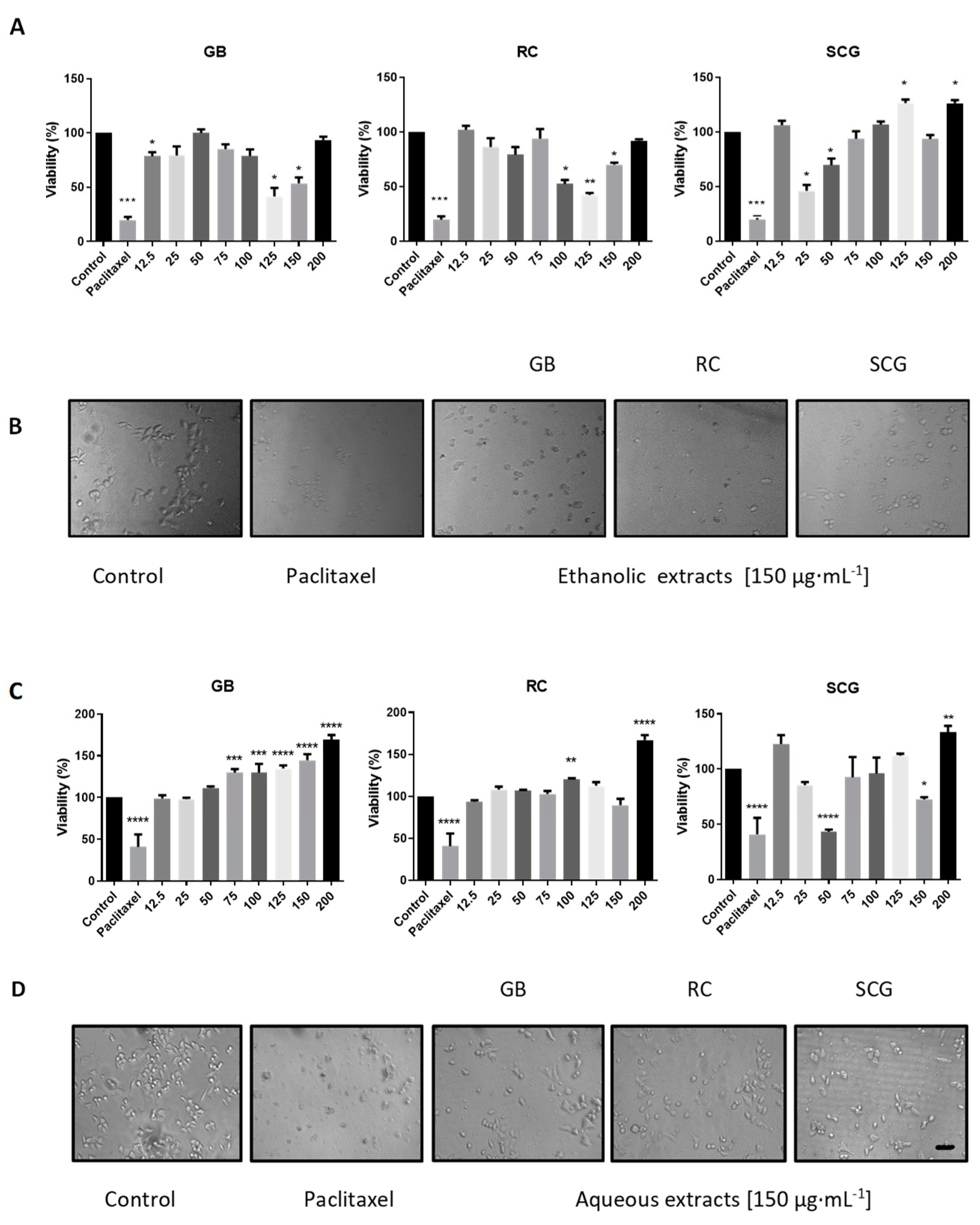
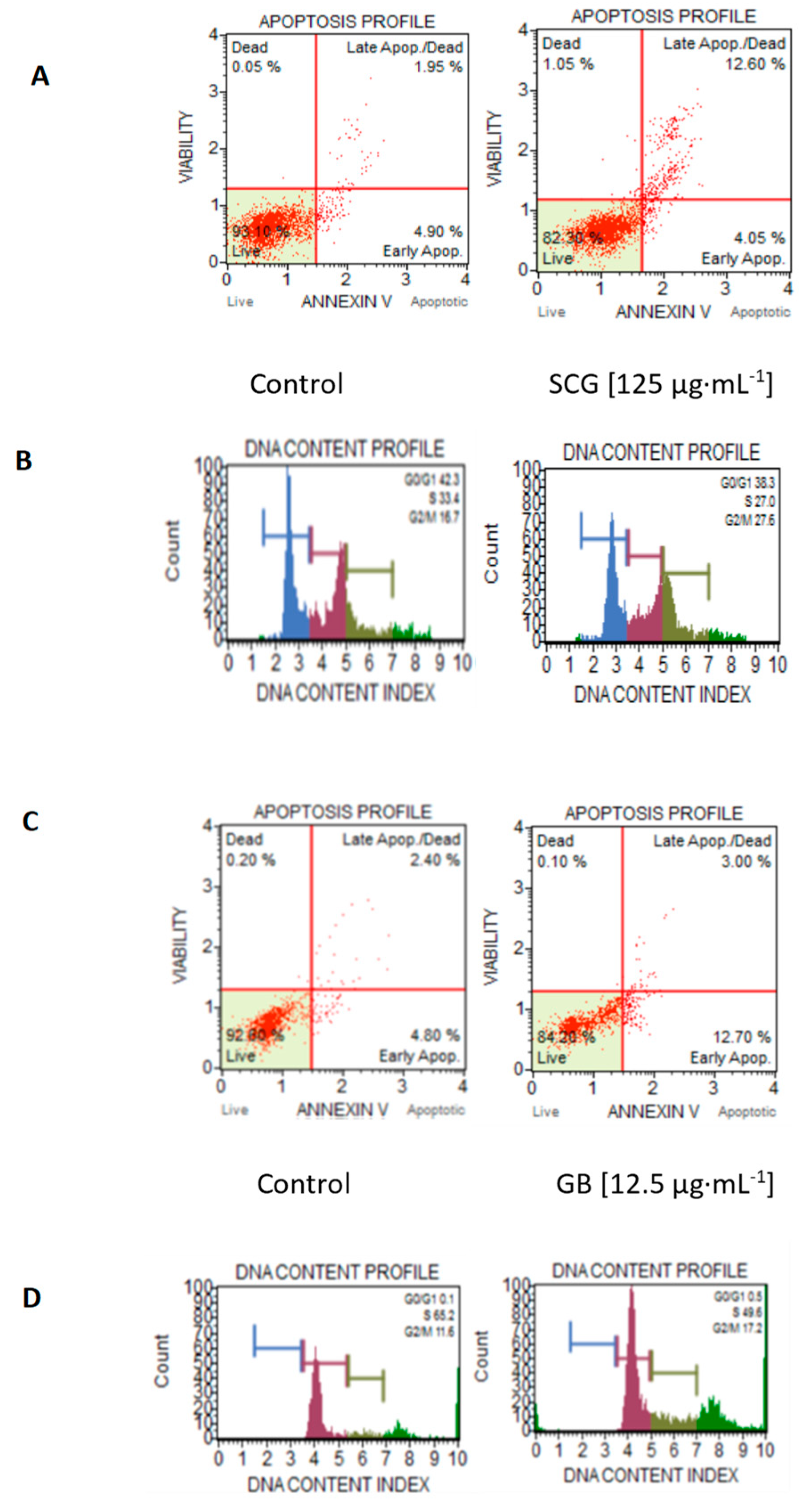
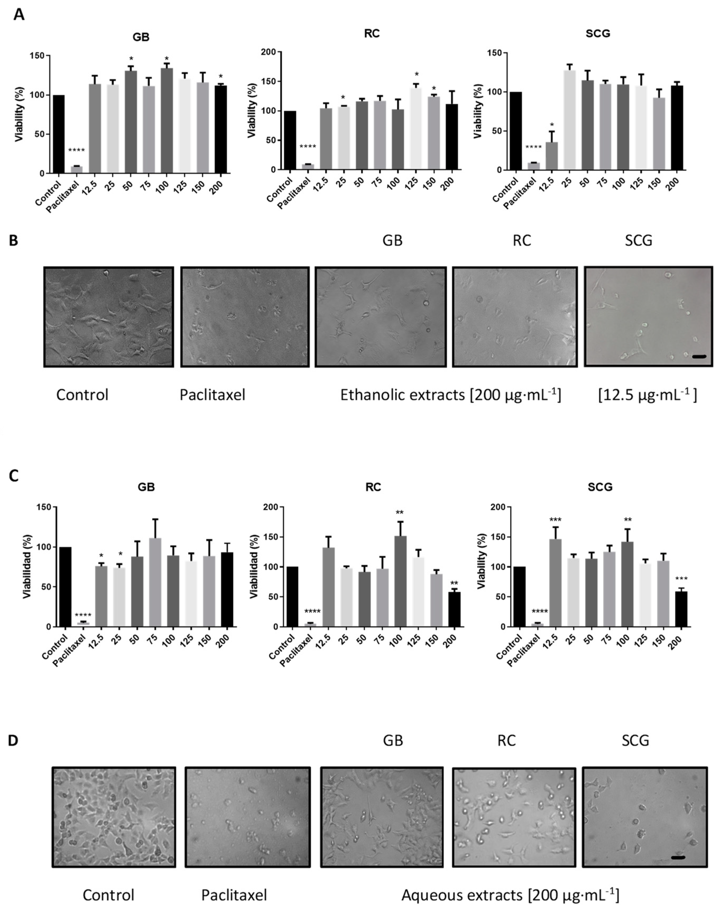
2.3. Analysis of In Vitro Antiproliferative Activity, Morphological Changes, Apoptosis, and Cell-Cycle Arrest

Cancers are characterized by uncontrolled cell proliferation and increased oxidative stress. Evidence has shown that some coffee metabolites can reduce viability and cell proliferation in transformed cells. We compared the antiproliferative effects of the aqueous and ethanolic extracts of GB, RC, and SCG. Our results show that the aqueous and ethanolic extracts from GB, RC, and SCG affected cell viability differently in A549 and C33A cell lines. In the antiproliferative activity assays, the SCG and RC extracts led to a significant decrease in viability in C33A cells (p < 0.001). The most significant effect in C33A was observed with ethanolic extracts from GB and RC; these extracts diminished cell viability by up to 58.5% and 57.6%, respectively. The most efficacious concentrations were 100, 125, and 150 µg·mL1. Notably, the ethanolic extract of SCG diminished cell proliferation in 30.5% of cells at 50 µg·mL−1, and in 53.9% of cells at 25 µg·mL−1 (Figure 1A).
The aqueous extract of SCG showed a reduction in cell viability by 56.7% and by 27.6% at 50 and 150 µg·mL1, respectively (Figure 1C).
Morphological changes associated with apoptosis include cellular size reduction, cytoplasmic inclusions, and cellular fragmentation [38]. We observed morphological changes in both cell lines treated for 24 h with the extracts; however, the changes were more evident in C33A cells. Ethanolic extracts caused cellular debris, as occurs with paclitaxel treatment, for most concentrations in C33A cells (Figure 1B). Furthermore, changes in cell morphology and reductions in the number of cells per field were evident, mainly at concentrations of 50 and 150 µg·mL−1 in all aqueous extracts, but mainly in extracts from SCG (Figure 1D). These results coincided with the effect determined in the cell viability assays where, at concentrations with the highest antiproliferative activity, the number of cells per field was reduced, with a greater effect on C33A cells. Notably, morphological changes in cells were correlated with extracts having the highest phenolic content.
In A549 cells, the ethanolic extract from SCG diminished up to 35.5% cell proliferation at 12.5 µg·mL−1, while the aqueous extract from SCG and RC only reduced cell viability at the highest concentration evaluated (200 µg·mL−1); see Figure 2A–C.
In C33A cells, aqueous GB or ethanolic SCG extracts seemed to increase cell viability; however, this does not necessarily indicate an increase in cell proliferation. Similar results have been described, as some metabolites contained in GB or SCG coffee extracts can increase enzymatic-mitochondrial activity, specifically a phytochemicals such as kahweol and cafestol, and CGAs can increase and potentiate mitochondrial-reducing enzymes and increased formazan products, which can result in higher percentages of cell viability [39]. Notably, the ethanolic extract from SCG diminished A549 cell viability only at 12.5 µg·mL−1, the lowest concentration evaluated. These results can be explained by the biphasic dose-response or hormesis, with a stimulatory effect at a low concentration, while high concentrations have the opposite effect. The hormesis effect has been widely documented in biological and pharmacological research [40,41].
It should be noted that we did not evaluate the effects of the coffee extracts on non-transformed or non-cancerous cells to determine their cytotoxicity. However, there are several comparative studies that have shown that the phytochemicals contained in coffee have a high specificity to decrease the cell viability of cancer cells without affecting non-transformed cells. It has been determined that, in fibroblast cell lines (CCD-18Co), hepatocytes (AML-12) and macrophages (RAW 264.7), the metabolites present in various coffee extracts do not have a cytotoxic effect; these findings suggest their selectivity to inhibit the proliferation and viability of cancer cells [9,42].
To assess whether the extracts increased the number of apoptotic cells or caused cell-cycle arrest in C33A and A549, which could explain the reduction in cell viability, we performed a flow cytometry analysis. We only evaluated the concentrations and extracts in which we observed a notable effect on the reduction of cell viability; as such, the aqueous extracts were not evaluated. In C33A cells, we evaluated ethanolic extracts at 25, 75, 125, and 200 µg·mL1 concentrations, with a differential effect on the increase in the number of apoptotic cells between 13.5% and 15.7%. The highest percentage of apoptotic cells was observed with the GB ethanolic extract at 12.5 µg·mL−1. In the analysis of cell-cycle changes, the ethanolic RC extract at 125 µg·mL−1 decreased the number of cells in the G1/S phase by 12.7%, promoting cell-cycle arrest (see Figure 3A,B; Table 3).
In A549 cells, the ethanolic extracts from GB, RC, and SCG were evaluated at 12.5, 150, and 200 µg·mL−1. The ethanolic extract from GB increased apoptotic cells by 8.7%. Furthermore, the aqueous extract from SCG exhibited the highest percentage of increase in apoptotic cells (10.3%). Regarding changes in the cell cycle, the ethanolic extracts from SCG and GB reduced G1/S-phase proliferative cells by 15.1% and 15.3%, respectively, in comparison with the control (see Figure 3C,D; Table 3).
The antiproliferative and proapoptotic effects of the ethanolic and aqueous extracts of GB, RC, and SCG can be attributed to coffee metabolites, such as caffeine, caffeic acid, trigonelline, chlorogenic acids, and diterpenes (e.g., cafestol and kahweol), as well as some molecular mechanisms that have been described in several cell lines [43,44,45,46,47]. However, coffee roasting and other processes can change the profile of the bioactive compounds and, therefore, can affect its bioactivity or physiological effects [48,49]. In this regard, few studies have been focused on extracts from SCG, and analysis of the compound profile in SCG has indicated a high content of melanoidins, chlorogenic acids, caffeic acid and, at a lesser concentration, caffeine [25,50]. Ramalakshmi et al. [51] reported a remarkable effect of reducing cell viability in P388 cancer cells treated with SCG extracts, compared with other coffee samples. Furthermore, studies have demonstrated the antitumor activity of SGC extracts in HT-29 colon cancer cells [11], as well as an induction of the expression of antioxidant proteins in Raw 264.7 cells [52]. Additionally, the induction of apoptosis has been described through DNA fragmentation, the increased expression of proapoptotic proteins, and activation of the caspase pathway [53], as well as through the reduction of reactive oxygen species (ROS) [54].
Some action mechanisms have been described, in terms of the anticancer activities of the bioactive metabolites contained in SCG extracts, such as melanoidins and phenolic compounds: they have antiproliferative and proapoptotic effects on cancer cells. Specifically, the chlorogenic acids and caffeic acid inhibited the phosphorylation of ERK1/2 in hepatic or colon cancer cell lines, this pathway’s signaling being related to cell proliferation and apoptosis [55,56]. Furthermore, it has been described that melanoidins reduce cell proliferation and induce cell cycle arrest in G0 or G1/S in cancer cell lines, where one of the mechanisms described for cell cycle arrest is alteration of the microtubule skeleton [57].

3. Materials and Methods

3.1. Materials

Green coffee beans (Coffea arabica L.), in a blend of typica and caturra (50:50) varieties, were supplied by local producers in Malinaltepec (Guerrero, Mexico). All reagents employed were of analytical grade. Standards of caffeic acid, Folin–Ciocalteu reagent, 2,2′-Azino-bis (3-ethylBenzoThiazoline-6-Sulfonic acid) diammonium salt (ABTS●+), 2,2-DiPhenyl-1-PicrilHidrazilo (DPPH), and 3-(4,5-dimethylthiazol-2-yl)-2,5-diphenyltetrazolium bromide (MTT), as well as solvents (ethanol and dimethylsulfoxide), were purchased from Sigma-Aldrich (St. Louis, MO, USA). Other analytical-grade reagents employed, including sodium bicarbonate, trypsin, phosphate-buffered saline (PBS), and trypsin, were purchased from J. T. Baker (Phillipsburg, NJ, USA) and Gibco (Waltham, MA, USA). Human lung A549 and human cervical C33A carcinoma cell lines were supplied by the Cell Bank of the Facultad de Ciencias Químico Biológicas at the Universidad Autónoma de Guerrero (FCQB, Guerrero, México).

3.2. Preparation of Coffee Sample Extracts

The green coffee blend was roasted at 200 ± 5 °C for 20 min. Later, the roasted coffee grains were ground in a mill (Krups GX410011 Coffee Grinder). Then, 10 g of roasted ground coffee was inserted into a 1-cup filter holder and percolated in an Oster® (BVSTDCDW12B) coffee machine with 500 mL of water. The obtained SCG was dried at 60 °C for 5 h. For preparation of the aqueous and ethanolic extracts, 5 g of green bean coffee (GB), roasted coffee (RC), or spent coffee grounds (SCG), along with 50 mL of water or ethanol (60%) was transferred into glass flasks, and the mixture was sonicated at 45 °C for 1 h. After centrifugation at 1800× g for 30 min, the top phases were removed, and the residues were added to 50 mL of water or ethanol (60%) and sonicated again. The supernatants were merged, filtered, evaporated, and lyophilized using a 4.5 L FreeZone freeze-drier model (Labconco, KS, USA). The yield was calculated utilizing the following equation: yield = (grams of extracts obtained × 100/grams of coffee beans macerated). The extracts obtained were stored at -20 °C until further use.

3.3. Determination of Total Phenolic Content and Antiradical Scavenging Activity

Working solutions employing GB, RC, and SCG extracts were prepared at 5 mg·mL−1 with distilled water. Total phenolic content (TPC) was determined in samples through the Folin–Ciocalteu method. The standard was caffeic acid (CA), and the results were expressed in mg of CA equivalents µg CAE∙mg−1 of dry extract.
Antioxidant activity in the coffee extracts was determined by antiradical scavenging activity, according to the DPPH and the ABTS●+ method described by Thaipong et al. [58,59]. Median inhibitory concentration (IC50) values, which are the concentrations of the coffee extract needed to inhibit 50% of the DPPH and ABTS●+ radical activity, were obtained from best-fit line plotting the concentration of coffee extracts vs. the percentage of DPPH or ABTS●+ inhibition ((A0-A1/A0) × 100), where A0 is the absorbance of DPPH or ABTS●+ solution and A1 is the absorbance of the extract). Statistical comparisons and non-linear curve fittings for IC50 determination were carried out using Origin® v.8.0724 software (OriginLab Corporation, Northampton, MA, USA).

3.4. Antibacterial Activity

Minimal inhibitory concentration (MIC) of coffee extracts was performed according to the method reported by Salazar-Pineda et al. [60], with slight modification.
Antibacterial activity was evaluated against a reference sensitive to antibiotic American Type Culture Collection (ATCC) strains Escherichia coli 25922; Staphylococcus aureus 25923; Enterococcus faecalis 29212; Enterobacter cloacae 700323; and Staphylococcus dublin 9676 and clinically isolated methicillin-resistant strains including Staphylococcus haemolitycus 1129, Staphylococcus hominis 433, Klebsiella pneumoniae 189, and Escherichia coli 1042. Briefly, a stock extract solution was prepared with 200 μL DMSO and 800 μL of sterile water. Serial dilutions were made to obtain concentrations of the extracts at 1, 2, 4, 6, and 8 mg·mL−1, adding 100 μL of Müeller–Hinton broth (MH) to each sterile 96-well microplate. Then, 3 μL of the inoculum of each bacterium (standard 0.5 of the McFarland scale 1.5 × 10⁸ CFU·mL−1) was added. Microplates were incubated at 37 °C for 24 h. After incubation, the MIC was determined, according to the coloration generated by the reaction with 30 μL of 3-(4,5-DiMethylThiazol-2-yl)-2,5-diphenylTetrazolium Bromide (MTT) added to each well at 37 °C for 15 min. The antibiotic utilized as a positive control (C+) was amikacin (100 µg·mL−1). The negative control (C−) was a mixture of MH, DMSO, and the inoculum. Evaluations were performed in triplicate.

3.5. Cell Proliferation Assay and Morphological Changes

Lung carcinoma (A549) and cervical carcinoma (C33A) cell cultures were maintained in DMEM-F12 growth media containing 10% fetal bovine serum, streptomycin (0.1 mg· mL−1), and penicillin (100 U·mL−1) at 37 °C with a 5% CO2 concentration. Inhibition of cell proliferation by coffee extracts was measured by 3-(4,5-DiMethylThiazol-2-yl)-2,5-diphenylTetrazolium Bromide (MTT) assay. A549 and C33A cells were plated in 96-well culture plates at a density of 4 × 10³. Then, the cells were exposed to different concentrations of coffee extracts obtained from GB, RC, or SCG (at 12.5, 25, 50, 75, 100, 125, 150, and 200 µg·mL−1) for 24 h, and with paclitaxel (120 nM) as a positive control. After that, 10 µL of MTT in PBS (5 mg·mL−1) was added. After incubation for 4 h at 37 °C, the purple formazan crystals were dissolved with 100 µL DMSO and the absorbance was measured at 545 nm on a plate reader (Stat-fax-100; Awareness Technology, Inc. Palm City, FL, USA), according to Jiménez-Hernández et al. [60]. Cytotoxicity was expressed using the IC50 value, defined as the concentration of aqueous and ethanolic coffee extracts inhibiting cell proliferation by 50%. In addition, morphological changes in A549 and C33A cells were visualized, employing an optical microscope (AE31E; Motic, Hong Kong) and documented using an MD500 digital camera (AmScope, Irvine, CA, USA).

3.6. Apoptosis and the Analysis of Cell-Cycle Arrest

A549 and C33A cells were cultured and incubated with ethanolic, or aqueous extract obtained from GB, RC, or SCG. Only the concentrations that exhibited the greatest inhibition of cell viability in each cell line were evaluated in A549 (12.5, 150, and 200 µg·mL1) and in C33A (25, 75, 125, and 200 µg·mL−1) over 24 h. After that, 100 μL of Muse™ Annexin V & Dead Cell reagent (MCH100105; Millipore, Darmstadt, Germany) and 100 μL of cell suspension were added to each tube. Finally, the cells were incubated for 20 min at room temperature. Subsequently, readings were conducted in a Muse® Cell Analyzer (Millipore, Darmstadt, Germany). To evaluate cell cycle arrest in A549 and C33A cells, an assay was performed in the Muse® Cell Analyzer, following the manufacturer’s instructions. Briefly, after the cell culture and incubation of the extract, 1 × 10⁶ cells were centrifuged and washed with PBS. Then, the cell pellet was resuspended, 1 mL of cold ethanol 70% was added, and the solution was incubated for 3 h at −20 °C. After that, 200 μL of fixed cells was added to a new tube, centrifuged, and washed with PBS. Then, 200 μL of Muse™ Cell Cycle reagent (MCH100106; Millipore, Darmstadt, Germany) was added and incubated for 30 min at room temperature in the dark. Finally, the cells were analyzed in the Muse® Cell Analyzer, and the results obtained were inserted into a graph.

3.7. Statistical Analysis

All results are expressed as mean ± standard deviation values (n = 3) and were analyzed by analysis of variance (ANOVA) utilizing the JMP v.9.0 statistical software package (SAS Institute, Inc., Cary, NC, USA). The experimental design was bifactorial, where the evaluated factors were the coffee extracts and the solvent employed. When significant F-values were obtained, group differences were evaluated by Tukey’s test. The results of the antiproliferative assay were analyzed through ANOVA followed by Dunnett’s test. The significance level was p < 0.05, unless otherwise indicated.

4. Conclusions

In this study, we showed that coffee ethanolic and aqueous extracts from green coffee beans, roasted coffee, and spent coffee grounds have different total phenolic contents, which are related to their respective biological activities, such as antiradical scavenging, antibacterial ability, and antiproliferative activity in cancer cells. Ethanolic extracts of coffee showed higher contents of total phenols. The SCG extracts presented lower values for the IC50 of free radicals. Furthermore, the SCG and GB extracts demonstrated a significant decrease in viability in C33A cells, inducing morphological changes, increasing apoptotic cells, and promoting cell cycle arrest. Notably, SCG extracts had the lowest MIC values in clinically isolated methicillin-resistant bacterial strains. Therefore, bioactive extracts derived from coffee processing have the potential to provide novel alternative molecules with antiproliferative, antiradical, and antibacterial activities.

Author Contributions

Conceptualization, R.S., M.R., J.J.-H., and P.A.-F.; methodology, G.C.D.-H., R.S., and Y.I.M.-A.; formal analysis, G.C.D.-H., R.S., M.R., and P.A.-F.; writing—original draft preparation, R.S., M.R., and P.A.-F.; writing—review and editing, and funding acquisition, R.S., M.R., I.P.-R., E.F.-A., and P.A.-F. All authors have read and agreed to the published version of the manuscript.

Funding

This work was partially supported by CONACYT grant A1-S-34290 to MR.

Institutional Review Board Statement

Not applicable.

Informed Consent Statement

Not applicable.

Data Availability Statement

All data generated during this study are included in this published article.

Acknowledgments

The authors would like to thank Mónica Lamas, who generously shared the Muse® equipment.

Conflicts of Interest

The authors declare no conflict of interest.

References

  1. Higdon, J.V.; Frei, B. Coffee and health: A review of recent human research. Crit. Rev. Food Sci. Nutr. 2006, 46, 101–123. [Google Scholar] [CrossRef] [PubMed]
  2. Esquivel, P.; Jiménez, V.M. Functional properties of coffee and coffee by-products. Food Res. Int. 2012, 46, 488–495. [Google Scholar] [CrossRef]
  3. Ludwig, I.A.; Clifford, M.N.; Lean, M.E.J.; Ashihara, H.; Crozier, A. Coffee: Biochemistry and potential impact on health. Food Funct. 2014, 5, 1695–1717. [Google Scholar] [CrossRef]
  4. Janissen, B.; Huynh, T. Chemical composition and value-adding applications of coffee industry by-products: A review. Resour. Conserv. Recycl. 2018, 128, 110–117. [Google Scholar] [CrossRef]
  5. Tilahun, A.; Soo, B. Influence of pretreatment and modifiers on subcritical water liquefaction of spent coffee grounds: A green waste valorization approach. J. Clean. Prod. 2017, 142, 3719–3727. [Google Scholar] [CrossRef]
  6. Kovalcik, A.; Obruca, S.; Marova, I. Valorization of spent coffee grounds: A review. Food Bioprod. Process. 2018, 110, 104–119. [Google Scholar] [CrossRef]
  7. Daglia, M.; Cuzzoni, M.T.; Dacarro, C. Antibacterial activity of coffee. J. Agric. Food Chem. 1994, 42, 2270–2272. [Google Scholar] [CrossRef]
  8. Monente, C.; Bravo, J.; Vitas, A.I.; Arbillaga, L.; De Peña, M.P.; Cid, C. Coffee and spent coffee extracts protect against cell mutagens and inhibit growth of food-borne pathogen microorganisms. J. Funct. Foods 2015, 12, 365–374. [Google Scholar] [CrossRef]
  9. Amigo-Benavent, M.; Wang, S.; Mateos, R.; Sarriá, B.; Bravo, L. Antiproliferative and cytotoxic effects of green coffee and yerba mate extracts, their main hydroxycinnamic acids, methylxanthine and metabolites in different human cell lines. Food Chem. Toxicol. 2017, 106, 125–138. [Google Scholar] [CrossRef] [PubMed] [Green Version]
  10. Jiménez-Zamora, A.; Pastoriza, S.; Rufián-Henares, J.A. Revalorization of coffee by-products. Prebiotic, antimicrobial and antioxidant properties. LWT-Food Sci. Technol. 2015, 61, 12–18. [Google Scholar] [CrossRef]
  11. Hernández-Arriaga, A.M.; Dave Oomah, B.; Campos-Vega, R. Microbiota source impact in vitro metabolite colonic production and anti-proliferative effect of spent coffee grounds on human colon cancer cells (HT-29). Food Res. Int. 2017, 97, 191–198. [Google Scholar] [CrossRef]
  12. Balzano, M.; Loizzo, M.R.; Tundis, R.; Lucci, P.; Nunez, O.; Fiorini, D.; Giardinieri, A.; Frega, N.G.; Pacetti, D. Spent espresso coffee grounds as a source of anti-proliferative and antioxidant compounds. Innov. Food Sci. Emerg. Technol. 2020, 59, 102254. [Google Scholar] [CrossRef]
  13. Zuorro, A.; Lavecchia, R. Spent coffee grounds as a valuable source of phenolic compounds and bioenergy. J. Clean. Prod. 2012, 34, 49–56. [Google Scholar] [CrossRef]
  14. Murthy, P.S.; Naidu, M.M. Recovery of phenolic antioxidants and functional compounds from coffee industry by-products. Food Bioprocess Technol. 2012, 5, 897–903. [Google Scholar] [CrossRef]
  15. Chaves-Ulate, E.C.; Esquivel-Rodríguez, P. Chlorogenic acids present in coffee: Antioxidant and antimicrobial capacity. Agron. Mesoam. 2019, 30, 299–311. [Google Scholar] [CrossRef]
  16. Farah, A.; Donangelo, C.M. Phenolic compounds in coffee. Braz. J. Plant Physiol. 2006, 18, 23–36. [Google Scholar] [CrossRef]
  17. Perrone, D.; Farah, A.; Donangelo, C.M. Influence of coffee roasting on the incorporation of phenolic compounds into melanoidins and their relationship with antioxidant activity of the brew. J. Agric. Food Chem. 2012, 60, 4265–4275. [Google Scholar] [CrossRef] [PubMed]
  18. Panusa, A.; Zuorro, A.; Lavecchia, R.; Marrosu, G.; Petrucci, R.; Farmaco, D.; Superiore, I.; Elena, V.R.; Rome, I.-; Chimica, I.; et al. Recovery of natural antioxidants from spent coffee grounds. J. Agric. Food Chem. 2013, 61, 4162–4168. [Google Scholar] [CrossRef]
  19. Coelho, C.; Ribeiro, M.; Cruz, A.C.S.; Domingues, M.R.M.; Coimbra, M.A.; Bunzel, M.; Nunes, F.M. Nature of phenolic compounds in coffee melanoidins. J. Agric. Food Chem. 2014, 62, 7843–7853. [Google Scholar] [CrossRef]
  20. Delgado-Andrade, C.; Rufián-Henares, J.A.; Morales, F.J. Assessing the antioxidant activity of melanoidins from coffee brews by different antioxidant methods. J. Agric. Food Chem. 2005, 53, 7832–7836. [Google Scholar] [CrossRef] [Green Version]
  21. Bartel, C.; Mesias, M.; Morales, F.J. Investigation on the extractability of melanoidins in portioned espresso coffee. Food Res. Int. 2015, 67, 356–365. [Google Scholar] [CrossRef] [Green Version]
  22. Andrade, K.S.; Gonalvez, R.T.; Maraschin, M.; Ribeiro-Do-Valle, R.M.; Martínez, J.; Ferreira, S.R.S. Supercritical fluid extraction from spent coffee grounds and coffee husks: Antioxidant activity and effect of operational variables on extract composition. Talanta 2012, 88, 544–552. [Google Scholar] [CrossRef]
  23. Getachew, A.T.; Cho, Y.J.; Chun, B.S. Effect of pretreatments on isolation of bioactive polysaccharides from spent coffee grounds using subcritical water. Int. J. Biol. Macromol. 2018, 109, 711–719. [Google Scholar] [CrossRef] [PubMed]
  24. Páscoa, R.N.M.J.; Magalhães, L.M.; Lopes, J.A. FT-NIR spectroscopy as a tool for valorization of spent coffee grounds: Application to assessment of antioxidant properties. Food Res. Int. 2013, 51, 579–586. [Google Scholar] [CrossRef]
  25. Ozuna, C.; Mulík, S.; Valdez-rodríguez, B.; Rosario, M.; Fernández-lópez, C.L. The effect of organic farming on total phenols, total flavonoids, brown compounds and antioxidant activity of spent coffee grounds from Mexico. Biol. Agric. Hortic. 2020, 36, 107–118. [Google Scholar] [CrossRef]
  26. Ballesteros, L.F.; Teixeira, J.A.; Mussatto, S.I. Chemical, functional, and structural properties of spent coffee grounds and coffee silverskin. Food Bioprocess Technol. 2014, 7, 3493–3503. [Google Scholar] [CrossRef] [Green Version]
  27. Głowacka, R.; Górska, A.; Irkowska-Wojdyła, M.; Wołosiak, R.; Majewska, E.; Derewiaka, D. The influence of brewing method on bioactive compounds residues in spent coffee grounds of different roasting degree and geographical origin. J. Food Sci. Technol. 2019, 54, 3008–3014. [Google Scholar] [CrossRef]
  28. Sedem, C.; Duan, Y.; Zhang, H.; Wen, C.; Zhang, J. The effects of ultrasound assisted extraction on yield, antioxidant, anticancer and antimicrobial activity of polyphenol extracts: A review. Food Biosci. 2020, 35, 100547. [Google Scholar] [CrossRef]
  29. Stefani, T.; Garza-González, E.; Rivas-Galindo, V.M.; Rios, M.Y.; Alvarez, L.; Camacho-Corona, M.D.R. Hechtia glomerata Zucc: Phytochemistry and activity of its extracts and major constituents against resistant bacteria. Molecules 2019, 24, 3434. [Google Scholar] [CrossRef] [PubMed] [Green Version]
  30. Almeida, A.A.P.; Farah, A.; Silva, D.A.M.; Nunan, E.A.; Glória, M.B.A. Antibacterial activity of coffee extracts and selected coffee chemical compounds against enterobacteria. J. Agric. Food Chem. 2006, 54, 8738–8743. [Google Scholar] [CrossRef]
  31. Torres-Valenzuela, L.S.; Ballesteros-Gómez, A.; Sanin, A.; Rubio, S. Valorization of spent coffee grounds by supramolecular solvent extraction. Sep. Purif. Technol. 2019, 228, 115759. [Google Scholar] [CrossRef]
  32. Khochapong, W.; Ketnawa, S.; Ogawa, Y.; Punbusayakul, N. Effect of in vitro digestion on bioactive compounds, antioxidant and antimicrobial activities of coffee (Coffea arabica L.) pulp aqueous extract. Food Chem. 2021, 348, 129094. [Google Scholar] [CrossRef] [PubMed]
  33. Martínez-Tomé, M.; Jiménez-Monreal, A.M.; García-Jiménez, L.; Almela, L.; García-Diz, L.; Mariscal-Arcas, M.; Murcia, M.A. Assessment of antimicrobial activity of coffee brewed in three different ways from different origins. Eur. Food Res. Technol. 2011, 233, 497–505. [Google Scholar] [CrossRef]
  34. Pedras, B.M.; Nascimento, M.; Sá-Nogueira, I.; Simões, P.; Paiva, A.; Barreiros, S. Semi-continuous extraction/hydrolysis of spent coffee grounds with subcritical water. J. Ind. Eng. Chem. 2019, 72, 453–456. [Google Scholar] [CrossRef]
  35. Rufián-Henares, J.A.; Morales, F.J. Antimicrobial activity of melanoidins against Escherichia coli is mediated by a membrane-damage. J. Agric. Food Chem. 2008, 56, 2357–2362. [Google Scholar] [CrossRef] [PubMed] [Green Version]
  36. Sinsinwar, S.; Vadivel, V. Catechin isolated from cashew nut shell exhibits antibacterial activity against clinical isolates of MRSA through ROS-mediated oxidative stress. Appl. Microbiol. Biotechnol. 2020, 104, 8279–8297. [Google Scholar] [CrossRef] [PubMed]
  37. Chung, P.Y.; Chung, L.Y.; Navaratnam, P. Potential targets by pentacyclic triterpenoids from Callicarpa farinosa against methicillin-resistant and sensitive Staphylococcus aureus. Fitoterapia 2014, 94, 48–54. [Google Scholar] [CrossRef]
  38. Diederich, M.; Cerella, C. Non-canonical programmed cell death mechanisms triggered by natural compounds. Semin. Cancer Biol. 2016, 40–41, 4–34. [Google Scholar] [CrossRef] [PubMed]
  39. Hutachok, N.; Koonyosying, P.; Pankasemsuk, T.; Angkasith, P.; Chumpun, C.; Fucharoen, S.; Srichairatanakool, S. Chemical Analysis, Toxicity Study, and Free-Radical Scavenging and Iron-Binding Assays Involving Coffee (Coffea arabica) Extracts. Molecules 2021, 26, 26. [Google Scholar] [CrossRef]
  40. Calabrese, E.J.; Mattson, M.P. How does hormesis impact biology, toxicology, and medicine? npj Aging Mech. Dis. 2017, 3, 13. [Google Scholar] [CrossRef] [Green Version]
  41. De Souza, L.D.S.; Carrero Horta, I.P.; De Souza Rosa, L.; Barbosa Lima, L.G.; Santos Da Rosa, J.; Montenegro, J.; Da Silva Santos, L.; Nana De Castro, R.B.; Freitas-Silva, O.; Teodoro, A.J. Effect of the roasting levels of: Coffea arabica L. extracts on their potential antioxidant capacity and antiproliferative activity in human prostate cancer cells. RSC Adv. 2020, 10, 30115–30126. [Google Scholar] [CrossRef]
  42. Jung, S.; Kim, M.H.; Park, J.H.; Jeong, Y.; Ko, K.S. Cellular antioxidant and anti-inflammatory effects of coffee extracts with different roasting levels. J. Med. Food 2017, 20, 626–635. [Google Scholar] [CrossRef]
  43. Cheng, Y.C.; Ding, Y.M.; Hueng, D.Y.; Chen, J.Y.; Chen, Y. Caffeine suppresses the progression of human glioblastoma via cathepsin B and MAPK signaling pathway. J. Nutr. Biochem. 2016, 33, 63–72. [Google Scholar] [CrossRef]
  44. Ku, B.M.; Lee, Y.K.; Jeong, J.Y.; Ryu, J.; Choi, J.; Kim, J.S.; Cho, Y.W.; Roh, G.S.; Kim, H.J.; Cho, G.J.; et al. Caffeine inhibits cell proliferation and regulates PKA/GSK3β pathways in U87MG human glioma cells. Mol. Cells 2011, 31, 275–279. [Google Scholar] [CrossRef] [Green Version]
  45. Choi, D.W.; Lim, M.S.; Lee, J.W.; Chun, W.; Lee, S.H.; Nam, Y.H.; Park, J.M.; Choi, D.H.; Kang, C.D.; Lee, S.J.; et al. The cytotoxicity of kahweol in HT-29 human colorectal cancer cells is mediated by apoptosis and suppression of heat shock protein 70 expression. Biomol. Ther. (Seoul) 2015, 23, 128–133. [Google Scholar] [CrossRef] [PubMed] [Green Version]
  46. Bułdak, R.J.; Hejmo, T.; Osowski, M.; Bułdak, Ł.; Kukla, M.; Polaniak, R.; Birkner, E. The impact of coffee and its selected bioactive compounds on the development and progression of colorectal cancer in vivo and in vitro. Molecules 2018, 23, 23. [Google Scholar] [CrossRef] [PubMed] [Green Version]
  47. Hu, G.L.; Wang, X.; Zhang, L.; Qiu, M.H. The sources and mechanisms of bioactive ingredients in coffee. Food Funct. 2019, 10, 3113–3126. [Google Scholar] [CrossRef]
  48. Montenegro, J.; dos Santos, L.S.; de Souza, R.G.G.; Lima, L.G.B.; Mattos, D.S.; Viana, B.P.P.B.; da Fonseca Bastos, A.C.S.; Muzzi, L.; Conte-Júnior, C.A.; Gimba, E.R.P.; et al. Bioactive compounds, antioxidant activity and antiproliferative effects in prostate cancer cells of green and roasted coffee extracts obtained by microwave-assisted extraction (MAE). Food Res. Int. 2021, 140, 110014. [Google Scholar] [CrossRef] [PubMed]
  49. Mojica, B.E.; Fong, L.E.; Biju, D.; Muharram, A.; Davis, I.M.; Vela, K.O.; Rios, D.; Osorio-Camacena, E.; Kaur, B.; Rojas, S.M.; et al. The impact of the roast levels of coffee extracts on their potential anticancer activities. J. Food Sci. 2018, 83, 1125–1130. [Google Scholar] [CrossRef] [PubMed]
  50. Campos-Vega, R.; Loarca-Piña, G.; Vergara-Castañeda, H.A.; Dave Oomah, B. Spent coffee grounds: A review on current research and future prospects. Trends Food Sci. Technol. 2015, 45, 24–36. [Google Scholar] [CrossRef]
  51. Ramalakshmi, K.; Rao, L.J.M.; Takano-Ishikawa, Y.; Goto, M. Bioactivities of low-grade green coffee and spent coffee in different in vitro model systems. Food Chem. 2009, 115, 79–85. [Google Scholar] [CrossRef]
  52. Yoon, C.S.; Kim, M.K.; Kim, Y.S.; Lee, S.K. In vitro protein expression changes in RAW 264.7 cells and HUVECs treated with dialyzed coffee extract by immunoprecipitation high performance liquid chromatography. Sci. Rep. 2018, 8, 13841. [Google Scholar] [CrossRef]
  53. Gouthamchandra, K.; Sudeep, H.V.; Venkatesh, B.J.; Shyam Prasad, K. Chlorogenic acid complex (CGA7), standardized extract from green coffee beans exerts anticancer effects against cultured human colon cancer HCT-116 cells. Food Sci. Hum. Wellness 2017, 6, 147–153. [Google Scholar] [CrossRef]
  54. Bravo, J.; Arbillaga, L.; De Peña, M.P.; Cid, C. Antioxidant and genoprotective effects of spent coffee extracts in human cells. Food Chem. Toxicol. 2013, 60, 397–403. [Google Scholar] [CrossRef] [PubMed]
  55. Kang, N.J.; Lee, K.W.; Kim, B.H.; Bode, A.M.; Lee, H.-J.; Heo, Y.-S.; Boardman, L.; Limburg, P.; Lee, H.J.; Dong, Z. Coffee phenolic phytochemicals suppress colon cancer metastasis by targeting MEK and TOPK. Carcinogenesis 2011, 32, 921–928. [Google Scholar] [CrossRef] [Green Version]
  56. Yan, Y.; Liu, N.; Hou, N.; Dong, L.; Li, J. Chlorogenic acid inhibits hepatocellular carcinoma in vitro and in vivo. J. Nutr. Biochem. 2017, 46, 68–73. [Google Scholar] [CrossRef] [PubMed]
  57. Marko, D.; Habermeyer, M.; Kemény, M.; Weyand, U.; Niederberger, E.; Frank, O.; Hofmann, T. Maillard reaction products modulating the growth of human tumor cells in vitro. Chem. Res. Toxicol. 2003, 16, 48–55. [Google Scholar] [CrossRef] [PubMed]
  58. Thaipong, K.; Boonprakob, U.; Crosby, K.; Cisneros-Zevallos, L.; Hawkins Byrne, D. Comparison of ABTS, DPPH, FRAP, and ORAC assays for estimating antioxidant activity from guava fruit extracts. J. Food Compos. Anal. 2006, 19, 669–675. [Google Scholar] [CrossRef]
  59. Salazar-Pineda, D.T.; Castro-Alarcón, N.; Moreno-Godínez, M.E.; del Pilar Nicasio-Torres, M.; Pérez-Hernández, J.; Alvarez-Fitz, P. Antibacterial and anti-inflammatory activity of extracts and fractions from agave cupreata. Int. J. Pharmacol. 2017, 13, 1063–1070. [Google Scholar] [CrossRef]
  60. Jiménez-Hernández, J.; Estrada-Bahena, E.B.; Maldonado-Astudillo, Y.I.; Talavera-Mendoza, Ó.; Arámbula-Villa, G.; Azuara, E.; Álvarez-Fitz, P.; Ramírez, M.; Salazar, R. Osmotic dehydration of mango with impregnation of inulin and piquin-pepper oleoresin. LWT-Food Sci. Technol. 2017, 79, 609–615. [Google Scholar] [CrossRef]
Figure 1. In vitro antiproliferative activity and morphological changes caused by ethanolic (A,B) and aqueous (C,D) extracts of GB, RC, and SCG in C33A cells. (A) Percentage of viability of C33A cells, treated with different concentrations of ethanolic extracts, to evaluate the effect on cell proliferation. Green bean, GB; roasted coffee, RC; spent coffee grounds, SCG; and paclitaxel as positive control (120 nM) during 24 h. (B) Morphological changes caused by coffee ethanolic extracts evaluated at 150 µg·mL−1. Reductions in the number of cells compared with control and intracellular inclusions, such as apoptotic bodies, can be observed. (C) Percentage of viability of C33A cells treated with different concentrations of aqueous extracts, to evaluate the effect of the extracts on cell proliferation. (D) Morphological changes caused by aqueous coffee extracts evaluated at 150 µg·mL−1. Calibration bar, 50 µm. Dunnett: ****, p < 0.0001; ***, p < 0.001; **, p < 0.01; and *, p < 0.05.
Applsci 12 01938 g001
Figure 2. In vitro antiproliferative activity and morphological changes caused by ethanolic (A,B) and aqueous (C,D) extracts from GB, RC, and SCG in A549 cells. (A) Percentage of the viability of A549 cells, treated with different concentrations of ethanolic extracts, to evaluate the effect on cell proliferation. Green bean coffee, GB; roasted coffee, RC; spent coffee grounds, SCG; paclitaxel as a positive control (120 nM) during 24 h. (B) Morphological changes caused by coffee ethanolic extracts evaluated at 200 µg·mL−1 in GB and RC; the last photograph presents the cells exposed to SCG at 12.5 µg·mL−1, a reduction in the number of cells compared with control and intracellular inclusions, such as apoptotic bodies, can be observed. (C) Percentage of viability of A549 cells treated with different concentrations of aqueous extracts, to evaluate the effect on cell proliferation. (D) Morphological changes caused by aqueous coffee extracts evaluated at 200 µg·mL−1. Calibration bar, 50 µm. Dunnett: ****, p < 0.0001; ***, p < 0.001; **, p < 0.01; and *, p < 0.05.
Applsci 12 01938 g002
Figure 3. Effect of ethanolic coffee extracts on apoptosis and cell-arrest cycle in C33A and A549 cells. (A) Representative result of Annexin V assay to detect apoptosis, by the addition of 125 µg·mL−1 of the SCG ethanolic extract, compared with control. (B) An example of the analysis of cell-cycle phases by flow cytometry; with the addition of 125 µg·mL−1, the SCG ethanolic extract caused cell-cycle arrest. Different concentrations of ethanolic and aqueous extracts from GB and SCG were evaluated in A549 cells. (C) Representative result of Annexin V assay to detect apoptosis, with induction of apoptosis by the addition of 12.5 µg·mL−1 of the GB ethanolic extract compared with control. (D) An example of the analysis of cell-cycle phases by flow cytometry, with the addition of 12.5 µg·mL−1 of the ethanolic extract.
Applsci 12 01938 g003
Table 1. Total phenols and the antiradical scavenging activity of coffee extracts.
SampleYield
(%)
TPC
(μg CAE·mg1 Dry Extract)
ABTS
(IC50 μg·mL−1)
DPPH
(IC50 μg·mL−1)
GB19.28 ± 0.21 b196.05 ± 1.21 c10.76 ± 0.14 bc87.60 ± 1.68 ab
EthanolicRC17.57 ± 0.10 c244.97 ± 8.66 b13.21 ± 3.60 b59.11 ± 7.11 c
SCG11.82 ± 0.34 e298.33 ± 1.41 a7.50 ± 0.58 c44.88 ± 5.81 c
GB22.17 ± 0.39 a160.57 ± 1.82 e19.54 ± 0.33 a96.79 ± 5.12 a
AqueousRC23.28 ± 0.79 a183.00 ± 3.10 d6.32 ± 1.45 c79.55 ± 8.70 b
SCG12.80 ± 0.42 d187.13 ± 1.01 cd9.99 ± 2.87 bc51.20 ± 6.13 c
GB: green coffee; RC: roasted coffee; SCG: spent coffee grounds. Mean values followed by a different letter in the same column are significantly different (p < 0.05).
Table 2. Minimal inhibitory concentration (MIC) of coffee extracts.
MIC (mg∙mL1)
ATCCMethicillin-Resistant
ExtractSampleEC1ECloSdSa1EfKpEC2ShShoSa2
GB8>8>8>8>8>88>8>88
EthanolicRC8>882844424
SCG8888822222
GB>8>8>8>8>8>8>8>8>8>8
AqueousRC>8>8>8>8>844444
SCG>8884824484
C−++++++++++
C+
GB: green bean coffee; RC: roasted coffee; SCG: spent coffee grounds. ATCC strains: EC1, Escherichia coli 25923; EClo, Enterobacter cloacae 700323; Sd Salmonella dublin 9676; Sa1, Staphylococcus aureus 25,923; Ef, Enterococcus faecalis 29212. Clinically isolated methicillin-resistant strains: Kp, Klebsiella pneumoniae; EC2, Escherichia coli; Sh, Staphylococcus haemolitycus; Sho, Staphylococcus hominis; and Sa2, Staphylococcus aureus. C−: negative control (+, bacterial growth); C+: positive control, amikacin at 100 µg·mL−1 (--, no bacterial growth).
Table 3. Determination of apoptosis and cell-cycle phases in C33A and A549 cells treated with the different coffee extracts.
ConcentrationApoptosis (%)Cell Cycle Phases (%)
Cell
Line
ExtractSample(µg·mL1)LCApSub G0/G1G1-SG2-M
Control----93.14.942.333.416.7
GB2590.5932.935.623.1
GB12585.813.547.521.226.6
C33AEthanolicRC2592.86.837.624.432
RC12585.513.942.720.732.2
SCG2592.47.436.622.835.1
SCG12582.012.638.32727.6
Control----92.64.80.165.211.6
EthanolicGB12.584.212.70.549.617.2
RC12.591.38.70.253.8 16
A549SCG12.595.14.70.950.115.5
AqueousGB20094.44.50.364.212
RC20091.27.40.962.6 17.5
SCG20087.110.30.558.7 14
LC: Live cells, Ap: apoptosis, GB: green coffee, RC: roasted coffee, SCG: spent coffee grounds, -- control DMSO vehicle.
Publisher’s Note: MDPI stays neutral with regard to jurisdictional claims in published maps and institutional affiliations.

