Review

Complementary Strategies for Deciphering the Information Contained in Ancient Parchment Documentary Materials

1
Institute of Natural Sciences and Technology in the Art, Academy of Fine Arts Vienna. Schillerplatz 3, A-1010 Vienna, Austria
2
Department of Slavonic Studies, University of Vienna. Spitalgasse 2-4, Hof 3, A-1090 Vienna, Austria
*
Author to whom correspondence should be addressed.
Appl. Sci. 2022, 12(20), 10479; https://doi.org/10.3390/app122010479
Submission received: 12 September 2022 / Revised: 6 October 2022 / Accepted: 9 October 2022 / Published: 17 October 2022
(This article belongs to the Special Issue Application of Biology to Cultural Heritage II)

Abstract

:
This article reviews the complementary strategies that are used to decipher the valuable information that is contained in ancient parchment documentary materials. A new trend is molecular analysis, which has given rise to the emerging field of biocodicology, comprising protein and DNA analysis for the identification of the biological origin of the skins that are used for their manufacture. In addition, DNA analysis can identify the microbiome that is present in the object under investigation, which adds value by providing information on its history and state of preservation. In any case, it is important to complement the biomolecular investigations with microscopical and physicochemical analyses. Some of the complementary analytical techniques that are reviewed here, such as elemental analysis by X-ray fluorescence (XRF) with compound-specific analytical methods such as Fourier transform infrared (FTIR) and Raman spectroscopy are advantageous as they can be applied in a non-invasive way and without inducing any changes in the objects.

1. Introduction

Parchment is a material that is part of our historical and cultural heritage as its written information represents an irreplaceable historical, artistic and social documentary source. However, in addition, the physical object of the parchment contains a considerable amount of biological information that can be used to learn about the history of the object’s manufacture and use, its state of preservation and its past storage history [1,2,3].
Parchments are elaborated from the skins of domestic animals, mainly calves, sheep and goats, but also lambs and pigs. The main chemical component of the parchment is collagen, a natural biopolymer with a relative molecular mass of 350 kDa [4]. In contrast to papermaking, their manufacture did not involve an industrial-scale production method, therefore, each specimen can be considered as unique. Parchment manufacture was carried out in different ways, depending on the geographical area and historical period of its production, but in general, animal skins go through a process of washing and salt-curing, which is followed by depilation, stretching, drying, scraping and pouncing. For instance, after the fourth century, hair removal was achieved by immersing the animal skins in a calcium hydroxide solution. Powders and pastes of calcium compounds were also used to help in the removal of grease to prevent applied inks from running. To give the parchment a smooth, white surface, lime, flour, egg whites and milk could be added to the surface. In addition, salting (using mainly sodium or potassium chlorides, ammonium chloride or sulphate in addition to lime) was carried out in the early stages of the manufacture, immediately after skinning the animal, to inhibit the microbial activity and prevent the putrefaction of the hides prior to their transformation into parchment. A salt treatment, in addition to the use of lime, was carried out either dry or in tanks where the hides were immersed in brine for a few days to allow for the deep penetration of the salt into the hides [5]. This manufacturing process produces a durable substrate that can survive and be undamaged for centuries.
In addition to the raw materials that are used for the manufacture of the parchment, most of the materials that were used for the further production of ancient codices or scrolls were of animal origin: alum-dyed hides, tanned leather, parchment, furs, silk linings, fish glue, casein glue, egg white finish, beeswax and even sinew for making thread [6]. It is worth mentioning that all these materials are prone to biodeterioration. Furthermore, parchment manuscripts were written with inks that were pigmented with iron gall or charcoal or printed with resinous printing inks; they were often illuminated with a wide range of mineral and organic pigments, and they were decorated with metals such as gold or silver and their alloys, depending on their date of manufacture and country of origin [4].
As mentioned above, although parchment is a fairly resilient and durable material, its deterioration may be due to a combination of undergoing exposure to light, high temperatures, humidity and atmospheric pollutants, thereby causing both chemical and structural changes. In addition, parchment is extremely hygroscopic, which makes it very vulnerable to the development of microorganisms if it is exposed to a high level of relative humidity.
To study the ancient parchment manuscripts in a comprehensive way, two scientific fields, codicology and the emerging field of biocodicology, can be brought together. Codicology is the study of books and ancient manuscripts as physical objects, especially those that are written on parchment in the codex form, dealing with the materials and techniques that were used to make the manuscripts. By closely examining the physical and chemical attributes of a manuscript, it is possible to establish the materials that were used for its production, writing and illumination, but also to infer its state of preservation, possible subsequent restoration or even its history and provenance. An emerging field that is currently expanding the scope of codicology is biocodicology, which studies the biological information stored in ancient manuscripts [6]. Biocodicology incorporates high-throughput molecular techniques such as proteomics [7], genomics and metagenomics [1,2,3,8]. These technologies make it possible to extract the biological information that has been stored for centuries in ancient parchment manuscripts to better understand how the manuscripts were produced and used throughout history and how this can help to focus our view of the past. We can say that these two fields do not have a dividing line, but rather the former has given rise to the latter field as the study of ancient parchment materials has increasingly captured the interest of a multidisciplinary community, involving materials scientists, biologists and even bioinformaticians. This interest has been boosted by the tremendous development of the non-invasive analytical approaches, as well as the biomolecular techniques in recent years, which is enabling analyses that were unthinkable a few years ago.
In this article, we review the latest trends in the molecular techniques as well as other complementary, mostly non-invasive methods, such as the microscopic, physicochemical and spectroscopic approaches to obtain the maximum of information that is contained in the ancient parchment documentary materials. We highlight in particular some of the recent case studies on ancient Slavonic codices that were analysed in our laboratory, and in addition, we present for the first time some biomolecular results from an additional codex, the Glagolita Clozianus, which we aim to compare with our previous studies. All these Slavonic codices date from between the 10th and 11th centuries.

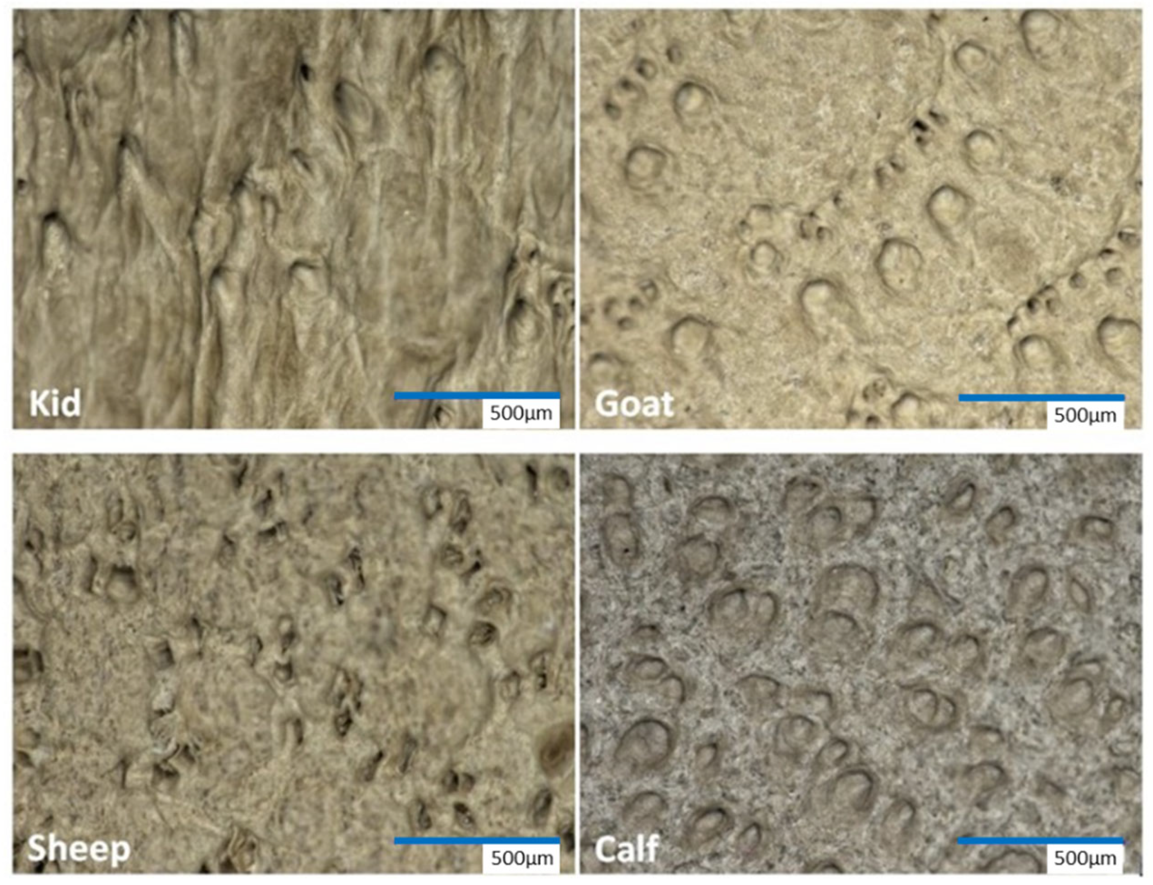
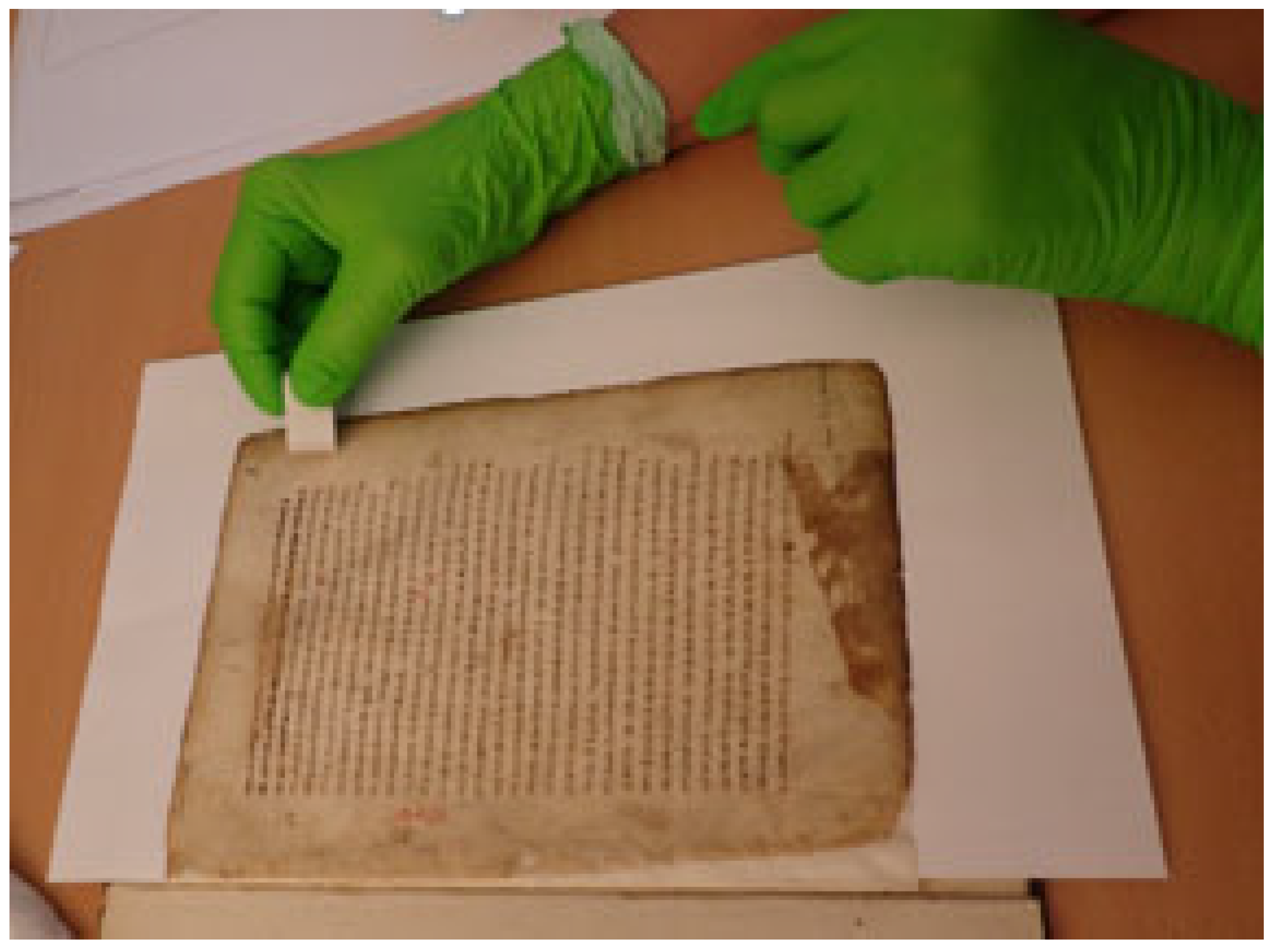
2. Visual and Microscopical Analyses

The first step when analyzing ancient parchment manuscripts is to perform a careful observation of them. Through a visual and microscopic inspection, it is possible to obtain the information about the production of the parchment and even sometimes the type of animal skin that was used by looking at the traces that were left during its manufacture. For example, cut marks that were left by the knife when one was skinning the animal, which can open up and create holes during the stretching of the skin on the frame, or even the marks from shaving it or the final surface treatment of the parchment [9]. The identification of the hair and flesh sides is also highly important as it relates to the different historical methods of construction [10].
The animal origin of the skins was previously identified by an evaluation of the size of it, its thickness, color, the levels of grease and the patterns of the follicles [11]. A detailed visual inspection using different types of microscopy and lighting sometimes can facilitate the identification of the animal skin patterns [12]. As described by Larsen et al. [13], variations in the distribution and frequency of the follicles (mammalian skin organs that produce hair) in the skins (Figure 1) that are used to produce parchments have served as a reference for the identification of the animal species since the early days of parchment production. However, the inspection requires a subjective user experience, which can lead to flaws being incorporated in the data, as a biological natural variation can lead to an incorrect identification. In addition, the follicle patterns are not always visible due to the surface treatments that were performed during the manufacture of parchment, and therefore, they cannot be used as an accurate or standard method of the species identification.
The microscopic level is defined as the structure that is observable under the light microscope, where the bundles of collagen fibers are visible. For this reason, microscopy has also been widely used to visualize the state of preservation of the parchment. Under the microscope, chemical and physical deterioration may be detected as frayed and fragmented fibers that may transform partly or fully into a sticky, jelly-like substance or gelatin during contact with water and heat. Visual changes such as getting transparent, cracking and the gelatinization of the fibers, can be correlated with the changes to the underlying structure of the collagen [13]. Several more sophisticated non-destructive microscopic techniques can be applied, thereby allowing researchers to deepen the investigation into the bulk of the material at a mesoscopic level. Polarized light microscopy can be used as a non-invasive technique, thereby allowing the identification of the gelatinized regions, layered or fibrous regions, the analysis of the lipid content, the observation of water diffusion and the detection of stress-induced patterns resulting from the preparation of the proteinaceous material [14]. For the assessment of the degradation level of the collagen, scanning electron microscopy (SEM) and micro-thermal analysis (micro-TA) can be applied [15,16]. SEM provides information on the overall morphology of the collagen fibers; micro-TA provides the thermo-mechanical information. Unfortunately, sample material is required for both of these techniques, which can be a limitation for such investigations due to the precious and unique character of the object. Parchment structural changes can, further be promoted by the presence of microorganisms that are present on the organic proteinaceous support. Optical microscopy and SEM are also applied for the identification and study of these microorganisms, which possibly can contaminate the parchment objects and cause the microbial degradation of the library materials [4,15,17]. As an example of the application of the SEM analyses in the parchment materials, Figure 2 reveals the microbial damage to the collagen fibers and the presence of characteristic cell chains that are typical of filamentous bacteria being on the surface of parchment from the Archimedes Palimpsest [18]. The SEM image shows that an outer layer of the parchment is still intact, but it is dotted with bacterial cells (mostly spore chains filaments that are smaller than one micron, which are likely to be actinomycetes). In the lower left corner of the image, the outer layer is eroded with collagen fibers, showing cavities and holes due to the occurrence of biological activity (collagenolytic exoenzymes can be deduced).
Another state-of-the-art technique is light transmission analysis (LTA), which is used to assess the damage that occurs to the native parchment collagen. The LTA equipment consists of a microscopy system that is used to register the images in polarized light, thus recording what happens during denaturation (i.e., shrinkage or loss of birefringence of the fibrous tissue). The images are then linked to the specific characteristics of the LTA curve [19], which is obtained during the experiment. The application of LTA for the analysis of collagen composite materials allows for the quantitative assessment of the state of integrity of the material by measuring its chemical stability through the hydrothermal denaturation temperature [19,20]. During the LTA analysis, the parchment fibers are heat-jellified in an aqueous solution, while a signal that is generated by light passing through the sample is recorded. The fluctuations of the signal during the transformation process are converted into a denaturation curve as a function of temperature. The curve is defined by specific peaks, which indicate the denaturation temperature and provide the semi-quantitative information on the chemical stability of the collagen pool. The higher the temperature that corresponds to the position of the peak is, then the higher the chemical stability is.
Recent studies have applied complementary analyses, including the LTA method, to investigate the phenomenon of purple stain biodeterioration on parchment [5,20,21,22]. These analyses have helped to highlight the nature of a microbial attack, which modifies the collagen populations throughout the biodegradation process. The results of this complementary approach pointed to a salt-mediated origin of the purple stain damage [20], revealed the presence of bacteriorhodopsin, and supported the presence of Halobacteria by detecting their DNA by using the high-throughput sequencing (NGS) technology (see Section 4.2). In addition, the LTA analysis revealed that the deterioration had spread to the highly structured collagen fibers over time.

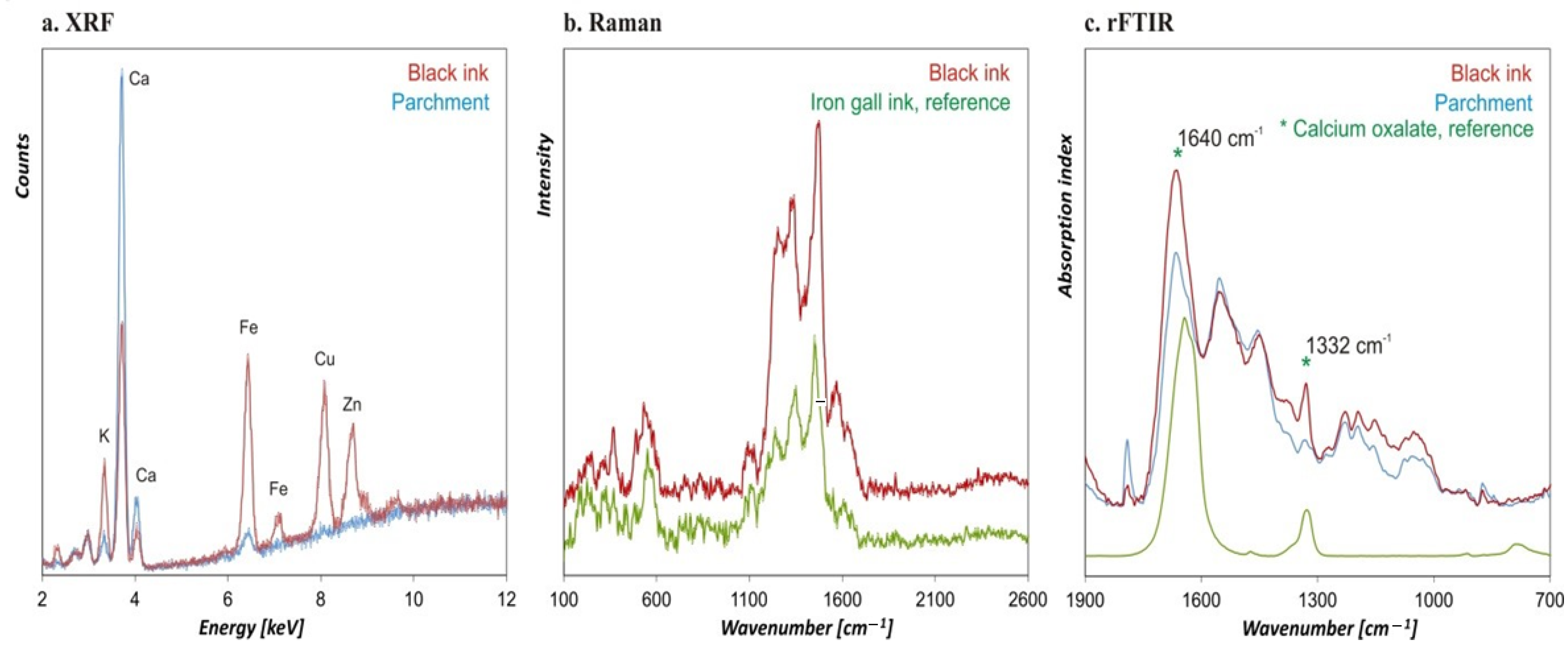
3. Physico-Chemical Analyses

The material analysis aims primarily to identify the composition of the support, the inks, pigments, dyes, and the binding media that were used for the manuscript production [23,24,25] as well as to detect the possible deterioration of the materials [26]. In order to obtain a complete picture of the composition and the preservation status of an object, it is important to correctly merge the results that are obtained by the use of the different analytical methods. Generally, the chemical-material analysis is based on the combination of three complementary spectroscopic methods: X-ray fluorescence analysis (XRF) for the detection of the chemical elements and Fourier Transform Infrared analysis (FTIR) as well as Raman spectroscopy, which are compound-specific methods [27]. The three analytical techniques have strengths and weaknesses [28], resulting on the one hand from the characteristic interactions of the applied radiation with the materials that are analyzed, the penetration depths of the radiation that is used, and on the other hand, from the design of the instruments (e.g., differences in the detectors or the different spot sizes). Therefore, their application is recommended to be performed in a complementary way [29]. The continuous progress in the development of suitable instrumentation for the analysis of cultural heritage items has led to the improvement and proposal of new devices, covering a broad area of materials and objects. The range goes from the study of colors and painting techniques, degradation processes, restoration practices, and the protection/degradation of objects of organic nature such as archaeological wood, paper, textile and proteinaceous materials such as parchment and leather. Moreover, the booming development of the analytical methods in recent decades has brought a great number of new instrumental micro-analytical techniques that are especially suitable for the study of membranous codices [30,31]. Transportable and handheld instruments allow, nowadays, not only the in situ investigations of the objects, but their sampling is not required anymore (thus, it is non-invasive). This means that there is no need for the original sample material to be taken.
The analytical approach starts by applying energy dispersive XRF in air, thereby yielding the qualitative and semi-quantitative results of the elements that are present in the materials. With this method, information about the class of the pigments and inks that were applied can be only deduced from the elements that are detected [32]. For example, the detection of sulfur (S) and mercury (Hg) in a red area leads, definitely, to the identification of the pigment vermilion (HgS). Another example would be the detection of copper (Cu) in a blue area, which points to the application of azurite, which is the only copper-based blue pigment. However, by applying XRF no compound-specific information can be obtained. Therefore, also in the case of calcium carbonate (CaCO3), no differentiation between the three modifications (crystallographic structures), calcite, vaterite, and aragonite, is possible. Moreover, artists were often mixing pigments with white to achieve a lighter hue. The mixtures of the inorganic pigments can hardly be completely characterized by elemental specific methods. The detection of lead (Pb) in a red paint layer indicates the presence of red lead (minium—lead oxide). In the case of possible mixture of minium with lead white (lead carbonate) the differentiation of the two materials cannot be clearly identified by just employing XRF. For this reason, a compound-specific analysis is necessary for the identification of the materials. FTIR and Raman spectroscopy have been employed widely in the field. Both techniques are carried out in air, and they are applicable in a non-invasive way.
The following are some relevant results that have been obtained from our recent studies of ancient Slavonic manuscripts. The investigations which were conducted on one of them, specifically the Kiev Folia, were performed directly in the Vernadsky National Library of Ukraine, and this included, first, the identification of the type of ink that was used in the text, which was iron gall ink [8]. This result was obtained by merging together the data of the XRF technique (showing a high number of counts in the quantity measure of elemental iron (Fe)) and Raman spectroscopy (Figure 3a,b). An FTIR analysis which was performed on the writings revealed the presence of calcium oxalates (Figure 3c), which were probably formed after the oxidative degradation of the ink components, which can occur due to a biological attack [15,17]. As described in more detailed in Section 4.2.2., as a result of this particular study, the microorganisms that were detected with the most plausible capacities to produce calcium oxalate precipitation were the species of the genera Staphylococcus and Streptococcus [8].
A high amount of calcium (Ca) was detected by the XRF method on the parchment folios. The complementarity of the approach revealed not only the presence of calcium carbonate (CaCO3) deriving from the manufacturing of the material, but also from the presence of calcium stearate (e.g., calcium soaps) which was probably formed during the depilation process by the reaction of the calcium in the lime bath and the lipidic content (subcutaneous fat) of the parchment [8]. This material was also found in another of the previously investigated Slavonic codices, the Codex Assemanianus. In fact, in this precious manuscript, calcium soaps and oxalates were also detected. Once again, further microbiological investigations were performed on this manuscript in order to better understand the conservation status of the item and support the hypothesis on the future risk of deterioration, as shown in the next sections [3]. Furthermore, all of the complementary methods that have been described and applied for the analysis of the mentioned manuscripts represent, in general, the optimal non-invasive tools for the identification of (I) inorganic and organic pigments that were used for illuminations in manuscripts (e.g., lapis lazuli, azurite, and indigo, lead white, vermilion, etc.), and extenders (e.g., chalk or kaolinite), (II) materials that were used for the preparation of the proteinaceous support (e.g., silicates due to the usage of pumice stone), as well as (III) substances that were applied during the later conservation processes (e.g., starch) [8,28,33].
Finally, it is important to emphasize that the combination of analytical techniques can render additional information about ancient manuscripts, as shown in the study of Bicchieri et al. [34]. The authors show how results that were obtained by scanning electron microscope imaging which was coupled with dispersive X-ray microanalysis, Raman microscopy and Fourier Transform Infrared Spectroscopy can reveal the presence of micro-objects, which are both inorganic and organic, and are associated with the parchment fibers or in the inks and dirt that have accumulated over the centuries between the pages. These observations can reveal important information about the manufacture of ancient documents, about their history or about the causes of ageing and deterioration of the materials which they contain. The authors also argue that some chemical components, which are normally attributed to the manufacture of materials or inks, may instead have a particular origin and nature, and therefore, lead, if they are not properly highlighted, to some misdiagnoses.

4. Molecular Analyses

Novel molecular analyses have been applied in recent years to the study of ancient parchment documentary materials. The choice of the molecular technique that is used depends to a large extent on the question that is posed. There are two types of molecular analysis which have been applied to the study of ancient parchment documentary materials. Those that focus on the proteins [6] and those focusing on the analysis of the genetic information that is contained in the parchment, which includes the ancient DNA (aDNA) of the original animal skin [1] and the DNA of the microorganisms that make up the microbiome of the parchment material [3].
Molecular analyses applied to cultural heritage have taken advantage of the enormous development in the field of so-called “omics” analyses [35,36], which now also offer suitable tools to decipher the biological information that is contained in the ancient parchments. These approaches allow for the analysis of all of the biomolecules (proteins and total DNA), thereby delivering a kind of “bio-archive” of the material that are investigated. In addition, this approach confers a more reliable quantitative analysis of the data. Nevertheless, the biggest challenge in the molecular analysis of the manuscripts and other objects of cultural heritage has been the development of non-invasive or minimal invasive sampling techniques [37]. These techniques include different tools that are aimed at obtaining the maximum amount of information with the minimum amount of invasiveness that is employed when one is studying the objects. The use of a destructive method is permissible only on the fragments that cannot undergo conservation or cannot be reunited (for example, the fragments from the margins of pages, the bore dust that is produced by insects and the parts that will certainly be eliminated during restoration such as parts of the binding or cover leaves). Especially in the case of the documentary material, the development of non-invasive sampling techniques allows access to previously unanalyzed documents with the permission of conservators and archivists. To this end, a minimally invasive method has recently been developed for the sampling of parchment using conventional PVC rubber bands [6,7]. Documents can be sampled in situ by gently rubbing the eraser across the surface of the material (Figure 4), and then, collecting the eraser fragments, without the need for abrasion or cutting. This sampling can easily be done by the conservators themselves without the need for specialized tools or equipment, and the sample can then be sent to the research laboratory for its analysis. Depending on the biocodicological analysis that is to be carried out (proteins or DNA), different amounts of eraser crumbs have to be collected, ranging from a 20–100 µL volume [6].

4.1. Protein Analysis and Applications

The study of proteins, or proteomics, is a powerful tool for the study of a parchment material, as it can provide not only a sensitive approach to characterize the protein composition of a given sample, but also to identify the species of the proteins themselves and thus, the protein-based material that was used to manufacture a specific object [38]. This molecular approach is gradually becoming a recognized method in archaeological research, as one of the major advantages of this technology is that it requires a small sample and it is less laborious and costly than DNA analysis is. This approach includes basic protein analyses or more in-depth proteomic analyses to identify the additional proteins on the surface of the parchment material [6].
Specifically, in the study of ancient parchment, a basic protein analysis has been successfully applied to determine the species of animal that was used in the parchment’s manufacture. This method is called peptide mass fingerprinting (PMF) and it uses enzymatic digestion to cleave the proteins at specific amino acid sites, thereby forming a peptide mixture. After the collection has been performed, each sample is chemically treated to allow for the extraction of collagen from the skin. Although all of animal skin contains collagen, the mixture of peptides in the collagen of each species is unique. Using mass spectrometry techniques, the molecular weights of the individual peptides in each sample can be determined, thereby allowing a peptide mass fingerprint (PMF) to be generated for each of them. The fingerprint of one particular protein that has been obtained using mass spectrometry can identify the species of the animal that it derived from [38].
Until recently, the protein analysis of parchment required its destructive sampling, which consequently severely limited the number of analyses that could be performed on a given document or on several documents in an archive. As an example, in the study by Toniolo et al. [39] on the 13th century “Marco Polo Bible”, the authors needed 5 mg of single-leaf parchment to identify that it was made of calfskin by the analysis of the peptide sequences. Thus, the same authors pointed out that the study was biased because more sheets could not be analyzed due to the destructive technique that was used and that it could have been made from different types of parchment, depending on what material was available at the time of its production. A further study by Kirby et al. [38] also showed the use of destructive sampling techniques to identify a Qur’an folio that is dated to the 9th c. as being made of sheep skin. Authors have also analyzed and compared some other objects that are made of animal skins, and they have stated that in all of the objects that were investigated, the ages and sample conditions had no significant effect on the resulting protein spectra and their subsequent identification. In order to avoid destructive techniques being used in the study of ancient manuscripts, Fiddyment et al. [7] went a step forward, using the non-invasive sampling method that is based on PVC polymer rubbers, which has the additional advantage of preserving the proteins in the PVC polymer residues at room temperature and without the need for the special storage of them. These authors performed a comparative analysis of the same sample using conventional MS techniques that require destructive sampling to occur, as well as the non-destructive electrostatic MS using the PVC eraser sampling method. They obtained the same, if not better, results with the PVC sampling than they did when they were using a real fragment of parchment. Thanks to the development of this non-invasive approach, the authors were able to extract the proteins from 513 parchment samples to resolve the animal origin of the suspected tissue paper-thin “uterine” vellum that was used in 13th century pocket Bibles and some additional parchment samples spanning this time period. The authors have found no evidence of the use of unexpected species, such as rabbit or squirrel, and they suggested that uterine vellum was the result of the technological production of it using the available animal resources, and that it would not have required unsustainable farming practices [7].
The ongoing development of proteomic techniques is enabling researchers to conduct more extensive studies to infer the animal origin of a multitude of parchment documents. Although, in principle, the selection of the skins seems to reflect the available livestock, and thus, the preferences of a geographical region, recent studies have deciphered that some animals were preferentially used for the production of certain types of documents. It has been shown that valuable books were made from a rather expensive calfskin, which was reserved for the highest quality books [7]. In contrast, Doherty et al. [40] analyzed 477 legal deeds from the sixteenth to the twentieth century in the United Kingdom using peptide mass fingerprinting, and they concluded that most of them were made from sheepskin. This was due not only to the abundance of sheepskin and the low cost of sheepskin parchment at this time in the UK [41], but also to the unique biological structure of sheepskin parchment, which consists of an inner layer of skin called the dermis. The authors conclude that the pre-modern writers preferred to use sheepskin parchment due to its inherent ability to make visible the fraudulent erasure of texts, which is attributed to the high fat content of the skin (30–50%) when it is compared to that of calfskin (2–3%) and goatskin (3–10%) [42,43]. The removing of the fat during the parchment-making process may cause the layers of sheepskin to detach more easily than those of other animals. In order to make fraudulent changes to the documents after signing them, the original text would have to be scraped off. This could cause the sheepskin parchment layers to separate, and it would leave a visible imprint on the document, thereby making the fraud easily noticeable [40].

4.2. DNA Analysis

DNA analyses have been applied for several years to recover the biological information that is contained in the ancient manuscripts on the parchment [44]. These analyses have been gradually developed to minimize the biases that are associated with destructive sampling in order to recover the genetic material that is contained in the parchment. Nevertheless, the amount of DNA that can be extracted from the parchment is generally low, and it may be degraded for a variety of reasons, such as the chemical processes that occur during its manufacture, the age of the parchment and the various alterations it may have undergone over the centuries due to the human manipulation or poor preservation of it. Several scientific publications have addressed this issue and all of them point to the need to develop accurate non-destructive and non-invasive sampling methodologies and precise DNA extraction protocols, as well as a careful DNA manipulation to avoid the occurrence of external contamination [1,2,45,46,47,48,49].
It is important to highlight that the field of biocodicology has experienced a unthinkable advance thanks to the development of next-generation sequencing (NGS) approaches, thus eliminating the intrinsic limitations of the methodologies that were used before NGS platforms became available. However, even before NGS analysis was widely used, numerous studies already highlighted the possibilities that are offered by genetic analysis of parchment [44]. Some of these studies focused only on ancient DNA (aDNA) for the sole purpose of identifying the animal skins that were used in the parchment production [46,48,50]. In contrast, other studies have focused on the microbiological research of manuscripts to identify the risk of potential damage that is exerted by microorganisms [17,18,51,52] as well as to establish restoration/conservation methods [53].
Nowadays, advances in non-invasive sampling techniques, together with the development of NGS approaches, are making it possible to investigate the genetic material of the parchment on a larger scale. These analyses are still far from being routinely applied on ancient parchment due to the laborious work that is required for sequencing techniques, but also due to the high price of such analyses, which can only be afforded within special research projects. Nevertheless, there are already some studies in which high-throughput NGS analyses coupled with bioinformatics have been applied to parchment manuscripts, thus allowing the researchers to identify the aDNA [2]. The compilation of these data has resulted in a specific microbiome for each investigated parchment or “bio-archive” [3,8,54], which can be considered to be a historical added value. The recovered biological information offers new possibilities to answer two main questions: (a) the type of skin/animal that was used to produce the parchments, and (b) the composition of the microbiome that is colonizing their surface, but also other relevant questions that are related to the manufacturing process of the book, its state of preservation and its historical use as well as the animal breeding history [1,2,3,5,6,8,20].
The workflow to answer these questions includes several steps that can be summarized as the use of non-invasive or minimally invasive sampling techniques which are followed by the DNA’s extraction, obtained directly from the sample material. This is a very delicate and difficult task that requires experience and care to be taken. There are strict criteria to ensure the reliability of the aDNA results. There are critical conditions for which the DNA extraction procedures can occur, such as it being performed in dedicated laboratories using special devices and equipment, and there are downstream steps that will depend on the selected sequencing platform. Primarily, the Illumina sequencing platform has been used for these analyses, as it is an ideal platform for sequencing short fragments of aDNA, it has good coverage and it shows a greater sensitivity and reliable results [1,55,56]. Bioinformatics analyses with different pipelines have also been developed to better analyze the huge dataset that results from this method [8]. Comparative analyses with DNA databases finally allow the identification of sequences and their affiliation to animals, bacteria, fungi, viruses and humans [2].

4.2.1. Identification of Animal Skins via DNA Analyses

One of the main applications of the genetic analysis of parchment is, and has been in the past, to discover the origin of the skins that were used for its manufacture. This involves the isolation of the ancient DNA (aDNA) that may still be preserved in the parchment. However, the alteration of the material throughout its history, including during its manufacture, preservation and analysis, affects the endogenous DNA of the parchment from the time when the skin was removed from the donor animal until it is extracted in the laboratory for the DNA analysis several hundred years later. When an organism dies, its DNA is generally degraded by endogenous nucleases, but chemical damage, such as oxidation or hydrolysis, also occurs. These processes lead to breaks in the DNA strands, the loss or alteration of the bases in the nucleotides, and the cross-linking of the strands [44]. Furthermore, microbial attack also contributes to the breakdown of the DNA molecules. However, it may be the case that under certain favorable circumstances, such as a low temperature, rapid desiccation, or high salt concentrations, the nucleases themselves may be destroyed or inactivated before all the nucleic acids are degraded [57,58,59]. The processes that were used in the manufacture of the parchment include some treatment conditions that may have allowed some of the DNA in the skin to remain intact [44], as previous research has suggested that both the mitochondrial and autosomal DNA may be well preserved in this material [1,45]. In fact, there is a compilation of research showing that ancient DNA can be detected in a variety of museum artefacts as well as biological and archival materials, including parchment [60].
As mentioned above, some of these studies were conducted before the NGS platforms were available and they used PCR-based assays using mitochondrial and/or nuclear markers for the identification of the animal skins that were used in the production of the parchment [46,48,50,61]. However, several shortcomings that are related to PCR-based assays have been documented, such as the control of the contamination risk, the high proportion of PCR chimeras and the questionable results that are obtained with the cross-contamination between individuals and species [48,61]. In addition, PCR facilitates the amplification of longer and less-damaged DNA fragments, thus it favours the modern-contaminating DNA over endogenous aDNA [1].
Next-generation sequencing (NGS) approaches allow a very thorough analysis of the ancient parchments to infer the proportion of the endogenous DNA (species of origin) that is retained in the different samples as well as its possible contamination with other animal species. Teasdale et al. [1] performed a first shotgun NGS study on parchment, and they showed that this material is a very usable substrate for aDNA recovery and analysis. They were able to retrieve 7–9% of the endogenous ovine genome from two of the investigated parchment documents dating from the 17th and 19th centuries at a high-quality and estimated-to-be-comparable or an even lower external contamination rate than that which was obtained from aDNA that was extracted from other materials. They assumed that the alignment rates of the non-source species were probably due to the homology between the genomes of the ruminants, as well as the damage that occurred to the aDNA molecules, resulting in a higher alignment with the non-source species. In a further study, Teasdale et al. [2] investigated samples that were derived from the 1000-year-old bifolia of the York Gospels, and they estimated that there was an average percentage of endogenous DNA of 19.3% (range 0.7–51.4%) in all of the samples. However, these calculations fell in the range of 0.2–5.7% when filtering for the quality of the mapping reads was applied. Later, Piñar et al. [3] investigated the animal origins of two ancient Slavonic codices dating back to the 11th century, and they found that the proportion of endogenous DNA was less than 0.25%. This proportion is relatively low, but it should be noted that the authors carried out an extensive filtering of the sequences that were obtained. The loss of the mapped reads occurs after the filtering process is performed, and this decrease indicates that there may be a bias in the preservation and/or recovery of the DNA sequences from the different manuscripts, depending on their state of conservation. This highlights the critical necessity to standardize the protocols for comparing metagenomic studies, including not only DNA sequencing protocols, but also bioinformatics downstream analyses, such as data filtering as well as comparable pipelines for the processing and statistical analysis of the data.
Recent studies that were carried out by our working group on ancient Slavonic codices have been able to identify the animal origin of the skins that were used for the manufacturing of several manuscripts [3,8] and, in particular, the latest investigated item: the Glagolita Clozianus (shown in this paper in Table 1). Our studies show that these ancient codices were made from animals that were available in the region of the origin of the manuscripts and that different folios of the same codex could be made from the skins of different animals [3]. Here, we show for the first time the data that were obtained from our latest analysis of the Glagolita Clozianus, an Old Church Slavonic codex in the Glagolitic script from the 11th century, consisting of a total of 14 leaves. Two of them are preserved in the library of the “Landesmuseum Ferdinandeum” in Innsbruck (Austria) and these are the ones that were investigated, and we reveal that they are made of sheepskin. We observed that there is a high proportion of human DNA (Table 1) in all of the investigated manuscripts, wherein the number of sequences that aligned only with the human genome were higher compared to those of the host animal, which could indicate the more recent origin of these sequences due to the ongoing human manipulation of the manuscripts, which is also showed by Teasdale et al. [2].
Finally, further applications of high-throughput sequencing techniques on parchment documents have been used to determine whether the different fragments of parchment belong to the same document through the genetic analysis of their aDNA, and this was the case in a study that was performed on the New Zealand’s founding document [56] or to show the unification of the scrolls from distinct geological locations, as Anava et al. [62] showed in their study about the Dead Sea Scrolls.

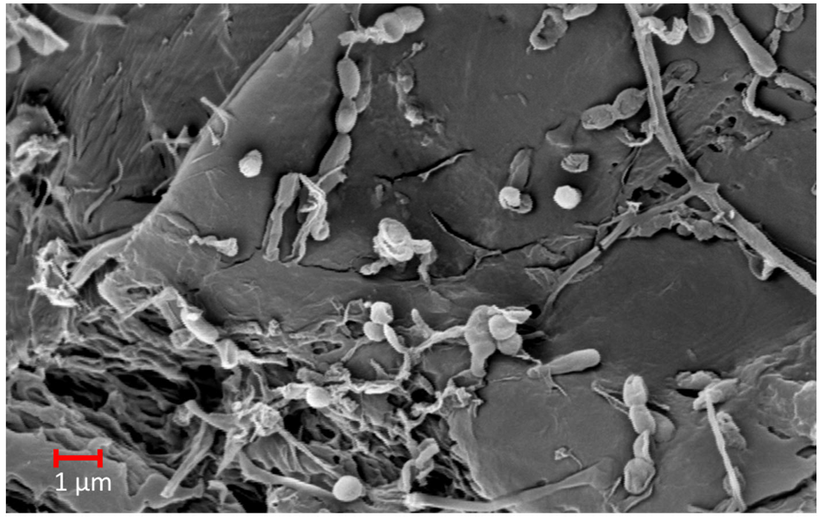
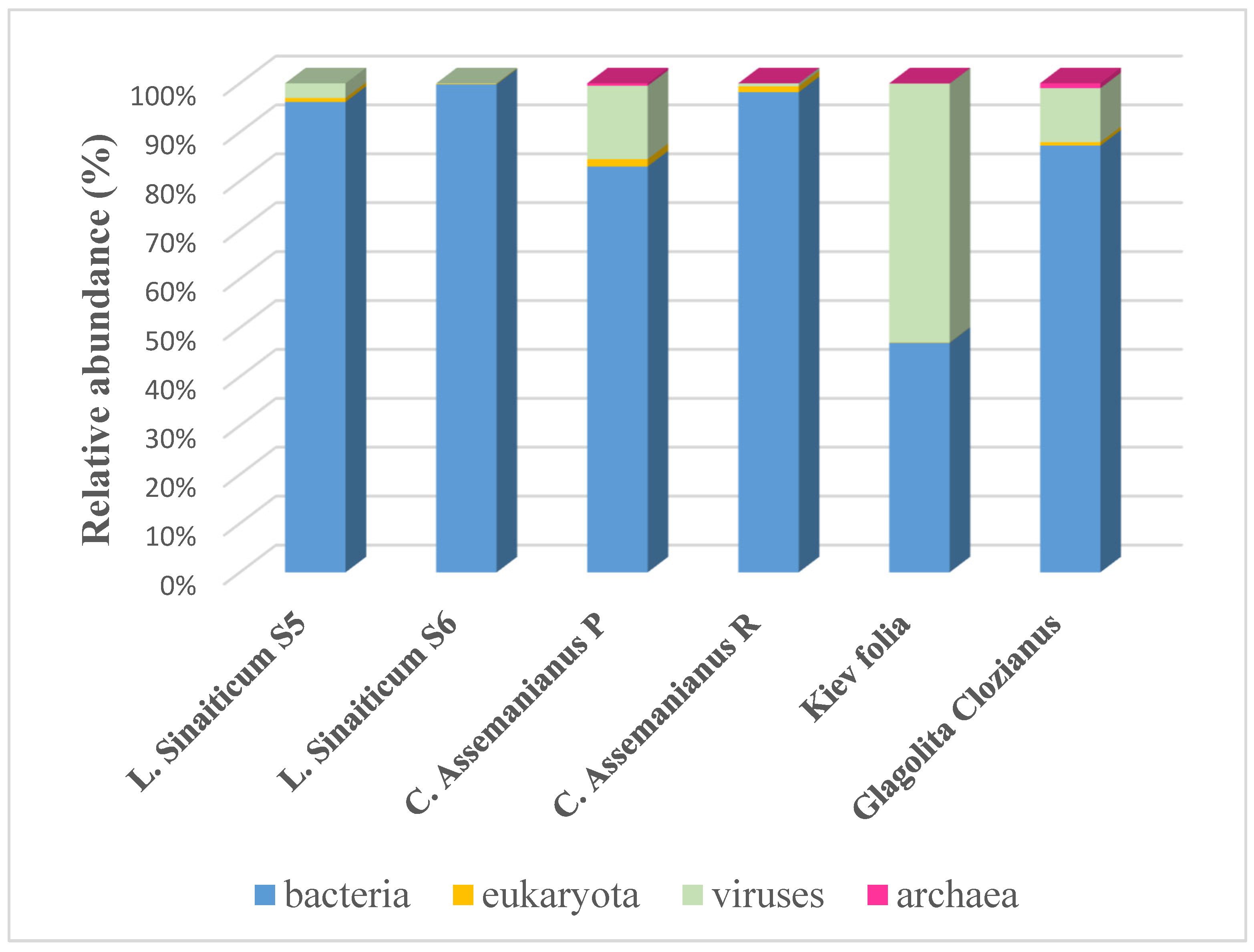
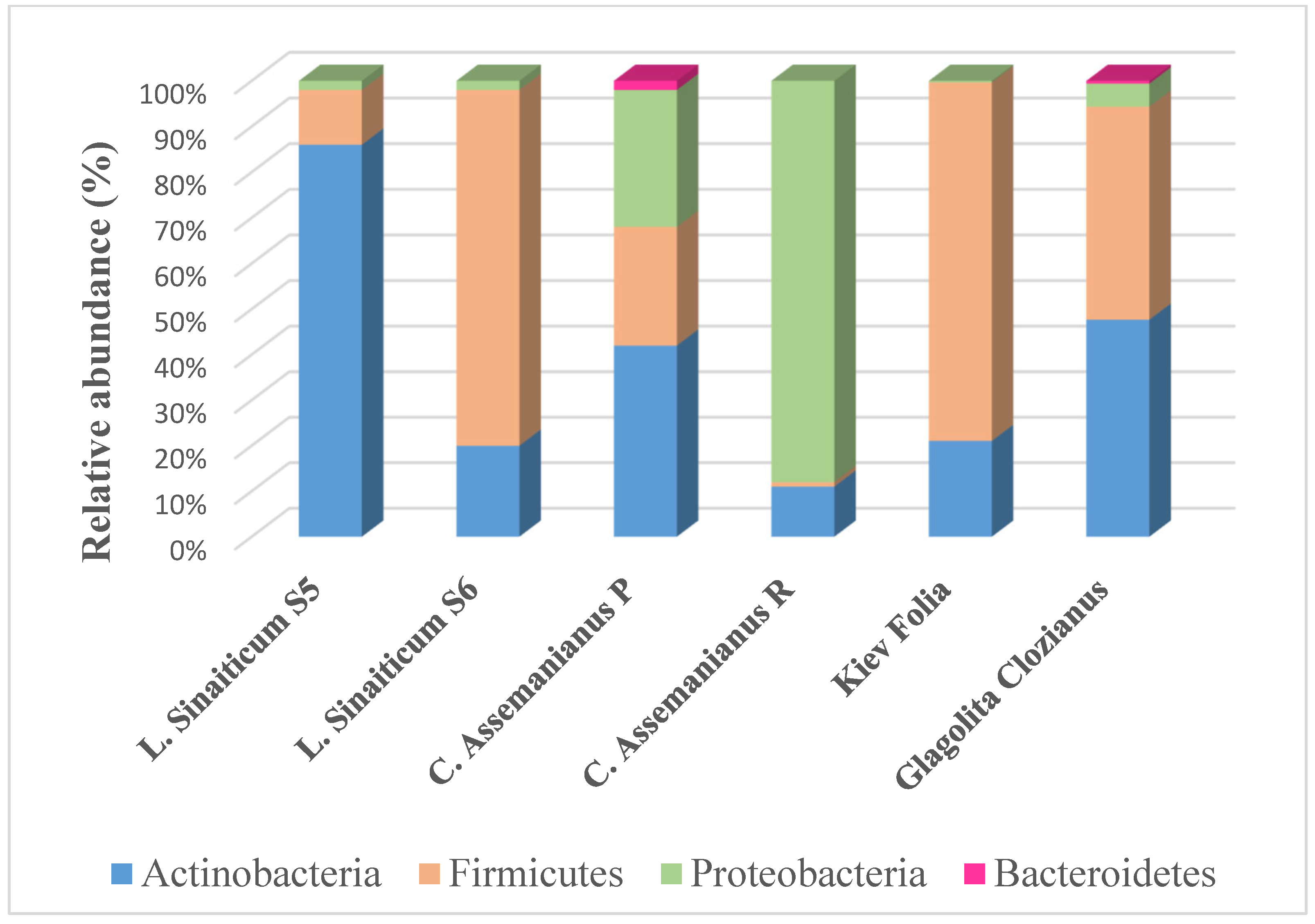
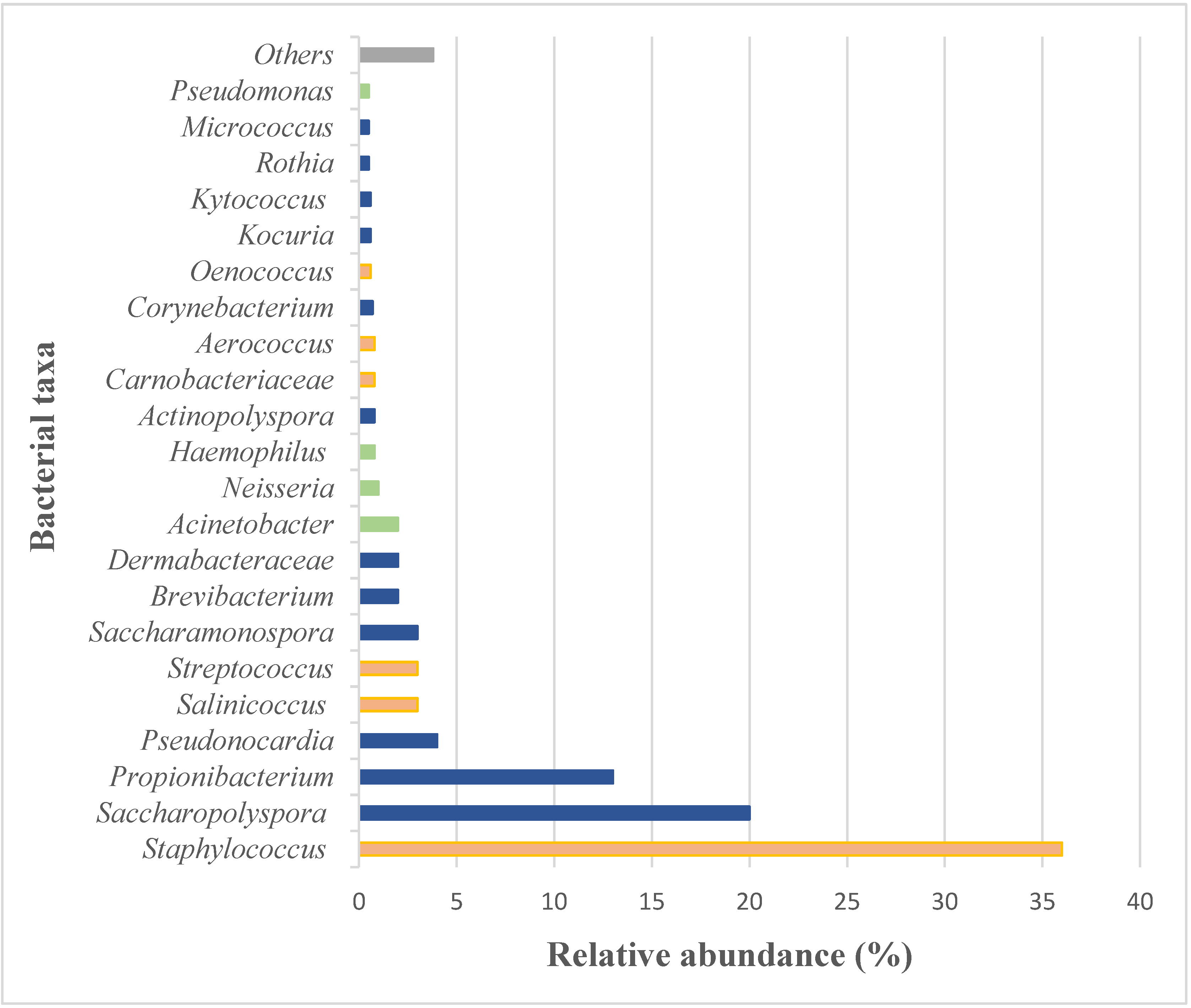
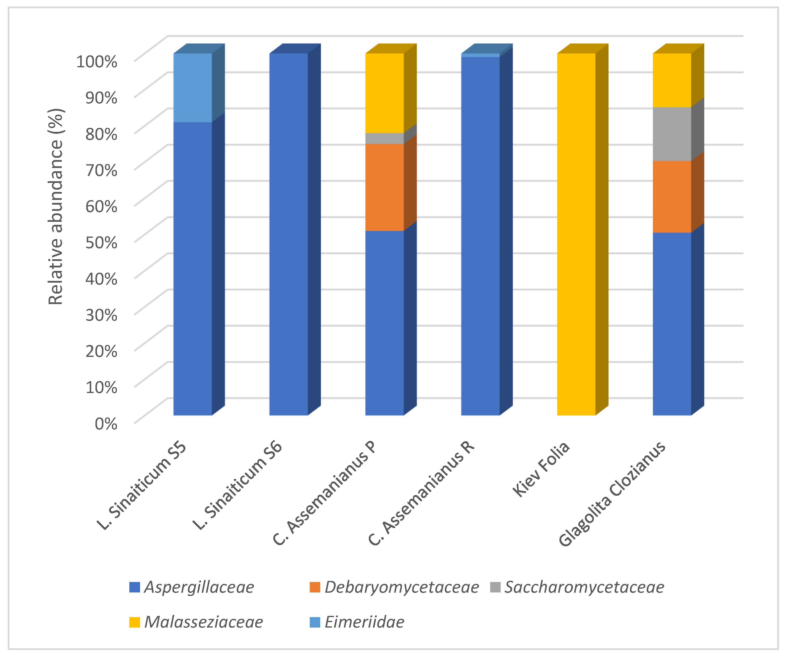
4.2.2. Microbiome Analyses and Applications

Since the technological development of the next-generation sequencing (NGS) techniques, in particular metagenomics, some more comprehensive studies have been published that allow for a very complete analysis not only of the animal origin of the parchment, but also of its microbiome [2,3,5,8,20,21]. The microbiome is the community of microorganisms (such as fungi, bacteria and archaea) and even viruses that exists in a given environment. In the specific case of parchments, it is the total community of microorganisms that has inhabited, or is inhabiting, the surface of the document. However, the question is: does a parchment have a specific microbiome? Reviewing the studies that have been carried out so far by molecular methods, we can say that the parchment possesses a core microbiome, which reveals on the one hand, the history of its manufacture and, on the other hand, the history of the use of the documents.
Metagenomics using target sequencing have been applied on parchments to reveal that halophilic/halotolerant archaea and bacteria are the main players in the parchment microbiome [5,20]. However, surprisingly, viruses have also been detected in relatively high proportions in the studies where non-targeted sequencing or the so-called “shotgun approach” has been used [2,3,8]. This last approach involves the sequencing of the total extracted DNA, without focusing on any target sequence, which allows the simultaneous identification of both the raw material (animal skin) that was used for the manufacture of the manuscript as well as the different life domains that make up the total microbiome inhabiting the object.
In this context, Figure 5 shows the microbiomes that were obtained in our latest studies on the ancient Slavonic codices, which are mentioned above, by shotgun sequencing. In all of the samples that were analyzed, it was possible to determine a dominance of bacteria, an unexpected relevance of viruses and a low relative abundance of eukaryota and archaea. As Figure 5 shows, a great advantage of performing shotgun sequencing is the visualization of the microbiome members with their real proportions in the community. However, despite these advantages, to date, there have been very few studies using this approach to study the microbiomes of parchments [2,3,8].

Prokaryotic Community in the Microbiome

The prokaryotic community that has been identified so far by molecular approaches in the parchment microbiome includes members that can be grouped into two main groups: those that are typical of a saline environment and those that are typical of the human microbiome.
The first group comes into contact with the parchment at a very early stage, i.e., during the manufacturing process [5,20,21,63]. As mentioned in the Introduction section, since ancient times, the salting of animal skins has been used wherever salt was affordable and available to prevent the skins from putrefying [64]. Salting was carried out either dry or in brine tanks, where the skins were left for several days. During the salting process, NaCl gradually penetrated the skin’s inner surface, thereby making the parchment’s environment extremely salty [65,66]. In addition, the salt that was used also introduced marine microorganisms that are present in natural sea salt, such as halophilic archaea and halotolerant bacteria, and it acted as a kind of culture/enrichment medium for these microorganisms [5]. Halophilic/halotolerant microorganisms were therefore, the “pioneers” in the colonization of parchments, as only these microorganisms can thrive in such extreme salinity conditions. In fact, Migliore et al. [5,20] proposed a model of microbial succession in which the halophilic archaea would be the first colonizers of the parchment, but subsequently, as the humidity increased and salinity decreased, these microorganisms would gradually disappear and give way to the growth of halotolerant bacteria, such as some groups of Actinobacteria, Proteobacteria, but also Firmicutes, which could grow consuming the haloarchaeal remains. In fact, this model fits very well with the observations that other authors have made and with our own results. In the data that were obtained from our studies on the old Slavonic documents, we could still detect traces of the pioneer colonizers, i.e., archaea, in all the documents except for L. Sinaiticum (see Figure 5). The retrieved sequences showed that they are affiliated with the family Halobacteriaceae, specifically with the genus Halobacterium and Halococcus [3,8] (also detected in the Glagolita Clozianus in this study). The low proportion of archaea in the samples may be due to the limited availability of the ancient DNA strands when assessing the colonization process from a dynamic microbial succession perspective. As Perini et al. [22] explain in their review, a metagenome analysis provides a two-dimensional representation of the colonization process on the parchment, which, in contrast, occurred in a three-dimensional perspective, including the layering of the generations of different microbes over the centuries. In this way, the most recent colonizers quantitatively outnumber the older ones, and they are easily detectable due to the well-preserved state of their DNA. Some of these more recent colonizers are halotolerant bacteria [5,20]. Our results also agree with these observations, and they show the same phyla that were mentioned by these authors in the microbial succession on the parchments (see Figure 6), but, in addition, we could detect members of the phylum Bacteroidetes in two of the samples that were analyzed, although in a very low proportion. The members of this last phylum were also detected in some of our previous studies which were devoted to ancient parchment manuscripts [51].
Figure 6 shows that the relative proportions of the above-mentioned phyla can vary in the different manuscripts that were investigated, even in the samples that were taken from different folios of the same codex, suggesting that this may reflect different stages in the microbial succession in each sampled specimen. As mentioned above, one part of the secondary colonizers of parchments are still the halotolerant bacteria belonging to the phyla Actinobacteria, Firmicutes and Proteobacteria. Regarding the Actinobacteria, the members of the order Pseudonocardiales have been suggested as being the dominant ones [5,20], with some of the identified genera being Saccharopolyspora, Saccharomonospora and Pseudonocardia [2,3,8,51] (which were also detected in Glagolita Clozianus in this study, see Figure 7). However, also, some species belonging to the genera Nocardiopsis, such as N. xinjiangensis and N. kunsanensis, which are tolerant to salt, alkalis and desiccation [67,68] and Actinopolyspora and species of Yaniella, as Y. halotolerans have been identified as being part of the core microbiome of the parchments [3,51].
Regarding the Firmicutes, some of the halotolerant members of the genera Gracilibacillus, Halobacillus [3,51] and Salinicoccus [8] have been identified in previous studies on parchments, as well as in the Glagolita Clozianus in this study (see Figure 7), but also of the genus Staphylococcus [2,3,8]. Specifically, the species S. equorum exhibits a high salt tolerance (up to 25% (w/v)), and it has been shown to be part of the core microbiome of several investigated parchment manuscripts [3,8] (and in this study, see Figure 7).
Several studies have also demonstrated the presence of Proteobacteria in the parchment microbiome, with most of the sequences being related to the Gammaproteobacteria class. Migliore et al. [5] reported on the halotolerant aquatic or marine Gammaproteobacteria in a study that was conducted on a scroll from the Vatican Secret Archives. The authors identified a high proportion of members of the order Vibrionales, but also relative proportions of Alteromonadales, Aeromonadales, Oceanospirillales and Chromatiales. Among them, the most frequent genera were: Aliivibrio, Allomonas, Photobacterium and Vibrio, but also, there were Aeromonas, Halomonas, Marinomonas, Pseudoalteromonas, Rheinheimera and Shewanella. In addition, the same authors reported on a relative high proportion of the order Pseudomonadales in this scroll. The presence of Proteobacteria has also been reported in our studies on the ancient Slavonic documents (see Figure 6), where we also detected the dominance of Gammaproteobacteria. However, the genera we found do not match the saline bacteria that were found by Migliore et al. [5]. Instead, we found sequences that are related to the genera Pantoea, Enhydrobacter, Pseudomonas, Acinetobacter and Haemophilus in our previous studies [3,8], as well as in the study of the Glagolita Clozianus (this study, see Figure 7). Some of these genera were also identified in other studies by molecular means [5,18], as well as cultivated from parchments in studies that were conducted by other authors [52,69]. The members of the Alphaproteobacteria and Betaproteobacteria have been also identified in parchments, but with lower proportions, with species of the genera Bartonella, Brevundimonas and Neisseria [3,5] (the last one also detected in this study, see Figure 7).
It is important to note that some of the halotolerant bacteria that were identified in the parchments pose a potential risk for this material. As some examples, the species of the genus Nocardiopsis produce a large number of bioactive compounds and excrete extracellular enzymes such as proteases and keratinases, which facilitate the utilization of the proteins in nature [70]. The species of Gracilibacillus are able to produce proteolytic enzymes that are capable of hydrolyzing gelatin [71,72], and elastases [73], thus cleaving the elastin, which together with collagen, determines the mechanical properties of the tissues. Staphylococcus equorum exhibits efficient enzymatic activities, including proteases and lipases [74,75]. Additionally, other species, such as Yaniella halotolerans and Halobacillus sp. are able to produce hydrolases [76,77]. Finally, Acinetobacter spp. are considered to have a great destructive potential for this material [8,52]. Nevertheless, the most intriguing finding is the repeated identification of Saccharopolyspora species in almost all of the ancient parchment manuscripts that have been analyzed by several research groups and using different molecular techniques, such as clone libraries and DGGE [52] as well as NGS using the target [5,20] or the shotgun approach [2,3,8] (and this study, see Figure 7). Saccharopolyspora has previously been described as a common microbial culprit that is detected in numerous ancient parchment documents, showing the so-called ‘measles-like’ parchment discoloration, consisting of purple spots that are linked to localized collagen damage and parchment deterioration [51]. However, later, Migliore et al. [20] documented the presence of the archaea Halobacterium salinarum in decayed documents that displayed the same phenomenon of purple spots. Furthermore, through a Raman analysis, the authors confirmed the presence of bacteriorhodopsin, which is the main photosynthetic protein of the archaeal H. salinarum, together with bacterioruberin, which is also produced by some of the species of Actinobacteria. In view of these findings, they postulated that the phenomenon of purple spots is triggered in the parchment initially by the haloarchaea and at a later stage of microbial succession, actinobacteria and fungi are implicated in the damage that occurs due to their ability to attack the collagen in depth. This assumption does not contradict the findings that were made by Piñar et al. [51], where the authors postulated that actinobacteria, specifically Saccharopolyspora, together with the Aspergillus species were responsible for the purple stains, but rather, these two assumptions reveal the two distinct stages of microbial succession leading to the same phenomenon of the biodeterioration of the parchment. A very similar phenomenon is the so-called “red heat”, which is a typical red coloration found as pitting on the flesh-side surface of salted hides after their storage during the leather manufacturing process. Some authors have recently confirmed that both, the presence of purple spots and the red heat, depends on the pioneer colonization of H. salinarum, thereby producing the red staining due to bacteriorhodopsin [21]. The sea salt that was used for the leather curing is responsible for the transmission of the halobacteria to the hides and skins.
The second group of prokaryotes that was found in the parchment microbiome are the secondary colonizers [5], and their distribution depends on the individual history of each parchment, including the specific environment where the parchment was stored (i.e., libraries, archives and churches) and its handling, history of use and the customs of the time [2,3,8]. Recent studies have shown that part of these secondary colonizers are typical bacteria of the human microbiome [2], and their presence on the parchment agrees with the fact that the skin microbiome is the most common component of the urban microbiome [78], and in particular, on handled surfaces. These bacteria belong mainly to two phyla, Actinobacteria and Firmicutes. Within the Actinobacteria, the most dominant group are the species of Propionibacterium, mainly P. acnes, but also of the genera Rothia, Corynebacterium and Brevibacterium as well as members of the family Dermabacteraceae [2,3,8], (and in this study, see Figure 7). Within the phylum Firmicutes, mainly the species of the genera Staphylococcus and Streptococcus, but also Leuconostoc, Lactobacillus and Gemella as well as the members of the family Carnobacteriaceae have been identified [2,3,8] (and this study, see Figure 7). It is important to highlight that most of the human microbial markers that have been found on parchments are characteristic of the microbiome of the skin and the oral [8] and nasal cavity [2], which points to the history of use of the parchments that have been investigated. The bacteria that were detected not only indicate the intensive handling of the parchments over the centuries, but also to the usage and devotional customs and habits of the past, such as the custom of kissing prayer books during religious ceremonies, which would explain the large number of bacteria that are typical of the oral cavity being present on the surface of religious books [8]. Nevertheless, these data should be taken with caution, as many of these bacteria, on the one hand, are potential human pathogens and, on the other hand, they may also be involved in the deterioration of the parchment. To name a few examples, it has been suggested that some Staphylococcus and Streptococcus species may be involved in the formation of calcium oxalates on parchment [8]. Additionally, the members of the genus Brevibacterium have been repeatedly detected in the parchment microbiome [2,3,69] (and this study, see Figure 7). Although the identification of them at species level was not always possible, in some cases B. linens was detected [3]. This species is abundant on human skin, but it is also well known for its ability to tolerate high salt concentrations. In addition, it produces carotenoid pigments and proteases, which are capable of damaging parchment [79], all together, posing a severe risk to the integrity of the parchment documentary material.

Eukaryotic Community in the Microbiome

The most recent studies using shotgun metagenomics have shown that eukaryotic microorganisms represent only a small proportion of the microbiome (typically below 2%) of parchment [2,3,8] (and this study, see Figure 5). This observation contrasts with those of the previous studies in which only some target regions of DNA were amplified and sequenced (18S rRNA and/or ITS sequences for the target amplification of fungi) to investigate the specific groups of microorganisms that are on the parchment [17,51], highlighting the greater accuracy of the shotgun approach to study the actual proportions of the different domains of life in the microbiomes.
As an example of the application of metagenomics with a shotgun approach, Figure 8 shows our previous and current studies on the ancient Slavonic manuscripts. The figure reveals that the small proportion of eukaryotic microorganisms in the microbiome of the investigated parchments are related, in most of the case studies, to the phylum Ascomycota, mainly to the families Aspergillaceae, Debaryomicetaceae and Saccharomycetaceae [2,3] (and in this study, see Figure 8) and they are specifically affiliated to sequences of the genera Penicillium, Debaryomyces and Saccharomyces, respectively (data not shown). In addition, the members of the phylum Basidiomycota have also been identified in three of our case studies [3,8], (and in Glagolita Clozianus in this study), with all of them being related to the Malassezia species. Finally, in two of the manuscripts that we have investigated, the Eimeria species belonging to the family Eimeriidae (phylum Apicomplexa) were identified instead [3].
Despite the low overall proportion of the eukaryotic microorganisms that were found in these ancient Slavonic manuscripts, the data that were recovered may also provide some interesting insights into the raw material that was used for the manufacture of some of the parchments which were investigated. For example, some of the sequences that were identified in two of the Slavonic manuscripts (L. Sinaiticum 5 and C. Assemanianus R, see Figure 8) are members of the family Eimeriidae (phylum Apicomplexa). The Apicomplexa are a diverse group of intracellular parasitic protists, which are responsible for widespread diseases in domestic animals [80]. The genus Eimeria, which was detected in these ancient manuscripts, comprises several species that are capable of causing coccidiosis in animals such as poultry, cattle and small ruminants, including sheep and goats [81]. The finding of DNA traces of this parasite on these two parchment manuscripts, which are made from sheepskin, may indicate that animals which were in poor health were used to manufacture the parchment.
However, most of the eukaryotic taxa that were identified raise a red flag about the state of the preservation of the investigated manuscripts. As shown in Figure 8, the majority of the eukaryotic sequences were related to fungi, which belong mainly to Ascomycota, and specifically to the members of the family Aspergillaceae. This finding is in line with the previous studies in which the members of this family, such as Penicillium and Aspergillus species, were repeatedly identified on deteriorated parchments [15,18,51,82,83]. In addition, members of the genus Penicillium have been found to exhibit a marked level of proteolytic activity on parchments [52,69]. Fungi are considered to be second colonizers of parchments [5,20], and their presence is linked to the individual history of their storage and the use of the parchments over the centuries. However, these microorganisms can be very active participants in the deterioration of parchments [17,84,85], and their detection indicates that there is a high latent risk of the deterioration of the investigated parchment if the environmental conditions become suitable for their germination.
Finally, the members of the Basidiomycota were also identified by NGS analysis using our shotgun approach (see Figure 8), with all of the members being related to the family Malasseziaceae. Malassezia, the only genus that was detected, occurs naturally on the skin surface of many animals, including humans [86]. Most of the Malassezia species are unable to synthesize fatty acids and therefore, they possess a set of gene-encoding lipases and phospholipases that are responsible for the release of fatty acids from various lipid compounds, such as those that are made by the sebaceous glands of the host skin [87]. The capture of these fatty acids and their further utilization in various lipid biosynthesis pathways is essential for the growth of Malassezia species [88]. For this reason, in our previous study on the Kiev Folia [8], we pointed out that the detection of Malassezia sequences could be related to the calcium soap that was detected in this codex and distributed throughout the different folia (see Section 3).

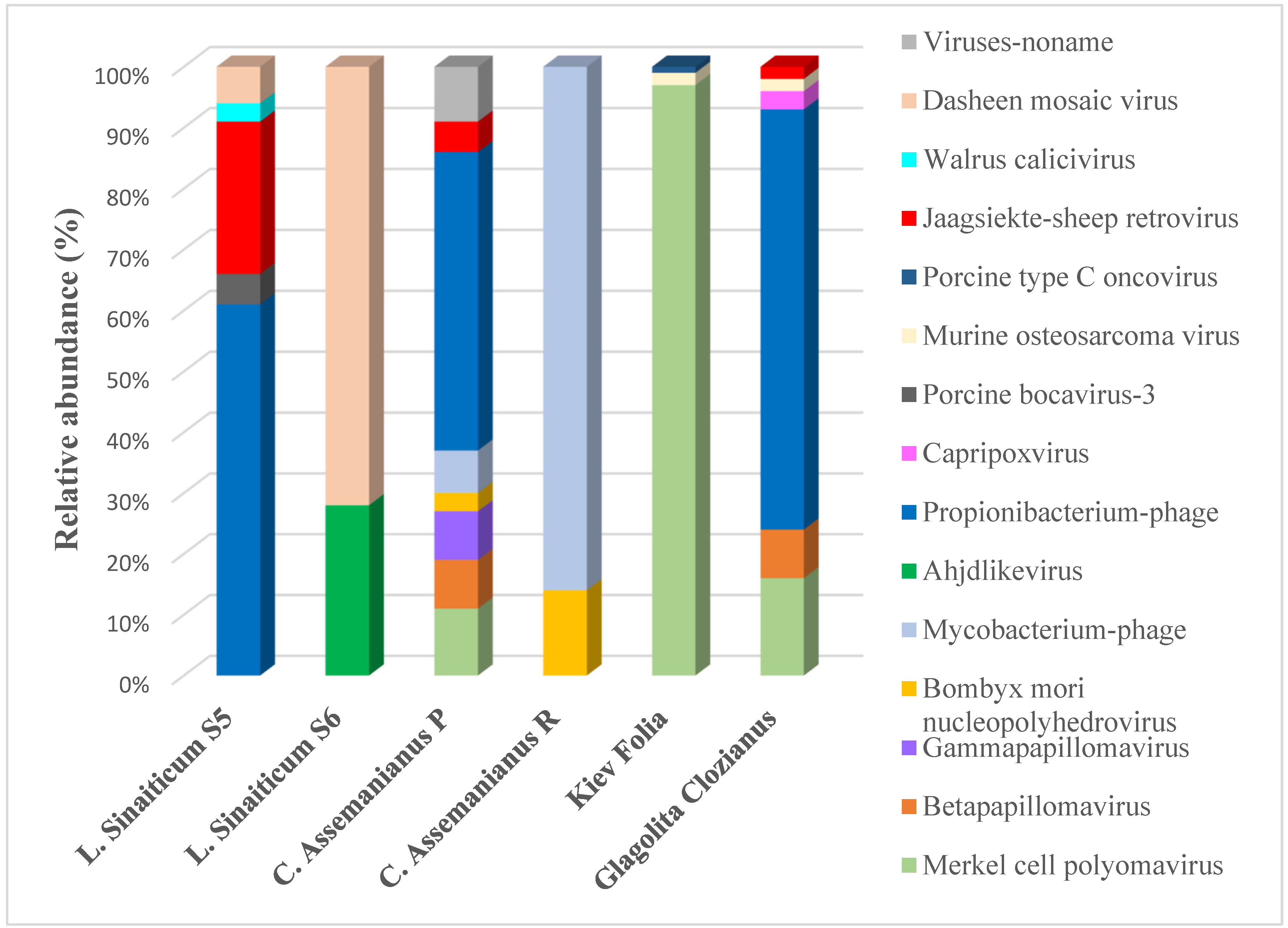
Viruses in Parchments

Until recently, viruses have not been investigated as an integral part of the microbiome of ancient parchment manuscripts. This is simply because they had previously been overlooked due to the use of technologies that did not allow for their detection, i.e., only cultivation or sequencing of DNA target regions. However, the few recent studies that have employed the shotgun NGS approach have revealed that viruses can account for a significant part of the parchment microbiome [2,3], with proportions of them that are as high as 50% of the sequences that have been obtained [8] (see Figure 5). Although these data are still very preliminary and require more advanced bioinformatics analyses, they indicate a new future direction in the emerging field of biocodicology.
Teasdale et al. [2] already showed the presence of viruses in the samples from the York Gospels, but they did not analyze these sequences in depth, showing only that some of them belonged to the Siphoviridae group. This observation has been confirmed in our studies on the ancient Slavonic parchment manuscripts, where the Siphoviridae group was widely represented in some of the manuscripts, such as the L. Sinaiticum S5, C. Assemanianus P and the Glagolita Clozianus, specifically with sequences being related to the Propionibacterium-phage (see Figure 9). This indicates a direct relationship with the bacteria that were found in these manuscripts, where Propionibacterium was detected in a relatively high proportion in their microbiomes [3,8] (and in Glagolita Clozianus in this study, see Figure 7), and this indicates that part of the viruses that were found are bacteriophages, i.e., they prey on the bacteria that colonize the parchment.
Figure 9 shows that diversity and proportion of the viruses can be very different in the investigated Slavonic manuscripts, which can also reveal the differences in their history of use. Besides the bacteriophages, a considerable proportion of the viruses that were found are partially members of the “virobiota”, i.e., viruses that are commonly found in healthy humans [89]. This is the case of the Merkel cell polyomavirus (MCV), which was widely distributed in some of the parchments, especially in the Kiev folia [8]. This virus causes most of the cases of Merkel cell carcinoma, a type of rare skin cancer [90]. However, it is also found on healthy skin [91]. The presence of the MCV on the surface of some of the parchments is most likely due to its close contact with human skin, considering its widespread manipulation throughout the centuries.
However, can viruses reveal any other information about the parchments beyond this? Our previous and current results suggest that they can, in fact, reveal or support important information about this material. Some of the viruses that were detected in our shotgun NGS analyses on the ancient Slavonic manuscripts provide fascinating information that confirms our conclusions about the source of the skins and even unveils the possible origins of some of the inks that were used for the elaboration of the manuscripts [3]. This was the case for the detection of the Jaagsiekte sheep retrovirus (JSRV) [92], which causes a contagious lung cancer in sheep, known as ovine pulmonary adenocarcinoma (OPA). JSRV has an endogenous counterpart, which is inserted in all of the sheep genomes, with about 27 copies of endogenous retroviruses (enJSRVs) per genome. Endogenous JSRVs have several functions in the development of domestic sheep, as they help in blocking the replication cycle of JSRV and have a key involvement in the maturation of the sheep embryo [93]. This virus was found in the L. Sinaiticum S5, the C. Assemanianus P [3] and the Glagolita Clozianus (this study, see Figure 9), all having been confirmed to be manufactured with sheepskin (see Table 1). The detection of JSRV in these manuscripts, through the application of the shotgun DNA analyses, suggests that what we have identified is the homologous endogenous sequences of the virus that are inserted in the genome of the sheep, which perfectly supports that the investigated folios were made from sheepskin. We could not detect this virus in the virobiota of the C. Assemanianus sample R, which was also identified as being from a sheep, but it can be explained by the lower retrieval of the genetic material from this sample due to the non-invasive sampling that was applied in this folio using erasers [3]. This virus was not detected in the samples of L. Sinaiticum S6 [3] and Kiev Folia [8], both of which were shown to be manufactured, fully and partially, respectively, from calfskin (see Table 1). The discovery of ovine retroviruses in the samples that were identified a priori as sheepskins opens an interesting perspective that could be further used to investigate the population genetics in the parchment materials by the performance of further bioinformatic analysis [94].
Another interesting case was the detection of the Dasheen mosaic virus (DsMV) in the microbiomes of the two samples that were investigated from the L. Sinaiticum (see Figure 9) [3]. This is a single-stranded RNA virus, belonging to a group of plant pathogenic viruses, specifically to the genus Potyvirus, which naturally infects the Taro plant (Colocasia esculenta) [95]. The detection of this ssRNA virus in a DNA sequencing analysis was explained by its integration into the genome of the host plant, Taro [3]. All types of viruses can become endogenous by integrating the viral copy sequences into the genomes of various host organisms. These sequences are generally referred to as endogenous viral elements (EVEs) [96]. The integration of potyvirus sequences into host plant genomes is well known [97], and it may be the only plausible explanation for this finding. However, the detection of DsMV in this codex led to an intriguing hypothesis through a literature search. Taro is known to have been cultivated in ancient Egypt, where it was an important crop plant [97]. At this time, this plant was mainly used for the consumption of edible leaves and corms, but also for its high concentration of anthocyanin [98]. Bicchieri et al. [99] previously described that some inks that were used for parchment illustration were composed of anthocyanins which were extracted from different plants, through a process in which the organic dye was precipitated with an inert binder, usually a metallic salt. Thus, could this be an indication that some of the inks that were used in this codex were composed of anthocyanin extracted from Taro? Well, one hypothesis to explain the detection of the DsMV in the Liturgiarium Sinaiticum could be that the Taro plant, which was very common in ancient Egypt, was used to extract the anthocyanins for the local production of the inks. In fact, previous studies on this codex, using X-ray fluorescence analysis (XRF) in a non-destructive and non-invasive manner, led to the conclusion that an organic dye had been applied for the yellow and a blue pigment [100]. Although only a hypothesis, the discovery of the Dasheen mosaic virus could indirectly support the previous theory, which some philologists suggest, that this codex was composed and written in the same place where it was found, namely in the monastery of St. Catherine in Sinai, Egypt [101], by using the materials that were available in this area.
Finally, the discovery of Capripoxvirus in our last investigated codex, the Glagolita Clozianus (see Figure 9), is also noteworthy. Capripoxvirus is a genus of viruses in the family Poxviridae, and it is among the most severe of all of the animal poxviruses. Sheep, goats and cattle are their natural hosts, where these viruses cause damage to their skins and wool [102,103,104,105,106,107]. The genus consists of three species: sheep pox virus (SPPV), goat pox virus (GTPV) and lumpy skin disease virus (LSDV). Unfortunately, it was not possible to identify this virus at the species level, which would have confirmed once again that this codex was made from sheepskin. Nevertheless, this virus could be an indication of the health status of the sheep that was used for the manufacture of the foil under investigation.
To our knowledge, our studies on the ancient Slavonic parchments are the first to analyze the viruses on this material in detail. The discovery of these viruses, which are taken as examples, illustrates the multiple applications and possibilities that are offered by shotgun metagenomics in the field of biocodicology for future studies and perhaps a new field of interest in the parchment “virobiota”.

5. Conclusions

In this review, we have shown the importance of applying a complementary analytical approach to the study of cultural heritage materials, in particular, parchment. Such a comprehensive investigation of the material composition of an object can be applied to: (a) assign an object to a particular historical context, (b) determine the correctness or incorrectness of the stated provenance, (c) explore the technology that was used for the preparation of it. Furthermore, the analytical research methods also provide important information about the state of the preservation and the possible deterioration reactions, which the human eye would not otherwise detect. The latest trend is to complement the analytical methods with biocodicological analyses (i.e., the study of the biological information that is contained in the parchment), which is opening up a new dimension in terms of the information that can be obtained by analyzing its proteins or its genetic material. This biological information is an added value or “individual bio-archive” that can help to infer not only the animal origin of the skins that were used for the manufacture of the parchments, but also their microbiome. The microbiome of the parchment is very specific, and it reveals, on the one hand, the history of its manufacture and, on the other hand, the history of the use of the documents. Metagenomic studies have shown that the microbiome is typically composed of halophilic/halotolerant microorganisms, which are the primary colonizers of this material, which can be considered in a way as an extreme environment due to its manufacturing process. Secondly, the secondary colonizers are directly related to the storage and use history of the documents and thus, most of the microorganisms are typical of the human microbiome. Finally, the recent discovery and investigation of the viruses on these parchments opens an interesting perspective and possibly a new direction of the research in biocodicological studies.

Supplementary Materials

The following supporting information can be downloaded at: https://www.mdpi.com/article/10.3390/app122010479/s1, Supplementary File S1. Methods employed with the new codex analysed in this study: the Glagolita Clozianus, which includes Figure S1. Workflow of quality control and pre-processing of the obtained reads using the Kneaddata pipeline v0.7.3.

Author Contributions

Conceptualization, G.P., F.C. and K.S.; analytical measurements, F.C. and W.V.; molecular analysis, G.P.; writing—original draft preparation, G.P. and F.C.; writing—review and editing, G.P., F.C., W.V., M.S., H.M. and K.S.; funding acquisition, H.M. All authors have read and agreed to the published version of the manuscript.

Funding

This work was funded by the Austrian Science Fund (FWF) project no. P29892 (The Origin of the Glagolitic-Old Church Slavonic Manuscripts).

Institutional Review Board Statement

Not applicable.

Informed Consent Statement

Not applicable.

Data Availability Statement

All data are available in public databases.

Acknowledgments

The authors express their gratitude to the Centre of Image and Material Analysis in Cultural Heritage (CIMA, www.cima.or.at) in the framework of which this article was composed. We thank Flavia Pinzari for her permission to publish an image that she produced (Figure 2) in this review. We would like to thank the “Tyrolean Landesmuseum Ferdinandeum” conservators for their availability and for the sampling of the Glagolita Clozianus. Finally, authors thank the VBCF NGS Unit (www.viennabiocenter.org/facilities) for sequencing and bioinformatic analyses.

Conflicts of Interest

The authors declare no conflict of interest.

References

  1. Teasdale, M.D.; van Doorn, N.L.; Fiddyment, S.; Webb, C.C.; O’Connor, T.; Hofreiter, M.; Collins, M.J.; Bradley, D.G. Paging through history: Parchment as a reservoir of ancient DNA for next generation sequencing. Philos. Trans. R. Soc. B Biol. Sci. 2015, 370, 20130379. [Google Scholar] [CrossRef] [PubMed]
  2. Teasdale, M.D.; Fiddyment, S.; Vnouček, J.; Mattiangeli, V.; Speller, C.; Binois, A.; Carver, M.; Dand, C.; Newfield, T.P.; Webb, C.C.; et al. The york gospels: A 1000-year biological palimpsest. R. Soc. Open Sci. 2017, 4, 170988. [Google Scholar] [CrossRef] [PubMed] [Green Version]
  3. Piñar, G.; Tafer, H.; Schreiner, M.; Miklas, H.; Sterflinger, K. Decoding the biological information contained in two ancient Slavonic parchment codices: An added historical value. Environ. Microbiol. 2020, 22, 3218–3233. [Google Scholar] [CrossRef] [PubMed]
  4. Pinzari, F.; Gutarowska, B. Extreme Colonizers and Rapid Profiteers: The Challenging World of Microorganisms That Attack Paper and Parchment. In Microorganisms in the Deterioration and Preservation of Cultural Heritage; Joseph, E., Ed.; Springer: Cham, Switzerland, 2021. [Google Scholar] [CrossRef]
  5. Migliore, L.; Thaller, M.C.; Vendittozzi, G.; Mejia, A.Y.; Mercuri, F.; Orlanducci, S.; Rubechini, A. Purple spot damage dynamics investigated by an integrated approach on a 1244 A.D. parchment roll from the Secret Vatican Archive. Sci. Rep. 2017, 7, 1–12. [Google Scholar] [CrossRef]
  6. Fiddyment, S.; Teasdale, M.D.; Vnouček, J.; Lévêque, É.; Binois, A.; Collins, M.J. So you want to do biocodicology? A field guide to the biological analysis of parchment. Herit. Sci. 2019, 7, 35. [Google Scholar] [CrossRef] [Green Version]
  7. Fiddyment, S.; Holsinger, B.; Ruzzier, C.; Devine, A.; Binois, A.; Albarella, U.; Fischer, R.; Nichols, E.; Curtis, A.; Cheese, E.; et al. Animal origin of 13th-century uterine vellum revealed using noninvasive peptide fingerprinting. Proc. Natl. Acad. Sci. USA 2015, 112, 15066–15071. [Google Scholar] [CrossRef] [Green Version]
  8. Cappa, F.; Piñar, G.; Brenner, S.; Frühmann, B.; Wetter, W.; Schreiner, M.; Engel, P.; Miklas, H.; Sterflinger, K. The Kiev Folia: An interdisciplinary approach to unravelling the past of an ancient Slavonic manuscript. Int. Biodeterior. Biodegrad. 2022, 167, 105342. [Google Scholar] [CrossRef]
  9. Fuchs, R. Des Widerspenstigen Zähmung-Pergament in Geschichte und Struktur. In Pergament–Geschichte, Struktur, Restaurierung, Herstellung; Rück, P., Ed.; J. Thorbecke: Sigmaringen, Germany, 1991; ISBN 3799542027. [Google Scholar]
  10. Bischoff, F.M. Pergamentdicke und Lagenordnung. Beobachtung zur Herstellungstechnik Helmarshausener Evangeliare des 11. und 12. Jahrhunderts. In Pergament–Geschichte, Struktur, Restaurierung, Herstellung; J. Thorbecke: Sigmaringen, Germany, 1991; pp. 97–144. [Google Scholar]
  11. Ryder, M.L. Parchment—its history, manufacture and composition. J. Soc. Arch. 1960, 2, 391–399. [Google Scholar] [CrossRef]
  12. Alvarez, A.M.F.; Bouhy, J.; Dieu, M.; Charles, C.; Deparis, O. Animal species identification in parchments by light. Sci. Rep. 2019, 9, 1–10. [Google Scholar] [CrossRef]
  13. Larsen, R.; Poulsen, D.V.; Odlyha, M.; De Groot, J.; Wang, Q.; Wess, T.; Hiller, J.; Kennedy, C. Damage assessment of parchment: Complexity and relations at different structural levels Giuseppe Della Gatta, Elena Badea and Admir Mas Soghomon Boghosian. In Proceedings of the ICOM-CC 14th Triennial Meeting, The Hague, The Netherlands, 12–16 September 2005; pp. 199–208. [Google Scholar]
  14. Vilde, V.; Fourneau, M.; Charles, C.; Van Vlaender, D.; Bouhy, J.; Poumay, Y.; Deparis, O. Use of Polarised Light Microscopy to Improve Conservation of Parchment. Stud. Conserv. 2019, 64, 284–297. [Google Scholar] [CrossRef]
  15. Pinzari, F.; Colaizzi, P.; Maggi, O.; Persiani, A.M.; Schütz, R.; Rabin, I. Fungal bioleaching of mineral components in a twentieth-century illuminated parchment. Anal. Bioanal. Chem. 2012, 402, 1541–1550. [Google Scholar] [CrossRef]
  16. De Groot, J.; Odlyha, M.; Bozec, L.; Horotn, M.; Masic, A.; Coluccia, S. Damage assessment of parchment by micro-thermal analysis and scanning electron miscroscopy. In Proceedings of the ICOM-CC 14th Triennial Meeting, Hague Volume II, The Hague, The Netherlands, 12–16 September 2005; pp. 759–765. Available online: http://www.bcin.ca/Interface/openbcin.cgi?submit=submit&Chinkey=238589 (accessed on 10 July 2022).
  17. Sterflinger, K.; Pinzari, F. The revenge of time: Fungal deterioration of cultural heritage with particular reference to books, paper and parchment. Environ. Microbiol. 2012, 14, 559–566. [Google Scholar] [CrossRef]
  18. Piñar, G.; Sterflinger, K.; Ettenauer, J.; Quandt, A.; Pinzari, F. A Combined Approach to Assess the Microbial Contamination of the Archimedes Palimpsest. Microb. Ecol. 2015, 69, 118–134. [Google Scholar] [CrossRef] [Green Version]
  19. Cicero, C.; Mercuri, F.; Paoloni, S.; Orazi, N.; Zammit, U.; Glorieux, C.; Thoen, J. Integrated adiabatic scanning calorimetry, light transmission and imaging analysis of collagen deterioration in parchment. Thermochim. Acta 2019, 676, 263–270. [Google Scholar] [CrossRef]
  20. Migliore, L.; Perini, N.; Mercuri, F.; Orlanducci, S.; Rubechini, A.; Thaller, M.C. Three ancient documents solve the jigsaw of the parchment purple spot deterioration and validate the microbial succession model. Sci. Rep. 2019, 9, 1–13. [Google Scholar] [CrossRef] [Green Version]
  21. Perini, N.; Mercuri, F.; Thaller, M.C.; Orlanducci, S.; Castiello, D.; Talarico, V.; Migliore, L. The stain of the original salt: Red heats on chrome tanned leathers and Purple spots on ancient parchments are two sides of the same ecological coin. Front. Microbiol. 2019, 10, 2459. [Google Scholar] [CrossRef] [Green Version]
  22. Perini, N.; Mercuri, F.; Orlanducci, S.; Thaller, M.C.; Migliore, L. The Integration of Metagenomics and Chemical Physical Techniques Biodecoded the Buried Traces of the Biodeteriogens of Parchment Purple Spots. Front. Microbiol. 2020, 11, 598945. [Google Scholar] [CrossRef]
  23. Cappa, F.; Fruehmann, B.; Schreiner, M. Raman Spectroscopy for the Material Analysis of Medieval Manuscripts. In Nanotechnologies and Nanomaterials for Diagnostic, Conservation, and Restoration of Cultural Heritage; Lazzara, G., Fakhrullin, R., Eds.; Elsevier: Amsterdam, The Netherlands, 2018; pp. 127–147. ISBN 9780128139103. [Google Scholar]
  24. Frühmann, B.; Cappa, F.; Vetter, W.; Schreiner, M. Father Petrus Multianalytical approach for the analysis of the Codices Millenarius Maior and Millenarius Minor in Kremsmuenster Abbey, Upper Austria. Herit. Sci. 2018, 6, 1–12. [Google Scholar] [CrossRef] [Green Version]
  25. Vetter, W.; Frühmann, B.; Cappa, F.; Schreiner, M. Materials and techniques used for the “Vienna Moamin”: Multianalytical investigation of a book about hunting with falcons from the thirteenth century. Herit. Sci. 2021, 9, 1–17. [Google Scholar] [CrossRef]
  26. Cappa, F.; Schreiner, M.; Zoleo, A. Preliminary Studies of Parchment and Ink Degradation in Medieval Manuscripts promoted by Light and Heat. In Ink Corrosion–the Missing Links; Berger, Ferdinand Verlag: Horn, Austria, 2021. [Google Scholar]
  27. Vetter, W.; Schreiner, M.; Frühmann, B.; Cappa, F. Three complementary non-invasive methods applied to historical manuscripts. In Manuscript cultures 11, Proceedings of the ‘Second International Conference on Natural Sciences and Technology in Manuscript Analysis’ at the University of Hamburg, Hamburg, Germany, 13–14 June 2018; University of Hamburg: Hamburg, Germany; pp. 97–108. Available online: https://www.csmc.uni-hamburg.de/publications/mc/mc11.html (accessed on 6 July 2022).
  28. Cappa, F.; Sterflinger, K. Non-Invasive Physico-Chemical and Biological Analysis of Parchment Manuscripts–An Overview. Restaur. Int. J. Preserv. Libr. Arch. Mater. 2022, 43, 127–142. [Google Scholar] [CrossRef]
  29. Doherty, B.; Daveri, A.; Clementi, C.; Romani, A.; Bioletti, S.; Brunetti, B.; Sgamellotti, A.; Miliani, C. The Book of Kells: A non-invasive MOLAB investigation by complementary spectroscopic techniques. Spectrochim. Acta-Part A Mol. Biomol. Spectrosc. 2013, 115, 330–336. [Google Scholar] [CrossRef] [PubMed]
  30. Badillo-Sanchez, D.; Chelazzi, D.; Giorgi, R.; Cincinelli, A.; Baglioni, P. Characterization of the secondary structure of degummed Bombyx mori silk in modern and historical samples. Polym. Degrad. Stab. 2018, 157, 53–62. [Google Scholar] [CrossRef]
  31. Magistro, F.; Majolino, D.; Migliardo, P.; Ponterio, R.; Rodriquez, M.T. Confocal Raman spectroscopic study of painted medieval manuscripts. J. Cult. Herit. 2001, 2, 191–198. [Google Scholar] [CrossRef]
  32. Eastaugh, N.; Walsh, V.; Chaplin, T. Pigment Compendium: A Dictionary of Historical Pigments; Elsevier: Oxford, UK, 2004. [Google Scholar]
  33. Thompson, D.V. The Materials and Techniques of Medieval Paintings; Dover Publ.: New York, NY, USA, 1956. [Google Scholar]
  34. Bicchieri, M.; Biocca, P.; Colaizzi, P.; Pinzari, F. Microscopic observations of paper and parchment: The archaeology of small objects. Herit. Sci. 2019, 7, 47. [Google Scholar] [CrossRef] [Green Version]
  35. Marvasi, M.; Cavalieri, D.; Mastromei, G.; Casaccia, A.; Perito, B. Omics technologies for an in-depth investigation of biodeterioration of cultural heritage. Int. Biodeterior. Biodegrad. 2019, 144, 104736. [Google Scholar] [CrossRef]
  36. Gutarowska, B. The use of -omics tools for assessing biodeterioration of cultural heritage: A review. J. Cult. Herit. 2020, 45, 351–361. [Google Scholar] [CrossRef]
  37. Piñar, G.; Sterflinger, K. Natural sciences at the service of art and cultural heritage: An interdisciplinary area in development and important challenges. Microb. Biotechnol. 2021, 14, 806–809. [Google Scholar] [CrossRef]
  38. Kirby, D.P.; Buckley, M.; Promise, E.; Trauger, S.A.; Holdcraft, T.R. Identification of collagen-based materials in cultural heritage. Analyst 2013, 138, 4849–4858. [Google Scholar] [CrossRef]
  39. Toniolo, L.; D’Amato, A.; Saccenti, R.; Gulotta, D.; Righetti, P.G. The Silk Road, Marco Polo, a bible and its proteome: A detective story. J. Proteom. 2012, 75, 3365–3373. [Google Scholar] [CrossRef]
  40. Doherty, S.P.; Henderson, S.; Fiddyment, S.; Finch, J.; Collins, M.J. Scratching the surface: The use of sheepskin parchment to deter textual erasure in early modern legal deeds. Herit. Sci. 2021, 9, 4–9. [Google Scholar] [CrossRef]
  41. Clark, G. Labour productivity in English agriculture, 1300–1860. In Land, Labour and livestock: Historical Studies in European Agricultural Productivity; Manchester University Press: Manchester, UK, 1991; pp. 211–235. [Google Scholar]
  42. Koppenhoefer, R.M. The lipids of sheepskins. I. Lipid of fresh sheepskin. J. Am. Leather. Chem. As. 1938, 15, 33–203. [Google Scholar]
  43. Palop, R. Sheepskin and Cattle Hide Degreasing; Cromogenia Linits: Barcelona, Spain, 2016. [Google Scholar]
  44. Bower, M.A.; Campana, M.G.; Checkley-Scott, C.; Knight, B.; Howe, C.J. The potential for extraction and exploitation of DNA from parchment: A review of the opportunities and hurdles. J. Inst. Conserv. 2010, 33, 1–11. [Google Scholar] [CrossRef]
  45. Burger, J.; Hummel, S.; Herrmann, B. Palaeogenetics and cultural heritage. Species determination and STR-genotyping from ancient DNA in art and artefacts. Thermochim. Acta 2000, 365, 141–146. [Google Scholar] [CrossRef]
  46. Poulakakis, N.; Tselikas, A.; Bitsakis, I.; Mylonas, M.; Lymberakis, P. Ancient DNA and the genetic signature of ancient Greek manuscripts. J. Archaeol. Sci. 2007, 34, 675–680. [Google Scholar] [CrossRef]
  47. Campana, M.G.; Bower, M.A.; Bailey, M.J.; Stock, F.; O’Connell, T.C.; Edwards, C.J.; Checkley-Scott, C.; Knight, B.; Spencer, M.; Howe, C.J. A flock of sheep, goats and cattle: Ancient DNA analysis reveals complexities of historical parchment manufacture. J. Archaeol. Sci. 2010, 37, 1317–1325. [Google Scholar] [CrossRef]
  48. Pangallo, D.; Chovanova, K.; Makova, A. Identification of animal skin of historical parchments by polymerase chain reaction (PCR)-based methods. J. Archaeol. Sci. 2010, 37, 1202–1206. [Google Scholar] [CrossRef]
  49. Lech, T. Ancient DNA in historical parchments-Identifying a procedure for extraction and amplification of genetic material. Genet. Mol. Res. 2016, 15. [Google Scholar] [CrossRef]
  50. Vuissoz, A.; Worobey, M.; Odegaard, N.; Bunce, M.; Machado, C.A.; Lynnerup, N.; Peacock, E.E.; Gilbert, M.T.P. The survival of PCR-amplifiable DNA in cow leather. J. Archaeol. Sci. 2007, 34, 823–829. [Google Scholar] [CrossRef]
  51. Piñar, G.; Sterflinger, K.; Pinzari, F. Unmasking the measles-like parchment discoloration: Molecular and microanalytical approach. Environ. Microbiol. 2015, 17, 427–443. [Google Scholar] [CrossRef] [Green Version]
  52. Lech, T. Evaluation of a parchment document, the 13th century incorporation charter for the city of Krakow, Poland, for microbial hazards. Appl. Environ. Microbiol. 2016, 82, 2620–2631. [Google Scholar] [CrossRef]
  53. Vadrucci, M.; Borgognoni, F.; Cicero, C.; Perini, N.; Migliore, L.; Mercuri, F.; Orazi, N.; Rubechini, A. Parchment processing and analysis: Ionizing radiation treatment by the REX source and multidisciplinary approach characterization. Appl. Radiat. Isot. 2019, 149, 159–164. [Google Scholar] [CrossRef]
  54. Piñar, G.; Sclocchi, M.C.; Pinzari, F.; Colaizzi, P.; Graf, A.; Sebastiani, M.L.; Sterflinger, K. The Microbiome of Leonardo da Vinci’s Drawings: A Bio-Archive of Their History. Front. Microbiol. 2020, 11, 1–22. [Google Scholar] [CrossRef]
  55. Krause, J.; Briggs, A.W.; Kircher, M.; Maricic, T.; Zwyns, N.; Derevianko, A.; Pääbo, S. A Complete mtDNA Genome of an Early Modern Human from Kostenki, Russia. Curr. Biol. 2010, 20, 231–236. [Google Scholar] [CrossRef] [Green Version]
  56. Shepherd, L.D.; Whitehead, P.; Whitehead, A. Genetic analysis identifies the missing parchment of New Zealand’s founding document, the Treaty of Waitangi. PLoS ONE 2019, 14, e0210528. [Google Scholar] [CrossRef]
  57. Paabo, S. Ancient DNA: Extraction, characterization, molecular cloning, and enzymatic amplification. Proc. Natl. Acad. Sci. USA 1989, 86, 1939–1943. [Google Scholar] [CrossRef] [Green Version]
  58. Pääbo, S.; Poinar, H.; Serre, D.; Jaenicke-Després, V.; Hebler, J.; Rohland, N.; Kuch, M.; Krause, J.; Vigilant, L.; Hofreiter, M. Genetic analyses from ancient DNA. Annu. Rev. Genet. 2004, 38, 645–679. [Google Scholar] [CrossRef] [Green Version]
  59. Hofreiter, M.; Serre, D.; Poinar, H.N.; Kuch, M.; Pääbo, S. Hofreiter_Ancient_NatRevGen_2001_1556040. Nat. Commun. 2001, 2, 3–9. [Google Scholar]
  60. Green, E.J.; Speller, C.F. Novel substrates as sources of ancient DNA: Prospects and hurdles. Genes 2017, 8, 180. [Google Scholar] [CrossRef] [Green Version]
  61. Campana, M.G.; Robinson, T.; Campos, P.F.; Tuross, N. Independent confirmation of a diagnostic sheep/goat peptide sequence through DNA analysis and further exploration of its taxonomic utility within the Bovidae. J. Archaeol. Sci. 2013, 40, 1421–1424. [Google Scholar] [CrossRef]
  62. Anava, S.; Neuhof, M.; Gingold, H.; Sagy, O.; Munters, A.; Svensson, E.M.; Afshinnekoo, E.; Danko, D.; Foox, J.; Shor, P.; et al. Illuminating Genetic Mysteries of the Dead Sea Scrolls. Cell 2020, 181, 1218–1231.e27. [Google Scholar] [CrossRef]
  63. Migliore, L.; Perini, N.; Alabiso, A. May metagenomics disclose the hidden secrets of the ancient damaged parchments? In Proceedings of the IMEKO TC4 International Conference on Metrology for Archaeology and Cultural Heritage, Online, 22–24 October 2020; pp. 590–593. [Google Scholar]
  64. Reed, R. The Nature and Making of Parchment; The Elemen: Leeds, UK, 1975. [Google Scholar]
  65. Bailey, D.G. The preservation of hides and skins. J. Am. Leath. Chem. Ass 2003, 98, 308–319. [Google Scholar]
  66. Hernàndez, E.; Marmer, W.; Kolomazník, K.; Cooke, P.; Dudley, R. Mathematical model of raw hide curing with brine. J. Am. Leath. Chem. Ass 2008, 103, 167–173. [Google Scholar]
  67. Chun, J.; Bae, K.S.; Moon, E.Y.; Jung, S.O.; Lee, H.K.; Kim, S.J. Nocardiopsis kunsanensis sp. nov., a moderately halophilic actinomycete isolated from a saltern. Int. J. Syst. Evol. Microbiol. 2000, 50, 1909–1913. [Google Scholar] [CrossRef] [PubMed]
  68. Li, M.G.; Li, W.J.; Xu, P.; Cui, X.L.; Xu, L.H.; Jiang, C.L. Nocardiopsis xinjiangensis sp. nov., a halophilic actinomycete isolated from a saline soil sample in China. Int. J. Syst. Evol. Microbiol. 2003, 53, 317–321. [Google Scholar] [CrossRef] [PubMed] [Green Version]
  69. Kraková, L.; Chovanová, K.; Selim, S.A.; Šimonovičová, A.; Puškarová, A.; Maková, A.; Pangallo, D. A multiphasic approach for investigation of the microbial diversity and its biodegradative abilities in historical paper and parchment documents. Int. Biodeterior. Biodegrad. 2012, 70, 117–125. [Google Scholar] [CrossRef]
  70. Bennur, T.; Kumar, A.R.; Zinjarde, S.; Javdekar, V. Nocardiopsis species: Incidence, ecological roles and adaptations. Microbiol. Res. 2015, 174, 33–47. [Google Scholar] [CrossRef]
  71. Waino, M. Gracilibacillus gen. nov., with description of Gracilibacillus halotolerans gen. nov., sp. nov.; transfer of Bacillus dipsosauri to Gracilibacillus dipsosauri comb, nov., and Bacillus salexigens to the genus Salibacillus gen. nov., as Salibacillus salexig. Int. J. Syst. Bacteriol. 1999, 49, 821–831. [Google Scholar] [CrossRef] [PubMed] [Green Version]
  72. Hirota, K.; Hanaoka, Y.; Nodasaka, Y.; Yumoto, I. Gracilibacillus alcaliphilus sp. nov., a facultative alkaliphile isolated from indigo fermentation liquor for dyeing. Int. J. Syst. Evol. Microbiol. 2014, 64, 3174–3180. [Google Scholar] [CrossRef] [Green Version]
  73. Varbanets, L.D.; Matseliukh, O.V.; Avdiiuk, K.V.; Hudzenko, O.V.; Nidialkova, N.A.; Romanovs’ka, V.O.; Tashirev, O.B. Hydrolytic activity of microorganisms of the Dead Sea coastal ecosystems. Mikrobiol. Z 2014, 76, 16–20. [Google Scholar]
  74. Jeong, D.W.; Heo, S.; Ryu, S.; Blom, J.; Lee, J.H. Genomic insights into the virulence and salt tolerance of Staphylococcus equorum. Sci. Rep. 2017, 7, 1–11. [Google Scholar] [CrossRef] [Green Version]
  75. Lee, J.H.; Heo, S.; Jeong, D.W. Genomic insights into Staphylococcus equorum KS1039 as a potential starter culture for the fermentation of high-salt foods. BMC Genom. 2018, 19, 136. [Google Scholar] [CrossRef] [Green Version]
  76. Müller, V.; Oren, A. Metabolism of chloride in halophilic prokaryotes. Extremophiles 2003, 7, 261–266. [Google Scholar] [CrossRef]
  77. Li, W.J.; Chen, H.H.; Xu, P.; Zhang, Y.Q.; Schumann, P.; Tang, S.K.; Xu, L.H.; Jiang, C.L. Yania halotolerans gen nov., sp. nov., a novel member of the suborder Micrococcineae from saline soil in China. Int. J. Syst. Evol. Microbiol. 2004, 54, 525–531. [Google Scholar] [CrossRef]
  78. Stephens, B. What Have We Learned about the Microbiomes of Indoor Environments? mSystems 2016, 1, 1–9. [Google Scholar] [CrossRef]
  79. Rattray, F.P.; Fox, P.F. Aspects of Enzymology and Biochemical Properties of Brevibacterium linens Relevant to Cheese Ripening: A Review. J. Dairy Sci. 1999, 82, 891–909. [Google Scholar] [CrossRef]
  80. Siński, E.; Behnke, J.M. Apicomplexan parasites: Environmental contamination and transmission. Polish J. Microbiol. 2004, 53, 67–73. [Google Scholar]
  81. Chartier, C.; Paraud, C. Coccidiosis due to Eimeria in sheep and goats, a review. Small Rumin. Res. 2012, 103, 84–92. [Google Scholar] [CrossRef]
  82. Polacheck, I.; Salkin, I.F.; Schenhav, D.; Ofer, L.; Maggen, M.; Haines, J.H. Aspergillus. Eur. J. Clin. Microbiol. Infect. Dis. 1989, 8, 413–437. [Google Scholar]
  83. Troiano, F.; Polo, A.; Villa, F.; Cappitelli, F. Assessing the microbiological risk to stored sixteenth century parchment manuscripts: A holistic approach based on molecular and environmental studies. Biofouling 2014, 30, 299–311. [Google Scholar] [CrossRef]
  84. Paiva de Carvalho, H.; Mesquita, N.; Trovão, J.; Peixoto da Silva, J.; Rosa, B.; Martins, R.; Bandeira, A.M.L.; Portugal, A. Diversity of fungal species in ancient parchments collections of the Archive of the University of Coimbra. Int. Biodeterior. Biodegrad. 2016, 108, 57–66. [Google Scholar] [CrossRef]
  85. Pinzari, F. Microbial processes involved in the deterioration of paper and parchment. In Biodeterioration and Preservation in Art, Archaeology and Architecture; Mitchell, R., Clifford, J., Eds.; Archetype Publications: London, UK, 2018; pp. 33–56. [Google Scholar]
  86. Vijaya Chandra, S.H.; Srinivas, R.; Dawson, T.L.; Common, J.E. Cutaneous Malassezia: Commensal, Pathogen, or Protector? Front. Cell. Infect. Microbiol. 2021, 10, 1–16. [Google Scholar] [CrossRef]
  87. Gemmer, C.M.; DeAngelis, Y.M.; Theelen, B.; Boekhout, T.; Dawson, T.L. Fast, noninvasive method for molecular detection and differentiation of Malassezia yeast species on human skin and application of the method to dandruff microbiology. J. Clin. Microbiol. 2002, 40, 3350–3357. [Google Scholar] [CrossRef] [Green Version]
  88. Triana, S.; de Cock, H.; Ohm, R.A.; Danies, G.; Wösten, H.A.B.; Restrepo, S.; González Barrios, A.F.; Celis, A. Lipid metabolic versatility in Malassezia spp. yeasts studied through metabolic modeling. Front. Microbiol. 2017, 8, 1–18. [Google Scholar] [CrossRef]
  89. Duerkop, B.A.; Hooper, L.V. Resident viruses and their interactions with the immune system. Nat. Immunol. 2013, 14, 654–659. [Google Scholar] [CrossRef]
  90. Rotondo, J.C.; Bononi, I.; Puozzo, A.; Govoni, M.; Foschi, V.; Lanza, G.; Gafa, R.; Gaboriaud, P.; Touzé, F.A.; Selvatici, R.; et al. Merkel cell carcinomas arising in autoimmune disease affected patients treated with biologic drugs including anti-TNF. Clin. Cancer Res. 2017, 23, 3929–3934. [Google Scholar] [CrossRef] [Green Version]
  91. Schowalter, R.M.; Pastrana, D.V.; Pumphrey, K.A.; Moyer, A.L.; Buck, C.B. Merkel cell polyomavirus and two previously unknown polyomaviruses are chronically shed from human skin. Cell Host Microbe 2010, 7, 509–515. [Google Scholar] [CrossRef]
  92. York, D.F.; Querat, G. A history of ovine pulmonary adenocarcinoma (jaagsiekte) and experiments leading to the deduction of the JSRV nucleotide sequence. Curr. Top. Microbiol. Immunol. 2003, 275, 1–23. [Google Scholar]
  93. Arnaud, F.; Varela, M.; Spencer, T.E.; Palmarini, M. Coevolution of endogenous betaretroviruses of sheep and their host. Cell Mol. Life Sci. 2008, 65, 3422–3432. [Google Scholar] [CrossRef] [Green Version]
  94. Chessa, B.; Pereira, F.; Arnaud, F.; Amorim, A.; Goyache, F.; Mainland, I. Revealing the history of sheep domestication using retrovirus integrations. Science 2009, 324, 532–536. [Google Scholar] [CrossRef] [Green Version]
  95. Yusop, M.S.M.; Saad, M.F.M.; Talip, N.; Baharum, S.N.; Bunawan, H. A Review on Viruses Infecting Taro. Pathogens 2019, 8, 56. [Google Scholar] [CrossRef] [Green Version]
  96. Takahashi, H.; Fukuhara, T.; Kitazawa, H.; Kormelink, R. Virus Latency and the Impact on Plants. Front. Microbiol. 2019, 10, 2764. [Google Scholar] [CrossRef] [PubMed]
  97. Grimaldi, I.M.; Muthukumaran, S.; Tozzi, G.; Nastasi, A.; Boivin, N.; Matthews, P.J.; Van Andel, T. Literary evidence for taro in the ancient Mediterranean: A chronology of names and uses in a multilingual world. PLoS ONE 2018, 13, e0198333. [Google Scholar] [CrossRef] [PubMed] [Green Version]
  98. Ghan, H.T.; Kao-Jao, H.C.; Nakayama, T.O.M. Anthocyanin Composition of Taro. J. Food Sci. 1977, 42, 19–21. [Google Scholar] [CrossRef]
  99. Bicchieri, M. The purple Codex Rossanensis: Spectroscopic characterisation and first evidence of the use of the elderberry lake in a sixth century manuscript. Environ. Sci. Pollut. Res. 2014, 21, 14146–14157. [Google Scholar] [CrossRef]
  100. Schreiner, M.; Hammerschmid, E.G.; Melcher, M. Materialanalytische Untersuchungen mittels portabler Röntgenfluoreszenzanalyse (RFA) an Sinaitischen glagolitischen Sakramentarfragmenten im Katharinen-Kloster, Sinai/Ägypten; Institute of Science and Technology in Art, Academy of Fine Arts Vienna: Vienna, Austria, 2008. [Google Scholar]
  101. Miklas, H. Zur editorischen Vorbereitung des sog. Missale Sinaiticum (Sin. slav. 5/N). In Glagolitica–Zum Ursprung der slavischen Schriftkultur; Österreichische Akademie der Wissenschaften: Wien, Austria, 2000; pp. 117–129. [Google Scholar]
  102. Babiuk, S.; Bowden, T.R.; Boyle, D.B.; Wallace, D.B.; Kitching, R.P. Capripoxviruses: An emerging worldwide threat to sheep, goats and cattle. Transbound. Emerg. Dis. 2008, 55, 263–272. [Google Scholar] [CrossRef] [Green Version]
  103. McIver, L.J.; Abu-Ali, G.; Franzosa, E.A.; Schwager, R.; Morgan, X.C.; Waldron, L.; Segata, N.; Huttenhower, C. bioBakery: A metageomic analysis environment. Bioinformatics 2018, 34, 1235–1237. [Google Scholar] [CrossRef] [Green Version]
  104. Truong, D.T.; Franzosa, E.; Tickle, T.L.; Scholz, M.; Weingart, G.; Pasolli, E. MetaPhlAn2 for enhanced metagenomic taxonomic profiling. Nat. Methods 2015, 12, 902–903. [Google Scholar] [CrossRef]
  105. Asnicar, F.; Weingart, G.; Tickle, T.L.; Huttenhower, C.; Segata, N. Compact graphical representation of phylogenetic data and metadata with GraPhlAn. PeerJ 2015, 3, e1029. [Google Scholar] [CrossRef]
  106. Ondov, B.D.; Bergman, N.H.; Phillippy, A.M. Interactive metagenomic visualization in a Web browser. BMC Bioinform. 2011, 12, 385. [Google Scholar] [CrossRef] [Green Version]
  107. Wingett, S.W.; Andrews, S. FastQ Screen: A tool for multi-genome mapping and quality control. F1000Research 2018, 7, 1338. [Google Scholar] [CrossRef]
Figure 1. Comparison of parchment follicle patterns from kid, goat, sheep and calf at 200× magnification. ©https://blogs.bl.uk/collectioncare/2013/09/index.html (Accessed on 19 July 2022).
Applsci 12 10479 g001
Figure 2. Scanning electron microscopical (SEM) image. The surface of parchment from the Archimedes Palimpsest [18]. The image was obtained in high vacuum mode using an EVO50 SEM (CarlZeiss) on a gold-metalized sample without further preparation. Picture shows the surface of collagen fibers of parchment at a magnification of about 20,000 X (20KX). Picture and legend: Dr. Flavia Pinzari.
Applsci 12 10479 g002
Figure 3. Analysis of black ink in the Kiev Folia performed with (a) XRF, showing its elemental composition with high counts in the quantity measure of iron (Fe) in comparison with the parchment (in blue), suggesting the presence of an iron-based ink. (b) Raman spectrum confirming the presence of an iron gall ink (green reference spectrum), and (c) rFTIR revealing the presence of calcium oxalate (green reference spectrum).
Applsci 12 10479 g003
Figure 4. Sampling of an ancient parchment manuscript using PVC erasers by gently rubbing the frame of the manuscript.
Applsci 12 10479 g004
Figure 5. Shotgun metagenomics showing the relative abundance (%) of bacteria, eukaryota, archaea and viruses in the microbiomes of ancient Slavonic codices [3,8] and in the Glagolita Clozianus (in this study).
Applsci 12 10479 g005
Figure 6. Shotgun metagenomics showing the relative abundance (%) of bacterial phyla in the microbiomes of ancient Slavonic codices [3,8] (and Glagolita Clozianus in this study).
Applsci 12 10479 g006
Figure 7. Relative abundance of the bacterial taxa (genera or family) showing ≥0.5% of total bacterial reads in the microbiome of the Glagolita Clozianus. Actinobacteria: blue (47% of bacteria); Firmicutes: orange (46% of bacteria); Proteobacteria: green (5% of bacteria). Others: grey (bacteria showing a relative proportion under 0.5% cut-off).
Applsci 12 10479 g007
Figure 8. Shotgun metagenomics showing the relative abundance (%) of eukaryotes in the microbiomes of ancient Slavonic codices at the family level [3,8] (and in Glagolita Clozianus in this study). The overall relative abundances of the eukaryotes in the microbiomes were: L. Sinaiticum S5: <1%; L. Sinaiticum S6: <1%; C. Assemanianus P: 1.5%; C. Assemanianus R:1.2%; Kiev Folia: <1%; Glagolita Clozianus: <1% (This study).
Applsci 12 10479 g008
Figure 9. Shotgun metagenomics showing the relative abundance (%) of viruses in the microbiomes of ancient Slavonic codices [3,8] (and in Glagolita Clozianus, this study). The overall relative abundances of the viruses in the microbiomes were: L. Sinaiticum S5: 3%; L. Sinaiticum S6: <1%; C. Assemanianus P: 15%; C. Assemanianus R: <1%; Kiev Folia: 53%; Glagolita Clozianus: 11% (This study).
Applsci 12 10479 g009
Table 1. Animal species identified in investigated ancient Slavonic codices showing the total numbers of reads affiliating with each animal species. The data concerning the Liturgiarium Sinaiticum and the Codex Assemanianus are published in Piñar et al. 2020 [3], the data from the Kiev folia in Cappa et al. [8] and the data from the Glagolita Clozianus are shown here for the first time. The whole methodological and data analysis procedure that was applied to the latter codex was done in exactly the same way as it was for the Kiev Folia [8] (for more details see supplementary Material File S1 and Figure S1). Bold marked: indicate the highest number of reads related to a specific animal besides humans in each sample.
CodexHomo sapiensBos taurusApplsci 12 10479 i001Ovis ariesApplsci 12 10479 i002Capra hircusApplsci 12 10479 i003
Liturgiarium Sinaiticum S5208,41615,20449,2238803
Liturgiarium Sinaiticum S664,3704630483174
Codex Assemanianus P7,852,95113,54455,14611,576
Codex Assemanianus R143,26819,95824,4653972
Kiev Folia5,903,087356,967289,404347,337
Glagolita Clozianus243,594180,518224,18859,737
Publisher’s Note: MDPI stays neutral with regard to jurisdictional claims in published maps and institutional affiliations.

