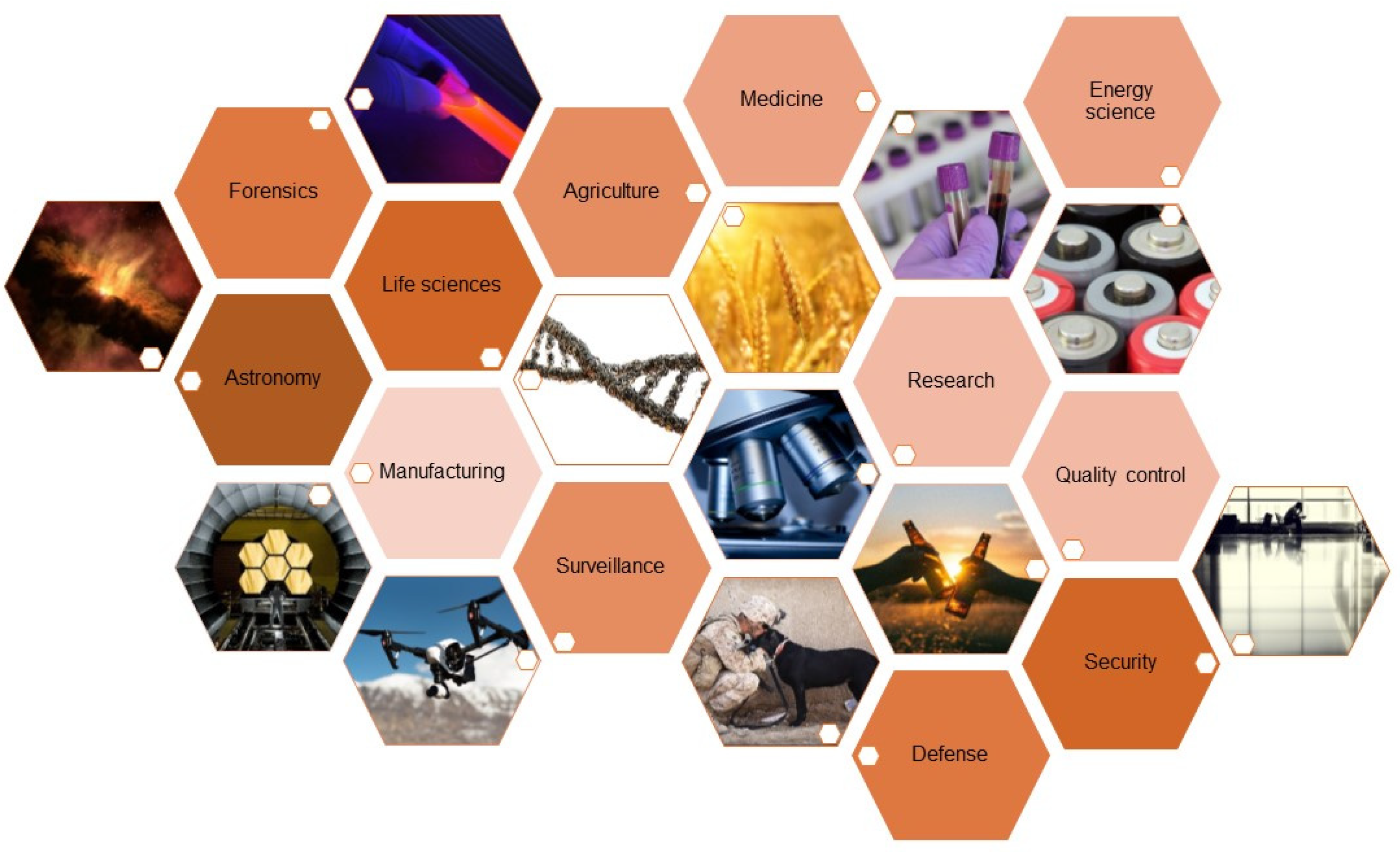
Review

Infrared Spectroscopy–Quo Vadis?

1
Institute of Analytical and Bioanalytical Chemistry, Ulm University, Albert-Einstein-Allee 11, D-89081 Ulm, Germany
2
Hahn-Schickard, Sedanstraße 14, 89077 Ulm, Germany
*
Author to whom correspondence should be addressed.
Appl. Sci. 2022, 12(15), 7598; https://doi.org/10.3390/app12157598
Submission received: 3 July 2022 / Revised: 22 July 2022 / Accepted: 26 July 2022 / Published: 28 July 2022
(This article belongs to the Special Issue Molecular Sensing Technologies)

Abstract

:
Given the exquisite capability of direct, non-destructive label-free sensing of molecular transitions, IR spectroscopy has become a ubiquitous and versatile analytical tool. IR application scenarios range from industrial manufacturing processes, surveillance tasks and environmental monitoring to elaborate evaluation of (bio)medical samples. Given recent developments in associated fields, IR spectroscopic devices increasingly evolve into reliable and robust tools for quality control purposes, for rapid analysis within at-line, in-line or on-line processes, and even for bed-side monitoring of patient health indicators. With the opportunity to guide light at or within dedicated optical structures, remote sensing as well as high-throughput sensing scenarios are being addressed by appropriate IR methodologies. In the present focused article, selected perspectives on future directions for IR spectroscopic tools and their applications are discussed. These visions are accompanied by a short introduction to the historic development, current trends, and emerging technological opportunities guiding the future path IR spectroscopy may take. Highlighted state-of-the art implementations along with novel concepts enhancing the performance of IR sensors are presented together with cutting-edge developments in related fields that drive IR spectroscopy forward in its role as a versatile analytical technology with a bright past and an even brighter future.

1. Introduction

The infrared (IR) spectral region provides the analytical spectroscopist with a vast amount of molecular information. Infrared spectroscopic measurement techniques are in general versatile non-destructive methods based on the interaction of infrared light with matter by the excitation of vibrational transitions in molecules. They are used in a variety of scenarios for sensing gases, liquids and solids. Due to this inherent molecular selectivity, IR spectroscopy can be used quantitatively and for obtaining chemical, structural and compositional information on analytes which can be analyzed by absorption and transmission techniques [1]. Since this would otherwise go beyond the scope of this review, we refer here to Griffith and Haseth [1], where the basic principles and main scientific drivers of infrared spectroscopy are described in detail. Commonly, the mid-infrared (MIR) is defined as the regime of electromagnetic waves with wavelengths ranging from about 2 µm to about 20 µm. Whereas the near-infrared (NIR) and the far infrared (FIR) are extending the MIR to the shorter wavelength (about 800 nm to 2 µm) and the longer wavelength (15 µm to about 1 mm) regime, respectively. However, in the MIR, wavenumbers are commonly used, as wavenumbers represent inverse wavelength and thus provide a clear overview of the important fingerprint region. As this spectral region covers a rather wide wavelength regime, the MIR is split in more detailed regions depending on the field of application with respective conventions via DIN (Deutsches Institut für Normung, German Institute for Standardization), CIE (International Commission on Illumination) or ISO (International Organization for Standardization). Hence, common classifications are short wave mid infrared (SWIR, IR-A, 1.4–3 µm), mid wave infrared (MWIR, IR-B, 3-8 µm) or the long wave infrared (LWIR, IR-C, 8–15 µm). Additionally, the infrared can be classified into bands-similar to the radio frequency bands-which is mostly used in infrared astronomy, following atmospheric transparent windows. In the infrared, there are bands such as the J, H and K bands that cover the near-infrared wavelengths as well as the L, M, N, and Q bands, which refer to the mid-infrared spectral region [2]. A brief overview of the respective band positions in the IR is given in Figure 1.
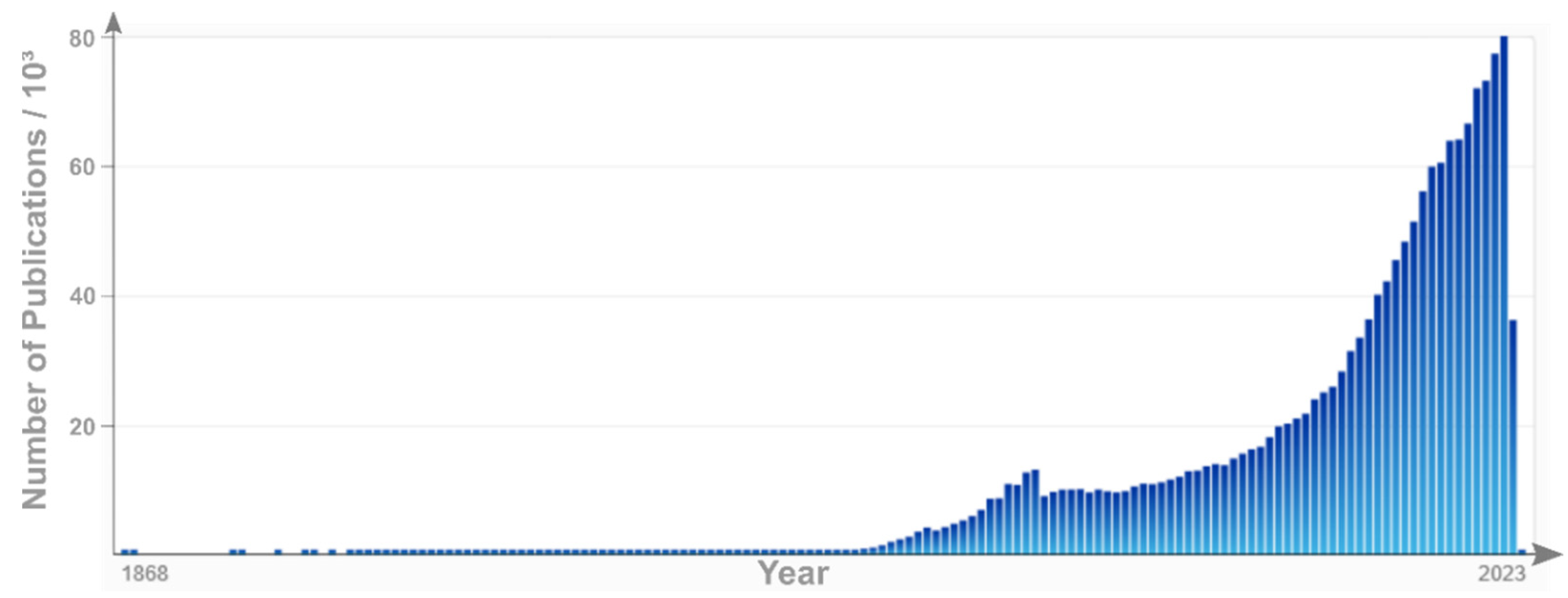
As evident by the manifold definitions of the IR, this spectral window is of growing interest (see Figure 2) and high importance to equally manifold fields of application. Given the recent evolution from an analytical method that was rather exclusively used by specialized laboratories, into a widely used routine method, the according development of IR-based tools is evident. To derive possible future applications of IR spectroscopy, such as medicine, the food industry, and the environment, as well as its potential as a non-invasive, on-line, fast, and in-situ analytical method, it is necessary to review and briefly discuss the historical development as well as the previous obstacles that have limited the broader application of IR spectroscopy. Furthermore, recent trends and adaptions of IR spectroscopy leading to future deployment scenarios will be elucidated. Finally, the potential role and positioning of IR spectroscopy in view of emerging challenges within the next decades is discussed in this perspective review.

2. Infrared Spectroscopy: The Past

Given the close resemblance to spectroscopy in the visible spectral region, NIR spectroscopy has been developed in a straightforward fashion. Fused silica is highly transparent in the NIR and thus is an excellent choice for NIR optics. Bright light sources as well as silicon based (e.g., line array) detectors for NIR spectroscopy are readily available at a fair price. A conceptual drawing of the basic elements of a spectroscopic setup is given in Figure 3. NIR spectroscopy provides excellent information, e.g., on the water content in analyte matrices [3]. Furthermore, carbohydrates, proteins, fats as well as plastics can be identified in the NIR [4]. Hence, industrial sorting machines are a main application of NIR spectroscopy as long as the broad and overlapping features in the NIR spectra are sufficient for identification, classification and quantification [5]. Hence, NIR has its capitalizing on highly sensitive and cheap photodetector technologies, which are not useable in MIR. Worthy of note is that the NIR spectral features are the overtones of the fundamental MIR vibrations, and thus, broader—and consequently more overlapping/less discriminatory—and significantly weaker in signal strength vs. the fundamental vibrational signatures prevalent in the MIR [6].
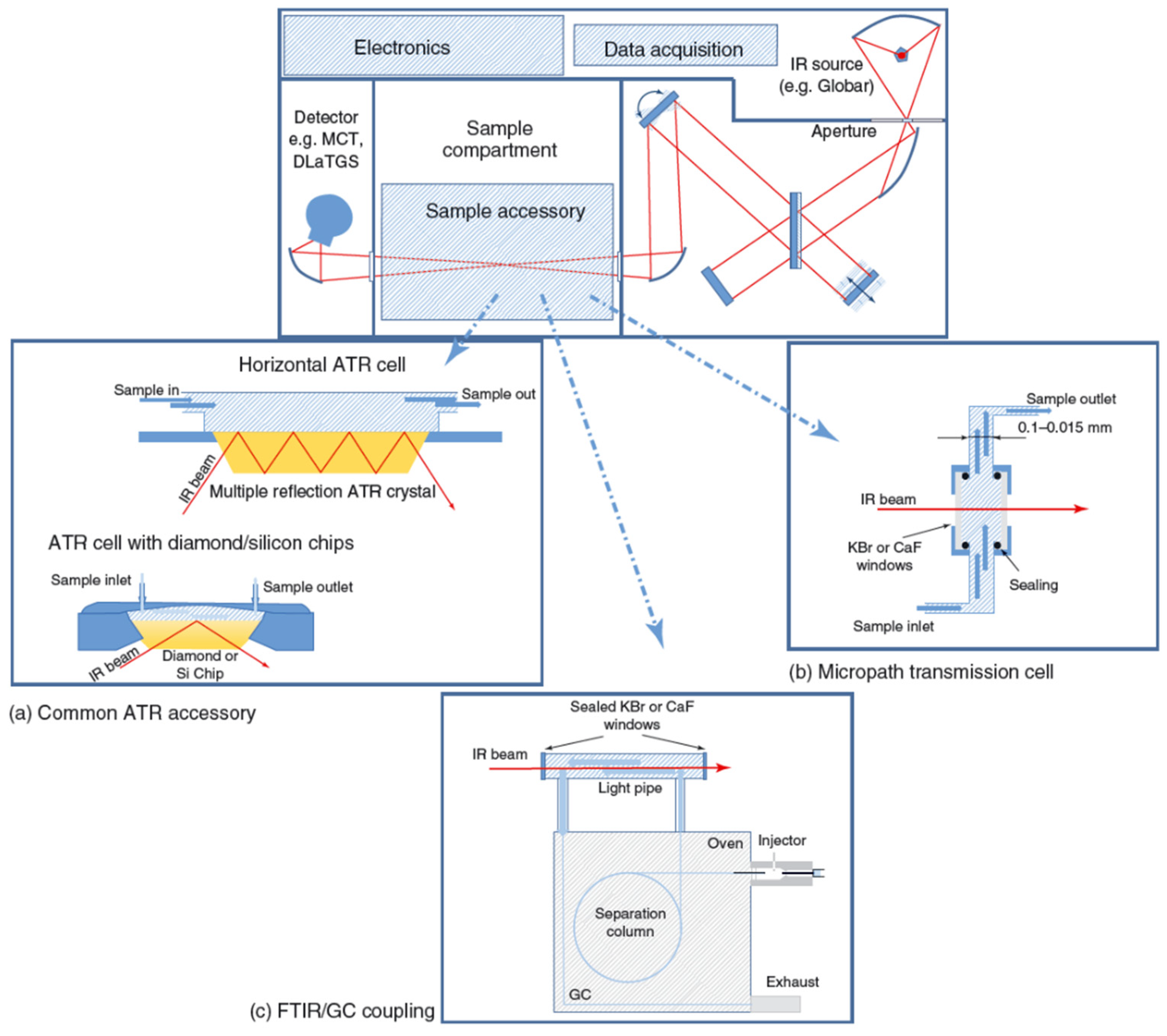
MIR spectroscopy has clearly found widespread use in academic research facilities such as universities as well as industrial laboratories. The basic concept of accessing fundamental rotational, vibrational and roto-vibrational molecular transitions has long matured into convenient-to-operate devices, albeit requiring more specialized optics, materials, light sources and detectors than in the NIR. With IR spectroscopy gaseous, liquid and solid phase (i.e., condensed phase) samples can be accessed. Here, MIR spectroscopy enables the non-destructive probing of analytes in various matrices via their well-pronounced MIR signatures providing fundamental transitions of much higher signal strength vs. the NIR. In the early days of MIR spectroscopy, bulky devices requiring sophisticated and delicate optics, including refractive and diffractive optical components, limited the applicability or the wide acceptance of MIR spectroscopic devices as on field tools, and complementary techniques-as for example NIR spectroscopy benefitting from the optical telecommunications revolution-had progressed faster towards lower cost and portable devices. Since then, slow scanning speeds, limited energy throughput and spectral resolution since have been overcome by the development of Fourier-transform infrared (FTIR) spectroscopy. The Fellgett (i.e., high signal-to-noise ratio), Jacquinot (i.e., high throughput) and Connes (i.e., wavelength accuracy) advantages are well-known hallmarks of FTIR technologies and are classical textbook knowledge. From a technological point of view, long-lived radiation sources such as silicon carbide-based blackbody radiators along with high wavelength stability ensured via narrow-band laser sources such as helium-neon (HeNe) lasers have led to a customer-friendly mode of operation. Further technological developments have enabled the implementation of highly sensitive room temperature operated pyroelectric deuterated (L-Alanine) tri-glycine sulphate (DTGS, DLaTGS) or thermoelectrically cooled photoelectric or photovoltaic mixed-semiconductor (e.g., mercury-cadmium-telluride; MCT) detectors facilitating the evolution of MIR spectroscopy not only for routine demands, but also as a high-end research tool. Further developments have led to exceedingly robust and miniaturized interferometers (e.g., Michelson interferometers) providing fast wavelength modulation with a compact device footprint. Hence, entire system footprints on the order of magnitude of a sheet of letter sized or A4 paper and even handheld devices have since entered the markets. However, device scaling remains associated with a trade-off between the highest spectral resolution (i.e., requiring a long travel pathlength of the moving interferometer mirror), fast scanning speed, and portability resulting in dedicated device technology–and performance figures-of-merit–adapted to specific application scenarios. Yet, potent computers enabling fast-Fourier transform algorithms basically in real-time, the introduction of attenuated total reflection (ATR) based sensing techniques, and the increased quality of optical components albeit still requiring specialized MIR transparent materials such as ZnSe, CsI, KBr, CaF2, etc. have led to MIR spectroscopy catching up with NIR methods in almost any field of application [7]. Figure 4 displays schematically the possible applications of such IR-transparent materials for the use of FTIR spectroscopes with an interferometer in combination with various sample accessories for the detection of gas, liquid and solid applications. The introduction of ATR techniques has significantly facilitated the probing of aqueous or water-containing samples, in any way, shape or form, as water itself is a pronouncedly strong IR absorber and commonly gives rise to a largely opaque sample matrix in the MIR. Additionally, probing solid samples has been eased in terms of sample preparation, i.e., usually encasing into KBr pellets, which has become almost obsolete in most application scenarios placed by ATR techniques. Consequently, MIR spectroscopy has increasingly ventured into applications requiring analytical spectroscopy with inherent molecular discrimination exemplarily—but not limited to—environmental monitoring, as hyphenated analytical tool (e.g., GC-IR, LC-IR, etc.), in food quality [8], for recycling of plastics [5], in wastewater monitoring [9], for surveillance, purposes in winery applications [10,11,12], for restauration [13], in industrial quality control (e.g., surface coatings quality) [14], in paint analysis, as well as for pharmaceuticals [15,16] and in oil/fuel analysis [17].
Lastly, FIR or THz spectroscopy at the other end of the spectrum primarily uses antenna technologies to take advantage predominantly of antenna, and has developed into a tool mainly used in space observation, as various gases provide distinct features in the THz region [18]. Similarly, FIR is applied to the study of low-energy lattice vibrations, such as minerals, but is increasingly adopted for investigating large (bio)molecular constructs as proteins and DNA. These distinct features are detected by the excitation of low-energy modes that cause all atoms in the molecule to move collectively and therefore lead to these spectral signatures of many organic molecules in the terahertz range. But when comparing THz/FIR spectroscopy with MIR spectroscopy, the development of THz/FIR spectroscopy has been delayed due to the difficulties in developing instruments in this range, which are due to obstacles such as the insufficient intensity of radiant energy from light sources in the THz/FIR spectral range. In addition to the development of light sources, the development of dedicated THz/FIR detectors has also been ongoing. This development benefited primarily from advances in ultrafast laser techniques for generating and detecting this radiation. Techniques for generating or detecting ultrafast THz laser beams include electro-optic generation/detection, photoconductive generation/detection, and generation in air. Furthermore, it was not easy to exclude the effects of water vapor in the THz/FIR region. Besides, the band assignments of the low-frequency oscillation modes were not clear, making it difficult to find applications for FIR spectroscopy.

3. Infrared Spectroscopy: The Present

Currently, basic instrumentation for NIR and MIR spectroscopic devices has been continuously refined and improved. Again, developments in several technological fields have led to significant improvements in IR spectroscopic instruments (see Figure 5). Again, from a technological point of view the introduction of silicon-based detectors and inexpensive thermopile detectors (i.e., low cost down to few euro-cents), as well as high-end sensitive room-temperature-operated MCT detectors has enabled rapid detection schemes with high signal to noise levels. Commonly, MCT detectors had to be cooled with liquid nitrogen, requiring the respective infrastructure, or at least thermoelectric cooling (TEC) via Peltier elements, which results in some bulky cooling components as well as the need for heat dissipation. In addition, various concepts that have been developed for UV/Vis and NIR spectroscopy are increasingly transitioned into MIR spectroscopic tools. NIR spectroscopy has benefited from development pressure at telecom wavelengths. Global data communication greatly benefits from glass fibers that provide ultra-low losses down to few dB/km at adequate manufacturing efforts. As a consequence, the cost and performance of optical components such as sources, detectors, lenses and mirrors have been optimized for wavelengths around 1300 nm. Furthermore, silicon-based source and detector concepts greatly benefit from advances in microfabrication technologies derived from integrated microelectronic circuits. Yet, these co-parallel developments, interconnections within microelectronics, and UV/Vis and NIR spectroscopy benefitting from communications technology are dictated by the utilized material platforms, which is by far and large silicon. Hence, first smartphone-based handheld spectroscopic devices have emerged on the consumer marked, e.g., for the detection of nutrition values as well as potential food spoilage based on NIR techniques.
Traditionally, IR spectroscopy has relied on broadband thermal infrared emitters that emit black body radiation via resistive heating across a certain wavelength band. The respective emission is dictated by the material (e.g., tungsten (W) or SiC, etc.) and the temperature of operation. With the increasing availability of light emitting diodes (LEDs) that are capable of emitting IR wavelengths, small footprint IR sources with a reduced power consumption have recently become available and will become increasingly common within the next decades [19,20]. Recent progress in microfabrication has led to MEMS based sources that provide a broad selection of emission wavelength/frequencies as well as high spectral energy densities [21]. In this context, non-coherent IR light sources such as NIR-LEDs, MEMS-based thermal infrared emitters, as well as coherent IR light sources such as inter-band cascade lasers (ICLs) [22], and quantum cascade lasers (QCL) [23,24], have since become high-performance alternatives replacing traditional lead salt laser diodes or CO2 lasers. ICLs are characterized by a particular low energy consumption, which enables applications in remote or wearable scenarios, e.g., for spectroscopic sensing of biomarkers in breath or volatile organic compounds in environmental monitoring [25,26]. QCLs, on the other hand, provide high emission power at the cost of increased power consumption. First, applications of QCLs for remote gas sensing or chemo/bio sensing in transmission mode, as well as spectroscopic sensing with ATR accessories have been shown to and will become more ubiquitous within the next few years. Rather limited spectral coverage of tuneable diode lasers (TDL) absorption spectroscopy (TDLAS) [27] scenarios has recently been brought to a new level, as, e.g., broadly tuning QCLs and QCL-arrays have become available that are capable of covering broad wavelength ranges of up to 1200 cm−1 [28]. Those lasers have gained interest for the development of exceedingly sensitive spectroscopic applications such as cavity ring down spectroscopy (CRDS) [26,29,30], providing well defined, short pulses combined with accurate wavelength selection at a compact form factor. Additionally, the 3D-printing of gas cells and even very complex optical structures such as multi-pass gas cells further increase the accessibility of complex TDLAS experiments [31,32,33]. Thus, trace gas detection via IR techniques continue to be an emerging topic, in the decades to come, as those lasers ideally fit the needs for sensing narrow rotational bands in complex matrices. Recent trends in laser resonator techniques allow for the envisioning of even broader spectral coverage with complex resonator structures such as tuneable external cavities [34] or multiple integrated DFB resonator concepts. An exciting strategy for such advanced structures are ring-or dual-ring-QCLs, whereby the quantum cascade structure is arranged in a circular way that facilitates high integration levels. The combination of a QCL source and QC detector in close vicinity are also interesting, e.g., for gas sensing scenarios and will provide an interesting alternative to, for example, existing non-dispersive infrared sensors [35,36]. Increased levels of control on the quality of quantum layers now enables the fabrication of devices with tailored electronic band structures at wafer scale production levels. Hence, low-cost MIR emitting IC-LEDs made, for example, from indium arsenide/antimonide phosphide (InAsSbP), are within reach. Furthermore, in the context of FTIR spectroscopy, diode lasers have recently been applied as stable and long-lived wavelength reference replacing He-Ne laser tubes, thus facilitating an additional reduction of device footprints.
Advancing from NIR wavelengths to longer wavelengths further into the mid-infrared spectral region results in demanding technological barriers, mainly concerning the development of suitable substrate materials or material mixes. Silicon (Si) and silicon dioxide (i.e., SiO2, silica, silica glass, quartz) are suitable materials for fabricating windows or lenses in the UV/Vis and NIR range, but become increasingly opaque in the MIR. On the other hand, micro-bolometers may be fabricated from silicon, which facilitates the straightforward integration of such bolometers into integrated circuits (ICs) for MIR light detection. Bulk manufacturing capabilities that have been developed for Si and SiO2 processing led to the availability of high-quality substrates that can be milled via computerized numerical control (CNC) machines. Such substrates are well suited for depositing such things as highly reflective metallic mirrors. Here, most metallic materials are useful for the fabrication of broadband reflective mirrors due to their high reflectivity in the MIR. It is worthy of note that, gold (Au), silver (Ag), aluminum (Al), copper (Cu) and alloys thereof (i.e., brass and bronze) are appropriate materials for the fabrication of highly reflective mirrors in the MIR. Here, the selection of the specific material is dependent on the required performance as resistivity towards, for example, corrosive media and the acceptable costs. For application in analytical spectroscopy in the MIR, substrate integrated hollow waveguides have been developed that provide a rugged and highly integrated material platform for analysing gaseous environmental and (bio)medical samples [37,38,39]. Another parameter that can be crucial for the material decisions may be weight, especially in application scenarios where the device payload is critical (e.g., drone-mounted IR sensing devices). Aviation, space and high acceleration applications demand exceedingly lightweight yet robust substrate materials [40,41]. Here, silicon carbide (SiC) has recently emerged as a stiff, durable and lightweight substrate material for metallic mirrors that are reflective in a wide spectral window, especially covering the MIR. Most recently, SiC Mirrors were used as the primary mirrors of the EUKLID telescope by the European Space Agency’s Cosmic Vision program. Within this project, a visible imager and a near-infrared spectrometer and photometer together with a Korsch Telescope were built [42].
As an alternative, dielectric mirrors can provide enhanced reflection performance at a narrower spectral range. However, such Bragg reflectors and photonic bandgap structures can be fabricated via gas phase deposition processes (i.e., chemical vapour deposition, CVD; physical vapour deposition PVD; molecular beam epitaxy, MBE; atomic layer deposition, ALD, etc.). Here, alternating layers of specific atomic composition can be deposited to specifically tune the reflected wavelength.
The very same principle has been introduced to prepare highly reflective coatings for MIR optics. Dichroic mirrors represent an implementation of the combination of dielectric mirrors and highly reflective coatings. With such mirrors, narrow-band radiation of different wavelengths can be combined into a single multi-colour beam. Such mirrors represent the standard way for beam combinations where diffractive elements (gratings) or tilted combinations cannot be deployed. Very recently, substrate integrated hollow waveguides (iHWGs) have been developed to achieve such a combination without the need to pass a dispersive element, i.e., the substrate material, of such a dielectric mirror [43].
With efforts towards the reduction of moving parts and the ideally monolithic integration linear variable filter (LVF) elements have also been introduced for IR spectroscopic demands. Such filters, in combination with detector arrays fabricated, for example., from lead zirconate titanate (PZT), provide promising wavelength selective elements for robust spectroscopic sensors in the IR. It is worthy of note that such alternating structures have also been introduced as light emitting and light detecting structures [44].
In general, dielectric materials are the material of choice in the MIR. Similar to UV/Vis and NIR optics, distinct dielectric materials are suitable for the fabrication of lenses, windows, beam-splitters, fibers and waveguides. Germanium (Ge), diamond, zinc selenide (ZnSe), zinc sulphide (ZnS), germanium based chalcogenide glasses (such as AMTIR-1 and AMTIR-3), other germanium based chalcogenide glasses (e.g., Schott IRG, Ge–Sb–Se, Ge–Ga–Sb–S)) [45,46], thallium bromine chloride or iodide (KRS-5, KRS-6), silver halides (AgX, AgClxBr1−x) [47,48,49,50], potassium bromide (KBr), sodium chloride (NaCl) and gallium arsenide (GaAs) [51,52] are only some examples of materials that have been implemented as routinely deployed dielectrics in MIR spectroscopy of late. Many other chalcogenide-based materials are the topics of today’s research on spectroscopy as shown in the articles by Churbanov et al. and Denker et al. [53,54].
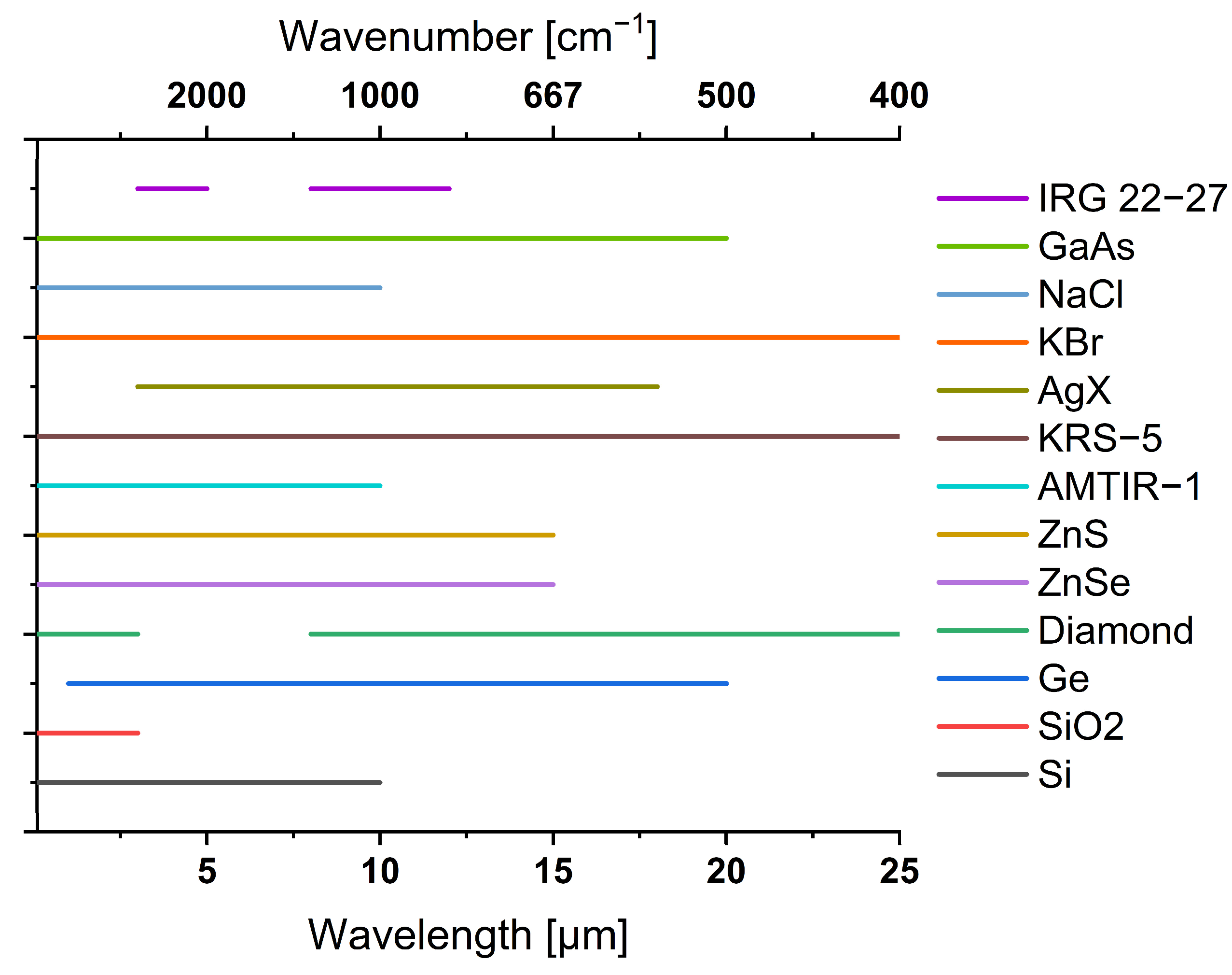
These materials are transparent in the MIR (the respective transparency regions are given in Figure 6) and provide sufficient robustness, inertness, hardness, and pH-stability. Diamond infrared optics have gained much interest in the military, such as for defence applications for night vision or guidance systems in the form of infrared domes or windows due to their excellent durability. The formerly used ZnS and ZnSe optics were cheaper to produce but much more prone to material damage, such as abrasion and darkening. As fabrication capabilities of diamonds have made this material increasingly available for civil stakeholders, diamond has become the quasi-gold standard for vacuum windows and ATR transducer materials in the MIR [55]. High-quality crystals as well as foreseeably reduced fabrication costs will certainly lead to a much wider implementation of diamond-based infrared optics given the advantage of being inert, non-toxic and will therefore be of increasing importance in food analysis and pharmaceutical analysis [56]. In addition, the fabrication of waveguides such as thin-film waveguides with envisioned applications in ICs requires the existence of optical insulators (i.e., optical buffers) for which such materials are suitable. Recently, smart combinations of, e.g., freestanding diamond waveguide strips [57,58] and membranes as well as AgX, ZnSe [59] or GaAs/AlGaAs [45] -based thin-film waveguides have been developed as efficient ATR transducer elements for MIR sensing tasks. Given these trends, refined fabrication techniques are clearly resulting in the elevated quality of the fabricated structures with existing materials as well as the development of more sophisticated waveguide structures. Some, still more exotic, waveguide designs based on, e.g., slot guiding [60], plasmonic concepts [61], bimodal waveguides [62], on-chip Mach-Zehnder Interferometers [63,64], micro-ring resonators [65] and photonic crystals (photonic bandgap structures) have recently been presented. These structures represent a well performing alternative to more traditional rib, strip and ridge designs of photonic chemo/bio sensors and will lead to highly sensitive implementations in chemo/bio sensors for various tasks. In combination with dedicated microfluidics, highly sensitive devices may be fabricated on a small footprint requiring only a few droplets of analyte and reaction chemicals [66].
Hollow or dielectrically filled waveguide technology with metallic waveguide walls is derived from microwave/terahertz (THz) and radar technology. Such waveguides work well for distinct reflections in dedicated MIR hollow waveguide structures. However, with reduced lateral dimensions towards single mode wave guiding, fast-alternating electric fields induce high losses in the waveguide walls, leading to limited lengths of such hollow waveguides [67,68,69]. Hence, dielectric waveguides (fibers) based on total internal reflection are more efficient as light guiding structures. In particular, waveguide fibers for localized light delivery are most commonly realized from solid materials instead of hollow core fibers. In this context, fibers can be utilized to route light in endoscopic scenarios, enabling IR spectroscopy to be used on inaccessible parts of a patient’s body. Such tools have already been introduced for cancer detection and cartilage evaluation [70,71,72,73]. With a foreseeable market entry of such tools, surgeons will be provided with a tool that facilitates diagnostic decision-making based on the molecular composition of the evaluated tissue. Apart from applications in medical scenarios, probes have been developed for the on-line monitoring of processes such as bio-fermenter status. IR spectroscopic monitoring facilitates the fast detection of reaction states as well as potential spoilage, allowing fast response times and elevated process control. The delivery of high intense laser light emitted from CO2 lasers for application in industrial welding or chirurgical environments benefits from a high damage threshold and low insertion losses of hollow waveguides. Therefore, some hollow core fibers with inner walls that are tailored towards low attenuation at 10.6 µm have emerged on the market.
In addition to advancing capabilities in fabrication and material sciences that lead to advanced application scenarios, novel technological concepts and their adaption in IR spectroscopic techniques provide opportunities that have previously been inconceivable.
Similarly, utilizing the photoacoustic effect in photoacoustic spectroscopy (PAS) has enabled the non-invasive analysis of glucose concentration in interstitial fluid [74]. Therefore, progress towards non-invasive diabetes care based on extended IR spectroscopy will be within reach in the next few of years. Additionally, PAS facilitates the acquisition of depth profiles of organic tissue and thus enables tomographic imaging. Here, screening for skin cancer is an important and emerging field for the application of IR spectroscopy.
At present, MIR spectroscopy is strongly connected to the latest developments in imaging technologies such as MIR microscopy. Previously, scanning techniques were realized by coupling an FTIR spectrometer to a movable ATR tip scanner or to a sharp atomic force microscopy (AFM) tip in so-called Nano-FTIR setups that are based on scattering type near field scanning near field optical microscopy (s-SNOM) techniques.
With the increasing availability of matrix detectors such as MCT arrays or micro-bolometer arrays in combination with fast scanning lasers, IR hyperspectral imaging has emerged from silicon based NIR techniques further into the MIR and have become a current hot-topic and will still be important in the near future.
As rapid image acquisition in reflection and transmission mode has become available, the application of this in medicine, surveillance and even defence is steadily increasing [75].
The combination with label-free and cost-effective detection based on minimal sample volumes makes IR spectroscopy especially suitable for routine clinical laboratory applications [76]. Considerable efforts have been made in recent years in FTIR spectroscopic imaging to study things such as the imaging of biopsy tissues [77], fixed cells [78] and live cells [79,80], including cancer cells [81]. In particular, synchrotron infrared sources coupled to FT-IR microscopy have immense potential for clinical research [82].
However, the long wavelengths of infrared radiation commonly limit lateral resolution in IR imaging. Very recently, smart concepts have been developed to surpass this limitation by exploiting the optical photothermal effect (O-PTIR) [83]. By using visible light as a probe, higher lateral resolution down to the submicron range is achieved for IR imaging and, as a side-effect, Raman scattering may be acquired in the very same experiment [84]. However, energy input is a major issue that hat to be considered when dealing with sensitive samples, as the ineffective Raman scattering process requires a considerable amount of photon flux.
The rich information in the NIR and especially MIR spectra often results in complex spectra. Hence, the interpretation of IR spectra greatly benefits from emerging powerful algorithms and software designed for interpretation and data mining. Tools that have been developed several years ago, such as the partial least squares (PLS) algorithm and principal components (PC) analysis have made their transition from specialized applications to a broad stakeholder community. Together with tools like random forests, K-nearest neighbours (KNN) or support vector machines (SVM), chemometrics with multivariate statistic tools, as well as more advanced extreme learning machines (ELM) [85,86] have become a main part of an IR spectroscopists tools in a variety of different applications [87,88,89,90,91,92,93,94].
Consequently, analytical application scenarios that have been dominated by highly sensitive hyphenated instruments like gas chromatography coupled to mass spectroscopy (GC-MS) are increasingly interested in IR spectroscopic alternatives. In application fields such as food monitoring, IR spectroscopy may provide a fast, simple to operate, inexpensive to maintain and non-destructive method with highly reliable results. First emerging applications of IR spectroscopy classifying mycotoxin spoilage based on regulatory limits pave the way for more wide spread application of IR spectroscopy in associated fields of profession such as life sciences, production of pharmaceuticals and polymer research.

4. Infrared Spectroscopy: The Future

With the most recent publications dealing with techniques based on IR spectroscopic technology, the elevated application and research interest on wavelength regimes is clearly anticipated. An emerging area of interest is NIR-based critical care monitoring. With the high penetration depth of NIR light in biological tissue, non-invasive monitoring of physiological markers is a prevalent application for NIR spectroscopy. So far, blood oxygenation, in combination with the pulse rate, can be monitored, in the fingertips of adult patients or on the cranium of newborns [95,96,97]. Such devices are commonly based on low cost LED emitters and can be readily modified due to their modular nature. Hence, bedside monitoring as well as personal health is a trending market for IR spectroscopic tools. Multiple biological markers, such as lactose or glucose, as well as pathogens, have strong signatures in the NIR and thus can be accessed with tailored tools. Given the recent developments in wearables and smartphone-based sensors, i.e., small-scale potent computers, tele-diagnostics is an emerging area with high potential for IR-based tools [98,99].
As infrared spectroscopy provides fast and non-invasive localized information, more imaging in the form of infrared provides extremely dense data cubes with tremendous molecular information that can be stored, transported and analyzed digitally [100,101]. Therefore, integrated databases with patient data will clearly be extended with IR spectral information in addition to more traditional X-ray or MIR images. Analogous to optical coherence tomography, infrared coherence tomography is a promising tool to extend the accessible tissue range and translate it to data. In this context, big data and artificial intelligence are important catchphrases. Data-based machine learning is of increasing interest as big databases are becoming increasingly available via the internet. Consequently, algorithms can be trained for elevated and reliable variances detection. Among plenty of tools such as PyTorch (https://pytorch.org/, accessed on 17 November 2021), orange (https://orange.biolab.si/, accessed on 17 November 2021) and scykit (https://scikit-learn.org/stable/, accessed on 17 November 2021) that are often available via open-source licensing, Google’s TensorFlow (https://www.tensorflow.org/, accessed on 17 November 2021) has become a widely used tool for the training of non-linear classification problems by users from various fields, including IR spectroscopy.
With the development of more powerful and miniaturized microprocessors/embedded systems (e.g., Raspberry Pi, Arduino, ESP32, ESP8266), which can already be purchased at a very low cost (i.e., up to 50$), a new tool for the evaluation of complex datasets (e.g., breath samples) is on the rise. Most of the already available systems have the ability to perform even complex calculations and use chemometric or deep learning methods for the evaluation of IR data. This is frequently made possible by the fact that popular programming languages (e.g., Python, R, C#, etc.) [102] can be executed on the corresponding platforms (i.e., ARM, ATMEL, PIC and others), and existing libraries are available (e.g., Tensorflow, PyChem, SciPy, NumPy) [103,104], providing an already developed workflow/ algorithm to analyze sensor data. In cases where the datasets are very complex or an array of different sensor data is fused, the collected/recorded data are evaluated over cloud computing, thereby, exploiting the computing power of mainframe computers [105].
Apart from increasing demands in global health care applications, recent great trends in human society are associated with climate change, micro plastics, environmental health, food production, sustainability, city growth, energy science as well as space exploration. For each of these great topics, IR spectroscopic tools provide valuable opportunities. The detection of micro-plastics in marine samples has recently been shown via MIR- based tools [106]. Furthermore, oil spills have been detected via FTIR based technologies, potentially matching or even outperforming commonly deployed GC based techniques in the future [94,107,108]. Additionally, airborne infrared imaging has become interesting for precision farming as well as for the analysis of soil quality.
Regarding soil quality, infrared-based tools have also found their way into space research. The US NASA mars rovers have been readily equipped with infrared spectrometers for spectral analysis of soil with the aim to find, traces of water [2]. Space exploration on probes such as orbiters is also a growing application for infrared-based technologies apart from stationary observatories that are screening the sky for infrared signatures of, organic components in the search for extra-terrestrial life (https://breakthroughinitiatives.org/, 17 November 2021). Utilizing IR radiation in the MIR in combination with a vector coronagraph enables discovery of exoplanets that are otherwise outshined by their main star [109,110,111,112]. With the recent entry of privately financed space travel and exploration, IR spectroscopy is certainly among the key technologies for exploration and safety measures among space travel.
The development in unmanned aerial vehicles on earth combined with the opportunity for environmental monitoring also provides future opportunities. The trade-off between the unmanned aerial vehicle demands and the IR sensor technology has yet to be further optimized [113].
All of these applications require bright and highly controllable light sources. For this purpose, QCLs and ICLs provide excellent technologies. A major drawback of QCL technology are the high currents that are required for operation. Heat dissipation as well as resistive losses commonly lead to a lower limit of miniaturization of the auxiliary electronics, although the QCL die can be downsized to a few µm in size. The development of high-performing alloys that provide conductivity beyond copper, ideally realizing room-temperature super conductance, would enable an unprecedented level of design freedom and the emission power of such lasers. Apart from application in more regular spectroscopy tasks, such laser light sources enable elevated acquisition speeds that are allowing single shot experiments [114,115,116]. In particular, frequency combs are promising candidates that allow high-power super continuum spectrum generation with precise control of the spectral characteristics [117]. These sources are ideal candidates for elucidating super-fast molecular transitions and thus visualizing the mechanisms of such processes to improve the understanding of them, especially in combination with further analytical techniques such as within spectro-electrochemistry [118,119,120,121]. Such processes are of crucial importance for, e.g., catalysis or energy conversion in batteries or fuel cells. In particular, the degradation of electrode, separator or electrolyte materials can be investigated via MIR spectroscopy.
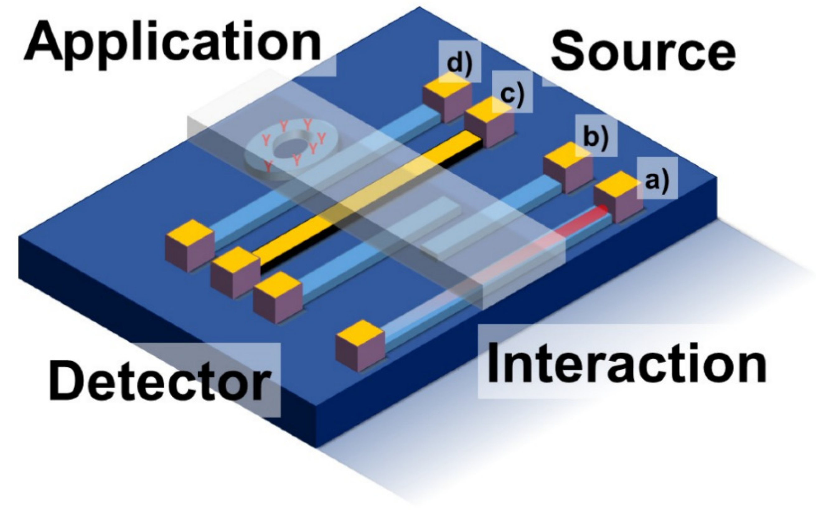
In addition, recent trends in additive manufacturing have led to new potential applications of IR spectroscopic tools. Three-dimensional printing has matured into high quality levels enabling the straightforward fabrication of optical quality structures such as waveguides [31,122]. Hence, low-cost, disposable IR analyzer tools are coming online within the next couple of years, similar to recent developments in electrochemical sensors. In addition, active structures that are fabricated and utilized similar to electronics in integrated circuits are on the horizon for applications in IR spectroscopy (Figure 7). Multiplexing analyte-dedicated recognition structures into highly integrated circuits is a mayor advantage and will lead to the elevated exploitation of IR tools in the future. Here, the development of the so-called ‘light or photonic computer concepts’ potentially operating with infrared radiation is a potential key technology that would reduce the cost of IR technology potentially to the level where electrochemical sensors are already at now.
Accessing low target analyte quantities or concentrations in the infrared can be challenging. There are different approaches to elevate signal interaction with the probing photons. First, the probe amount can be increased. Hence, specific concepts for pre-concentration via adsorption, absorption into membranes, porous films [123] or fishing molecules via recognition motives out of a bulk volume are possible for, e.g., virus detection [124,125,126,127]. Second, analytes can be converted into species that provide stronger interaction with infrared radiation, like converting H2S into SO2 [128,129]. Alternatively, sample interactions can be designed so that multiple interactions are achieved. Common approaches via multi-pass cells are accompanied with an increasing number of integrated microscopic structures. Here, structures can be designed to locally elevate electric fields instead of increasing the signal response via multiplied interaction. Standing wave resonators, via, micro-tori [130] and whispering gallery modes [131] are one example of such resonant structures. Surface enhancement in the infrared (SEIRAS; surface enhanced infrared spectroscopy) can be exploited as in Raman spectroscopy (SERS; surface enhanced Raman spectroscopy). To achieve signal enhancement factors ranging from about 10 to 100, plasmonic nanostructures can be deposited on surfaces to locally elevate electric field strength. Normally, noble metal nanoparticles are used for that purpose [132]. The most common shape of spherical nanoparticles has been extended by cubes, rods, and stars [133,134,135] that provide sharp tips as nano-antennas [136]. In addition, alternative materials such as less expensive silver, copper or iron oxide nano particles have been implemented for signal enhancement [137,138]. With the introduction of graphene-based signal enhancement [139,140,141] and quantum dots made from dielectrics and metallic alloys, a wide field of potential materials has been opened. With such materials, the precise tuning of the enhancement as well as the surface decoration thereof will lead to exquisite signal enhancing tools that are potentially non-toxic for applications in biological and medical scenarios.
Quantum dots have been recently developed for realizing bright light panels in television screens at optical frequencies, namely red, green and blue (RGB). As the emission wavelength of such quantum dots can be precisely tuned by their size, their increased diameters lead to the emission of infrared light. Moreover, such dots can be stimulated in a way to emit highly coherent light, also providing entangled photons in, e.g., N00N-states or squeezed states [142,143,144,145,146]. Such non-classical light enables interferometric sensing beyond the classical noise limit, reaching the quantum limit and thus providing ultra-sensitive sensor tools that provide limits of detection inaccessible by any classical tool, thus defining the ultimate goals for IR spectroscopic tools [147,148].

5. Conclusions

Although IR spectroscopy has been around for a long time, recent developments have evidently boosted the level of performance, integration and technology readiness levels (TRL) of increasingly specialized tools and concepts in IR spectroscopy and sensing. These developments distinctly align with the latest developments in solid-state physics, photonics, electronics, materials sciences, engineering, medicine and informatics. Virtually every component of an IR spectroscopic device has been recently further evolved and is subject to ongoing improvements regarding performance and level of integration. Rapid, non-destructive and label-free access of molecular transitions to date renders IR spectroscopy an indispensable analytical tool applicable in virtually any considered application scenario. Advanced control on the material properties, which is currently reaching atomic levels, facilitates the fabrication of next-generation light sources, signal transducers and detectors, allowing for the task-specific tuning of the device performance to access yet unprecedented levels of sensitivity and precision. Hence, IR spectroscopic measurement strategies can be found in application scenarios ranging from demanding, harsh environments such as monitoring within industrial manufacturing processes and even outer space exploration scenarios, to the ultra-fast detection of catalysis products and biological/biomedical analysis targeting, as with protein dynamics. Consequently, the answer to the question “Infrared Spectroscopy-Quo Vadis?” may be that not even the sky is the limit!

Author Contributions

M.H. and J.H. have prepared the manuscript fundament. R.S., V.K. and A.T. have extended the manuscript with their expert knowledge and their visions. M.D. proofread and edited the manuscript, and B.M. has proofread and prepared, edited the final manuscript for submission and publication. All authors have read and agreed to the published version of the manuscript.

Funding

Preparation of this article was in part supported by the European Union via the Horizon 2020 framework projects MIRACLE (780598), VOGAS (824986) and PHOTONFOOD (824986), the BMBF project GRADIA (03XP0206A), the DFG project PULMOSENS (GRK 2203), and the DFG project CATALIGHT (SFB/TRR 234).

Acknowledgments

The authors want to acknowledge valuable support from their colleagues from the Institute of Analytical and Bioanalytical Chemistry (IABC) at Ulm University as well as from their colleagues from Hahn-Schickard regarding fruitful discussions as well as highly rated expert knowledge, opinions and assessments. This study was particularly supported by the Europe Union’s Horizon 2020 Research and Innovation Programme project PHOTONFOOD (Grant Agreement Number 101016444) and is part of the Photonics Public Private Partnership.

Conflicts of Interest

There is no competing interest to declare.

Dedication

This article is dedicated to the 80th birthday of Prof. Peter Griffiths, to whom B. Mi-zaikoff is thankful for decades in mentorship, support, and friendship. Way to go, Peter!

References

  1. Griffiths, P.R.; de Haseth, J.A. Fourier Transform Infrared Spectrometry; Wiley-Interscience: Hoboken, NJ, USA, 2007; ISBN 9780471194040. [Google Scholar]
  2. Haas, J.; Mizaikoff, B. Advances in Mid-Infrared Spectroscopy for Chemical Analysis. Annu. Rev. Anal. Chem. 2016, 9, 45–68. [Google Scholar] [CrossRef] [PubMed]
  3. Müller, C.M.; Molinelli, A.; Karlowatz, M.; Aleksandrov, A.; Orlando, T.; Mizaikoff, B. Infrared Attenuated Total Reflection Spectroscopy of Quartz and Silica Micro- and Nanoparticulate Films. J. Phys. Chem. C 2012, 116, 37–43. [Google Scholar] [CrossRef]
  4. Karn, A.; Heim, C.; Flint-Garcia, S.; Bilyeu, K.; Gillman, J. Development of Rigorous Fatty Acid Near-Infrared Spectroscopy Quantitation Methods in Support of Soybean Oil Improvement. J. Am. Oil Chem. Soc. 2017, 94, 69–76. [Google Scholar] [CrossRef]
  5. van den Broek, W.H.A.M.; Derks, E.P.P.A.; van de Ven, E.W.; Wienke, D.; Geladi, P.; Buydens, L.M.C. Plastic Identification by Remote Sensing Spectroscopic NIR Imaging Using Kernel Partial Least Squares (KPLS). Chemom. Intell. Lab. Syst. 1996, 35, 187–197. [Google Scholar] [CrossRef] [Green Version]
  6. Wilks, P. NIR vs. Mid-IR-How to Choose. Spectroscopy 2006, 21, 43–46. [Google Scholar]
  7. Kim, S.-S.; Young, C.; Mizaikoff, B. Miniaturized Mid-Infrared Sensor Technologies. Anal. Bioanal. Chem. 2008, 390, 231–237. [Google Scholar] [CrossRef] [Green Version]
  8. McMullin, D.; Mizaikoff, B.; Krska, R. Advancements in IR Spectroscopic Approaches for the Determination of Fungal Derived Contaminations in Food Crops. Anal. Bioanal. Chem. 2015, 407, 653–660. [Google Scholar] [CrossRef] [Green Version]
  9. Nikolaou, A.D.; Golfinopoulos, S.K.; Kostopoulou, M.N.; Kolokythas, G.A.; Lekkas, T.D. Determination of Volatile Organic Compounds in Surface Waters and Treated Wastewater in Greece. Water Res. 2002, 36, 2883–2890. [Google Scholar] [CrossRef]
  10. Klein, O.; Roth, A.; Dornuf, F.; Schöller, O.; Mäntele, W. The Good Vibrations of Beer. The Use of Infrared and UV/Vis Spectroscopy and Chemometry for the Quantitative Analysis of Beverages. Z. Nat. B 2012, 67, 1005–1015. [Google Scholar] [CrossRef] [Green Version]
  11. Culbert, J.; Cozzolino, D.; Ristic, R.; Wilkinson, K. Classification of Sparkling Wine Style and Quality by MIR Spectroscopy. Molecules 2015, 20, 8341–8356. [Google Scholar] [CrossRef]
  12. Kumar, K.; Giehl, A.; Schweiggert, R.; Patz, C.-D. Multidimensional Scaling Assisted Fourier-Transform Infrared Spectroscopic Analysis of Fruit Wine Samples: Introducing a Novel Analytical Approach. Anal. Methods 2019, 11, 4106–4115. [Google Scholar] [CrossRef]
  13. Daffara, C.; Pampaloni, E.; Pezzati, L.; Barucci, M.; Fontana, R. Scanning Multispectral IR Reflectography SMIRR: An Advanced Tool for Art Diagnostics. Acc. Chem. Res. 2010, 43, 847–856. [Google Scholar] [CrossRef]
  14. Umeno, M.; Yoshimoto, M.; Shimizu, H.; Amemiya, Y. Ellipsometric and Infrared Spectroscopic Studies of Oxide Film on GaAs Surface. Surf. Sci. 1979, 86, 314–321. [Google Scholar] [CrossRef]
  15. Taghavi, S.; Ayatollahi, S.; Alibolandi, M.; Lavaee, P.; Ramezani, M.; Abnous, K. A Novel Label Free Cocaine Assay Based on Aptamer-Wrapped Single-Walled Carbon Nanotubes. Nanomed. J. 2014, 1, 100–106. [Google Scholar]
  16. da Silva, V.H.; da Silva, J.J.; Pereira, C.F. Portable Near-Infrared Instruments: Application for Quality Control of Polymorphs in Pharmaceutical Raw Materials and Calibration Transfer. J. Pharm. Biomed. Anal. 2017, 134, 287–294. [Google Scholar] [CrossRef]
  17. Stach, R.; Mizaikoff, B. Fuel Performance Specifications, Mid-Infrared Analysis Of. In Encyclopedia of Anal. Chem.; John Wiley & Sons, Ltd.: Chichester, UK, 2017; pp. 1–16. [Google Scholar]
  18. Razeghi, M.; Lu, Q.Y.; Bandyopadhyay, N.; Zhou, W.; Heydari, D.; Bai, Y.; Slivken, S. Quantum Cascade Lasers: From Tool to Product. Opt. Express 2015, 23, 8462. [Google Scholar] [CrossRef]
  19. Malyutenko, V.; Zinovchuk, A. Midinfrared LEDs Versus Thermal Emitters in IR Dynamic Scene Simulation Devices; Proc. SPIE 6368, Optoelectronic Devices: Physics, Fabrication, and Application III, 63680D; SPIE: Boston, MA, USA, 2006. [Google Scholar]
  20. Hlavatsch, M.; Mizaikoff, B. Advanced Mid-Infrared Lightsources above and beyond Lasers and Their Analytical Utility. Anal. Sci. 2022, 1–15. [Google Scholar] [CrossRef] [PubMed]
  21. Wolffenbuttel, R.F. MEMS-Based Optical Mini- and Microspectrometers for the Visible and Infrared Spectral Range. J. Micromech. Microeng. 2005, 15, S145–S152. [Google Scholar] [CrossRef]
  22. Vurgaftman, I.; Weih, R.; Kamp, M.; Meyer, J.R.; Canedy, C.L.; Kim, C.S.; Kim, M.; Bewley, W.W.; Merritt, C.D.; Abell, J.; et al. Interband Cascade Lasers. J. Phys. D Appl. Phys. 2015, 48, 123001. [Google Scholar] [CrossRef]
  23. Brandstetter, M.; Lendl, B. Tunable Mid-Infrared Lasers in Physical Chemosensors towards the Detection of Physiologically Relevant Parameters in Biofluids. Sens. Actuators B Chem. 2012, 170, 189–195. [Google Scholar] [CrossRef]
  24. Childs, D.T.D.; Hogg, R.A.; Revin, D.G.; Rehman, I.U.; Cockburn, J.W.; Matcher, S.J. Sensitivity Advantage of QCL Tunable-Laser Mid-Infrared Spectroscopy Over FTIR Spectroscopy. Appl. Spectrosc. Rev. 2015, 50, 822–839. [Google Scholar] [CrossRef] [Green Version]
  25. Jaeschke, C.; Glöckler, J.; El Azizi, O.; Gonzalez, O.; Padilla, M.; Mitrovics, J.; Mizaikoff, B. An Innovative Modular ENose System Based on a Unique Combination of Analog and Digital Metal Oxide Sensors. ACS Sens. 2019, 4, 2277–2281. [Google Scholar] [CrossRef] [PubMed]
  26. De, A.; Banik, G.D.; Maity, A.; Pal, M.; Pradhan, M. Continuous Wave External-Cavity Quantum Cascade Laser-Based High-Resolution Cavity Ring-down Spectrometer for Ultrasensitive Trace Gas Detection. Opt. Lett. 2016, 41, 1949. [Google Scholar] [CrossRef]
  27. José Gomes da Silva, I.; Tütüncü, E.; Nägele, M.; Fuchs, P.; Fischer, M.; Raimundo, I.M.; Mizaikoff, B. Sensing Hydrocarbons with Interband Cascade Lasers and Substrate-Integrated Hollow Waveguides. Analyst 2016, 141, 4432–4437. [Google Scholar] [CrossRef] [PubMed] [Green Version]
  28. Haas, J.; Catalán, E.V.; Piron, P.; Karlsson, M.; Mizaikoff, B. Infrared Spectroscopy Based on Broadly Tunable Quantum Cascade Lasers and Polycrystalline Diamond Waveguides. Analyst 2018, 143, 5112–5119. [Google Scholar] [CrossRef] [Green Version]
  29. Kosterev, A.; Wysocki, G.; Bakhirkin, Y.; So, S.; Lewicki, R.; Fraser, M.; Tittel, F.; Curl, R.F. Application of Quantum Cascade Lasers to Trace Gas Analysis. Appl. Phys. B 2008, 90, 165–176. [Google Scholar] [CrossRef]
  30. Banik, G.D.; Som, S.; Maity, A.; Pal, M.; Maithani, S.; Mandal, S.; Pradhan, M. An EC-QCL Based N 2 O Sensor at 5.2 Μm Using Cavity Ring-down Spectroscopy for Environmental Applications. Anal. Methods 2017, 9, 2315–2320. [Google Scholar] [CrossRef]
  31. Stach, R.; Haas, J.; Tütüncü, E.; Daboss, S.; Kranz, C.; Mizaikoff, B. PolyHWG: 3D Printed Substrate-Integrated Hollow Waveguides for Mid-Infrared Gas Sensing. ACS Sens. 2017, 2, 1700–1705. [Google Scholar] [CrossRef]
  32. Liu, Z.; Zheng, C.; Zhang, T.; Li, Y.; Ren, Q.; Chen, C.; Ye, W.; Zhang, Y.; Wang, Y.; Tittel, F.K. Midinfrared Sensor System Based on Tunable Laser Absorption Spectroscopy for Dissolved Carbon Dioxide Analysis in the South China Sea: System-Level Integration and Deployment. Anal. Chem. 2020, 92, 8178–8185. [Google Scholar] [CrossRef]
  33. Dong, L.; Li, C.; Sanchez, N.P.; Gluszek, A.K.; Griffin, R.J.; Tittel, F.K. Compact CH4 Sensor System Based on a Continuous-Wave, Low Power Consumption, Room Temperature Interband Cascade Laser. Appl. Phys. Lett. 2016, 108, 011106. [Google Scholar] [CrossRef] [Green Version]
  34. Alcaráz, M.R.; Schwaighofer, A.; Kristament, C.; Ramer, G.; Brandstetter, M.; Goicoechea, H.; Lendl, B. External-Cavity Quantum Cascade Laser Spectroscopy for Mid-IR Transmission Measurements of Proteins in Aqueous Solution. Anal. Chem. 2015, 87, 6980–6987. [Google Scholar] [CrossRef]
  35. Tütüncü, E.; Kokoric, V.; Szedlak, R.; MacFarland, D.; Zederbauer, T.; Detz, H.; Andrews, A.M.; Schrenk, W.; Strasser, G.; Mizaikoff, B. Advanced Gas Sensors Based on Substrate-Integrated Hollow Waveguides and Dual-Color Ring Quantum Cascade Lasers. Analyst 2016, 141, 6202–6207. [Google Scholar] [CrossRef] [Green Version]
  36. Szedlak, R.; Harrer, A.; Holzbauer, M.; Schwarz, B.; Waclawek, J.P.; MacFarland, D.; Zederbauer, T.; Detz, H.; Andrews, A.M.; Schrenk, W.; et al. Remote Sensing with Commutable Monolithic Laser and Detector. ACS Photonics 2016, 3, 1794–1798. [Google Scholar] [CrossRef] [Green Version]
  37. Perez-Guaita, D.; Kokoric, V.; Wilk, A.; Garrigues, S.; Mizaikoff, B. Towards the Determination of Isoprene in Human Breath Using Substrate-Integrated Hollow Waveguide Mid-Infrared Sensors. J. Breath Res. 2014, 8, 026003. [Google Scholar] [CrossRef] [PubMed]
  38. Wilk, A.; Carter, J.C.; Chrisp, M.; Manuel, A.M.; Mirkarimi, P.; Alameda, J.B.; Mizaikoff, B. Substrate-Integrated Hollow Waveguides: A New Level of Integration in Mid-Infrared Gas Sensing. Anal. Chem. 2013, 85, 11205–11210. [Google Scholar] [CrossRef] [PubMed]
  39. Seichter, F.; Tütüncü, E.; Hagemann, L.T.; Vogt, J.; Wachter, U.; Gröger, M.; Kress, S.; Radermacher, P.; Mizaikoff, B. Online Monitoring of Carbon Dioxide and Oxygen in Exhaled Mouse Breath via Substrate-Integrated Hollow Waveguide Fourier-Transform Infrared-Luminescence Spectroscopy. J. Breath Res. 2018, 12, 036018. [Google Scholar] [CrossRef] [PubMed] [Green Version]
  40. Mersen Boostec® Silicon Carbide SiC. Available online: https://www.mersen.com/products/graphite-specialties/boostec-silicon-carbide-sic (accessed on 1 July 2021).
  41. European Southern Observatory Extremely Large Telescope. Available online: https://elt.eso.org/ (accessed on 1 July 2021).
  42. Bougoin, M.; Lavenac, J.; Gerbert-Gaillard, A.; Pierot, D. The SiC Primary Mirror of the EUCLID Telescope. In Proceedings of the International Conference on Space Optics—ICSO 2016, Biarritz, France, 18–21 October 2016; Karafolas, N., Cugny, B., Sodnik, Z., Eds.; SPIE: Bellingham, WA, USA, 2017; p. 48. [Google Scholar]
  43. Haas, J.; Pleyer, M.; Nauschütz, J.; Koeth, J.; Nägele, M.; Bibikova, O.; Sakharova, T.; Artyushenko, V.; Mizaikoff, B. IBEAM: Substrate-Integrated Hollow Waveguides for Efficient Laser Beam Combining. Opt. Express 2019, 27, 23059. [Google Scholar] [CrossRef] [PubMed]
  44. Pyreos Ltd. Thin Film Pyroelectric IR Line Sensor Array Demonstrator Kit; Pyreos Ltd.: Edinburgh, UK, 2009; Available online: https://pyreos.com/evaluation-kits/ (accessed on 26 June 2021).
  45. Kim, H.; Lee, J.H.; Lee, J.I.; Ko, S.Y.; Choi, Y.G. Compositional Dependence of Infrared Transmission in Ge-Based Chalcogenide Glasses. Phys. Status Solidi B 2020, 257, 2000164. [Google Scholar] [CrossRef]
  46. Schott AG IRG Chalcogenide Glasses. Available online: https://www.schott.com/en-gb/products/ir-materials-p1000261/product-variants?tab=ir-materials-irg-chalcogenide-glasses-irg-22-27 (accessed on 22 July 2022).
  47. Kraft, M.; Jakusch, M.; Karlowatz, M.; Katzir, A.; Mizaikoff, B. New Frontiers for Mid-Infrared Sensors: Towards Deep Sea Monitoring with a Submarine FT-IR Sensor System. Appl. Spectrosc. 2003, 57, 591–599. [Google Scholar] [CrossRef] [PubMed]
  48. Rauh, F.; Luzinova, Y.; Katzir, A.; Pfeiffer, J.; Mizaikoff, B. A Gas Hydrate Autoclave Suitable for In-Situ Fiber Optic Infrared Spectroscopic Studies. In Proceedings of the 7th International Conference on Gas Hydrates, Edinburgh, UK, 17–21 July 2011. [Google Scholar]
  49. Schädle, T.; Eifert, A.; Kranz, C.; Raichlin, Y.; Katzir, A.; Mizaikoff, B. Mid-Infrared Planar Silver Halide Waveguides with Integrated Grating Couplers. Appl. Spectrosc. 2013, 67, 1057–1063. [Google Scholar] [CrossRef] [PubMed]
  50. Dettenrieder, C.; Raichlin, Y.; Katzir, A.; Mizaikoff, B. Toward the Required Detection Limits for Volatile Organic Constituents in Marine Environments with Infrared Evanescent Field Chemical Sensors. Sensors 2019, 19, 3644. [Google Scholar] [CrossRef] [Green Version]
  51. Wang, X.; Kim, S.-S.; Roßbach, R.; Jetter, M.; Michler, P.; Mizaikoff, B. Ultra-Sensitive Mid-Infrared Evanescent Field Sensors Combining Thin-Film Strip Waveguides with Quantum Cascade Lasers. Analyst 2012, 137, 2322. [Google Scholar] [CrossRef] [PubMed]
  52. Sieger, M.; Haas, J.; Jetter, M.; Michler, P.; Godejohann, M.; Mizaikoff, B. Mid-Infrared Spectroscopy Platform Based on GaAs/AlGaAs Thin-Film Waveguides and Quantum Cascade Lasers. Anal. Chem. 2016, 88, 2558–2562. [Google Scholar] [CrossRef] [PubMed]
  53. Churbanov, M.F.; Denker, B.I.; Galagan, B.I.; Koltashev, V.V.; Plotnichenko, V.G.; Sukhanov, M.V.; Sverchkov, S.E.; Velmuzhov, A.P. First Demonstration of ~ 5 µm Laser Action in Terbium-Doped Selenide Glass. Appl. Phys. B 2020, 126, 117. [Google Scholar] [CrossRef]
  54. Denker, B.I.; Galagan, B.I.; Koltashev, V.V.; Plotnichenko, V.G.; Snopatin, G.E.; Sukhanov, M.V.; Sverchkov, S.E.; Velmuzhov, A.P. Continuous Tb-Doped Fiber Laser Emitting at ∼5.25 µm. Opt. Laser Technol. 2022, 154, 108355. [Google Scholar] [CrossRef]
  55. Haas, J.; Catalán, E.V.; Piron, P.; Nikolajeff, F.; Österlund, L.; Karlsson, M.; Mizaikoff, B. Polycrystalline Diamond Thin-Film Waveguides for Mid-Infrared Evanescent Field Sensors. ACS Omega 2018, 3, 6190–6198. [Google Scholar] [CrossRef]
  56. Diamond Materials Diamond Crystals. Available online: https://www.diamond-materials.com/en/ (accessed on 15 September 2021).
  57. López-Lorente, Á.I.; Wang, P.; Sieger, M.; Vargas Catalan, E.; Karlsson, M.; Nikolajeff, F.; Österlund, L.; Mizaikoff, B. Mid-Infrared Thin-Film Diamond Waveguides Combined with Tunable Quantum Cascade Lasers for Analyzing the Secondary Structure of Proteins. Phys. Status Solidi A 2016, 213, 2117–2123. [Google Scholar] [CrossRef] [Green Version]
  58. Wang, X.; Karlsson, M.; Forsberg, P.; Sieger, M.; Nikolajeff, F.; Österlund, L.; Mizaikoff, B. Diamonds Are a Spectroscopist’s Best Friend: Thin-Film Diamond Mid-Infrared Waveguides for Advanced Chemical Sensors/Biosensors. Anal. Chem. 2014, 86, 8136–8141. [Google Scholar] [CrossRef] [PubMed]
  59. Dobbs, G.T.; Balu, B.; Young, C.; Kranz, C.; Hess, D.W.; Mizaikoff, B. Mid-Infrared Chemical Sensors Utilizing Plasma-Deposited Fluorocarbon Membranes. Anal. Chem. 2007, 79, 9566–9571. [Google Scholar] [CrossRef]
  60. Nedeljkovic, M.; Khokhar, A.Z.; Hu, Y.; Chen, X.; Penades, J.S.; Stankovic, S.; Chong, H.M.H.; Thomson, D.J.; Gardes, F.Y.; Reed, G.T.; et al. Silicon Photonic Devices and Platforms for the Mid-Infrared. Opt. Mater. Express 2013, 3, 1205. [Google Scholar] [CrossRef] [Green Version]
  61. El Shamy, R.S.; Mossad, H.; Swillam, M.A. Long-Range All-Dielectric Plasmonic Waveguide in Mid-Infrared. Appl. Phys. A 2017, 123, 52. [Google Scholar] [CrossRef]
  62. Zinoviev, K.E.; Gonzalez-Guerrero, A.B.; Dominguez, C.; Lechuga, L.M. Integrated Bimodal Waveguide Interferometric Biosensor for Label-Free Analysis. J. Lightwave Technol. 2011, 29, 1926–1930. [Google Scholar] [CrossRef] [Green Version]
  63. Lechuga, L.M.; Sepulveda, B.; Sanchez del Rio, J.; Blanco, F.; Calle, A.; Dominguez, C. Integrated Micro- and Nano-Optical Biosensor Silicon Devices CMOS Compatible. In Optoelectronic Integration on Silicon; Robbins, D.J., Jabbour, G.E., Eds.; SPIE: New York, NY, USA, 2004; pp. 96–110. [Google Scholar]
  64. Sieger, M.; Balluff, F.; Wang, X.; Kim, S.-S.; Leidner, L.; Gauglitz, G.; Mizaikoff, B. On-Chip Integrated Mid-Infrared GaAs/AlGaAs Mach–Zehnder Interferometer. Anal. Chem. 2013, 85, 3050–3052. [Google Scholar] [CrossRef] [PubMed]
  65. Haas, J.; Artmann, P.; Mizaikoff, B. Mid-Infrared GaAs/AlGaAs Micro-Ring Resonators Characterized via Thermal Tuning. RSC Adv. 2019, 9, 8594–8599. [Google Scholar] [CrossRef] [PubMed] [Green Version]
  66. Blanco, F.J.; Agirregabiria, M.; Berganzo, J.; Mayora, K.; Elizalde, J.; Calle, A.; Dominguez, C.; Lechuga, L.M. Microfluidic-Optical Integrated CMOS Compatible Devices for Label-Free Biochemical Sensing. J. Micromech. Microeng. 2006, 16, 1006–1016. [Google Scholar] [CrossRef]
  67. Doradla, P.; Joseph, C.S.; Kumar, J.; Giles, R.H. Characterization of Bending Loss in Hollow Flexible Terahertz Waveguides. Opt. Express 2012, 20, 19176. [Google Scholar] [CrossRef]
  68. Abel, T.; Hirsch, J.; Harrington, J.A. Hollow Glass Waveguides for Broadband Infrared Transmission. Opt. Lett. 1994, 19, 1034. [Google Scholar] [CrossRef]
  69. Harrington, J.A.; Hirsch, J.; Abel, T.; Matsuura, Y. Small-Bore Hollow Waveguide for Delivery of near Singlemode IR Laser Radiation. Electron. Lett. 1994, 30, 1688–1690. [Google Scholar] [CrossRef]
  70. Wang, P.; Balko, J.; Lu, R.; López-Lorente, Á.I.; Dürselen, L.; Mizaikoff, B. Analysis of Human Menisci Degeneration via Infrared Attenuated Total Reflection Spectroscopy. Analyst 2018, 143, 5023–5029. [Google Scholar] [CrossRef]
  71. Töyräs, J.; Lyyra-Laitinen, T.; Niinimäki, M.; Lindgren, R.; Nieminen, M.T.; Kiviranta, I.; Jurvelin, J.S. Estimation of the Young’s Modulus of Articular Cartilage Using an Arthroscopic Indentation Instrument and Ultrasonic Measurement of Tissue Thickness. J. Biomech. 2001, 34, 251–256. [Google Scholar] [CrossRef]
  72. Mao, Z.-H.; Yin, J.-H.; Zhang, X.-X.; Wang, X.; Xia, Y. Discrimination of Healthy and Osteoarthritic Articular Cartilage by Fourier Transform Infrared Imaging and Fisher’s Discriminant Analysis. Biomedical Opt. Express 2016, 7, 448. [Google Scholar] [CrossRef] [Green Version]
  73. Rieppo, L.; Töyräs, J.; Saarakkala, S. Vibrational Spectroscopy of Articular Cartilage. Appl. Spectrosc. Rev. 2017, 52, 249–266. [Google Scholar] [CrossRef]
  74. Pleitez, M.; von Lilienfeld-Toal, H.; Mäntele, W. Infrared Spectroscopic Analysis of Human Interstitial Fluid in Vitro and in Vivo Using FT-IR Spectroscopy and Pulsed Quantum Cascade Lasers (QCL): Establishing a New Approach to Non Invasive Glucose Measurement. Spectrochim. Acta Part A Mol. Biomol. Spectrosc. 2012, 85, 61–65. [Google Scholar] [CrossRef]
  75. Mohar, B. Real-Time Mid-IR Chemical Imaging of Dynamic Processes: Proton-Deuteron Exchange within a Micro Fluidic System Using the Spero TM QCL Based Microscope. Daylight Solut. 2015. Available online: https://daylightsolutions.com/wp-content/uploads/2017/09/Spero-AN201501-Real-Time-Chemical-Imaging-of-Microfluidic-System.pdf (accessed on 26 June 2021).
  76. Beć, K.B.; Grabska, J.; Huck, C.W. Biomolecular and Bioanalytical Applications of Infrared Spectroscopy–A Review. Anal. Chim. Acta 2020, 1133, 150–177. [Google Scholar] [CrossRef] [PubMed]
  77. Findlay, C.R.; Wiens, R.; Rak, M.; Sedlmair, J.; Hirschmugl, C.J.; Morrison, J.; Mundy, C.J.; Kansiz, M.; Gough, K.M. Rapid Biodiagnostic Ex Vivo Imaging at 1 Μm Pixel Resolution with Thermal Source FTIR FPA. Analyst 2015, 140, 2493–2503. [Google Scholar] [CrossRef] [Green Version]
  78. Wrobel, T.P.; Marzec, K.M.; Majzner, K.; Kochan, K.; Bartus, M.; Chlopicki, S.; Baranska, M. Attenuated Total Reflection Fourier Transform Infrared (ATR-FTIR) Spectroscopy of a Single Endothelial Cell. Analyst 2012, 137, 4135. [Google Scholar] [CrossRef]
  79. Chan, K.L.A.; Altharawi, A.; Fale, P.; Song, C.L.; Kazarian, S.G.; Cinque, G.; Untereiner, V.; Sockalingum, G.D. Transmission Fourier Transform Infrared Spectroscopic Imaging, Mapping, and Synchrotron Scanning Microscopy with Zinc Sulfide Hemispheres on Living Mammalian Cells at Sub-Cellular Resolution. Appl. Spectrosc. 2020, 74, 544–552. [Google Scholar] [CrossRef] [PubMed] [Green Version]
  80. Chan, K.L.A.; Kazarian, S.G. Attenuated Total Reflection Fourier-Transform Infrared (ATR-FTIR) Imaging of Tissues and Live Cells. Chem. Soc. Rev. 2016, 45, 1850–1864. [Google Scholar] [CrossRef]
  81. Chan, K.L.A.; Kazarian, S.G. Correcting the Effect of Refraction and Dispersion of Light in FT-IR Spectroscopic Imaging in Transmission through Thick Infrared Windows. Anal. Chem. 2013, 85, 1029–1036. [Google Scholar] [CrossRef] [Green Version]
  82. Finlayson, D.; Rinaldi, C.; Baker, M.J. Is Infrared Spectroscopy Ready for the Clinic? Anal. Chem. 2019, 91, 12117–12128. [Google Scholar] [CrossRef] [Green Version]
  83. Banas, A.; Banas, K.; Lo, M.K.F.; Kansiz, M.; Kalaiselvi, S.M.P.; Lim, S.K.; Loke, J.; Breese, M.B.H. Detection of High-Explosive Materials within Fingerprints by Means of Optical-Photothermal Infrared Spectromicroscopy. Anal. Chem. 2020, 92, 9649–9657. [Google Scholar] [CrossRef]
  84. Photothermal Spectroscopy Corp Optical Photothermal Infrared (O-PTIR) Spectroscopy. Available online: https://www.photothermal.com/mirage/ (accessed on 6 August 2021).
  85. Ouyang, T.; Wang, C.; Yu, Z.; Stach, R.; Mizaikoff, B.; Huang, G.-B.; Wang, Q.-J. NOx Measurements in Vehicle Exhaust Using Advanced Deep ELM Networks. IEEE Trans. Instrum. Meas. 2021, 70, 1–10. [Google Scholar] [CrossRef]
  86. Ouyang, T.; Wang, C.; Yu, Z.; Stach, R.; Mizaikoff, B.; Liedberg, B.; Huang, G.-B.; Wang, Q.-J. Quantitative Analysis of Gas Phase IR Spectra Based on Extreme Learning Machine Regression Model. Sensors 2019, 19, 5535. [Google Scholar] [CrossRef] [PubMed] [Green Version]
  87. Öner, T.; Thiam, P.; Kos, G.; Krska, R.; Schwenker, F.; Mizaikoff, B. Machine Learning Algorithms for the Automated Classification of Contaminated Maize at Regulatory Limits via Infrared Attenuated Total Reflection Spectroscopy. World Mycotoxin J. 2019, 12, 113–122. [Google Scholar] [CrossRef]
  88. Sieger, M.; Kos, M.; Sulyok, M.; Godejohann, M.; Krsha, R.; Mizaikoff, B. Mid-Infrared Spectroscopy Based on GaAs Thin-Film Waveguide and Quantum Cascade Laser Technology as a Tool for the Detection of Deoxynivalenol (DON) in Maize Extracts. In Proceedings of the 8th Conference on World Mycotoxin Forum, Martina Franca, Italy, 27–31 May 2013; p. 314018. [Google Scholar]
  89. Sieger, M.; Kos, G.; Sulyok, M.; Godejohann, M.; Krska, R.; Mizaikoff, B. Portable Infrared Laser Spectroscopy for On-Site Mycotoxin Analysis. Sci. Rep. 2017, 7, 44028. [Google Scholar] [CrossRef] [PubMed]
  90. Knittel, P.; Stach, R.; Yoshikawa, T.; Kirste, L.; Mizaikoff, B.; Kranz, C.; Nebel, C.E. Characterisation of Thin Boron-Doped Diamond Films Using Raman Spectroscopy and Chemometrics. Anal. Methods 2019, 11, 582–586. [Google Scholar] [CrossRef] [Green Version]
  91. da Silva Oliveira, V.; Honorato, R.S.; Honorato, F.A.; Pereira, C.F. Authenticity Assessment of Banknotes Using Portable near Infrared Spectrometer and Chemometrics. Forensic Sci. Int. 2018, 286, 121–127. [Google Scholar] [CrossRef]
  92. Vasilikos, I.; Haas, J.; Teixeira, G.Q.; Nothelfer, J.; Neidlinger-Wilke, C.; Wilke, H.; Seitz, A.; Vavvas, D.G.; Zentner, J.; Beck, J.; et al. Infrared Attenuated Total Reflection Spectroscopic Surface Analysis of Bovine-tail Intervertebral Discs after UV-light-activated Riboflavin-induced Collagen Cross-linking. J. Biophotonics 2020, 13. [Google Scholar] [CrossRef]
  93. Kos, G.; Sieger, M.; McMullin, D.; Zahradnik, C.; Sulyok, M.; Öner, T.; Mizaikoff, B.; Krska, R. A Novel Chemometric Classification for FTIR Spectra of Mycotoxin-Contaminated Maize and Peanuts at Regulatory Limits. Food Addit. Contam. Part A 2016, 33, 1596–1607. [Google Scholar] [CrossRef] [PubMed]
  94. Stach, R.; Pejcic, B.; Crooke, E.; Myers, M.; Mizaikoff, B. Mid-Infrared Spectroscopic Method for the Identification and Quantification of Dissolved Oil Components in Marine Environments. Anal. Chem. 2015, 87, 12306–12312. [Google Scholar] [CrossRef] [PubMed]
  95. Wahr, J.A.; Tremper, K.K.; Samra, S.; Delpy, D.T. Near-Infrared Spectroscopy: Theory and Applications. J. Cardiothorac. Vasc. Anesth. 1996, 10, 406–418. [Google Scholar] [CrossRef]
  96. Wolf, M.; Ferrari, M.; Quaresima, V. Progress of Near-Infrared Spectroscopy and Topography for Brain and Muscle Clinical Applications. J Biomed. Opt. 2007, 12, 062104. [Google Scholar] [CrossRef]
  97. Muehlemann, T.; Haensse, D.; Wolf, M. Wireless Miniaturized In-Vivo near Infrared Imaging. Opt. Express 2008, 16, 10323. [Google Scholar] [CrossRef] [PubMed] [Green Version]
  98. Lee, H.; Ko, H.; Lee, J. Reflectance Pulse Oximetry: Practical Issues and Limitations. ICT Express 2016, 2, 195–198. [Google Scholar] [CrossRef] [Green Version]
  99. Zhang, X.; Jing, Y.; Zhai, Q.; Yu, Y.; Xing, H.; Li, J.; Wang, E. Point-of-Care Diagnoses: Flexible Patterning Technique for Self-Powered Wearable Sensors. Anal. Chem. 2018, 90, 11780–11784. [Google Scholar] [CrossRef] [PubMed]
  100. Camacho, N.P.; West, P.; Torzilli, P.A.; Mendelsohn, R. FTIR Microscopic Imaging of Collagen and Proteoglycan in Bovine Cartilage. Biopolymers 2001, 62, 1–8. [Google Scholar] [CrossRef]
  101. Weida, M.J.; Yee, B. Quantum Cascade Laser-Based Replacement for FTIR Microscopy. In Imaging, Manipulation, and Analysis of Biomolecules, Cells, and Tissues IX; Farkas, D.L., Nicolau, D.V., Leif, R.C., Eds.; SPIE: Bellingham, WA, USA, 2011; p. 79021C. [Google Scholar]
  102. Cagnoni, S.; Cozzini, L.; Lombardo, G.; Mordonini, M.; Poggi, A.; Tomaiuolo, M. Emotion-Based Analysis of Programming Languages on Stack Overflow. ICT Express 2020, 6, 238–242. [Google Scholar] [CrossRef]
  103. Jarvis, R.M.; Broadhurst, D.; Johnson, H.; O’Boyle, N.M.; Goodacre, R. PYCHEM: A Multivariate Analysis Package for Python. Bioinformatics 2006, 22, 2565–2566. [Google Scholar] [CrossRef] [PubMed]
  104. Osorio, J.D.; Vilches, S.; Zappe, H. Diffuse Reflectance Spectroscopy as a Monitoring Tool for Gastric Mucosal Devitalization Treatments with Argon Plasma Coagulation. J. Biophotonics 2020, 13, e201960125. [Google Scholar] [CrossRef] [PubMed]
  105. Heraud, P.; Chatchawal, P.; Wongwattanakul, M.; Tippayawat, P.; Doerig, C.; Jearanaikoon, P.; Perez-Guaita, D.; Wood, B.R. Infrared Spectroscopy Coupled to Cloud-Based Data Management as a Tool to Diagnose Malaria: A Pilot Study in a Malaria-Endemic Country. Malar. J. 2019, 18, 348. [Google Scholar] [CrossRef] [PubMed]
  106. Käppler, A.; Windrich, F.; Löder, M.G.J.; Malanin, M.; Fischer, D.; Labrenz, M.; Eichhorn, K.-J.; Voit, B. Identification of Microplastics by FTIR and Raman Microscopy: A Novel Silicon Filter Substrate Opens the Important Spectral Range below 1300 cm−1 for FTIR Transmission Measurements. Anal. Bioanal. Chem. 2015, 407, 6791–6801. [Google Scholar] [CrossRef]
  107. Schädle, T.; Pejcic, B.; Myers, M.; Mizaikoff, B. Fingerprinting Oils in Water via Their Dissolved VOC Pattern Using Mid-Infrared Sensors. Anal. Chem. 2014, 86, 9512–9517. [Google Scholar] [CrossRef] [PubMed]
  108. Pejcic, B.; Boyd, L.; Myers, M.; Ross, A.; Raichlin, Y.; Katzir, A.; Lu, R.; Mizaikoff, B. Direct Quantification of Aromatic Hydrocarbons in Geochemical Fluids with a Mid-Infrared Attenuated Total Reflection Sensor. Org. Geochem. 2013, 55, 63–71. [Google Scholar] [CrossRef]
  109. Mawet, D.; Absil, O.; Delacroix, C.; Girard, J.H.; Milli, J.; O’Neal, J.; Baudoz, P.; Boccaletti, A.; Bourget, P.; Christiaens, V.; et al. L’-Band AGPM Vector Vortex Coronagraph’s First Light on VLT/NACO. Astron. Astrophys. 2013, 552, L13. [Google Scholar] [CrossRef] [Green Version]
  110. Absil, O.; Mawet, D.; Karlsson, M.; Carlomagno, B.; Christiaens, V.; Defrère, D.; Delacroix, C.; Femenía Castella, B.; Forsberg, P.; Girard, J.; et al. Three Years of Harvest with the Vector Vortex Coronagraph in the Thermal Infrared. In Ground-Based and Airborne Instrumentation for Astronomy VI; Evans, C.J., Simard, L., Takami, H., Eds.; SPIE: Bellingham, WA, USA, 2011; p. 99080Q. [Google Scholar]
  111. Delacroix, C.; Absil, O.; Forsberg, P.; Mawet, D.; Christiaens, V.; Karlsson, M.; Boccaletti, A.; Baudoz, P.; Kuittinen, M.; Vartiainen, I.; et al. Laboratory Demonstration of a Mid-Infrared AGPM Vector Vortex Coronagraph. Astron. Astrophys. 2013, 553, A98. [Google Scholar] [CrossRef]
  112. Vargas Catalan, E.; Forsberg, P.; Absil, O.; Karlsson, M. Controlling the Profile of High Aspect Ratio Gratings in Diamond. Diam. Relat. Mater. 2016, 63, 60–68. [Google Scholar] [CrossRef]
  113. Rutkauskas, M.; Asenov, M.; Ramamoorthy, S.; Reid, D.T. Multi-Species Environmental Gas Sensing Using Drone-Based Fourier-Transform Infrared Spectroscopy. In Proceedings of the 2019 Conference on Lasers and Electro-Optics Europe & European Quantum Electronics Conference (CLEO/Europe-EQEC), Munich, Germany, 23–27 June 2019. [Google Scholar]
  114. Rodriguez, E.; Mottaghizadeh, A.; Gacemi, D.; Palaferri, D.; Asghari, Z.; Jeannin, M.; Vasanelli, A.; Bigioli, A.; Todorov, Y.; Beck, M.; et al. Room-Temperature, Wide-Band, Quantum Well Infrared Photodetector for Microwave Optical Links at 4.9 Μm Wavelength. ACS Photonics 2018, 5, 3689–3694. [Google Scholar] [CrossRef]
  115. Klocke, J.L.; Mangold, M.; Allmendinger, P.; Hugi, A.; Geiser, M.; Jouy, P.; Faist, J.; Kottke, T. Single-Shot Sub-Microsecond Mid-Infrared Spectroscopy on Protein Reactions with Quantum Cascade Laser Frequency Combs. Anal. Chem. 2018, 90, 10494–10500. [Google Scholar] [CrossRef] [PubMed] [Green Version]
  116. Calabrese, C.; Stingel, A.M.; Shen, L.; Petersen, P.B. Ultrafast Continuum Mid-Infrared Spectroscopy: Probing the Entire Vibrational Spectrum in a Single Laser Shot with Femtosecond Time Resolution. Opt. Lett. 2012, 37, 2265. [Google Scholar] [CrossRef] [PubMed] [Green Version]
  117. Picqué, N.; Hänsch, T.W. Frequency Comb Spectroscopy. Nat. Photonics 2019, 13, 146–157. [Google Scholar] [CrossRef]
  118. Hagemann, L.T.; Repp, S.; Mizaikoff, B. Hybrid Analytical Platform Based on Field-Asymmetric Ion Mobility Spectrometry, Infrared Sensing, and Luminescence-Based Oxygen Sensing for Exhaled Breath Analysis. Sensors 2019, 19, 2653. [Google Scholar] [CrossRef] [Green Version]
  119. Martin, H.B.; Morrison, P.W. Application of a Diamond Thin Film as a Transparent Electrode for In Situ Infrared Spectroelectrochemistry. Electrochem. Solid-State Lett. 2001, 4, E17. [Google Scholar] [CrossRef]
  120. Neubauer, D.; Scharpf, J.; Pasquarelli, A.; Mizaikoff, B.; Kranz, C. Combined in Situ Atomic Force Microscopy and Infrared Attenuated Total Reflection Spectroelectrochemistry. Analyst 2013, 138, 6746. [Google Scholar] [CrossRef]
  121. Lins, E.; Read, S.; Unni, B.; Rosendahl, S.M.; Burgess, I.J. Microsecond Resolved Infrared Spectroelectrochemistry Using Dual Frequency Comb IR Lasers. Anal. Chem. 2020, 92, 6241–6244. [Google Scholar] [CrossRef] [PubMed]
  122. Rusling, J.F. Developing Microfluidic Sensing Devices Using 3D Printing. ACS Sens. 2018, 3, 522–526. [Google Scholar] [CrossRef] [PubMed] [Green Version]
  123. Baumgartner, B.; Hayden, J.; Schwaighofer, A.; Lendl, B. In Situ IR Spectroscopy of Mesoporous Silica Films for Monitoring Adsorption Processes and Trace Analysis. ACS Appl. Nano Mater. 2018, 1, 7083–7091. [Google Scholar] [CrossRef] [Green Version]
  124. Schartner, J.; Hoeck, N.; Güldenhaupt, J.; Mavarani, L.; Nabers, A.; Gerwert, K.; Kötting, C. Chemical Functionalization of Germanium with Dextran Brushes for Immobilization of Proteins Revealed by Attenuated Total Reflection Fourier Transform Infrared Difference Spectroscopy. Anal. Chem. 2015, 87, 7467–7475. [Google Scholar] [CrossRef]
  125. Schartner, J.; Güldenhaupt, J.; Mei, B.; Rögner, M.; Muhler, M.; Gerwert, K.; Kötting, C. Universal Method for Protein Immobilization on Chemically Functionalized Germanium Investigated by ATR-FTIR Difference Spectroscopy. J Am Chem Soc 2013, 135, 4079–4087. [Google Scholar] [CrossRef] [PubMed]
  126. Jakusch, M.; Janotta, M.; Mizaikoff, B.; Mosbach, K.; Haupt, K. Molecularly Imprinted Polymers and Infrared Evanescent Wave Spectroscopy. A Chemical Sensors Approach. Anal. Chem. 1999, 71, 4786–4791. [Google Scholar] [CrossRef]
  127. Fernandes, R.S.; Dinc, M.; Raimundo, I.M., Jr.; Mizaikoff, B. Molecularly Imprinted Core–Shell Hybrid Microspheres for the Selective Extraction of Vanillin. Anal. Methods 2017, 9, 2883–2889. [Google Scholar] [CrossRef]
  128. Flávio da Silveira Petruci, J.; Fortes, P.R.; Kokoric, V.; Wilk, A.; Raimundo, I.M.; Cardoso, A.A.; Mizaikoff, B. Monitoring of Hydrogen Sulfide via Substrate-Integrated Hollow Waveguide Mid-Infrared Sensors in Real-Time. Analyst 2014, 139, 198–203. [Google Scholar] [CrossRef] [PubMed]
  129. Petruci, J.F.D.S.; Cardoso, A.A.; Wilk, A.; Kokoric, V.; Mizaikoff, B. ICONVERT: An Integrated Device for the UV-Assisted Determination of H 2 S via Mid-Infrared Gas Sensors. Anal. Chem. 2015, 87, 9580–9583. [Google Scholar] [CrossRef] [PubMed]
  130. Jouy, P.; Mangold, M.; Tuzson, B.; Emmenegger, L.; Chang, Y.-C.; Hvozdara, L.; Herzig, H.P.; Wägli, P.; Homsy, A.; de Rooij, N.F.; et al. Mid-Infrared Spectroscopy for Gases and Liquids Based on Quantum Cascade Technologies. Analyst 2014, 139, 2039–2046. [Google Scholar] [CrossRef] [PubMed]
  131. Canning, J.; Padden, W.; Boskovic, D.; Naqshbandi, M.; Costanzo, L.; de Bruyn, H.; Sum, T.H.; Crossley, M.J. Enhanced Evanescent Field Spectroscopy at Waveguide Surfaces Using High Index Nano and Near-Nano Layers. Opt. Mater. 2010, 1, 192–200. [Google Scholar] [CrossRef] [Green Version]
  132. Garcia-Araez, N.; Rodriguez, P.; Bakker, H.J.; Koper, M.T.M. Effect of the Surface Structure of Gold Electrodes on the Coadsorption of Water and Anions. J. Phys. Chem. C 2012, 116, 4786–4792. [Google Scholar] [CrossRef]
  133. Bibikova, O.; Haas, J.; López-Lorente, Á.I.; Popov, A.; Kinnunen, M.; Ryabchikov, Y.; Kabashin, A.; Meglinski, I.; Mizaikoff, B. Surface Enhanced Infrared Absorption Spectroscopy Based on Gold Nanostars and Spherical Nanoparticles. Anal. Chim. Acta 2017, 990, 141–149. [Google Scholar] [CrossRef] [PubMed] [Green Version]
  134. Bibikova, O.; Haas, J.; López-Lorente, Á.I.; Popov, A.P.; Bykov, A.; Kinnunen, M.; Tuchin, V.V.; Meglinski, I.; Mizaikoff, B. Plasmonic Nanostars as Signal Enhancers for Surface-Enhanced Vibrational Spectroscopy and Optical Imaging (Conference Presentation). In Proceedings of the Nanoscale Imaging, Sensing, and Actuation for Biomedical Applications XIV, San Francisco, CA, USA, 30 January–1 February 2017; Cartwright, A.N., Nicolau, D.V., Fixler, D., Eds.; SPIE: Bellingham, WA, USA, 2017; p. 17. [Google Scholar]
  135. Khlebtsov, B.; Panfilova, E.; Khanadeev, V.; Bibikova, O.; Terentyuk, G.; Ivanov, A.; Rumyantseva, V.; Shilov, I.; Ryabova, A.; Loshchenov, V.; et al. Nanocomposites Containing Silica-Coated Gold–Silver Nanocages and Yb–2,4-Dimethoxyhematoporphyrin: Multifunctional Capability of IR-Luminescence Detection, Photosensitization, and Photothermolysis. ACS Nano 2011, 5, 7077–7089. [Google Scholar] [CrossRef]
  136. Srajer, J.; Schwaighofer, A.; Ramer, G.; Rotter, S.; Guenay, B.; Kriegner, A.; Knoll, W.; Lendl, B.; Nowak, C. Double-Layered Nanoparticle Stacks for Surface Enhanced Infrared Absorption Spectroscopy. Nanoscale 2014, 6, 127–131. [Google Scholar] [CrossRef] [PubMed]
  137. Cai, Q.; Hu, F.; Lee, S.-T.; Liao, F.; Li, Y.; Shao, M. Controllable Fe 3 O 4 /Au Substrate for Surface-Enhanced Infrared Absorption Spectroscopy. Appl. Phys. Lett. 2015, 106, 023107. [Google Scholar] [CrossRef]
  138. Kellner, R.; Mizaikoff, B.; Jakusch, M.; Wanzenböck, H.D.; Weissenbacher, N. Surface-Enhanced Vibrational Spectroscopy: A New Tool in Chemical IR Sensing? Appl. Spectrosc. 1997, 51, 495–503. [Google Scholar] [CrossRef]
  139. Hu, Y.; López-Lorente, Á.I.; Mizaikoff, B. Versatile Analytical Platform Based on Graphene-Enhanced Infrared Attenuated Total Reflection Spectroscopy. ACS Photonics 2018, 5, 2160–2167. [Google Scholar] [CrossRef]
  140. Kumar, N.; Hu, Y.; Singh, S.; Mizaikoff, B. Emerging Biosensor Platforms for the Assessment of Water-Borne Pathogens. Analyst 2018, 143, 359–373. [Google Scholar] [CrossRef] [PubMed]
  141. Hu, Y.; Chen, Q.; Ci, L.; Cao, K.; Mizaikoff, B. Surface-Enhanced Infrared Attenuated Total Reflection Spectroscopy via Carbon Nanodots for Small Molecules in Aqueous Solution. Anal. Bioanal. Chem. 2019, 411, 1863–1871. [Google Scholar] [CrossRef] [PubMed]
  142. Rozema, L.A.; Bateman, J.D.; Mahler, D.H.; Okamoto, R.; Feizpour, A.; Hayat, A.; Steinberg, A.M. Scalable Spatial Superresolution Using Entangled Photons. Phys. Rev. Lett. 2014, 112, 223602. [Google Scholar] [CrossRef] [Green Version]
  143. Cable, H.; Dowling, J.P. Efficient Generation of Large Number-Path Entanglement Using Only Linear Optics and Feed-Forward. Phys. Rev. Lett. 2007, 99, 163604. [Google Scholar] [CrossRef] [PubMed] [Green Version]
  144. Müller, M.; Bounouar, S.; Jöns, K.D.; Glässl, M.; Michler, P. On-Demand Generation of Indistinguishable Polarization-Entangled Photon Pairs. Nat. Photonics 2014, 8, 224–228. [Google Scholar] [CrossRef] [Green Version]
  145. Rengstl, U.; Schwartz, M.; Herzog, T.; Hargart, F.; Paul, M.; Portalupi, S.L.; Jetter, M.; Michler, P. On-Chip Beamsplitter Operation on Single Photons from Quasi-Resonantly Excited Quantum Dots Embedded in GaAs Rib Waveguides. Appl. Phys. Lett. 2015, 107, 021101. [Google Scholar] [CrossRef]
  146. Jöns, K.D.; Rengstl, U.; Oster, M.; Hargart, F.; Heldmaier, M.; Bounouar, S.; Ulrich, S.M.; Jetter, M.; Michler, P. Monolithic On-Chip Integration of Semiconductor Waveguides, Beamsplitters and Single-Photon Sources. J. Phys. D Appl. Phys. 2015, 48, 085101. [Google Scholar] [CrossRef]
  147. Haas, J.; Schwartz, M.; Rengstl, U.; Jetter, M.; Michler, P.; Mizaikoff, B. Chem/Bio Sensing with Non-Classical Light and Integrated Photonics. Analyst 2018, 143, 593–605. [Google Scholar] [CrossRef] [Green Version]
  148. Vanselow, A.; Kaufmann, P.; Zorin, I.; Heise, B.; Chrzanowski, H.; Ramelow, S. Mid-Infrared Frequency-Domain Optical Coherence Tomography with Undetected Photons. In Proceedings of the Quantum Information and Measurement (QIM) V: Quantum Technologies, Roma, Italy, 4–6 April 2019; OSA: Washington, DC, USA, 2019; p. T5A.86. [Google Scholar]
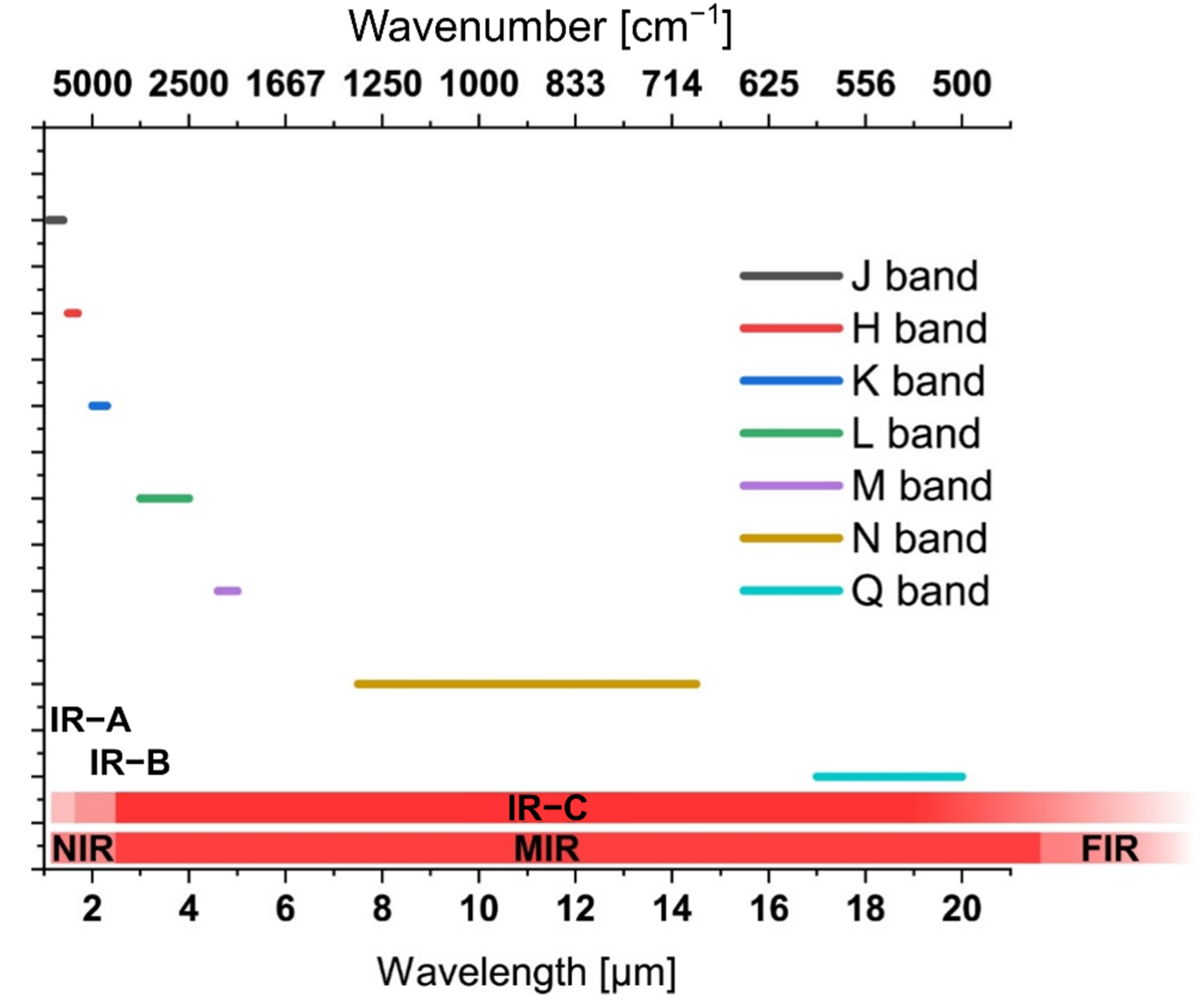
Figure 1. Electromagnetic wavelength regime defined as ‘infrared’ with associated classification of transitions.
Applsci 12 07598 g001
Figure 2. Growing number of scientific publications concerned with infrared spectroscopy. Results were retrieved from a SciFinder search querying for the term “infrared”, limiting the results to scientific journal publications. With a total number of over 1.2 M publications, the growth of infrared-related topics from the year 1924 to 2022 (2023 preliminary) is clearly evident. CAplus. CAS. https://scifinder.cas.org/ (accessed on 6 May 2022).
Applsci 12 07598 g002
Figure 3. Conceptual illustration of an instrument for infrared spectroscopy. Commonly, such a setup comprises a light source, a wavelength selective element, a signal transducer, which defines the interaction path length, and a radiation detection device. This core setup is typically accompanied by sampling accessories such as sample feed conduits, flow cells, etc. as well as a robust housing and system control, signal acquisition and data evaluation electronics and appropriate–in current times multivariate–data evaluation, mining, and classification software packages.
Applsci 12 07598 g003
Figure 4. Combination of FTIR spectroscopy with (a) an ATR system, (b) a micropath transmission cell, and (c) a GC (gas chromatography) detector (e.g., DTLaGS or MCT). Reprinted with permission from Ref. [17]. Copyright © 2017, John Wiley and Sons.
Applsci 12 07598 g004
Figure 5. Selected application scenarios where infrared spectroscopy is routinely applied, and which provide growing market shares for infrared spectroscopic tools. Selected images were used from pixabay (pixabay.com; accessed on 18 October 2020).
Applsci 12 07598 g005
Figure 6. Spectral transparency range of selected materials used for waveguiding applications in the infrared.
Applsci 12 07598 g006
Figure 7. Conceptual design of infrared spectroscopy/sensor devices with a high level/on chip integration providing all necessary components hybridized or monolithically integrated onto a single chip-sized die derived from CMOS/IC technology. Such chip-scale devices can be realized, for example, via on-chip laser sources and detectors. Analyte interaction may be realized via various approaches including but not limited to evanescent field interaction via straight strip dielectric waveguides (a), more sophisticated waveguide structures such as slot, bimodal or MZI waveguides (b), plasmonic waveguides (c), or by using resonant cavities with the possible addition of analyte scavenging/enriching motifs (d). Defined sample-photon interaction may be realized via microfluidic channels or hollow structures accommodating gas/vapor, liquid or solid sample matrices, respectively.
Applsci 12 07598 g007
Publisher’s Note: MDPI stays neutral with regard to jurisdictional claims in published maps and institutional affiliations.