Article

Valorization of Pineapple Residues from the Colombian Agroindustry to Produce Cellulose Nanofibers

by
Marcelo A. Guancha-Chalapud
1,
Liliana Serna-Cock
2,* and
Diego F. Tirado
3,*
1
Centro Nacional de Asistencia Técnica a la Industria (ASTIN), Servicio Nacional de Aprendizaje (SENA), Cali 760004, Colombia
2
Faculty of Engineering and Administration, Universidad Nacional de Colombia, Sede Palmira, Palmira 763533, Colombia
3
Dirección Académica, Universidad Nacional de Colombia, Sede de La Paz, La Paz 202017, Colombia
*
Authors to whom correspondence should be addressed.
Appl. Sci. 2022, 12(14), 6956; https://doi.org/10.3390/app12146956
Submission received: 12 June 2022 / Revised: 29 June 2022 / Accepted: 5 July 2022 / Published: 9 July 2022
(This article belongs to the Special Issue Knowledge-Based Biotechnology for Food, Agriculture and Fisheries)

Abstract

:
Cellulose nanofiber is the world’s most advanced biomass material. Most importantly, it is biodegradable. In this work, nanofibers were obtained from pineapple leaves, a large solid waste in Colombia, using a combined extraction method (chemical procedures and ultrasound). The native fibers were bleached, hydrolyzed, treated with ultrasound, and characterized by scanning electron microscopy (SEM), infrared analysis (FTIR), thermogravimetric analysis (TGA), and transmission electron microscopy (TEM). As a comparison, a commercial microcrystalline cellulose sample was analyzed, which demonstrated the efficiency of cellulose extraction. The nanofibers had a diameter and a length of 18 nm and 237 nm, respectively, with a maximum degradation temperature of 306 °C. The analysis showed the efficiency of acid treatment combined with ultrasound to obtain nanofibers and confirmed that pineapple residues can be valorized by this method. These results indicate that lignocellulosic matrices from pineapple leaves have potential application for obtaining polymeric-type composite materials. Due to their morphology and characteristic physical properties, the cellulose nanofibers obtained in this work could be a promising material for use in a wealth of fields and applications such as filter material, high gas barrier packaging material, electronic devices, foods, medicine, construction, cosmetics, pharmacy, and health care, among others.

1. Introduction

The urgency of today’s environmental problems has led academia and the industry to focus on increasing research and production of natural biodegradable materials [1,2,3]. In this sense, the availability of biopolymers, due to their abundance and accessibility in nature and their low cost, have led to their use in different interesting applications [3,4]. A clear example of biopolymers with these advantages is cellulose [3,5,6,7].
Aside from being one of the easiest organic compounds to obtain, cellulose is the most abundant natural, renewable, and biodegradable polysaccharide in the world [4,5], with an annual production yield greater than 10 × 1011 tons [2,8].
Cellulose contains amorphous regions in its structure that are susceptible to acid attack and allow the formation of nanofibrils [6]. These nanofibrils show excellent water dispersibility and are easy to obtain, handle, and shape. Moreover, nanofibrils are able to form interactions between units at the nanoscale, creating permeable networks connected by hydrogen bonds, which results in good dispersion in the polymer matrix [9].
Cellulose nanofibers are sustainable materials with high elastic modulus, tensile strength, transparency, chemical resistance, aspect ratio (length/diameter), and specific surface area. In addition, they have a low thermal expansion coefficient and remarkable optical, rheological, and film-forming properties [5,10]. Because of these properties, cellulose nanomaterials hold different applications such as absorbents in water treatment and antimicrobial materials. In fact, cellulose nanofibers have been used to turn contaminated water into drinking water; they have also been shown to be antimicrobial materials in protective healthcare equipment, food packaging, and ultrafiltration membranes [8].
Cellulose nanofibers have had a recent impact on industrial applications [5,7], mainly in the Japanese market. The cellulose nanofiber market size was valued at USD 346 million in 2021 and is projected to reach USD 1424 million by 2030, growing at a compound annual growth rate (CAGR) of 19% from 2022 to 2030 [11].
Cellulose nanofibers are generally obtained from agricultural sources such as wheat straw, cotton, coconut husk fibers, sisal fiber, bamboo waste, and sugarcane bagasse, among others [5,12]. Agro-industrial waste could also be a good source of cellulose nanofibers [13,14,15]. In this regard, circular economy [16,17] and revalorization strategies [18,19] are capable of revalorizing abundant and biodegradable agro-industrial waste that could play an important role in a globally sustainable future economy.
Colombia is one of the ten pineapple-producing countries in the world. Colombian pineapple production has increased at an annual rate of 12%, reaching over 1.2 million tons in 2021 [20]. Pineapple leaves are an abundant waste during pineapple transformation since leaves represent between 65% and 80% of the total crop; most of these come from the crown (leaves from the upper part of the fruit) and the stem. A pineapple plant in the harvested state contains 25 to 30 leaves, with diameters ranging from 45 µm to 205 µm. These leaves contain 2% to 3% fiber and are composed of approximately 70–80% cellulose, 17% hemicellulose, and 5% lignin [21].
Concentrated chemicals and mechanical methods over long periods of time degrade the cellulose during the isolation of nanofibers, reducing the degree of chain polymerization. The use of combined methods could decrease cellulose degradation and optimizes morphology and aspect ratio [14]; therefore, this work aimed to obtain cellulose nanofibers from pineapple waste using chemical methods combined with ultrasound. Then, to demonstrate the efficiency of these methods, fibers were characterized by scanning electron microscopy (SEM), infrared analysis (FTIR), thermogravimetric analysis (TGA), and transmission electron microscopy (TEM).

2. Materials and Methods

2.1. Reagents

Hydrochloric acid solution (HCl, 37% w v−1, Sigma-Aldrich, St. Louis, MO, USA), ammonium hydroxide (Sigma-Aldrich, St. Louis, MO, USA), sodium hydroxide (NaOH, pellets, Merck, Darmstadt, Germany), sodium chlorite (NaClO2, crystals, Sigma-Aldrich, St. Louis, MO, USA), glacial acetic acid (CH3COOH, 99.7%, Merck, Darmstadt, Germany), sulfuric acid solution in water type II (H2SO4, 96%, Merck, Darmstadt, Germany), and nitrogen (99.99%, Linde, Dublin, Ireland) were used. All materials were used as received. The water used in this work was distilled and came from our laboratory.

2.2. Pineapple Waste

Pineapple leaf samples were obtained from the Bengala Agrícola S.A. company (Pradera, Valle del Cauca, Colombia). Samples were washed with distilled water and cut with stainless steel scissors into pieces of about 10 mm. Then, samples were subjected to freeze-drying (Freezone 6 Plus, Labconco, Kansas City, MO, USA) at −40 °C and 0.01 mbar for 24 h. The dried samples were ground in a blade mill to a size of 38 µm (400 mesh), placed in airtight polyethylene bags, and stored in a desiccator at room temperature.

2.3. Obtention of Cellulose Nanofibers

2.3.1. Native Nanofibers

First, native fibers (PL-N) were obtained. For this, dried samples were stirred with a hot plate (Corning®, PC-420D, Corning, NY, USA) in 0.01 N HCl solution (1:10 ratio) at 600 rpm and 70 °C for 2 h to remove fat and pectin from the samples [22]. Subsequently, ammonium hydroxide was added until the pH value of the mixture was adjusted to 9.5 [23]. Finally, the fibers were vacuum filtered, washed with distilled water to neutral pH, and dried at 50 °C in a forced convection oven (UN 110, Memmert, Schwabach, Germany) until constant weight was achieved, obtaining PL-N.

2.3.2. Obtention of Nanofibers by Chemical Methods Combined with Ultrasound

Cellulose nanofibers from native fibers were obtained according to previous reports [13,14]. For this, PL-N were both delignified and blanched (PL-DB). To delignify, the native fiber was mixed with a 4% w v−1 aqueous NaOH solution (1:25 ratio) at 600 rpm and 90 °C for 4 h. For blanching, the delignified material was mixed (1:20 ratio) with a solution resulting from the mixture (1:1 ratio) of a 1.7% w v−1 aqueous NaClO2 solution and an acetate buffer solution (27 g NaOH + 75 mL CH3COOH L water−1). Afterwards, this solution was stirred at 600 rpm and 80 °C for 6 h. Finally, the fibers were vacuum filtered (Millipore, Merck, Darmstadt, Germany), washed with water (1:1 ratio) until neutral pH was reached, and dried in an oven at 50 °C until constant weight was achieved.
To obtain nanofibers through acid hydrolysis (PL-H) combined with ultrasound (PL-HUS), 5 g of previously obtained samples was stirred at 400 rpm with 125 mL of H2SO4 6.5 M for 24 h at 50 °C. Then, the mixture was diluted in 300 mL of water and neutralized with 1600 mL of 4% aqueous NaOH solution. Afterwards, the precipitate was vacuum filtered (Millipore, Merck, Darmstadt, Germany), washed with water (1:1 ratio), and dried at 50 °C for 24 h. Using this dried sample, 100 mL of an aqueous suspension (1% w v−1) was prepared and mixed well with an ULTRA-TURRAX® (T25, IKA, Staufen, Germany) at 10,000 rpm for 15 min. At this point, the PL-H nanofibers were obtained. Afterwards, suspensions were ultrasonically homogenized (SFX550, Branson, Barcelona, Spain) with a ½″ diameter cylindrical probe at 20 kHz and 550 W for 30 min. The sample was kept between 25 °C and 30 °C in an ice bath. The obtained nanofiber suspensions were freeze-dried at an ice condenser temperature of −40 °C and a pressure of 0.01 mbar for 24 h. At this point, the PL-HUS nanofibers were obtained and stored in hermetic containers at 5 °C for further analysis.

2.4. Characterization of the Nanofibers

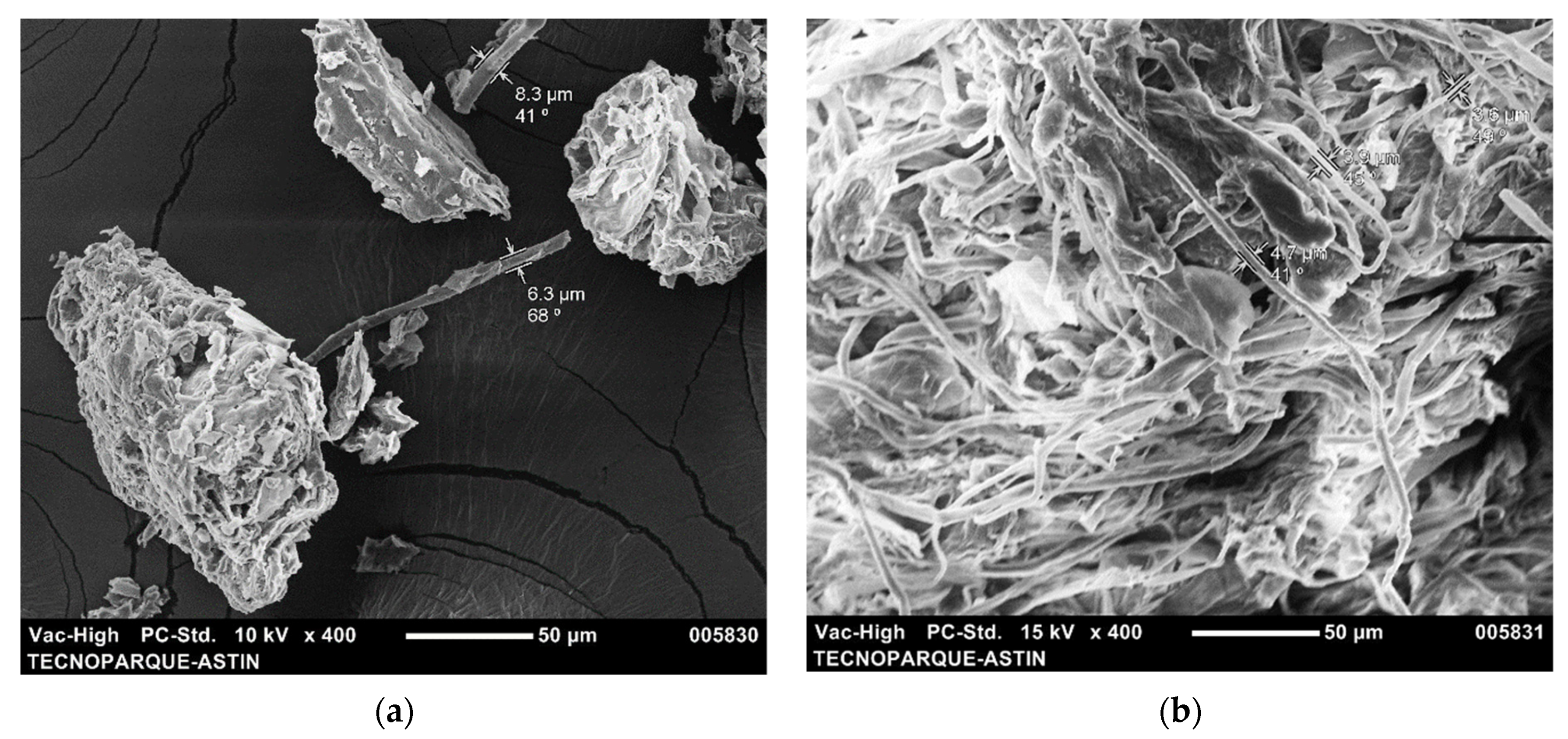
2.4.1. Scanning Electron Microscopy (SEM)

The surface morphology of the fibers was analyzed with a scanning electron microscope (SEM, JEOL Model JSM-6490, Akishima, Japan). For this, the sample was fixed on a double-sided carbon tape attached to the sample holder and then it was coated with gold (layer thickness: 5 nm) using a Cressington 108 Auto Sputter Coater (Ted Pella, Redding, CA, USA).

2.4.2. Infrared Analysis (FITR)

FTIR spectra were obtained between 400 cm−1 and 4000 cm−1. Commercial microcrystalline cellulose (CC, cellulose microcrystalline, Merck, Darmstadt, Germany) was used as a control treatment.

2.4.3. Thermogravimetric Analysis (TGA)

Thermograms (DSC/TGA 2 Star system, Mettler Toledo, Columbus, OH, USA) were obtained between 30 °C and 600 °C at 10 °C min−1 and 20 mL nitrogen min−1. CC was used as a control treatment.

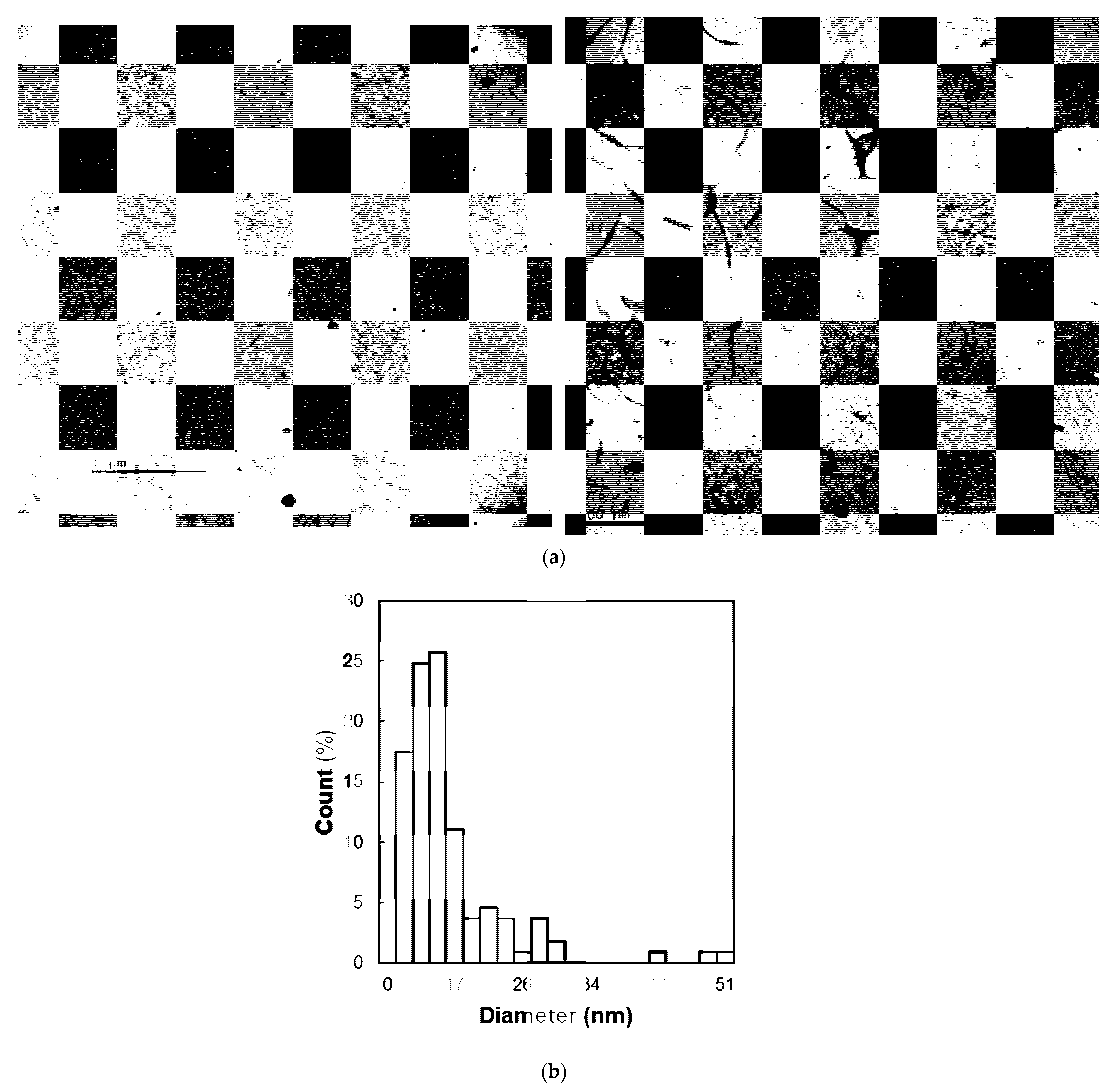
2.4.4. Transmission Electron Microscopy (TEM)

Dispersions of 0.1% w w−1 nanofibers in water were prepared and mounted on copper grids. Then, samples were photographed with a transmission electron microscope (TEM, JEOL JEM 1.011, Akishima, Japan). Finally, to estimate the diameter and length of these samples, three representative TEM micrographs were analyzed using an image processing software (Image J, National Institutes of Health, Bethesda, MD, USA) [14].

3. Results and Discussion

3.1. Morphology Analysis

In order to verify that the fiber bleaching process was efficient to eliminate the lignin present in native nanofiber, the obtained samples were analyzed by SEM. Figure 1 shows SEM images of native (see PL-N in Figure 1a) and delignified–blanched nanofibers (see PL-DB in Figure 1b). As can be seen in Figure 1a, PL-N were non-homogeneous due to the presence of compounds (such as lignin, hemicellulose, and pectin, among others) which acted as binders, preventing the defibrillation of the cellulose chains [24]. In Figure 1b, on the other hand, a morphological change in fibrils with a defined and uniform diameter with a size of roughly 3.6 µm was observed, indicating that the lignin and hemicellulose that enveloped the cellulose fibers had been eliminated by the effect of delignification and blanching [25].

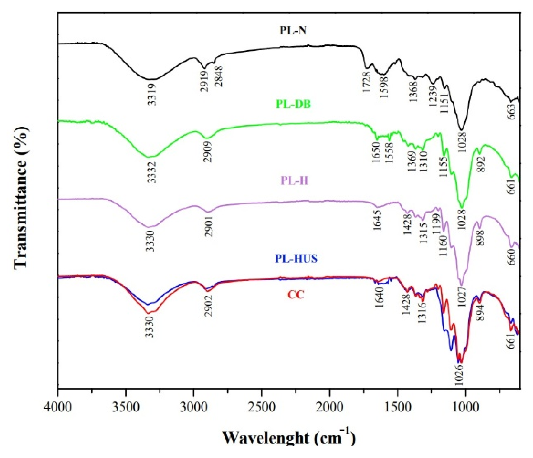
3.2. Functional Groups Analysis

Infrared (see Figure 2) and thermal stability (see Figure 3) analyses of PL-N, PL-DB, PL-H, and PL-HUS nanofibers were performed to demonstrate the efficiency of bleaching, hydrolysis acid, and ultrasound treatments. Regarding PL-N, two absorption zones were observed (see Figure 2): 600 cm−1–1750 cm−1 and 2800 cm−1–3400 cm−1. The absorption band found at 3332 cm−1 corresponded to the stretching of the hydroxyl groups (-OH). The peak at 2860 cm−1 corresponded to the C-H stretching present in cellulose, hemicellulose, and lignin [26]. Meanwhile, the peak at 1739 cm−1 corresponded to the C=O vibrations in the ester groups (-COO- and H3CO-) characteristic of hemicellulose, or in the ester bond of the carboxylic group (-COOH) characteristic of the ferulic and p-coumaric acids belonging to lignin [26,27]. The peak at 1240 cm−1 was related to the -C-O-C- vibration found in the bonds between the aromatic ring and methoxy groups in lignin [28]. The peaks at 1647 cm−1, 1562 cm−1, and 1460 cm−1 referred to the C=C stretching in the aromatic rings in lignin [24,25,26,28]. Finally, the peak at 1028 cm−1 corresponded to the C-O-C stretching of the β-1,4-glycosidic ring bonds between the D-glucose units in cellulose [24].
Figure 2 also includes infrared spectra of PL-DB, PL-H, PL-HUS, and CC. Figure 2 shows that the bands around 1240 cm−1, 1647 cm−1, and 1740 cm−1 disappeared after the delignification–bleaching process (see PL-DB in Figure 2), which was proof of the removal of lignin and hemicellulose during the treatment [29]. Additionally, Figure 2 shows the disappearance of the peak around 2848 cm−1 for PL-DB, PL-H, and PL-US. According to Salim et al. [30], a peak around 2850 cm−1 corresponds to the C-H stretching vibration corresponding to the methyl (-CH3) and methoxy (O-CH3) groups that form the aromatic ring of lignin. Consequently, a disappearance of this peak after treatment could be attributed to lignin removal.
Meanwhile, FTIR spectra of PL-HUS (see Figure 2) show a peak around 1000 cm−1, which was related to the C-O-C vibration of the β-1,4-glycosidic bond between the D-glucose units in the cellulose [24]. Figure 2 also shows an increase in the intensity of the bands around 1000 cm−1, 3300 cm−1, and 2900 cm−1, which is typical of the functional groups of cellulose, which were prominent in the PL-H and PL-HUS spectra. In these last ones, the appearance of bands at 1100 cm−1, 1300 cm−1, and 890 cm−1 was also noted, which became more intense due to the effect of the treatment with H2SO4 and ultrasound. Additionally, a band around 1400 cm−1 and the increase in the intensity of the bands around 1360 cm−1 in the PL-HUS spectrum were evident, which agreed with that reported by Julie Chandra et al. [24], who indicated that sharp signals around 1426 cm−1–897 cm−1 and 1370 cm−1–2900 cm−1 reflect the cellulose crystalline band.

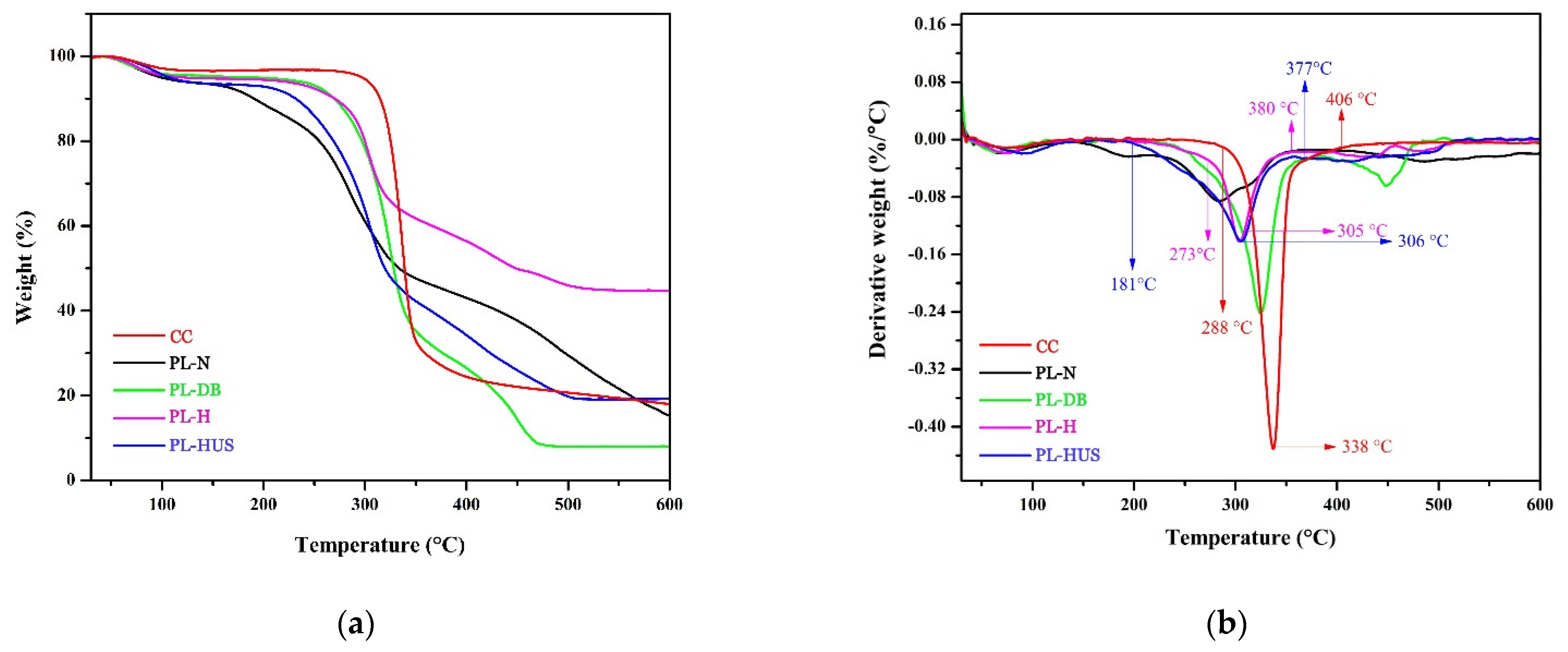
3.3. Thermal Stability Analysis

To demonstrate the efficiency of the chemical methods combined with ultrasound to produce cellulose nanofibers, thermal stability analyses (see Figure 3) were performed. Figure 3a,b show the mass loss and thermogravimetric derivatives, respectively, of PL-N, PL-DB, PL-H, and PL-HUS nanofibers.
A first weak peak was found between 50 °C and 150 °C for all samples in Figure 3. This peak was related to the water evaporation. Another pronounced peak was found between 400 °C and 500 °C, and maximum degradation points were observed in the range of 200 °C–400 °C. For PL-H and PL-HUS, a single maximum degradation point was found at around 306 °C, compared to PL-N, indicating higher thermal stability due to the removal of less thermally stable substances such as hemicellulose and pectin. These results were attributed to the chemical treatments combined with ultrasound [31]. However, it was also evident in Figure 3 that the degradation temperature of PL-HUS was lower with reference to the PL-DB sample (315 °C). As reported by Vanderfleet et al. [32] this lower degradation temperature of PL-HUS could be attributed to the fact that the acid treatment resulted in shorter cellulose chains and, consequently, in a lower degree of polymerization and in an increase in the surface charge density, which led to a lower degradation temperature.
Additionally, it was observed that the maximum degradation temperature of CC was higher (338 °C), compared to the produce nanofibers (LP-HUS, 306 °C), which could be attributed to the number of free terminal chains, which decompose at lower temperatures [33,34]. Hydrolysis with H2SO4 caused a significant decrease in the degradation temperature and an increase in the maximum degradation temperature range for PL-HUS between 181 °C and 360 °C, caused by the inclusion of sulfate groups in the cellulose chains [33,35].
The drying method is another factor that affects the thermal stability of cellulose nanofibers due to fiber rearrangements caused by water removal. By conventional sample drying, nanofibers are free to form a homogeneous phase with defined particle spacing, whereas freeze-drying requires initial freezing. In this state, the nanofibrils cannot be rearranged during water sublimation, resulting in dispersed nanofibril clusters and a large specific surface area, which affects thermal stability [36]. It was important to consider the aforementioned in this work, as the samples were dried in a circulating air oven after the hydrolysis process was completed and by freeze-drying after sonication.

3.4. Particle Size Analysis of Cellulose Nanofibers

Figure 4 shows the TEM micrographs (Figure 4a) and size distribution (Figure 4b) of the PL-HUS nanofibers obtained by H2SO4 treatment combined with ultrasound. In agreement with other studies [37,38] where it was claimed that cellulose nanofibers had a typical elongated shape, whose diameter varied from 10 nm to 100 nm, the length and diameter of the nanofibers in this work were 237 nm ± 33 nm and 18 nm ± 7 nm, respectively.
Some authors have discussed the reasons for the effect of ultrasound applied after acidic hydrolysis on the production of smaller nanofiber particle sizes. According to Csiszar et al. [39], sonication has a crucial impact on the particles in liquid suspension of nanocellulose since the microjets involved in the sonicated suspension can disintegrate the large micron-sized aggregates of nanowhiskers, decreasing the particle size gradually, as can be seen in Figure 4a. Likewise, in their study of nanocellulose preparation from ginkgo seed shells, Li et al. [40] indicated that ultrasonic treatments can change the particle size due to the sound energy provided by acoustic cavitation. These authors mentioned that the violent collapse of bubbles produces microjets and shock waves on the surfaces of suspended cellulose particles, resulting in the scission effect on micron-sized cellulose particles. This scission effect can break the relatively weak interfaces between the fibers, which are bound together mainly by van der Waals forces and hydrogen bonds.

4. Conclusions and Perspectives

Due to its strong mechanical and structural functions, cellulose nanofiber has gained popularity for sustainable production in recent years [2]. The use of cellulose nanofiber from natural sources has grown significantly as the industry shifts away from unsustainable fossil fuel resources and looks toward a softer and more sustainable environmental approach.
Colombia currently produces more than one million tons of pineapple per year [20], which indicates that the country has a production potential of 800.000 tons of solid waste that is up to 80% cellulose [41], which gives this waste commercialization potential and the possibility of adding value to a Colombian pineapple waste.
In Colombia, the use of petrochemical plastics has recently been regulated, which calls for the development of new biodegradable packaging materials. Such materials could be reinforced with nanocellulose fibers from pineapple leaves, which could mechanically reinforce thermosets and thermoplastics. Hydrophobic properties could also be enhanced by adding cellulose nanofibers as nanocomposites for adhesion in fiber-reinforced composites such as epoxies.
Due to the morphology and characteristic physical properties of the obtained materials, cellulose nanofiber produced in this work could be a promising material for use in a wealth of fields and applications such as filter material, high gas barrier packaging material, electronic devices, foods, medicine, construction, cosmetics, pharmacy, and health care [2]: (i) due to their large specific surface area, cellulose nanofibers can accumulate small dust particles, which demonstrates their potential for air filtration (they could even support the development of nanofoams and aerogels to filter particles from the air); (ii) cellulose nanofibers are a great barrier against oxygen (due to their > 40% crystallinity) and have low thermal expansion, properties that give them potential for use in food production and preservation since cellulose nanofibers could act as gas barrier films to reduce air flow and to maintain food freshness, thus acting as biodegradable packaging; (iii) for construction, nanocellulose fiber coating materials could protect from ultraviolet rays and oxygen better than acrylic resin, so it has a higher potential to suppress the generation of radicals, such as discoloration and cracking; (iv) the thermal stability of cellulose nanofibers makes them excellent materials for use in electronic devices as they exhibit low porosity and roughness, increase print resolution, make electronic devices more conductive, and can prevent short circuits by distorting water permeation in electronic circuits; and finally, (v) these produced materials could be used as adsorbent, antimicrobial material and for controlled drug delivery.

Author Contributions

Conceptualization, M.A.G.-C., L.S.-C. and D.F.T.; data curation, M.A.G.-C.; formal analysis, M.A.G.-C., L.S.-C. and D.F.T.; funding acquisition, M.A.G.-C., L.S.-C. and D.F.T.; investigation, M.A.G.-C.; methodology, M.A.G.-C., L.S.-C. and D.F.T.; project administration, L.S.-C. and D.F.T.; resources, M.A.G.-C., L.S.-C. and D.F.T.; software, M.A.G.-C., L.S.-C. and D.F.T.; supervision, L.S.-C. and D.F.T.; validation, M.A.G.-C., L.S.-C. and D.F.T.; visualization, M.A.G.-C., L.S.-C. and D.F.T.; writing—original draft, M.A.G.-C., L.S.-C. and D.F.T.; writing—review and editing, D.F.T. All authors have read and agreed to the published version of the manuscript.

Funding

This research was funded by ASTIN-SENA (project reference 11796).

Institutional Review Board Statement

Not applicable.

Informed Consent Statement

Not applicable.

Data Availability Statement

The data that support the findings of this study are available from all authors upon request.

Acknowledgments

The authors thank the Research Group on Development of Materials and Products (GIDEMP, ASTIN), students of the Technology in Chemistry Applied to Industry (SENA), and the biotechnology laboratories of Tecnoparque Nodo Cali.

Conflicts of Interest

The authors declare no conflict of interest. The funders had no role in the design of the study; in the collection, analyses, or interpretation of data; in the writing of the manuscript; or in the decision to publish the results.

References

  1. Abazari, M.F.; Gholizadeh, S.; Karizi, S.Z.; Birgani, N.H.; Abazari, D.; Paknia, S.; Derakhshankhah, H.; Allahyari, Z.; Amini, S.M.; Hamidi, M.; et al. Recent advances in cellulose-based structures as the wound-healing biomaterials: A clinically oriented review. Appl. Sci. 2021, 11, 7769. [Google Scholar] [CrossRef]
  2. Lam, W.S.; Lee, P.F.; Lam, W.H. Cellulose nanofiber for sustainable production: A bibliometric analysis. Mater. Today Proc. 2022, 62 Pt 12, 6460–6467. [Google Scholar] [CrossRef]
  3. Spagnol, C.; Rodrigues, F.H.A.; Neto, A.G.V.C.; Pereira, A.G.B.; Fajardo, A.R.; Radovanovic, E.; Rubira, A.F.; Muniz, E.C. Nanocomposites based on poly(acrylamide-co-acrylate) and cellulose nanowhiskers. Eur. Polym. J. 2012, 48, 454–463. [Google Scholar] [CrossRef]
  4. Uusi-Tarkka, E.-K.; Skrifvars, M.; Haapala, A. Fabricating sustainable all-cellulose composites. Appl. Sci. 2021, 11, 10069. [Google Scholar] [CrossRef]
  5. Moohan, J.; Stewart, S.A.; Espinosa, E.; Rosal, A.; Rodríguez, A.; Larrañeta, E.; Donnelly, R.F.; Domínguez-Robles, J. Cellulose nanofibers and other biopolymers for biomedical applications. A review. Appl. Sci. 2019, 10, 65. [Google Scholar] [CrossRef] [Green Version]
  6. Aguilar-Rosero, J.; Urbina-López, M.E.; Rodríguez-González, B.E.; León-Villegas, S.X.; Luna-Cruz, I.E.; Cárdenas-Chávez, D.L. Development and characterization of bioadsorbents derived from different agricultural wastes for water reclamation: A review. Appl. Sci. 2022, 12, 2740. [Google Scholar] [CrossRef]
  7. Jarpa-Parra, M.; Chen, L. Applications of plant polymer-based solid foams: Current trends in the food industry. Appl. Sci. 2021, 11, 9605. [Google Scholar] [CrossRef]
  8. Phuong, H.T.; Thoa, N.K.; Tuyet, P.T.A.; Van, Q.N.; Hai, Y.D. Cellulose nanomaterials as a future, sustainable and renewable material. Crystals 2022, 12, 106. [Google Scholar] [CrossRef]
  9. Spagnol, C.; Rodrigues, F.; Pereira, A.; Fajardo, A.; Rubira, A.; Muniz, E. Superabsorbent hydrogel composite made of cellulose nanofibrils and chitosan-graft-poly(acrylic acid). Carbohydr. Polym. 2012, 87, 2038–2045. [Google Scholar] [CrossRef] [Green Version]
  10. Xie, Y.; Kurita, H.; Ishigami, R.; Narita, F. Assessing the flexural properties of epoxy composites with extremely low addition of cellulose nanofiber content. Appl. Sci. 2020, 10, 1159. [Google Scholar] [CrossRef] [Green Version]
  11. Research and Markets. The Global Market for Cellulose Nanofibers to 2031; Future Markets, Inc.: Dublin, Ireland, 2022. [Google Scholar]
  12. Soni, B.; Hassan, E.B.; Mahmoud, B. Chemical isolation and characterization of different cellulose nanofibers from cotton stalks. Carbohydr. Polym. 2015, 134, 581–589. [Google Scholar] [CrossRef] [PubMed]
  13. Guancha-Chalapud, M.A.; Serna-Cock, L.; Tirado, D.F. Hydrogels are reinforced with Colombian fique nanofibers to improve techno-functional properties for agricultural purposes. Agriculture 2022, 12, 117. [Google Scholar] [CrossRef]
  14. Guancha-Chalapud, M.A.; Gálvez, J.; Serna-Cock, L.; Aguilar, C.N. Valorization of Colombian fique (Furcraea bedinghausii) for production of cellulose nanofibers and its application in hydrogels. Sci. Rep. 2020, 10, 11637. [Google Scholar] [CrossRef] [PubMed]
  15. Serna Cock, L.; Guancha-Chalapud, M.A. Natural fibers for hydrogels production and their applications in agriculture. Acta Agronómica 2017, 66, 495–505. [Google Scholar] [CrossRef] [Green Version]
  16. Pedrazzoli, P.; Sorlini, M.; Rovere, D.; Lazaro, O.; Malò, P.; Fiorello, M. Challenges and founding pillars for a manufacturing platform to support value networks operating in a circular economy framework. Appl. Sci. 2022, 12, 2995. [Google Scholar] [CrossRef]
  17. Righi, C.; Barbieri, F.; Sgarbi, E.; Maistrello, L.; Bertacchini, A.; Andreola, F.N.; D’Angelo, A.; Catauro, M.; Barbieri, L. Suitability of porous inorganic materials from industrial residues and bioproducts for use in horticulture: A multidisciplinary approach. Appl. Sci. 2022, 12, 5437. [Google Scholar] [CrossRef]
  18. Adler, I.; Kotta, J.; Tuvikene, R.; Kaldre, K. Optimizing the processing of shellfish (Mytilus edulis and M. trossulus hybrid) miomass cultivated in the low salinity region of the Baltic sea for the extraction of meat and proteins. Appl. Sci. 2022, 12, 5163. [Google Scholar] [CrossRef]
  19. Fernandes, A.; Cruz-Lopes, L.; Dulyanska, Y.; Domingos, I.; Ferreira, J.; Evtuguin, D.; Esteves, B. Eco valorization of Eucalyptus globulus bark and branches through liquefaction. Appl. Sci. 2022, 12, 3775. [Google Scholar] [CrossRef]
  20. Martinez, C.; Menjívar, J.; Saavedra, R. Soils erosion in pineapple (Ananas comosus L. Merr) producing areas. Rev. Ciencias Agrícolas 2022, 39, 6366. [Google Scholar]
  21. Nashiruddin, N.I.; Mansor, A.F.; Rahman, R.A.; Ilias, R.M.; Yussof, H.W. Process parameter optimization of pretreated pineapple leaves fiber for enhancement of sugar recovery. Ind. Crops Prod. 2020, 152, 112514. [Google Scholar] [CrossRef]
  22. Cheng, S.; Panthapulakkal, S.; Sain, M.; Asiri, A. Aloe vera rind cellulose nanofibers-reinforced films. J. Appl. Polym. Sci. 2014, 131, 40592. [Google Scholar] [CrossRef]
  23. Ramezani Kakroodi, A.; Cheng, S.; Sain, M.; Asiri, A. Mechanical, thermal, and morphological properties of nanocomposites based on polyvinyl alcohol and cellulose nanofiber from Aloe vera rind. J. Nanomater. 2014, 2014, 903498. [Google Scholar] [CrossRef] [Green Version]
  24. Julie Chandra, C.S.; George, N.; Narayanankutty, S.K. Isolation and characterization of cellulose nanofibrils from arecanut husk fibre. Carbohydr. Polym. 2016, 142, 158–166. [Google Scholar] [CrossRef]
  25. Xie, J.; Hse, C.-Y.; Li, C.; Shupe, T.F.; Hu, T.; Qi, J.; De Hoop, C.F. Characterization of microwave liquefied bamboo residue and its potential use in the generation of nanofibrillated cellulosic fiber. ACS Sustain. Chem. Eng. 2016, 4, 3477–3485. [Google Scholar] [CrossRef]
  26. Zhang, P.P.; Tong, D.S.; Lin, C.X.; Yang, H.M.; Zhong, Z.K.; Yu, W.H.; Wang, H.; Zhou, C.H. Effects of acid treatments on bamboo cellulose nanocrystals. Asia-Pacific J. Chem. Eng. 2014, 9, 686–695. [Google Scholar] [CrossRef]
  27. Li, R.; Fei, J.; Cai, Y.; Li, Y.; Feng, J.; Yao, J. Cellulose whiskers extracted from mulberry: A novel biomass production. Carbohydr. Polym. 2009, 76, 94–99. [Google Scholar] [CrossRef]
  28. Xie, J.; Hse, C.; De Hoop, C.F.; Hu, T.; Qi, J.; Shupe, T.F. Isolation and characterization of cellulose nanofibers from bamboo using microwave liquefaction combined with chemical treatment and ultrasonication. Carbohydr. Polym. 2016, 151, 725–734. [Google Scholar] [CrossRef]
  29. Célino, A.; Gonçalves, O.; Jacquemin, F.; Fréour, S. Qualitative and quantitative assessment of water sorption in natural fibres using ATR-FTIR spectroscopy. Carbohydr. Polym. 2014, 101, 163–170. [Google Scholar] [CrossRef] [Green Version]
  30. Md Salim, R.; Asik, J.; Sarjadi, M.S. Chemical functional groups of extractives, cellulose and lignin extracted from native Leucaena leucocephala bark. Wood Sci. Technol. 2021, 55, 295–313. [Google Scholar] [CrossRef]
  31. Carrier, M.; Loppinet-Serani, A.; Denux, D.; Lasnier, J.; Ham-Pichavant, F.; Cansell, F.; Aymonier, C. Thermogravimetric analysis as a new method to determine the lignocellulosic composition of biomass. Biomass Bioenergy 2011, 35, 298–307. [Google Scholar] [CrossRef]
  32. Vanderfleet, O.M.; Reid, M.S.; Bras, J.; Heux, L.; Godoy-Vargas, J.; Panga, M.K.R.; Cranston, E.D. Insight into thermal stability of cellulose nanocrystals from new hydrolysis methods with acid blends. Cellulose 2019, 26, 507–528. [Google Scholar] [CrossRef]
  33. Barbash, V.A.; Yaschenko, O.V.; Alushkin, S.V.; Kondratyuk, A.S.; Posudievsky, O.Y.; Koshechko, V.G. Effect of mechanochemical treatment of cellulose on characteristics of nanocellulose films. Springer Proc. Phys. 2016, 183, 513–521. [Google Scholar] [CrossRef] [Green Version]
  34. Hashaikeh, R.; Abushammala, H. Acid mediated networked cellulose: Preparation and characterization. Carbohydr. Polym. 2011, 83, 1088–1094. [Google Scholar] [CrossRef]
  35. Roman, M.; Winter, W.T. Effect of sulphate groups from sulphuric acid hydrolysis on the thermal degradation behaviour of bacterial cellulose. Biomacromolecules 2004, 1671–1677. [Google Scholar] [CrossRef] [PubMed]
  36. Rämänen, P.; Penttilä, P.A.; Svedström, K.; Maunu, S.L.; Serimaa, R. The effect of drying method on the properties and nanoscale structure of cellulose whiskers. Cellulose 2012, 19, 901–912. [Google Scholar] [CrossRef]
  37. Song, Y.K.; Leng Chew, I.M.; Yaw Choong, T.S.; Tan, J.; Tan, K.W. Isolation of Nanocrystalline Cellulose from oil palm empty fruit bunch—A response surface methodology study. MATEC Web Conf. 2016, 60, 04009. [Google Scholar] [CrossRef]
  38. Vieyra, H.; Figueroa-López, U.; Guevara-Morales, A.; Vergara-Porras, B.; San Martín-Martínez, E.; Aguilar-Mendez, M.Á. Optimized monitoring of production of cellulose nanowhiskers from Opuntia ficus-indica (nopal cactus). Int. J. Polym. Sci. 2015, 2015, 871345. [Google Scholar] [CrossRef]
  39. Csiszar, E.; Kalic, P.; Kobol, A.; de Paulo Ferreira, E. The effect of low frequency ultrasound on the production and properties of nanocrystalline cellulose suspensions and films. Ultrason. Sonochem. 2016, 31, 473–480. [Google Scholar] [CrossRef]
  40. Ni, Y.; Li, J.; Fan, L. Effects of ultrasonic conditions on the interfacial property and emulsifying property of cellulose nanoparticles from ginkgo seed shells. Ultrason. Sonochem. 2021, 70, 105335. [Google Scholar] [CrossRef]
  41. Cherian, B.M.; Leão, A.L.; de Souza, S.F.; Costa, L.M.M.; de Olyveira, G.M.; Kottaisamy, M.; Nagarajan, E.R.; Thomas, S. Cellulose nanocomposites with nanofibres isolated from pineapple leaf fibers for medical applications. Carbohydr. Polym. 2011, 86, 1790–1798. [Google Scholar] [CrossRef]
Figure 1. Scanning electron micrographs (SEM) of (a) native (PL-N) and (b) delignified–blanched (PL-DB) nanofibers from pineapple leaves.
Applsci 12 06956 g001
Figure 2. Infrared spectrum (FITR) of native (PL-N), delignified–blanched (PL-DB), hydrolyzed (PL-H), and ultrasound (PL-HUS) nanofibers from pineapple waste, along with the commercial microcrystalline cellulose (CC).
Applsci 12 06956 g002
Figure 3. (a) Thermogravimetric and (b) derivative thermogravimetric analysis of native (PL-N), delignified–blanched (PL-DB), hydrolyzed (PL-H), and ultrasound (PL-HUS) nanofibers from pineapple leaves, along with the commercial microcrystalline cellulose (CC).
Applsci 12 06956 g003
Figure 4. (a) Transmission electron micrographs (TEM) and (b) size distribution (mean diameter 18 ± 7) of produced nanofibers (PL-HUS) from pineapple waste.
Applsci 12 06956 g004
Publisher’s Note: MDPI stays neutral with regard to jurisdictional claims in published maps and institutional affiliations.

