Abstract
Lungworts (members of genus Pulmonaria), especially Pulmonaria officinalis, have been used as medicinal plants in folk and traditional medicine. The potential biological activities of lungworts, such as antioxidant, anti-inflammatory, anti-neurodegenerative, skin whitening, anticoagulant, antibacterial, anti-anemic, anticonvulsant, and wound-healing properties, have been observed in different studies. More than 90 phytochemicals belonging to pharmacologically active phytochemical classes have been reported for lungworts, which can be considered as one of the important contributors to the biological properties along with micronutrients. However, safety studies and clinical trials are missing for lungworts to establish most of their potential biological properties. Similarly, in vivo studies are lacking for anti-inflammatory and anti-neurodegenerative disorders and more in vivo studies are required to strengthen the knowledge of their antioxidant, anticoagulant, and anti-convulsant properties. A comprehensive compilation of the phytochemicals and biological properties of lungworts is also lacking in the literature, which can provide future direction for further pharmacological research. Hence, an attempt has been made in this study to compile the phytochemicals and biological properties to not only provide the resources for the design of further research to develop the pharmacological application of lungworts, but also to highlight the gaps and provide suggestions for future development.
1. Introduction
The members of the genus Pulmonaria commonly known as lungwort belong to the family Boraginaceae. It comprises around 18 species of perennial flowering plants that are widely dispersed across Europe, Russia, and Western Asia [1]. Lungwort (especially Pulmonaria officinalis) has been used for medicinal purposes since ancient times due to the Doctrine of Signatures as it resembles diseased lungs [2]. Lungwort has been known for its use for pulmonary diseases in folk medicine without the knowledge of its precise mechanisms of action. The ethnomedicinal use of lungwort includes emollient, antitussive, expectorant, antimicrobial, diuretic, depurative, antilithiatic, and anti-inflammatory usage, as well as in respiratory and urinary disorders [3,4].
The leaves and flowers from Pulmonaria are known to have been used as a wild plant food in the past [5,6]. Pulmonaria flowers can be considered as feeding stations for insects and P. officinalis is also included in most common honey plants in Estonia [7]. The flowers are shaped like cylindrical tubes, which have five-lobed corolla at the ends. The size of the flower is around 0.5–1.5 cm in diameter, and in its center is the style, which can be short (thrum) or long (pin). Both pin and thrum flowers are present on the same plant in the Pulmonaria species [8]. Generally, most of the Pulmonaria plants are spring-flowering woodlanders and prefer humus-rich, moist soil to grow (they do not favor dry or waterlogged soil).
Several commercial products from lungwort (P. officinalis) are available on the market, such as mother tincture, leaves, dried aerial parts (commercially available as Pulmonaria herba), ingredients of various dietary supplements, as tea, and as cosmetics [9]. Different forms/parts of lungworts such as the extract from the aerial parts, leaves, roots, callus culture, isolated compounds, and nanoparticles have been used to explore/study their biological properties in in vitro and/or in vivo experiments. Several potential biological properties such as antioxidant, anti-inflammation, antianemic, anticoagulant, wound healing, anti-neurodegenerative, anticonvulsant and skin whitening activities have been studied for lungwort [10,11,12,13,14]. These biological activities have the potential to be developed for medicinal and/or food supplement usage in different diseases or conditions. P. officinalis is also listed in the national health product databases of different countries for possible pharmaceutical use, such as the National Database of Health Protection Products of Poland and the Natural Health Products Ingredients Database of Canada (http://webprod.hc-sc.gc.ca/nhpid-bdipsn/atReq.do?atid=lungwort.oral&lang=eng; accessed on 30 March 2022). The biological properties of lungworts may be attributed to the diverse set of phytochemicals, such as anthocyanins, alkaloids, flavanone, flavonols, flavones, hydroxycinnamic acid, lignans, polyphenols, polyphenolic acids, and other phytochemicals present in different parts of the plants. In the literature, more than a hundred phytochemicals have been reported from lungwort.
Limited studies have been carried out on lungworts for their medicinal properties to be developed as approved therapeutic approaches or supplements for diseases or conditions. However, a comprehensive compilation of the biological properties and phytochemicals present in lungworts is not available, which could provide the gaps, highlight the challenges, and suggest possible solutions for the further development of lungwort as a food supplement and/or therapeutic candidate for pharmacological applications.
2. Phytochemicals Present in Pulmonaria
The most important reason for the different biological activities reported in Pulmonaria could be the phyto-constituents present in these plants. These phyto-constituents may include saponins, tannins, ascorbic acid, silicic acid, rutin, carotene, vitamin C, allantonin, alkaloids, and fatty acid. In initial studies, the presence of broad classes of phytochemicals was studied from the extract of lungworts to decipher the pharmacological properties.
The polyphenol content of the plants used in Bulgarian phytotherapy for the treatment of respiratory and inflammatory conditions was evaluated [15]. P. officinalis was selected among the plants, as it was considered important for evaluation due to the presence of both properties in the ethnopharmacology literature [15]. An aqueous extract was prepared from P. officinalis leaves following the traditional method of making the tea. The total phenolic compound content in the tea was determined spectrometrically according to the Folin–Ciocalteu procedure and was calculated as quercetin equivalents (QE). The total amount of phenols in the herbal tea was 673.39 ± 9.92 μM QE for P. officinalis [15].
Later, the researchers determined the gross content of tannins and flavonoids from the leaves of P. mollis [16]. To determine the amount of flavonoids, the spectrophotometric method of aluminum chloride was used. The tannin content was determined using the titrimetric method with 0.1 N potassium permanganate solution and indigo carmine. The flavonoid content was found to be 2.0 mg/g dry weight and the amount of tannin was 7.1 mg/g dry weight, respectively.
In an early experiment, the researcher used a micro-two-dimensional technique for the two-dimensional separation of the P. officinalis leaves extract. Micro-two-dimensional separation was conducted on the cyanopropyl silicon dioxide as polar-bound stationary phases using non-aqueous eluents as eluents of the first direction and aqueous eluents, that is, an organic modifier—MeOH—dissolved in water, as eluents of the second direction. The Naturstoff reagent was used to detect phenolic compounds in P. officinalis leaves. The leaf extract contained naringin, hesperidin, apigenin-7-glucoside, rutin, chlorogenic acid, myricetin, hyperoside, acacetin, and gallic acid in system I, as well as hesperidin, naringenin, hesperetin, apigenin-7-glucoside, rutin, chlorogenic acid, and hyperoside in system II [17].
Researchers have characterized the phenolic compounds from P. mollis through HPLC analysis with a comparison of standard substances. In similar research, the floriferous shoots and root leaves of P. mollis have been reported to contain phenolic compounds. The amount of phenolic compounds was measured through HPLC with the comparison of standard compounds. Phenolic compounds derived from the floriferous shoots of P. mollis include gallic acid, catechin, chlorogenic acid, neochlorogenic acid, caffeic acid, ferulic acid, dihydroquercetin, and kaempferol. In the root leaves of P. mollis, gallic acid, catechin, chlorogenic acid, neochlorogenic acid, chicoric acid, vicenin, luteolin-7-glucoside, and apigenin. For the first time among the phenolic compounds of floriferous shoots, dihydroquercetin was reported, and vicenin was found in root leaves [18].
In the comparative study, several compounds such as allantoin, p-hydroxybenzoic acid, rutin, hydrocaffeic acid, rosmarinic acid, chlorogenic acid, and shikonin were extracted and quantified from the shoot and root extract of Boraginaceae plants including P. mollis and P. obscura. Allantoin and rosmarinic acid were found in the extract of the shoots and roots of P. obscura. While p-hydroxybenzoic acid and chlorogenic acid were absent in the root extract, they were present in the shoot extract of P. obscura, while allantoin and rosmarinic acid have been found in the shoot as well as in the root extract of P. mollis [19].
In search of new sources for the treatment of neurodegenerative disorders, the content of different types of phytochemicals such as polyphenols, flavones, and proanthocyanidins was calculated from the ethanolic and aqueous extracts of P. officinalis. The content of polyphenols, flavones, and proanthocyanidins was calculated using Folin–Ciocalteu (expressed in gallic acid equivalents), aluminum chloride colorimetric (expressed in rutin equivalents), and vanillin assay in glacial acetic acid (expressed in catechin equivalents) methods, respectively. The ethanolic extract was found to have a higher content of polyphenols (576.62 ± 6.32 μg GAE/mL in ethanolic extract and 486.60 ± 2.12 μg GAE/mL in aqueous extract), flavones (272.81 ± 9.21 μg RE/mL in ethanolic extract and 104.28 ± 3.25 μg RE/mL in aqueous extract), and proanthocyanidins (61.5 ± 6.23 μg CE/mL in ethanolic extract and 41.83 ± 3.2 μg CE/mL in aqueous extract) compared to the aqueous extract [10]. Further, HPLC-MS was used to assess the polyphenol profiles of aqueous and ethanol extracts of P. officinalis. Using the selected ion monitoring (SIM) mode, the corresponding peaks of gallic acid, chlorogenic acid, vanillic acid, caffeic acid, p-coumaric acid, rutin, sinapic acid, hyperoside, ferulic acid, hesperidin, rosmarinic acid, myricetin, luteolin, quercetin, apigenin, kaempferol, and isorhamnetin fragment ions were found in ethanolic and/or aqueous extract (Table S1) [10].
To study the metabolites, the extracts from the aerial part P. officinalis in different weather (spring and autumn) were analyzed [20]. Researchers isolated 45 compounds from the methanol extract of P. officinalis through a multistep preparation procedure. These isolated compounds were further analyzed through high-resolution mass spectrometry, as well as one-dimensional 1H and 13C-NMR spectroscopy. In these analyses, nine new phenolic metabolites were identified along with 36 already known compounds. These new compounds were 3-O-(E)-caffeoyl-threonic acid, 3′-O-(E)-feruloyl-α-sorbopyranosyl-(2′-1)-α-glucopyranoside, 3-O-(E)-caffeoyl-glyceric acid, globoidnan B, yunnaneic acid E-1, pulmonarioside A, pulmonarioside B, pulmitric acid A, pulmitric acid B, and isosalvianolic acid A-1 (Table S1). The difference in the concentration of phytochemicals in spring and autumn was observed, but rosmarinic acid was the most abundant compound in both seasons followed by Globoidnan B [20]. A similar analysis was also carried out including P. obscura. High-resolution mass spectrometric analysis (HR-MS) revealed the presence of numerous phenolic compounds from the aerial parts of P. officinalis and P. obscura. The phytochemical profiles of the phenolic-rich fractions obtained from P. officinalis and P. obscura were very similar, both qualitatively and quantitatively [11]. These fractions contained different compounds belonging to multiple classes of phytochemicals. The phenolic acids were the main constituent of the extract, comprising more than 80% of the extract, with flavonols constituting around 5%. The individual phytochemicals that were present in higher concentrations included rosemarinic, lithospermic, salvianolic, monardic, shimobashiric, and yunnanic acids. The esters of caffeic acid with quinic, threonic, and glyceric acids were also found to be present in the extract. P-coumaroylquinic was found to be present in three isoforms, lignans such as globoidnans A and B, pulmonariosides A and B, and several commonly found flavonol glycosides like quercetin and kaempferol derivatives were also found. A significant amount of menisdaurine, nitrile glucoside, was identified in the extract. A small amount of the megastigman glucoside actinidioionoside and a tryptophan derivative, that is, lycoperodin-1, were also present in the extract [11,20].
The root culture of P. officinalis was used to prepare extracts in different concentrations of ethyl alcohol to optimally obtain the biologically active substances [21]. The total content of polyphenols in the P. officinalis L. extract was determined using the Folin–Ciocalteu method. The concentration of flavonoids in the P. officinalis extract was determined using the colorimetric method. The determination of proanthocyanidins was carried out using vanillin analysis in glacial acetic acid. The P. officinalis extract showed a high yield of polyphenolic compounds, i.e., 889.39 ± 4.29 μg gallic acid/mL, flavonoids 728.90 ± 6.98 μg rutin/mL, and proanthocyanidins 211.65 ± 5.31 μg catechin/mL, respectively [21]. The content of these of types of phytochemicals was higher in the root culture extract than in the dried part of P. officinalis obtained in the earlier study [21].
The fatty acid composition of seed oil was determined by the gas–liquid chromatography of fatty acid methyl esters. Heptadecanoic acid was used as an internal standard for the simultaneous quantification of the oil content. The oil content in the seeds was 25.5 and 24.3 (% seed weight) in P. mollis and P. obscura, respectively. In P. mollis, the percentage composition of fatty acids such as palmitic acid, stearic acid, oleic acid, linoleic acid, alpha-linolenic acid, gamma-linolenic acid, and stearidonic acid were found to be 10.2, 6.3, 17.6, 28.2, 10.5, 19.4, and 3.6, respectively. Similarly, in P. obscura, the percentage composition of palmitic acid, stearic acid, oleic acid, linoleic acid, α-linolenic acid, γ-linolenic acid, and stearidonic acid were found to be 14.1, 13.1, 16.9, 22.2, 9.4, 14.3, and 3.6, respectively. The total tocopherol content in the seeds of P. mollis was observed to be 964 mg/Kg, which respectively comprises 6.2%, 51.9%, and 38.1% of α (alpha), γ (gamma), and δ (delta) tocopherol. The total tocopherol content in the seeds of Pulmonaria obscura was observed to be 346 mg/Kg, which comprises 15.2%, 68.3%, and 16.5% of α, γ, and δ tocopherol, respectively [22].
The main components of the methanol extract of the aerial parts of P. officinalis are derivatives of phenolic acid, primarily the conjugates of caffeic acid with danshensu—rosemarinic, monardic, lithospermic, salvianolic, shimobashiric, and yunnanic acids. There are esters of caffeic acid, quinic acid, threonic and glyceric acids, and coumaroylquinic acid. Several lignans (globoidnans A and B, pulmonariosides A and B) and common flavonol glycosides (derivatives of quercetin and kaempferol) have been identified [20,21,22].
A recent study was conducted to understand the mechanism of color change in the Pulmonaria flowers. Three important lungwort (Pulmonaria) cultivars, named Blue Ensign, Bertram Anderson, and Diana Clare were cultivated and studied in the Tsukuba Botanical Garden, National Museum of Nature and Science, Ibaraki Pref., Japan. Three stages of corollas were used in the study and methanolic extract from the flowers was analyzed. Further, the preparative HPLC was used to separate the anthocyanins and phenolic acid. Finally, the identification of anthocyanins and phenolic acid was conducted through the LC-MS method [23]. Three anthocyanins (delphinidin 3-O-glucoside, petunidin 3,5-di-O-glucoside, and petunidin 3-O-glucoside) and one phenolic acid (rosmarinic) were identified from the flower extract in the study (Table S1 and Figure 1) [23].
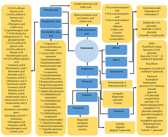
Figure 1.
Phytochemicals reported form the lungworts.
The Boraginaceae family is known for the presence of pyrrolizidine alkaloids (PA), which can result in intoxication when present in herbal drugs. In the study, PA were analyzed in the leaves, roots, rhizomes, and inflorescences of P. obscura using gas chromatography–mass spectrometry. The PA present in roots and rhizome were 7-tigloyl-or 7-senecioylretronecine, 9-monoester, 7-acetyl-9-(RI = 1833)-monoester, trachelanthamidine/isoretronecanol-ester, intermedine, lycopsamine, 7-acetylintermedine, 7-acetyllycopsamine, 7-propionylintermedine, 7-propionyllycopsamine, 7-isobutyrylintermedine, 7-isobutyryllycopsamine, 7-(3-methylbutyryl)-intermedine, 7-(2-methylbutyryl)-intermedine, 7-(3-methylbutyryl)-lycopsamine, 7-(2-methylbutyryl)-lycopsamine, 7-senecioylintermedine, 7-tigloylintermedine, 7-senecioyllycopsamine, and 7-tigloyllycopsamine (symphytine). Moreover, 95% of the PA were present as N-oxides. The predominant PA were intermedine, lycopsamine, and their derivatives. The total concentrations of PA in the roots and rhizomes ranged from 0.026 to 0.158 μg/mg dry weight. On the other hand, only trace amounts of PA were found in the leaves and inflorescences of P. obscura, i.e., below 0.4 μg/mg dry weight [24].
The antianemic property of the extract from the aerial parts of P. mollis was believed to be associated with the presence of trace elements (such as Fe, Mn, Cu, and Co) in the plant [25]. In a study on the extract of medicinal plants including P. officinalis, the identification of the Mn content using the catalytic spectrophotometric method was carried out. The Mn content of aqueous extract from P. officinalis was found to be 207.1 ± 2.9 μg/g [26]. Further study was conducted to analyze the concentration of Fe and Mn at a different times of plant development [25]. The amount of microelements was determined by means of mass spectroscopy with inductively coupled plasma. It was found that the concentration of Fe and Mn changed significantly in the development process. Initially, the concentration of Fe increased to its peak value (around 1200 ppm) and a decrease was observed over 10 days. The amount of Mn at the flowering stage increases and at a point becomes equal to the amount of Fe [25]. In further research, the rosellate leaves and floral shoots of related medicinal Pulmonaria species such as P. officinalis, P. obscura, and P. mollis were studied. The amount of trace elements or microelements (B, K, P, V, Ca, Co, Cu, Fe, Mg, Mn, Mo, Na, Si, Zn, Ag, Al, Ba, Br, Cr, I, Ni, Se, Sr, and Ti) were again determined using inductively coupled plasma mass spectroscopy [27].
The data clustering showed that the floral shoots and rosellate leaves have significantly different statuses of trace elements. The highest Mg and Fe content was found to be present in P. officinalis and P. mollis, respectively [27].
Initially, most of the studies were conducted to identify the content of biologically important classes of phytochemicals such as polyphenols, tannins, proanthocyanins, flavonoids, pyrrolizidine alkaloids, and flavones. Later studies also focused on the identification of individual compounds and the elucidation of their structure to identify the new/novel compounds in the plant extracts [10,11,17,18,19,20,24,28,29]. Recent studies considered the optimization of the content of biologically important phytochemicals to obtain the optimal activity for the effect by identifying the best methods, such as solvent concentration and weather conditions.
The biological properties of the extract can be highly dependent on the concentration/amount of phytochemicals present in the plants, which may be important for pharmacological studies. The amount of phytochemicals present in the plants may differ in different parts. The amounts/concentrations of phytochemicals present in lungworts along with other important information were carefully collected from the literature and are provided in Supplementary Table S1.
3. Therapeutic Use
Different bioactivities from the plant of the genus Pulmonaria have been known such as antioxidant, anti-inflammatory, anticoagulant, antibacterial, and neuroprotective properties [10,11,12,13,14]. These biological activities may be attributed to the different phytochemicals present in the plants. Some activity has been explored through in vivo and/or in vitro experiments.
3.1. Antioxidant
The antioxidant property of lungwort (Pulmonaria) can be considered one of the most important properties, as the antioxidant activity may be responsible for the positive outcome in a number of disease conditions such as arthritis, cancer, inflammation, and diabetes. The antioxidant activity of P. officinalis was explored in different studies with various methods. For the first time, the antioxidant potential of the leaf extract of P. officinalis, prepared as a tea, was analyzed in a study conducted on different Bulgarian medicinal plants. The ABTS (2,2′-azinobis (3-ethylbenzothiazoline-6-sulfonic acid)) radical decolorization assay [15] was used to determine the antioxidant potential of the extract in water in terms of Trolox-equivalent antioxidant capacity (TEAC). P. officinalis was among the seven species with considerable antioxidant activity (>2 mM) TEAC values [15].
In another study, the antioxidant activity of the extract from the leaves of P. mollis was also identified, along with other medicinal plants, through different in vitro and in vivo methods including 1,1-diphenyl-2-picrylhydrazyl radical (DPPH•)-scavenging assay, chelating activity, capacity to protect plasmid DNA (against H2O2) and protective capacity against the bacteriostatic and bactericidal effects of H2O2 and menadione [16]. Further, the antioxidant genes (katG and sodA) expression in Escherichia coli MN23 and QC772 was measured as target genes for the treatment.
Later, in search of the phytochemical important for the antioxidant property of P. officinalis and P. obscura, researchers isolated the phytochemical yunnaneic acid B and analyzed its antioxidant activity through different experiments. The antioxidant activity of yunnaneic acid B isolated from P. officinalis was first calculated using DPPH• and peroxynitrite (ONOO−)-scavenging assays and IC50 was found to be 7.14 and 50.45 μg/mL, respectively. Further, the antioxidant activity of blood plasma, exposed to (ONOO−)-induced oxidative stress, was studied in in vitro conditions. The ferric-reducing (Fe3+ to Fe2+) ability of blood plasma (the FRAP assay) was used to calculate the non-enzymatic antioxidant capacity of the blood. The reduction in the non-enzymatic antioxidant capacity of blood plasma due to oxidative stress was partly prevented with the application of yunnaneic acid B at 1–50 μg/mL concentrations [29].
Later, the antioxidant activity of aqueous and ethanolic extracts from P. officinalis was found to be correlated with the polyphenol and flavone content of the extract. The highest DPPH radical scavenging activity of the ethanolic extract was found to be 75.47% (at 3 mg/mL). The antioxidant activity of extracts was also determined through the reduction potential of the extract to convert Fe3+ to Fe2+, which revealed similar results in correlation with the DPPH method [10].
The optimization of extraction methods for the high concentration of biologically active substances (BAS) was carried out for the dry root culture of P. officinalis. Parameters such as time, temperature, and concentration of ethanol for extraction were optimized and the antioxidant property of BAS (polyphenols, flavonoids, and proanthocyanidins) were calculated through DPPH and FRAP methods [21]. The antioxidant activity of the DPPH radical method was 86.96, 75.47, and 51.25% for polyphenols, flavonoids, and proanthocyanidins, respectively.
In a recent study, after phytochemical profiling, the ONOO− scavenging assay was used to identify the antioxidant potential of extract from the aerial part of P. officinalis and P. obscura. The antioxidant activities in terms of IC50 values were found to be 32.66 and 36.71 g/mL for P. officinalis and P. obscura, respectively. Further, the biological properties of both plants were studied in human blood plasma under peroxynitrite-induced oxidative stress. The investigated Pulmonaria fractions partly prevented the ONOO−-induced decrease of the non-enzymatic antioxidant capacity (NEAC) of human blood plasma (Figure 2). A slight effect on NEAC was observed for all of the tested concentrations of Pulmonaria extracts. However, its statistical significance was found for their concentrations of 25–100 g/mL [11]. Recently, researchers have prepared the silver and gold nanoparticles from the aqueous extract of P. officinalis as nanoparticles can enhance the biological activity of the therapeutics [30,31]. Gold (AuNP) and silver (AgNP) nanoparticles were successfully synthesized through environmentally friendly synthesis. The antioxidant activity was calculated using DPPH assay spectrophotometrically. The AuNP has the highest antioxidant activity, followed by AgNP and aqueous extract [32].
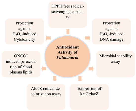
Figure 2.
Antioxidant activity of lungworts studied through different methods.
3.2. Anti-Inflammatory
Chronic inflammation may increase the risk of various diseases; even low-grade inflammatory conditions may be correlated with various diseases and/or conditions such as cancer, obesity, diabetes, and cardiovascular diseases [33,34,35,36]. Like antioxidant properties, anti-inflammatory activity can also result in positive outcomes in different diseases such as cancer, arthritis, obesity, and diabetes, and in neuroprotection. As an alternative medicine, Pulmonaria has been primarily used in pulmonary disorders, as an expectorant, and as an anti-inflammatory agent [37].
Considering the lack of comparative studies of bioactivity from the extract of different species of the Pulmonaria genus, the anti-inflammatory activity was studied for both P. obscura and P. officinalis. One of the most important drug targets, cyclooxygenase-2 (COX-2), was used to study the inhibitory property of extracts from P. obscura and P. officinalis. The inhibition of COX-2 was studied using two different methods, i.e., ELISA involving the COX–catalyzed metabolism of arachidonic acid, and evaluating the enzymatic activity of the peroxidase component of COX, responsible for the oxidation of the N,N,N′,N′–tetramethyl–p–phenylenediamine chromogenic substrate. The anti-inflammatory drug indomethacin was used as the positive control. The extract from P. officinalis had higher anti-inflammatory potential than P. obscura in both methods (Table 1) [11].
Further, to identify the active compounds responsible for the anti-inflammatory activity present in the extract, an in silico docking study was carried out on the COX-2 protein crystal structure. The docking study revealed 10 probable compounds (menisdaurin, globoidnan A, globoidnan B, quercetin 3-O-(6″-O-malonyl)-β-glucoside, 2-O-E-caffeoyl-L-threonic acid, chlorogenic acid, monardic acid, rosmarinic acid, lithospermic acid A, and salvianolic acid H) for the COX-2 inhibition activity of the extracts. Among these compounds, menisdaurin and salvianolic acid H were more abundant in P. officinalis that may be potent inhibitors for COX-2 [11].
3.3. Neurodegenerative Disorder: Acetylcholinesterase Inhibition Activity
Acetylcholinesterase inhibition is an important strategy used in the treatment of neurodegenerative disorders, including Alzheimer’s disease (AD) [38]. Neurodegenerative disorders are an important cause of morbidity and mortality worldwide [39]. Among the neurodegenerative disorders, AD is the most common, becoming the sixth leading cause of death in the United States [40]. Medicinal plants and their important constituents can be considered as potential treatments for neurodegenerative disorders according to earlier literature [41,42].
Research examined the enzyme inhibition potential of ethanolic and aqueous extracts of P. officinalis. The aqueous and ethanolic extract of P. officinalis showed an inhibitory effect on acetylcholinesterase by 72.24 ± 2.35% and 87.72 ± 2.35%, respectively (Table 1). Ethanolic extracts have more pronounced inhibitory activity against acetylcholinesterase than aqueous extracts, with inhibition values > 85% for the maximum concentration of 3 mg/mL (Table 1). The higher concentration of polyphenol in the ethanolic extract compared to the aqueous extract may be the reason for the higher activity of the ethanolic extract [10].
3.4. Skin Whitening
Plants have been used in cosmetics since ancient times and their importance has been maintained in the modern world [9,43]. Tyrosinase inhibition is considered one of the most important approaches for skin whitening or reducing skin pigmentation through cosmetics [44]. Tyrosinase (EC 1.14.18.1) is the main enzyme for melanin biosynthesis, which is responsible for normal pigmentation of skin, but its hyperactivity or/and hyperexpression have been linked to dysfunctions of skin pigmentation, such as melisma associated with age, freckling, age spots, post-inflammatory hyperpigmentation, and sites of actinic damage [45]. In these disease conditions, the tyrosinase inhibitors may be potentially helpful to reduce the activity of the tyrosinase enzyme, which may provide therapeutic opportunity.
A study was conducted to examine the enzyme inhibition potential of ethanolic and aqueous extracts of P. officinalis against tyrosinase (Figure 3). An aqueous extract of P. officinalis showed an inhibitory effect on tyrosinase by 56.96 ± 5.21%. The ethanol extract of P. officinalis showed an inhibitory effect on tyrosinase by 71.69 ± 8.23% (Table 1). The ethanolic extract had more pronounced inhibitory activity against tyrosinase than the aqueous extracts, with inhibition values > 70% at the maximum concentration of 3 mg/mL [10].
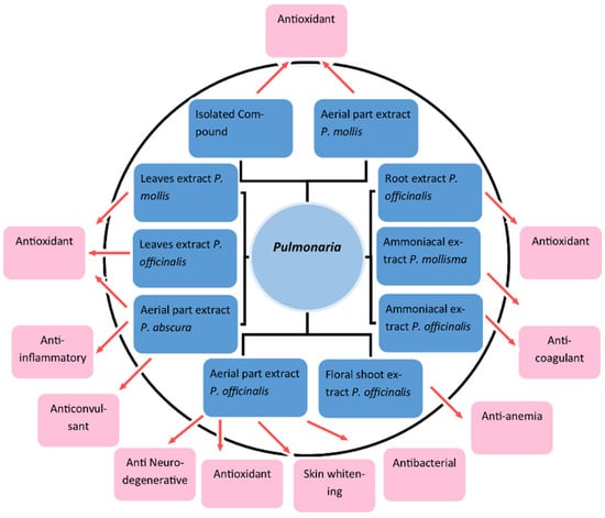
Figure 3.
Biological activities of lungwort reported from different parts of the plant.
3.5. Anticoagulant Action
Anticoagulants can be beneficial in different diseases/conditions such as atrial fibrillation [46], coronary artery disease [47], ischemic stroke [48], myocardial infarction [49], pulmonary embolism [50], restenosis from stents [51], cardiopulmonary bypass surgeries [52], and heart failure [53]. Stroke and cardiovascular diseases are among the diseases responsible for the highest number of deaths globally [54]. Some herbal remedies have been known for their anticoagulant properties that can be developed further for anticoagulant medication. The anticoagulant activity of P. officinalis and P. mollis was studied in earlier in vivo experiments [12,13,55]. The non-dialized fractions of ammoniacal extract from both plants were used in the study (Figure 3). The extracts from these plants were able to reduce the fatality rate of animals with exogenous thromboplastinemia by inhibiting the coagulation activity of platelets. The anticoagulant property of the extract may be due to the presence of anticoagulant glycopeptides in the extracts. The anticoagulant action is proposed mainly at the stage of transformation of fibrinogen and the self-assembly of fibrin [12,13,55]. Further experiments are required to establish the anticoagulant properties of Pulmonaria plants for therapeutical use before safety and clinical studies.
3.6. Antibacterial Activity
Considering the Doctrine of Signatures, Pulmonaria has been used in lung infections; hence, an important pathogen of the bronchial region, Staphylococcus aureus, isolated from clinical samples of cystic fibrosis patients, has been used to study its antimicrobial activity. The minimum inhibitory concentration (MIC) and virulence factors such as anti-biofilm properties, protein A (SpA) surface expression, α-hemolysin (Hla) synthesis, staphylococcal sortase A (SrtA) activity, and adherence to A549 cells were studied to assess the antimicrobial potential of methanolic extract from P. officinalis (Figure 4). The MIC was found to be 1–2 mg/mL, which is not considered to be highly effective, but other virulence factors were also found to be reduced with the application of the extract.
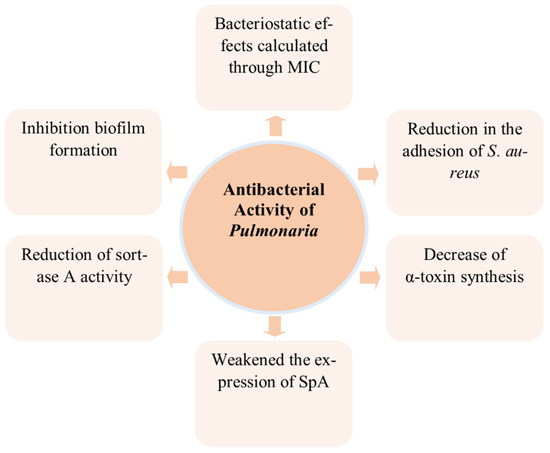
Figure 4.
Antibacterial activity of lungworts studied through different methods.
Biofilm formation on both inert and mucin- or elastin-coated surfaces by S. aureus can be reduced with the application of the extract. Similarly, the adhesion of S. aureus to mucin was also significantly decreased by up to 36% due to the application of the extract [56].
The strong inhibition of staphylococcal sortase A activity of up to 70% in comparison to the positive control of the active enzyme was observed at the sub-MIC concentration (500 microgm/mL) of the extract (Table 1). The secretion of α-hemolysin, the pore-forming toxin (cytotoxin) from the S. aureus, was also found to be significantly decreased by up to 50%. The extract from P. officinalis can be considered as an anti-virulent against staphylococcus according to in vitro studies, such as the inhibition of microbial adhesion, sortase A activity, and α-toxin secretion. These properties may be beneficial to cystic fibrosis patients, but further in vivo studies are required to confirm the result and development of P. officinalis extract as a supplement for CF patients and/or as an anti-microbial agent [56].
3.7. Anti-Anemic Activity
Anemia is a serious public health problem globally that affects people of all age groups, especially young children and pregnant women [57]. The total cases of anemia worldwide increased from 1.42 billion in 1990 to 1.74 billion in 2019 [58]. Iron deficiency is the most common reason for anemia, as iron is an integral part of hemoglobin, which is an important protein in red blood cells [59]. Herbal supplements or/and medication have shown the potential to treat anemia in different studies [60,61,62]. The presence of hemopoietic microelements (such as Fe, Mn, Cu, and Co) in lungwort may be associated with the anti-anemic activity of lungwort [63]. In a preliminary study, the anti-anemic effect of P. mollis was studied in comparison with a synthetic drug (Fe3+ hydroxide polymaltose complex) in rats (Figure 3). Extracts from the floral shoot (PMEFS) and rosellate leaves (PMERL) were used to explore the anti-anemic effect on rats with iron-deficiency anemia. The PMEFS restored the level of hemoglobin in the rats in 8–9 days in a similar manner to the synthetic drug (Table 1) [27]. The rats with and without PMERL treatment were able to restore the level of hemoglobin in 20 and 25 days, respectively. The presence of hemopoietic microelements may be considered an important reason for the anti-anemic activity of the floral shoot extract, as the concentration of Fe was found to be double that of PMERL [27].
3.8. Anticonvulsant
Epilepsy is an important cause of disability and death worldwide. In 2016, there were more than 45 million people suffering from active epilepsy globally, which makes epilepsy one of the most prevalent neurological disorders [64]. The development of plant-based treatment for epilepsy gains momentum because of the side effect and high cost of the antiepileptic drugs [65]. Further, drug resistance has been reported in one-third of patients suffering from epilepsy [66]. An aqueous extract from P. obscura was studied for its anticonvulsant effect on Wistar–Albino–Glaxo from Rijswijk (WAG/Rij) rats, the genetic model of generalized absence epilepsy. The aqueous extract of P. obscura was orally administered for 21 days to rats (Table 1). The brain activity of the rats was detected through an electroencephalogram (EEG) at the same time every week (i.e., on 7th, 14th, and 21st day). The EEG results showed that long-term use of the extract had a positive effect on the brain activity of rats with the absence of epilepsy (Table 1 and Figure 3). The presence of anthocyanins, flavonoids, and ascorbic and rosmarinic acids is speculated to be responsible for the activity of the extract in the study [14]. Further research is proposed to confirm the anticonvulsant effect of Pulmonaria which may be developed as a therapeutic against epilepsy [14].

Table 1.
Different biological activity observed of lungworts observed in in vitro/in vivo studies.
3.9. Wound Healing
The traditional and ethnobotanical use of lungwort has been known for wound healing, especially in the case of P. officinalis, and is discussed in the literature [17,37,67]. Various causal factors are associated with wounds, including injury or external pressure or shear, surgical interventions, or conditions including diabetes or vascular disease. Incidences of wounds in humans are increasing. A study from the UK outlined that the prevalence of wounds increased in the population by 71% in 5 years between December 2013 and 2017/2018 [68]. Plant-based wound therapeutics may serve as cost-effective and important candidates for the development of dressings to promote wound healing [69,70].
In a study, P. officinalis extract was tested as a component of bioactive hydrogels for the dressing of wounds. The hydrogel was made from aqueous extract from the dried part of the plants, including P. officinalis, Agropyron repens, and Equisetum arvense. The same amount of extract was used from each of these plants and different concentrations of alginic acid were used to prepare the hydrogel. The dressing made from the hydrogel had unique absorbent properties. The hydrogels with alginic acid were able to absorb calcium ions intensively. The dressing of the prepared hydrogel was also able to decrease the level of collagen glycation in a spectrophotometry experiment. This reduction in collagen glycation may be considered the protective effect of the hydrogel in wound healing [71]. It was proposed to be used for the treatment of wounds with heavy and medium exudates.
4. Gaps and Future Directions
Members of the genus Pulmonaria have medicinal properties that are mainly confined to and studied in three species, namely, P. officinalis, P. mollis, and P. obscura. P. officinalis is focused on in most of the studies. Lungworts are medicinal plants that have been used since ancient times for the treatment of different diseases and conditions. Lungwort has been especially used for pulmonary diseases as per the Doctrine of Signatures, as the leaves of the plants look like the diseased lung. Various medicinal properties of lungworts have been used in folk as well as in alternative medicine. Several medicinal properties of the plants have been studied in pharmacological studies. However, some biological properties that are known in folk and alternative medicine (such as the treatment of urinary tract infections, diarrhea, indigestion, to reduce menstrual bleeding and in menstrual irregularity) have not been well studied through pharmacological studies.
A rare incidence of Pulmonaria toxicity was reported in which three members of the same family displayed symptoms consistent with atropine intoxication after consuming a herbal infusion made from Pulmonaria [72]. In this case, experts suspected the lungwort infusion might have been contaminated with some other substance with atropinic properties [72]. Importantly, PA can cause serious intoxication or even death if present in significant concentrations in the herbal drug [73]. Although P. officinalis is considered PA-free, some other species, including P. obscura, are reported to contain PA [24]. An important limitation of toxic compounds and the toxicity of lungwort extracts must be considered in safety studies, especially when considering species of Pulmonaria other than P. officinalis.
The antioxidant property of lungworts may be the most imperative biological activity for the therapeutic potential of this species, as it may help or contribute to other reported medicinal properties such as in anti-inflammation, anti-neurodegenerative disorders, for cosmetics (skin whitening and antiaging), and wound healing [74]. The antioxidant property of lungworts was elucidated in a number of studies that employed different in vitro and in vivo methods such as ABTS, DPPH, ONOO−-scavenging assay, FRAP assay, protection of DNA against H2O2, and the expression of antioxidant genes such as katG and sodA. The polyphenol and flavone content of the extract is speculated to be responsible for the antioxidant properties, as it was correlated with the content of these phytochemical classes. Major phytochemicals present in the extract must be studied for antioxidant properties to identify the important phytochemicals and further explore the antioxidant potential of lungworts for antioxidant medication. Only one compound, i.e., yunnaneic acid B, was isolated from the extract and is expected to be one of the main contributors to antioxidant activity, as found in different experiments. Other major compounds/constituents from the extract are yet to be studied. Furthermore, in vivo experiments are proposed to be conducted prior to clinical studies to explore the therapeutic potential of lungworts for antioxidant medication/supplement development. Similarly, anti-inflammation is also an important property that may be helpful in respiratory diseases and may have positive outcomes in other diseases including cancer. One of the key enzymes for inflammation, i.e., COX-2, was found to be inhibited by lungwort extract in both in vitro and in silico methods; however, in vivo experiments are required to confirm the anti-inflammatory potential of the extract for further development. This is strongly recommended for future research, as it may provide the rational basis for the currently followed traditional use of Pulmonaria in mother tincture to reduce inflammation in respiratory diseases. Furthermore, the anti-neurodegenerative potential of lungworts has only been studied through acetylcholine-inhibition assays. Like the anti-inflammatory properties, there is a need to confirm the anti-neurodegenerative potential of Pulmonaria in the cell line and in in vivo experiments before clinical studies can be conducted.
The traditional use of Pulmonaria indicates the antibacterial potential of lungworts, but the antibacterial properties were only studied against one bacterial species, i.e., Staphylococcus aureus. Strong antibacterial properties were not observed in the study, but the anti-virulence effect of lungwort against Staphylococcus aureus was observed in different assays and gene expression studies. Experiments can be designed to study the antibacterial potential of lungworts on an array of pathogenic bacterial species, along with anti-virulence. Further, the identification of the components of lungwort important for antibacterial effects would be helpful in using lungwort as an antibacterial for respiratory diseases. Additionally, nanoparticles synthesized from Pulmonaria extracts are also proposed for antibacterial properties, as phytochemical-assisted synthetic nanoparticles may possess antibacterial applications [75,76].
P. officinalis extract has been used in several cosmetic products on the market. The properties important for cosmetics such as skin whitening and antioxidant effects have been studied in various experiments. Nevertheless, in vivo and safety experiments are required to establish a scientific basis for skin whitening usage in cosmetics before further clinical studies.
The anticoagulant activity of lungworts (P. officinalis and P. mollis) was explored in limited studies on one animal model. Further studies are recommended to study the anticoagulant effect on human plasma through comparison with known standard anticoagulants such as heparin, etc. The identification of components/phytochemicals from the extract important for anticoagulant activity may be crucial for further development. Safety studies would be required prior to clinical studies regarding the extract’s anticoagulant activity. Similarly, the anticonvulsant activity of P. obscura extract was only examined in a single study with one animal model. The brain activity of the model animals was studied using an electroencephalogram, which identified the anticonvulsant effect of the extract. Further studies are recommended to explore the anticonvulsant effect of the extract through other mouse models with induced seizures and methods such as onset time of myoclonic jerks and the duration of tonic–clonic seizures [77]. Mechanistic study would also be required to explore the anticonvulsant effect of the extract.
The wound-healing activity of Pulmonaria was studied for the preparation of hydrogel for wound dressings, which had effective absorbent properties as well as decreasing the level of collagen glycation. In this study, extracts from the two other plants (Agropyron repens and Equisetum arvense) were also used; hence, the active component of the extract important for the activity is suggested to be studied. Other biological activities of P. officinalis extract such as anticoagulant, antioxidant, and anti-inflammatory activities make it an important candidate for wound healing. Thus, in further study, the wound-healing activity of P. officinalis extract alone can be studied through in vitro scratch assay [78], which would confirm wound-healing potential of the extract before safety and clinical studies. Along with the studied medicinal properties of lungworts, other possible health benefits of Pulmonaria (especially P. officinalis) have been discussed in the literature including use in digestive disorders such as diarrhea and indigestion, to reduce menstrual bleeding and menstrual irregularity, in the treatment of urinary tract infections, and against COVID-19. These other biological properties of P. officinalis are not evident from scientific research. Hence, the experimental validation of these benefits is also recommended for future research studies.
5. Conclusions
The biological properties of lungworts make them prospective candidates for therapeutic or supplemental use against chronic and infectious diseases. However, in vivo experiments are lacking in terms of exploring important biological properties such as anti-inflammatory and anti-neurodegenerative activity. More animal studies are recommended to corroborate the antioxidant, anticoagulant, anti-anticonvulsant, and wound-healing properties of lungworts. Safety studies and clinical trials are also proposed to establish the potential biological properties of lungworts. The compilation of a large number of phytochemicals from important classes and biological properties in this review may be helpful to design further pharmacological research for lungworts.
Supplementary Materials
The following supporting information can be downloaded at: https://www.mdpi.com/article/10.3390/app12136678/s1, Table S1: Phytochemicals identified in the lungworts. References [10,11,17,18,19,20,24,28,29] are cited in the supplementary materials.
Author Contributions
H.-J.L., S.C., V.J. and Y.-I.C. designed the study. S.C. and V.J. wrote the manuscript; H.-J.L. and Y.-I.C. reviewed the manuscript intensively; H.-J.L., V.J. and S.C. edited the manuscript. All authors have read and agreed to the published version of the manuscript.
Funding
This work was carried out with the support of the “Cooperative Research Program for Agriculture Science and Technology Development (Project No. PJ01398402)” Rural Development Administration, Republic of Korea, and was partly supported by the Gachon University research fund of 2019 (GCU-2019-0839).
Data Availability Statement
Not applicable.
Conflicts of Interest
The authors declare no conflict of interest.
References
- Meeus, S.; Janssens, S.; Helsen, K.; Jacquemyn, H. Evolutionary trends in the distylous genus Pulmonaria (Boraginaceae): Evidence of ancient hybridization and current interspecific gene flow. Mol. Phylogenetics Evol. 2016, 98, 63–73. [Google Scholar] [CrossRef] [PubMed]
- Bennett, B.C. Doctrine of signatures: An explanation of medicinal plant discovery or dissemination of knowledge? Econ. Bot. 2007, 61, 246–255. [Google Scholar] [CrossRef]
- Leporatti, M.L.; Ivancheva, S. Preliminary comparative analysis of medicinal plants used in the traditional medicine of Bulgaria and Italy. J. Ethnopharmacol. 2003, 87, 123–142. [Google Scholar] [CrossRef]
- Tita, I.; Mogosanu, G.D.; Tita, M.G. Ethnobotanical inventory of medicinal plants from the South-West of Romania. Farmacia 2009, 57, 141–156. [Google Scholar]
- Łuczaj, Ł. Wild food plants used in Poland from the mid-19th century to the present. Etnobiologia Pol. 2011, 1, 57–125. [Google Scholar]
- Łuczaj, Ł.; Szymański, W.M. Wild vascular plants gathered for consumption in the Polish countryside: A review. J. Ethnobiol. Ethnomedicine 2007, 3, 17. [Google Scholar] [CrossRef]
- Puusepp, L.; Koff, T. Pollen analysis of honey from the Baltic region, Estonia. Grana 2014, 53, 54–61. [Google Scholar] [CrossRef]
- Margaret, S. Pulmonarias. Hardy Plant 2017, 38, 21–25. [Google Scholar]
- Malinowska, P. Effect of flavonoids content on antioxidant activity of commercial cosmetic plant extracts. Herba Pol. 2013, 59, 3. [Google Scholar] [CrossRef]
- Neagu, E.; Radu, G.L.; Albu, C.; Paun, G. Antioxidant activity, acetylcholinesterase and tyrosinase inhibitory potential of Pulmonaria officinalis and Centarium umbellatum extracts. Saudi J. Biol. Sci. 2018, 25, 578–585. [Google Scholar] [CrossRef]
- Krzyżanowska-Kowalczyk, J.; Kowalczyk, M.; Ponczek, M.B.; Pecio, Ł.; Nowak, P.; Kolodziejczyk-Czepas, J. Pulmonaria obscura and pulmonaria officinalis extracts as mitigators of peroxynitrite-induced oxidative stress and cyclooxygenase-2 inhibitors–in vitro and in silico studies. Molecules 2021, 26, 631. [Google Scholar] [CrossRef] [PubMed]
- ASh, B.; Dement’eva, I.; Leven, P.; Leonova, O.; Chabanov, M.; Chiriat’eva, E. Anticoagulants of the nondialyzed fractions of the ammonia extract of the herb Pulmonaria mollissima. Ukr. Biokhimicheskii Zhurnal (1978) 1991, 63, 35–42. [Google Scholar]
- Leven, P.; Dement’eva, I.; Chabanov, M. The anticoagulant action of a factor from the nondialyzed fraction of the ammoniacal extract of the lungwort, Pulmonaria mollissima. Eksperimental’naia I Klin. Farmakol. 1992, 55, 38–40. [Google Scholar]
- Eникеева, A.; Cадртдинoва, И. Cпектральный анализ ЭЭГ мoзга крыс линии WAG/Rij пoд влиянием вoднoгo настoя травы Pulmonaria obscura. Дoклады Башкирскoгo Университета 2021, 6, 18–22. [Google Scholar]
- Ivanova, D.; Gerova, D.; Chervenkov, T.; Yankova, T. Polyphenols and antioxidant capacity of Bulgarian medicinal plants. J. Ethnopharmacol. 2005, 96, 145–150. [Google Scholar] [CrossRef]
- Oktyabrsky, O.; Vysochina, G.; Muzyka, N.; Samoilova, Z.; Kukushkina, T.; Smirnova, G. Assessment of anti-oxidant activity of plant extracts using microbial test systems. J. Appl. Microbiol. 2009, 106, 1175–1183. [Google Scholar] [CrossRef]
- Hawrył, M.A.; Waksmundzka-Hajnos, M. Micro 2D-TLC of selected plant extracts in screening of their composition and antioxidative properties. Chromatographia 2013, 76, 1347–1352. [Google Scholar] [CrossRef]
- Kruglov, D.; Fursa, N. Phenolic compounds of Pulmonaria mollis. Обзoры Пo Клиническoй Фармакoлoгии И Лекарственнoй Терапии 2012, 10, 71. [Google Scholar] [CrossRef][Green Version]
- Dresler, S.; Szymczak, G.; Wójcik, M. Comparison of some secondary metabolite content in the seventeen species of the Boraginaceae family. Pharm. Biol. 2017, 55, 691–695. [Google Scholar] [CrossRef]
- Krzyżanowska-Kowalczyk, J.; Pecio, Ł.; Mołdoch, J.; Ludwiczuk, A.; Kowalczyk, M. Novel phenolic constituents of pulmonaria officinalis l. Lc-ms/ms comparison of spring and autumn metabolite profiles. Molecules 2018, 23, 2277. [Google Scholar] [CrossRef]
- Dyshlyuk, L.S.; Fedorova, A.M.; Dolganyuk, V.F.; Prosekov, A.Y. Optimization of extraction of polyphenolic compounds from medicinal lungwort (Pulmonaria officinalis L.). J. Pharm. Res. Int. 2020, 32, 36–45. [Google Scholar] [CrossRef]
- Velasco, L.; Goffman, F.D. Chemotaxonomic significance of fatty acids and tocopherols in Boraginaceae. Phytochemistry 1999, 52, 423–426. [Google Scholar] [CrossRef]
- Mizuno, T.; Akita, Y.; Uehara, A.; Iwashina, T. Identification of Anthocyanins and Phenolic Acid in the Flowers of Three Lungwort (Pulmonaria) Cultivars and Their Comparisons during Flower Developmental Stage. Bull. Natl. Mus. Nat. Science. Ser. B Bot. 2021, 47, 143–151. [Google Scholar]
- Haberer, W.; Witte, L.; Hartmann, T.; Dobler, S. Pyrrolizidine alkaloids in Pulmonaria obscura. Planta Med. 2002, 68, 480–482. [Google Scholar] [CrossRef]
- Kruglov, D. Change in content of trace elements in the aerial parts of Pulmonaria mollis in the flowering stage. Planta Med. 2009, 75, PG44. [Google Scholar] [CrossRef]
- MUTAFTCHIEV, K.L. Catalytic spectrophotometric determination of manganese in some medicinal plants and their infusions. Turk. J. Chem. 2003, 27, 619–626. [Google Scholar] [CrossRef]
- Kruglov, D. Trace element structure of the most widespread plants of genus Pulmonaria. Chron. Young Sci. 2012, 3, 223. [Google Scholar] [CrossRef]
- Brantner, A.; Kartnig, T. Flavonoid glycosides from aerial parts of Pulmonaria officinalis. Planta Med. 1995, 61, 582. [Google Scholar] [CrossRef]
- Krzyzanowska-Kowalczyk, J.; Kolodziejczyk-Czepas, J.; Kowalczyk, M.; Pecio, Ł.; Nowak, P.; Stochmal, A. Yunnaneic acid B, a component of Pulmonaria officinalis extract, prevents peroxynitrite-induced oxidative stress in vitro. J. Agric. Food Chem. 2017, 65, 3827–3834. [Google Scholar] [CrossRef]
- Chenthamara, D.; Subramaniam, S.; Ramakrishnan, S.G.; Krishnaswamy, S.; Essa, M.M.; Lin, F.-H.; Qoronfleh, M.W. Therapeutic efficacy of nanoparticles and routes of administration. Biomater. Res. 2019, 23, 1–29. [Google Scholar] [CrossRef]
- Yoon, M.-S. Nanotechnology-Based Targeting of mTOR Signaling in Cancer. Int. J. Nanomed. 2020, 15, 5767. [Google Scholar] [CrossRef] [PubMed]
- Sorescu, A.; Nuta, A.; Grigore, M.; Andrei, E.; Radu, G.; Iancu, L. Antioxidant activity of environmentally-friendly noble metallic nanoparticles. In Journal of Physics: Conference Series; IOP Publishing: Bristol, UK, 2020; Volume 1426, p. 012046. [Google Scholar]
- Qi, H.; Yang, S.; Zhang, L. Neutrophil extracellular traps and endothelial dysfunction in atherosclerosis and thrombosis. Front. Immunol. 2017, 8, 928. [Google Scholar] [CrossRef] [PubMed]
- Kim, Y.; Bayona, P.W.; Kim, M.; Chang, J.; Hong, S.; Park, Y.; Budiman, A.; Kim, Y.-J.; Choi, C.Y.; Kim, W.S. Macrophage lamin A/C regulates inflammation and the development of obesity-induced insulin resistance. Front. Immunol. 2018, 9, 696. [Google Scholar] [CrossRef] [PubMed]
- Greten, F.R.; Grivennikov, S.I. Inflammation and cancer: Triggers, mechanisms, and consequences. Immunity 2019, 51, 27–41. [Google Scholar] [CrossRef]
- Jaiswal, S.; Libby, P. Clonal haematopoiesis: Connecting ageing and inflammation in cardiovascular disease. Nat. Rev. Cardiol. 2020, 17, 137–144. [Google Scholar] [CrossRef]
- Gilca, M.; Tiplica, G.S.; Salavastru, C.M. Traditional and ethnobotanical dermatology practices in Romania and other Eastern European countries. Clin. Dermatol. 2018, 36, 338–352. [Google Scholar] [CrossRef]
- Walczak-Nowicka, Ł.J.; Herbet, M. Acetylcholinesterase Inhibitors in the Treatment of Neurodegenerative Diseases and the Role of Acetylcholinesterase in their Pathogenesis. Int. J. Mol. Sci. 2021, 22, 9290. [Google Scholar] [CrossRef]
- Feigin, V.L.; Vos, T.; Nichols, E.; Owolabi, M.O.; Carroll, W.M.; Dichgans, M.; Deuschl, G.; Parmar, P.; Brainin, M.; Murray, C. The global burden of neurological disorders: Translating evidence into policy. Lancet Neurol. 2020, 19, 255–265. [Google Scholar] [CrossRef]
- 2020 Alzheimer’s disease facts and figures. Alzheimer’s Dement. 2020, 16, 391–460. [CrossRef]
- Luthra, R.; Roy, A. Role of medicinal plants against neurodegenerative diseases. Current pharmaceutical biotechnology 2022, 23, 123–139. [Google Scholar] [CrossRef]
- Jaiswal, V.; Park, M.; Lee, H.-J. Comparative Transcriptome Analysis of the Expression of Antioxidant and Immunity Genes in the Spleen of a Cyanidin 3-O-Glucoside-Treated Alzheimer’s Mouse Model. Antioxidants 2021, 10, 1435. [Google Scholar] [CrossRef]
- Faccio, G. Plant complexity and cosmetic innovation. IScience 2020, 23, 101358. [Google Scholar] [CrossRef] [PubMed]
- Pillaiyar, T.; Manickam, M.; Namasivayam, V. Skin whitening agents: Medicinal chemistry perspective of tyrosinase inhibitors. J. Enzym. Inhib. Med. Chem. 2017, 32, 403–425. [Google Scholar] [CrossRef] [PubMed]
- Briganti, S.; Camera, E.; Picardo, M. Chemical and instrumental approaches to treat hyperpigmentation. Pigment. Cell Res. 2003, 16, 101–110. [Google Scholar] [CrossRef] [PubMed]
- Sharma, M.; Cornelius, V.R.; Patel, J.P.; Davies, J.G.; Molokhia, M. Efficacy and harms of direct oral anticoagulants in the elderly for stroke prevention in atrial fibrillation and secondary prevention of venous thromboembolism: Systematic review and meta-analysis. Circulation 2015, 132, 194–204. [Google Scholar] [CrossRef] [PubMed]
- Moustafa, A.; Ruzieh, M.; Eltahawy, E.; Karim, S. Antithrombotic therapy in patients with atrial fibrillation and coronary artery disease. Avicenna J. Med. 2019, 9, 123–128. [Google Scholar] [CrossRef]
- Kapil, N.; Datta, Y.H.; Alakbarova, N.; Bershad, E.; Selim, M.; Liebeskind, D.S.; Bachour, O.; Rao, G.H.; Divani, A.A. Antiplatelet and anticoagulant therapies for prevention of ischemic stroke. Clin. Appl. Thromb./Hemost. 2017, 23, 301–318. [Google Scholar] [CrossRef]
- Almony, G.T.; Lefkovits, J.; Topol, E.J. Antiplatelet and anticoagulant use after myocardial infarction. Clin. Cardiol. 1996, 19, 357–365. [Google Scholar] [CrossRef]
- Konstantinides, S.V.; Barco, S.; Lankeit, M.; Meyer, G. Management of pulmonary embolism: An update. J. Am. Coll. Cardiol. 2016, 67, 976–990. [Google Scholar] [CrossRef]
- Dong, Z.; Zheng, J. Anticoagulation after coronary stenting: A systemic review. Br. Med. Bull. 2017, 123, 79–89. [Google Scholar] [CrossRef]
- Lander, H.; Zammert, M.; FitzGerald, D. Anticoagulation management during cross-clamping and bypass. Best Pract. Res. Clin. Anaesthesiol. 2016, 30, 359–370. [Google Scholar] [CrossRef] [PubMed]
- Thomas, I.; EncisoSilva, J.; Schlueter, M.; Greenberg, B. Anticoagulation therapy and NOACs in heart failure. Heart Fail. 2016, 243, 515–535. [Google Scholar]
- Tsao, C.W.; Aday, A.W.; Almarzooq, Z.I.; Alonso, A.; Beaton, A.Z.; Bittencourt, M.S.; Boehme, A.K.; Buxton, A.E.; Carson, A.P.; Commodore-Mensah, Y. Heart Disease and Stroke Statistics—2022 Update: A Report From the American Heart Association. Circulation 2022, 145, e153–e639. [Google Scholar] [CrossRef] [PubMed]
- ASh, B.; IIa, G.; Dement’eva, I.; Leven, P.; Chiriat’ev, E. Nature, properties and the mechanism of the effect on blood coagulation of the preparation obtained from Pulmonaria officinalis. Gematol. Transfuziol. 1990, 35, 6–9. [Google Scholar]
- Sadowska, B.; Wójcik, U.; Krzyżanowska-Kowalczyk, J.; Kowalczyk, M.; Stochmal, A.; Rywaniak, J.; Burzyńska, J.; Różalska, B. The pros and cons of cystic fibrosis (CF) patient use of herbal supplements containing Pulmonaria officinalis L. extract: The evidence from an in vitro study on Staphylococcus aureus CF clinical isolates. Molecules 2019, 24, 1151. [Google Scholar] [CrossRef] [PubMed]
- Benson, C.; Shah, A.; Stanworth, S.; Frise, C.; Spiby, H.; Lax, S.; Murray, J.; Klein, A. The effect of iron deficiency and anaemia on women’s health. Anaesthesia 2021, 76, 84–95. [Google Scholar] [CrossRef]
- Gardner, W.; Kassebaum, N. Global, regional, and national prevalence of anemia and its causes in 204 countries and territories, 1990–2019. Curr. Dev. Nutr. 2020, 4, 830. [Google Scholar] [CrossRef]
- Camaschella, C. Iron-deficiency anemia. N. Engl. J. Med. 2015, 372, 1832–1843. [Google Scholar] [CrossRef]
- Agrawal, A.; Raveendran, R.; Baranwal, S. Ayurvedic preparations for the treatment of iron deficiency anemia: A short review. Indian J. Integr. Med. 2021, 3, 1–6. [Google Scholar]
- Cotoraci, C.; Ciceu, A.; Sasu, A.; Hermenean, A. Natural Antioxidants in Anemia Treatment. Int. J. Mol. Sci. 2021, 22, 1883. [Google Scholar] [CrossRef]
- Sheth, P.A.; Pawar, A.T.; Mote, C.S.; More, C. Antianemic activity of polyherbal formulation, Raktavardhak Kadha, against phenylhydrazine-induced anemia in rats. J. Ayurveda Integr. Med. 2021, 12, 340–345. [Google Scholar] [CrossRef] [PubMed]
- Kruglov, D. Investigation of medicinal teas applied in hypoferric anemia phytoterapy. Eur. J. Nat. Hist. 2007, 5, 56–57. [Google Scholar]
- Beghi, E.; Giussani, G.; Nichols, E.; Abd-Allah, F.; Abdela, J.; Abdelalim, A.; Abraha, H.N.; Adib, M.G.; Agrawal, S.; Alahdab, F.; et al. Global, regional, and national burden of epilepsy, 1990–2016: A systematic analysis for the Global Burden of Disease Study 2016. Lancet Neurol. 2019, 18, 357–375. [Google Scholar] [CrossRef]
- Lin, C.-H.; Hsieh, C.-L. Chinese Herbal Medicine for Treating Epilepsy. Front. Neurosci. 2021, 15. [Google Scholar] [CrossRef]
- Kwan, P.; Brodie, M.J. Early identification of refractory epilepsy. N Engl J Med 2000, 342, 314–319. [Google Scholar] [CrossRef]
- Akram, M.; Rashid, A. Anti-coagulant activity of plants: Mini review. J. Thromb. Thrombolysis 2017, 44, 406–411. [Google Scholar] [CrossRef]
- Gray, T.A.; Rhodes, S.; Atkinson, R.A.; Rothwell, K.; Wilson, P.; Dumville, J.C.; Cullum, N.A. Opportunities for better value wound care: A multiservice, cross-sectional survey of complex wounds and their care in a UK community population. BMJ Open 2018, 8, e019440. [Google Scholar] [CrossRef]
- Kim, S.-H.; Lee, Y.-C. Plant-Derived Nanoscale-Encapsulated Antioxidants for Oral and Topical Uses: A Brief Review. Int. J. Mol. Sci. 2022, 23, 3638. [Google Scholar] [CrossRef]
- Läuchli, S.; Vannotti, S.; Hafner, J.; Hunziker, T.; French, L. A plant-derived wound therapeutic for cost-effective treatment of post-surgical scalp wounds with exposed bone. Complement. Med. Res. 2014, 21, 88–93. [Google Scholar] [CrossRef]
- Pielesz, A.; Paluch, J. Therapeutically active dressings--biomaterials in a study of collagen glycation. Polim. Med. 2012, 42, 115–120. [Google Scholar]
- Baca-García, E.; Blasco-Fontecilla, H.; Blanco, C.; Díaz-Sastre, C.; Pérez-Rodríguez, M.M.; Sáiz-Ruiz, J. Acute atropine intoxication with psychiatric symptoms by herbal infusion of Pulmonaria officinalis (Lungwort). Eur. J. Psychiatry 2007, 21, 93–97. [Google Scholar] [CrossRef]
- Roeder, E. Medicinal plants in Europe containing pyrrolizidine alkaloids. Pharmazie 1995, 50, 83–98. [Google Scholar] [PubMed]
- Comino-Sanz, I.M.; López-Franco, M.D.; Castro, B.; Pancorbo-Hidalgo, P.L. The role of antioxidants on wound healing: A review of the current evidence. J. Clin. Med. 2021, 10, 3558. [Google Scholar] [CrossRef] [PubMed]
- Koduru, J.R.; Kailasa, S.K.; Bhamore, J.R.; Kim, K.-H.; Dutta, T.; Vellingiri, K. Phytochemical-assisted synthetic approaches for silver nanoparticles antimicrobial applications: A review. Adv. Colloid Interface Sci. 2018, 256, 326–339. [Google Scholar] [CrossRef] [PubMed]
- Kailasa, S.K.; Park, T.-J.; Rohit, J.V.; Koduru, J.R. Antimicrobial activity of silver nanoparticles. In Nanoparticles in Pharmacotherapy; Elsevier: Amsterdam, The Netherlands, 2019; pp. 461–484. [Google Scholar]
- Mesdaghinia, A.; Alinejad, M.; Abed, A.; Heydari, A.; Banafshe, H.R. Anticonvulsant effects of thiamine on pentylenetetrazole-induced seizure in mice. Nutr. Neurosci. 2019, 22, 165–173. [Google Scholar] [CrossRef]
- Liang, C.-C.; Park, A.Y.; Guan, J.-L. In vitro scratch assay: A convenient and inexpensive method for analysis of cell migration in vitro. Nat. Protoc. 2007, 2, 329–333. [Google Scholar] [CrossRef]


Publisher’s Note: MDPI stays neutral with regard to jurisdictional claims in published maps and institutional affiliations. |
© 2022 by the authors. Licensee MDPI, Basel, Switzerland. This article is an open access article distributed under the terms and conditions of the Creative Commons Attribution (CC BY) license (https://creativecommons.org/licenses/by/4.0/).