Article

Exposure to Phthalates and Alternative Plasticizers Is Associated with Methylation Changes of ESR1 and PGR in Uterine Leiomyoma: The ELENA Study

1
Center for Environmental Health Sciences, Department of Biomedical and Pharmaceutical Sciences, The University of Montana, Missoula, MT 59812, USA
2
Center for Health Promotion, Department of Family Medicine, Inje University Ilsan Paik Hospital, Goyang-si 10380, Korea
3
Department of Health, Environment and Safety, School of Human & Environmental Service, Eulji University, Seongnam-si 13135, Korea
4
Chemical Safety Research Center, Chemical Platform Technology Division, Korean Research Institute of Chemical Technology, Daejeon 34114, Korea
5
Da Vinci College of General Education, Chung-Ang University, Seoul 06911, Korea
6
Department of Obstetrics and Gynecology, Inje University Ilsan Paik Hospital, Goyang-si 10380, Korea
*
Author to whom correspondence should be addressed.
Appl. Sci. 2021, 11(9), 4234; https://doi.org/10.3390/app11094234
Submission received: 1 April 2021 / Revised: 30 April 2021 / Accepted: 3 May 2021 / Published: 7 May 2021

Abstract

:
Uterine leiomyomas are estrogen-dependent benign tumors with unknown etiologies. Phthalates are endocrine-disrupting chemicals and ubiquitous in the environment; thus, it has been suggested that they play a role in the development of uterine leiomyoma. We aimed to investigate whether the pathogenesis of uterine leiomyoma is related to methylation changes in promoter regions of estrogen receptor α (ESR1) and progesterone receptor (PGR) genes in response to phthalates and alternative plasticizers exposure. Urinary concentrations of 20 phthalate metabolites and seven metabolites of di-2-ethylhexyl terephthalate (DEHTP) and di (isononyl) cyclohexane-1,2-dicarboxylate (DINCH) were measured by UHPLC-MS/MS in thirty leiomyoma patients, who provided both paired leiomyoma and myometrium tissues. Methylation levels of ESR1 and PGR were analyzed by pyrosequencing assay. A total of 12 phthalate metabolites and 5 alternative metabolites (3 DEHTP and 2 DINCH) were detected >70% among study participants. The methylation of ESR1 and PGR were significantly lower in leiomyoma tissues compared to those in myometrium (18.10 ± 4.41 vs. 28.72 ± 4.95; 2.32 ± 0.81 vs. 3.27 ± 0.56, respectively). ESR1 methylation in leiomyoma was negatively associated with mono-2-carboxylmethyl-hexyl phthalate (2cx-MMHP) and mono-3-carbocyl-propyl phthalate (MCPP) after adjusting for confounding factors. However, 1-mono-2-ethyl-5-oxohexyl-benzene-1,4-dicarboxylate (5OXO-MEHTP), one of the alternatives, showed positive association with ESR1 methylation in leiomyoma. PGR methylation in leiomyoma was significantly associated with mono butyl phthalate (MnBP), but negatively associated with cyclohexane-1,2-dicarboxylate-mono-7-hydroxy-4-methyl-heptyl ester (cx-MINCH). Our results suggest that phthalates exposure may contribute to leiomyoma pathogenesis via ESR1 and PGR methylation changes.

1. Introduction

Phthalates are high-volume-produced chemicals that have been used in many settings, including industrial, commercial, personal, and household applications. As a result, phthalates have become ubiquitous in the environment, attributable to their widespread and frequent use in daily life [1]. Phthalates are known to be estrogenic and androgenic endocrine disruptors; thus, adverse effects of phthalates on the human reproductive system have been reported [2,3,4,5,6,7]. Considering the negative health effects of phthalates to animals and humans, there is a need to develop and/or use alternative plasticizers with low toxicity and less severe consequences for human health [8,9]. Subsequently, alternative plasticizers have been recently and rapidly used; however, there is very limited information regarding their toxicity and health effects [10]. One murine study showed that prenatal exposure to di-iso-nonyl-cyclohexane-1,2-dicarboxylate (DINCH), one of the alternative plasticizers, was associated with premature aging of testes and impaired liver metabolic capacity in offspring [11]. Therefore, some animal studies may raise the concern that adverse human health effects will emerge with increasing use and exposure to these materials.
Uterine leiomyomas are estrogen-dependent benign tumors with unknown etiologies; thus, it has been suggested that endocrine-disrupting chemicals (EDC), including phthalates, play a role in the development of uterine leiomyoma. Our group previously reported that exposures to phthalate metabolites, including mono-2-ethylhexyl phthalate (MEHP), mono-2-ethyl-5-hydrohexyl phthalate (MEHHP), mono-2-carboxylmethylhexyl phthalate (2cx-MMHP), ∑MEHP-3 (combining MEHP, MEHHP, and mono-2-ethyl-5-oxohexyl phthalate (MEOHP)), and ∑MEHP-5 (∑3-DEHP plus mono-2-ethyl-5-carboxypentyl phthalate (5cx-MEPP), and 2cx-MMHP) were associated with the prevalence of leiomyoma in Korean women [12]. Several studies also examined the effects of phthalates on the development of uterine leiomyoma; however, the results are controversial and the underlying mechanisms remain unclear. [12,13,14,15,16,17].
It is becoming increasingly evident that environmental influences can result in physiological changes through epigenetics, which is the study of heritable changes in gene expression that occur without directly altering the DNA sequence. Epigenetic alterations are associated with the development and progression of numerous pathological states and diseases [18]. Thus, there is a great deal of potential for the use of epigenetics as biomarkers to better understand the early-stage biological response and molecular mechanisms by which environmental exposures to phthalates and alternative plasticizers lead to uterine leiomyoma. Several studies have reported that estrogen receptor α gene (ESR1) expression in leiomyoma tissues is regulated by DNA methylation [19,20]. It was also reported through a study on genome-wide DNA methylation analysis that a methylation change of ESR1 contributed to an aberrant response to estrogen, and this was subsequently associated with development of uterine leiomyoma [21].
Overexpression of ESR1 and aromatase was shown in myoma tissues compared to normal myometrium tissues for development of uterine leiomyomas. Estrogen is known to support both progesterone receptor (PR) induction and the facilitation of PR ligands’ action on target cells. Thus, estradiol’s primary role in uterine leiomyoma was suggested to be maintenance of PR levels, with progesterone promoting uterine leiomyoma growth and progression [22]. In the present study, we aimed to investigate whether the pathogenesis of uterine leiomyoma is related to methylation changes in promoter regions of ESR1 and PR gene (PGR) in response to phthalates and alternative plasticizers exposure.
In the present study, we analyzed urinary concentrations of twenty phthalate metabolites as well as four di-2-ethylhexyl terephthalate (DEHTP) and three DINCH metabolites as alternative plasticizers, which have rapidly increased in use [23,24]. We also measured methylation levels of ESR1 and PGR in leiomyoma tissues from 30 cases, and these methylation levels were compared to those in myometrium tissues—adjacent normal tissues—as internal controls from the same patients.

2. Materials and Methods

2.1. Clinical Subjects

This study was performed utilizing data and bio-samples of patients from the study of Environmental exposure for Leiomyoma, ENdometriosis, and Adeomyosis of reproductive women (ELENA). We recruited patients who had undergone laparoscopic surgery and exploratory laparotomy between March 2017 and February 2019 at the department of Obstetrics & Gynecology in Inje University Ilsan Paik Hospital. Written informed consents were obtained from each patient using consent forms. The study was conducted according to the guidelines of the Declaration of Helsinki, and approved by the Institutional Review Board of Inje University Ilsan Paik Hospital (Goyang-si, Korea, Code: 2017-06-024, Date of approval: 17 August 2017).
All study participants for the ELENA study were initially examined by gynecologic ultrasonography to detect uterine leiomyoma and any other gynecologic problems including intramural, submucosal, and subserosal fibroids before surgery. Final uterine leiomyoma diagnoses were confirmed by direct visualization during operation and subsequent pathological analysis. Medical and pain/disease histories of all study subjects were assessed and recorded in a standard format. Patients with a history of occupational exposure to reproductive toxicants, hormone therapy, any malignancy, or other reproductive treatment within the last 3 months were excluded from this study. The patients for the ELENA study consisted of 97 patients, including those with leiomyoma (n = 45), leiomyoma with endometriosis (n = 29), leiomyoma with adenomyosis (n = 10), and leiomyoma with endometriosis and adenomyosis (n = 13). Thus, among a total of 97 patients, we randomly selected 30 patients, from whom samples of both leiomyoma and adjacent normal myometrium tissues were obtained.

2.2. Data and Biospecimens (Urine and Tissues) Collection

The demographic data of patients were obtained from an interviewed questionnaire before operation. The questions included age, body mass index (BMI), waist circumference, age of menarche, duration of menstrual cycle, history of dysmenorrhea and operation, gravidity, parity, cigarette smoking, alcohol consumption, exposure to secondhand smoke, and exercise.
Once the presence of fibroids and need for surgery were confirmed in each patient, all subjects were requested to undergo blood tests in preparation for their surgery. A 20-30 mL urine sample was collected in a phthalate-free polypropylene container at the time of blood tests as previously described [25,26]. Samples were immediately centrifuged and stored at −80 °C until further analysis and after measurement of creatinine levels. Paired leiomyoma and myometrium tissues of the study participants who underwent hysterectomy and myomectomy were dissected immediately after the removal of the uterus or leiomyoma, immersed in liquid nitrogen, and stored at −80 °C until further analysis.

2.3. Analysis of Urinary Phthalate and Alternative Plasticizer Metabolites

Twenty urinary phthalates and seven alternative plasticizers (four DEHTP and three DINCH) were analyzed in the urine samples as previously described [25,26]. Standards of phthalate metabolites, including: MEHP, mono-butyl phthalate (MnBP), mono-isobutyl phthalate (MiBP), mono-benzyl phthalate (MBzP), mono-cyclo-hexyl phthalate (MCHP), MEHHP, MEOHP, 5cx-MEPP, 2cx-MMHP, mono-n-octyl-phthalate (MnOP), mono-iso-nomy phthalate (MiNP), mono-3-carbocyl-propyl phthalate (MCPP), mono-isodecyl phthalate (MiDP), mono-ethyl phthalate (MEP), mono-methyl phthalate (MMP), mono-n-pentyl phthalate (MnPP), mono-isopropyl phthalate (MiPrP), mono-hexyl phthalate (MHxP), mono-hydrocyl-isononyl phthalate (OH-MiNP), and mono-carboxy-isodecyl phthalate (cx-MiDP); and alternative plasticizer metabolites, including: mono-2-ethylhexyl terephthalate (MEHTP), 1-mono-2-ethyl-5-carboxylpentyl-benzene-1,4-dicarboxylate (5cx-MEHTP), 1-mono-2-ethyl-5-hydroxylhexyl-benzene-1,4-dicarboxylate (5OH-MEHTP), 1-mono-2-ethyl-5-oxohexyl-benzene-1,4-dicarboxylate (5OXO-MEHTP), cyclohexane-1,2-dicarboxylate-mono-4-methyloctyl ester (MINCH), cyclohexane-1,2-dicarboxylate-mono-7-hydroxy-4-methyl-octyl ester (OH-MINCH), and cyclohexane-1,2-dicarboxylate-mono-7-hydroxy-4-methyl-heptyl ester (cx-MINCH); as well as their respective 13C4 or 13C2-labeled internal standards, were purchased from Cambridge Isotope Laboratory (Cambridge Isotope Laboratory, Cambridge, MA, USA) and Toronto Research Chemical (North York, ON, Canada). After enzymatic hydrolysis, samples were pretreated using the Strata X SPE Cartridge (30 mg/cc, Phenomenex, Torrance, CA, USA), following a method by Servaes et al. [27] with a minor modification. Ultrahigh performance liquid chromatography mass spectrometry (UHPLC-MS/MS, UHPLC, Nexera X2, Shimadzu Corporation, Kyoto, Japan and API 4500, AB SCIEX, ON, Canada) was used for measurement.
The limits of detection (LOD) ranged from 0.01 ng/mL (2cx-MMHP) to 0.8 ng/mL (MiDP) as shown in Table S1. For each instrumental run, reagent blanks and QC samples were included for quality control. The method accuracies were between 80% and 120%, and precisions were lower than 10% relative standard deviation (RSD) for high and low concentrations of spiked samples (n = 7).

2.4. DNA Methylation Analysis

Genomic DNA was isolated from the frozen leiomyoma and myometrium tissues according to the manufacturer’s protocol with the DNeasy Blood & Tissue Kit (Qiagen, Valencia, CA, USA). The DNA concentration and purity ratio were measured using an ND1000 spectrophotometer (Thermo Scientific, Waltham, MA, USA). Subsequently, 1µg of the genomic DNA was sodium bisulfite-converted using the EZ DNA MethylationTM Kit (Zymo Research, Irvine, CA, USA) for Pyrosequencing assay.
Pyrosequencing assay was used for selected gene-specific methylation analysis. Gene specific primers were designed for ESR1 and PGR using PyroMark Assay Design 2.0 software (Qiagen) as previously described [28]. Table S2 shows the detail of the primers and PCR conditions used in this study. Gene regions were located using NCBI and GenBank.
PCR amplification of the bisulfite converted DNA was conducted and PCR reactions were performed using Pyromark PCR Kit (Qiagen) with cycling parameters consisting of denaturation at 95 °C for 5 min, and 45 cycles of 95 °C for 30 s, 51–53 °C for 30 s, 72 °C for 30 s, and a final extension of 72 °C for 5 min, and with the annealing temperature varying slightly for each gene-specific primer set. Pyrosequencing was conducted using a PyroMark Q96 MD instrument (Qiagen), with subsequent quantification of methylation levels determined by the PyroMark-CpG software (Qiagen). All samples were duplicated; each pyrosequencing run included a non-template control, and repeatability of the assay was confirmed using human methylated and non-methylated DNA sets (Zymo Research).

2.5. Statistical Analysis

We summarized the distribution of urinary phthalate metabolites using median and interquartile range. For statistical analysis, t-test, ANOVA, and Spearman correlation were performed using software from the R Foundation for Statistical Computing (version 4.0.4) and SPSS statistical software (version 26.0, Chicago, IL, USA). The urinary concentrations of four phthalate metabolites (i.e., MiPrP, MHxP, OH-MiNp, and cx-MIDP) and alternative plasticizers were not fully analyzed from all study participants (only n = 21 were available for those), attributable to insufficient urine samples. Thus, correlation and regression analyses were performed with <30 samples for the listed metabolites above. Phthalate metabolites that were detected at concentrations of more than 70% were selected for further analyses. All of the phthalate metabolites data were assessed by the Kolmogorov–Smirnov test to evaluate whether they were normally distributed. Since the distribution of phthalate metabolite concentrations was most likely right-skewed, all the phthalate metabolite measurements were logarithm-transformed to approximate normal distribution. The total concentration of phthalate and alternative plasticizer metabolites was creatinine standardized using the following formula to address urinary dilution as stated in previous studies: Total [phthalate metabolites] = 100× phthalate (ng/mL)/creatinine (mg/dL) [29,30].
Multiple regression analyses were performed to identify the association of methylation changes (by leiomyoma and myometrium tissues type) with each urinary phthalate and alternative plasticizer metabolite level. A separate regression model was conducted for each log-transformed urinary phthalate and alternative plasticizer metabolite due to a possible correlation with each other. The regression model was adjusted for age, dysmenorrhea, menstrual phase, smoking status, alcohol drinking, secondhand smoking, and BMI. The p values less than 0.05 were considered statistically significant.

3. Results and Discussion

3.1. Clinical Characteristics of Patients

The demographic and clinical characteristics of patients with uterine leiomyoma are shown in Table 1. The mean age (±SD) and BMI were 44.67 (4.99) years old and 24.25 (4.43), respectively. The size of leiomyomas ranged from 12 mm to 108 mm and the mean number of leiomyoma was 3.93 (3.16). Sixteen leiomyoma patients were accompanied by endometriosis (n = 3), adenomyosis (n = 9), or endometriosis and adenomyosis (n = 4).

3.2. Urinary Concentrations of Phthalate and Alternative Plasticizer Metabolites

Twenty phthalate metabolites and seven alternative plasticizer metabolites (i.e., 4 DEHTP and 3 DINCH) are shown in Table 2. A total of 12 phthalate metabolites (di-2-ethylhexyl phthalate (DEHP) (i.e., MEHP, MEOHP, MEHHP, 2cx-MMHP, and 5cx-MEPP); di-n-octyl phthalate (DnOP), and others (i.e., MBzP, MiBP, MEP, MnBP, MMP, MCPP, and OH-MiNP)) were detected >73%, while MiNP, MnOP, MCHP, MiDP, MnPP, MiPrP, MHxP, and cx-MiDP were rarely detected. Urinary concentrations of 4 DEHTP and 3 DINCH metabolites were measured, while their monoesters such as MEHTP and MINCH were rarely detected. Three DEHTP metabolites (i.e., 5cx-MEHTP, 5OH-MEHTP, and 5OXO-MEHTP) and two DINCH metabolites (i.e., OH MINCH and cx MINCH) were detected >75%, showing that alternative plasticizers are widespread and frequently exposed.
In phthalates, the creatinine-adjusted levels of 5cx-MEPP (35.47± 33.24 µg/g creatinine) were the highest, followed by MnBP, MEHHP, 2cx-MMHP, MiBP, MEOHP, MEHP, MEP, MBzP, MMP, OH-MiNP, and MCPP. Among alternative plasticizers, creatinine-adjusted levels of 5cx-MEHTP (28.78 ±16.31µg/g creatinine) were the highest, followed by 5OH-MEHTP, 5OXO-MEHTP, cx-MINCH and OH-MINCH. Both 5cx-MEPP and 5cx-MEPTP are carboxylated monoester metabolites and major oxidized metabolites excreted in urine.
Metabolites of DEHP (i.e., MEOHP, MEHHP, 2cx-MMHP), DEHTP (i.e., 5cx-MEHTP, 5OH-MEHTP, 5OXO-MEHTP), and DINCH (i.e., OH-MINCH, and cx-MINCH) were significantly correlated within each group as shown in Figure S1. In particular, the most widely and frequently used phthalates, including di-ethyl phthalate (DEP), DEHP, and di-n-butyl phthalate (DnBP), showed significant correlations among their metabolites (Spearman’s rho = 0.55 for MEP vs. MEHP, 0.39 for MEP vs. MCPP, rho = 0.53 for MiBP vs. MnBP, rho = 0.83 for MnBP and MCPP). This could be attributable to various phthalates that are mixed and used in products. Therefore, factor analyses were further performed to consider the possible mixing effects; however, there were no significant associations, as demonstrated by the single phthalate–leiomyoma association (data not shown).
There has recently been a shift from the use of traditional phthalates to alternative plasticizers due to environmental and health concerns [31]. One of the well-known alternative plasticizers is DEHTP, which is a structural isomer and a direct substitute for high-molecular-weight plasticizers of DEHP. Like DEHP, DEHTP is rapidly metabolized to its monoester and then further modified by a side-chain hydroxylation and oxidation reaction (5OH-MEHTP, 5oxo-MEHTP, 5cx-MEPTP, and 2cx-MMHTP) [32]. Another alternative plasticizer, DINCH, is a non-aromatic compound similar in viscosity and flexibility to DEHP [33]. OH-MINCH is the predominant metabolite, followed by cx-MINCH and cyclohexane-1 [34]. DINCH has chemical properties comparable to those of DEHP; however, it has been reported to have very limited toxic effects in animal studies. Therefore, DINCH has been approved for the production of food packaging plastic material [35], and as a result it has almost completely replaced DEHP in plastic production in Europe [23].
Although there is increased use of alternatives and political focus on the phasing-out of phthalates, little information is available regarding the metabolic pathways as well as distribution of exposure to alternative plasticizers. From one birth cohort of Puerto Rico, DEHTP metabolites were detected in at least 82% of samples, representing widespread prenatal exposure [36]. DINCH metabolites were also detected in 18% to 35% of samples, but detection rates of DINCH metabolites also increased over time. Our results of alternative plasticizers had a similar pattern; however, the exposed levels of phthalate and alternative plasticizer metabolites should be considered as those in the specialized leiomyoma patients’ group.

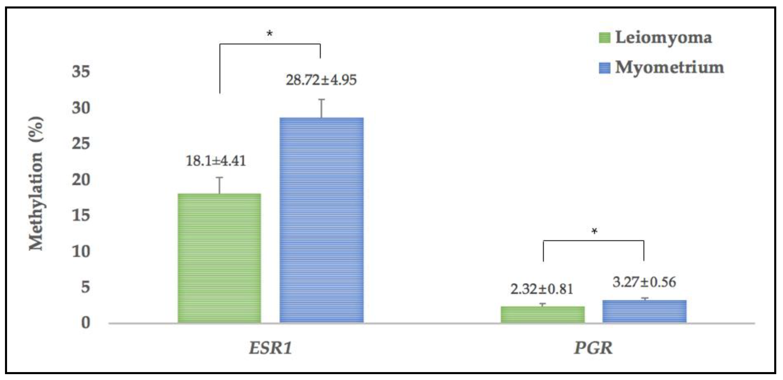
3.3. Methylation of ESR1 and PGR in Leiomyoma and MYOMETRIUM Tissues

Methylation levels of ESR1 and PGR in both leiomyoma tissues and myometrium tissues are shown in Figure 1. The methylation levels of ESR1 (18.10 ± 4.41vs. 28.72 ± 4.95) and PGR (2.32 ± 0.81 vs. 3.27 ± 0.56) were significantly lower in leiomyoma tissue compared to those in myometrium tissue.
The methylation of ESR1 and PGR showed no significant differences by status of parity, menstrual phases, menstrual pattern, smoking, indirect smoking, education, and family incomes. However, there was significant difference in the level of PGR methylation between patients with and without dysmenorrhea (2.07 ± 0.71 vs. 2.51 ± 0.85) (Table S3).
Epigenetic changes are known to play an important role in the pathogenesis of uterine leiomyoma. DNA methylation profiles in leiomyoma tissue are clearly distinguished from those in myometrium tissue, while quite similar DNA methylation profiles are observed in myometrium tissues in women with and without leiomyomas [21]. Furthermore, significant methylation changes in genome-wide DNA and X chromosomes, as well as promoter regions of specific genes, have been observed in leiomyoma tissues [37,38,39,40,41]. Asada et al. [20] reported that highly expressed ESR1, attributable to ESR1 hypomethylation, is associated with pathogenesis of uterine leiomyoma. Our results—significant hypomethylation of ESR1 and PGR in leiomyoma tissues compared to adjacent myometrium tissues—are in concord with their finding.
PR’s role in pathogenesis of leiomyoma is crucial, but methylation of PGR have not been extensively reported. Recently, one paper showed that PR integrates the effects of mutated mediator complex subunit 12 (MED12) and altered DNA methylation to stimulate receptor activator of nuclear factor kappa-B ligand (RANKL) expression and stem cell proliferation in uterine leiomyoma [41]. Progesterone action is essential for uterine leiomyoma growth and development via interaction with PR. Thus, our results could explain pathogenesis of uterine leiomyoma partially, but further investigations should be performed to validate this.

3.4. Methylation Changes and Exposure to Phthalates and Alternative Plasticizers

Our group previously reported that the urinary concentrations of MEHP, MEHHP, 2cx-MMHP, ∑MEHP-3, and ∑MEHP-5 were associated with risk of uterine leiomyoma, suggesting that phthalates could have the potent effects on pathogenesis of uterine leiomyoma [12]. Further associations between exposure to phthalates and alternative plasticizers and DNA methylation changes in two different tissues were analyzed as shown in Table 3. ESR1 methylation in leiomyoma was negatively associated with 2cx-MMHP (β = −9.389, 95% CI: −15.425, −3.353) and MCPP (β = −10.670, 95% CI: −20571, 0.769) after adjusting for confounding factors. However, 5OXO-MEHTP, one of the alternatives, showed positive association with ESR1 methylation in leiomyoma (β = 17.928, 95% CI: 6.420, 29.437). PGR methylation in leiomyoma was significantly associated with MnBP (β = 1.128, 95% CI: 0.055, 2.201), but negatively associated with cx-MINCH, another alternative metabolite (β = −1.220, 95% CI: −2.210, −0.230). In addition, PGR methylation in myometrium was also positively associated with MEP (β = 0.637, 95% CI: 0.093, 1.181) and MCPP (β = 1.259, 95% CI: 0.250, 2.268).
The epigenetic effect of phthalates was reported in several experimental and animal studies. One murine study showed that maternal exposure to DEHP caused testicular dysfunction via DNA hypermethylation, leading to increased expression of DNA methyltransferases (Dnmts) and downregulated expression of insulin-like hormone-3 (Insl3) [42]. Exposure to phthalates mixture induced abnormal expression of miR-141–3p and miR-184 in rats, and their downregulated target genes were associated with prostate development and oncogenesis [43]. Another study found that MCF7 cells treated with dibutyl phthalate (DBP) or butyl benzyl phthalate (BBP) led to demethylation of promoter regions of ESR1 [44]. In addition, phthalates are known to adversely affect estrogen signaling by interacting with ERα and ERβ [45]. Growth of uterine leiomyoma depends on ovarian steroids, estrogen and progesterone [46,47]. However, there are very limited studies to determine the association between phthalates and expression of ESR1 and PGR in relation to uterine leiomyoma.
Our results showed that two phthalate metabolites (i.e., 2cx-MMHP and MCPP) showed significant association with hypomethylation of ESR1 in leiomyoma tissue, while MnBP was associated with hypermethylation of PGR, suggesting that phthalate metabolites might contribute to pathogenesis of uterine leiomyoma via increased expression of ESR1. A recent study by Zota et al. [48] showed that mono-hydroxybutyl phthalate (MEBP) and MEHHP are significantly associated with overexpression of miR-10a-5p and miR-577 in leiomyoma patients. The consequent target genes of phthalates-associated miRNAs are linked to multiple fibroid-related processes including angiogenesis, apoptosis, and proliferation of connective tissues [48]. This study supports our findings suggesting that epigenetic alteration is associated with phthalates exposure and uterine leiomyoma development. Furthermore, EDC, including phthalates, are known to induce oxidative stress and its associated burden such as DNA methylation changes (i.e., hypermethylation and hypomethylation). EDC-induced reactive oxygen species (ROS) interact with androgen and ERs, leading to sex-hormone pathologies [49]. Thus, phthalates-generated ROS induce methylation changes in ESR1; this, in turn, may cause estrogen pathologies and uterine fibroids.
There is little data related to phthalates and PGR expression; a few mechanistic, preclinical, and clinical studies reported that phthalates interfere with normal function of PR. DEHP treatment inhibited progesterone secretion from human luteal cells [50] and decreased secretion of progesterone in mice [51]. One study on the structural binding mechanism of DEHP and its metabolites with PR found that phthalates induced dysfunction of progesterone signaling and adverse effects due to their high binding affinity [52]. Our results also showed that phthalates seemed to inhibit PGR expression in leiomyoma; however, additional studies are needed to confirm this.
In this study, we also analyzed alternative plasticizer metabolites in leiomyoma patients and examined the association between exposed levels of alternative plasticizers and methylation of ESR1 and PGR in leiomyoma. Interestingly, alternative metabolite 5OXO-MEHTP had an opposing association with ESR1 methylation compared to phthalate metabolites, while cx-MINCH showed another opposing association with PGR methylation in leiomyoma. DINCH and DEHTP have been used as the substitute for DEHP, but their metabolites’ effects on methylation changes in ESR1 and PGR are opposed to those of DEHP metabolites. These differences in biological activity at the molecular level may be associated with different structures and physical properties with different chemodynamics. Furthermore, DINCH showed significant impacts on steroidogenesis, testicular development, and function in some studies [53,54], and an in vitro study by Engel et al. showed that DINCH metabolites had a weak estrogenic property [55]; these results support our results. However, further findings, including in vivo and in vitro should be done to confirm the correct epigenetic mechanism.
There are some limitations in our study; the relatively small sample size with large variation in disease status may restrict the statistical power of the results. We also obtained only one spot urine sample from each of the cases, which would be only moderately predictive for long-term exposure, and exposure misclassification cannot be fully ruled out. Some of the study specimens accompany endometriosis and adenomyosis, which may be associated with phthalates exposure. Therefore, further study for only leiomyoma cases is needed. Despite these limitations, this study, to our knowledge, is the first to report methylation changes of ESR1 and PGR in related to uterine leiomyoma and exposure to phthalates and alternative plasticizers.

4. Conclusions

Our results suggest that exposure to some phthalates and alternative plasticizers is associated with ESR1 and PGR methylation changes, which may contribute to leiomyoma pathogenesis. However, further studies with larger sample size and additional genes are needed for strengthening these results.

Supplementary Materials

The following are available online at https://www.mdpi.com/article/10.3390/app11094234/s1, Figure S1: Correlations between metabolites of target phthalates and alternatives from leiomyoma patients Table S1: Levels of limit of detection for phthalate and alternative plasticizer metabolites, Table S2: Primer sequences and PCR conditions for gene-specific methylation analysis, Table S3: Methylation of ESR1 and PGR in leiomyoma and myometrium tissues by characteristics.

Author Contributions

Conceptualization, Y.A.K. and Y.H.C.; Methodology, M.S.K., W.K. and Y.K. Formal Analysis, Y.S.Y. and S.K.; Investigation, H.C., E.J.C., J.W.K. and K.C.C.; Data Curation, Y.A.K., Y.H.C. and Y.J.K.; Writing—Original Draft Preparation, Y.H.C. and Y.A.K. Writing—Review and Editing, Y.H.C., Y.A.K., Y.K., S.K. and Y.J.K.; Supervision, Y.H.C. and Y.A.K. All authors have read and agreed to the published version of the manuscript.

Funding

This research was supported by Basic Science Research Program through the National Research Foundation of Korea founded by the Ministry of Education (2017R1D1A3B03035836).

Institutional Review Board Statement

The study was conducted according to the guidelines of the Declaration of Helsinki, and approved by the Institutional Review Board of Inje University Ilsan Paik Hospital (Code: 2017-06-024, Date of approval: 17 August 2017).

Informed Consent Statement

All study participants signed a written informed consent, and pseudonymization was applied during data entry and analysis.

Acknowledgments

We thank Elizabeth Cole and Caroline Maughan (University of Montana) for their technical support for epigenetic analysis.

Conflicts of Interest

The authors declare no conflict of interest.

References

  1. Kluwe, W.M. Overview of phthalate ester pharmacokinetics in mammalian species. Environ. Health Perspect. 1982, 45, 3–9. [Google Scholar] [CrossRef] [PubMed]
  2. Cobellis, L.; Latini, G.; De Felice, C.; Razzi, S.; Paris, I.; Ruggieri, F.; Mazzeo, P.; Petraglia, F. High plasma concentrations of di-(2-ethylhexyl)-phthalate in women with endometriosis. Hum. Reprod. 2003, 18, 1512–1515. [Google Scholar] [CrossRef] [PubMed]
  3. Reddy, B.S.; Rozati, R.; Reddy, B.V.; Raman, N.V. Association of phthalate esters with endometriosis in Indian women. BJOG Int. J. Obstet. Gynaecol. 2006, 113, 515–520. [Google Scholar] [CrossRef] [PubMed]
  4. Chou, Y.Y.; Huang, P.C.; Lee, C.C.; Wu, M.H.; Lin, S.J. Phthalate exposure in girls during early puberty. J. Pediatr. Endocrinol. Metab. JPEM 2009, 22, 69–77. [Google Scholar] [CrossRef]
  5. Colón, I.; Caro, D.; Bourdony, C.J.; Rosario, O. Identification of phthalate esters in the serum of young Puerto Rican girls with premature breast development. Environ. Health Perspect. 2000, 108, 895–900. [Google Scholar] [CrossRef] [Green Version]
  6. Durmaz, E.; Ozmert, E.N.; Erkekoglu, P.; Giray, B.; Derman, O.; Hincal, F.; Yurdakök, K. Plasma phthalate levels in pubertal gynecomastia. Pediatrics 2010, 125, e122–e129. [Google Scholar] [CrossRef] [PubMed] [Green Version]
  7. López-Carrillo, L.; Hernández-Ramírez, R.U.; Calafat, A.M.; Torres-Sánchez, L.; Galván-Portillo, M.; Needham, L.L.; Ruiz-Ramos, R.; Cebrián, M.E. Exposure to phthalates and breast cancer risk in northern Mexico. Environ. Health Perspect. 2010, 118, 539–544. [Google Scholar] [CrossRef]
  8. Atek, D.; Belhaneche-Bensemra, N.; Turki, M. Migration of epoxidized sunflower oil and dioctyl phthalate from rigid and plasticized poly (vinyl chloride). Int. J. Polym. Mater. 2010, 59, 342–352. [Google Scholar] [CrossRef]
  9. Beach, E.S.; Weeks, B.R.; Stern, R.; Anastas, P.T. Plastics additives and green chemistry. Pure Appl. Chem. 2013, 85, 1611–1624. [Google Scholar] [CrossRef]
  10. Lioy, P.J.; Hauser, R.; Gennings, C.; Koch, H.M.; Mirkes, P.E.; Schwetz, B.A.; Kortenkamp, A. Assessment of phthalates/phthalate alternatives in children’s toys and childcare articles: Review of the report including conclusions and recommendation of the Chronic Hazard Advisory Panel of the Consumer Product Safety Commission. J. Expo. Sci. Environ. Epidemiol. 2015, 25, 343–353. [Google Scholar] [CrossRef]
  11. Campioli, E.; Lee, S.; Lau, M.; Marques, L.; Papadopoulos, V. Effect of prenatal DINCH plasticizer exposure on rat offspring testicular function and metabolism. Sci. Rep. 2017, 7, 11072. [Google Scholar] [CrossRef]
  12. Kim, Y.A.; Kho, Y.; Chun, K.C.; Koh, J.W.; Park, J.W.; Bunderson-Schelvan, M.; Cho, Y.H. Increased urinary phthalate levels in women with uterine leiomyoma: A case-control study. Int. J. Environ. Res. Public Health 2016, 13, 1247. [Google Scholar] [CrossRef] [Green Version]
  13. Huang, P.-C.; Li, W.-F.; Liao, P.-C.; Sun, C.-W.; Tsai, E.-M.; Wang, S.-L. Risk for estrogen-dependent diseases in relation to phthalate exposure and polymorphisms of CYP17A1 and estrogen receptor genes. Environ. Sci. Pollut. Res. 2014, 21, 13964–13973. [Google Scholar] [CrossRef]
  14. Huang, P.-C.; Tsai, E.-M.; Li, W.-F.; Liao, P.-C.; Chung, M.-C.; Wang, Y.-H.; Wang, S.-L. Association between phthalate exposure and glutathione S-transferase M1 polymorphism in adenomyosis, leiomyoma and endometriosis. Hum. Reprod. 2010, 25, 986–994. [Google Scholar] [CrossRef] [PubMed] [Green Version]
  15. Luisi, S.; Latini, G.; De Felice, C.; Sanseverino, F.; Di Pasquale, D.; Mazzeo, P.; Petraglia, F. Low serum concentrations of di-(2-ethylhexyl) phthalate in women with uterine fibromatosis. Gynecol. Endocrinol. 2006, 22, 92–95. [Google Scholar] [CrossRef] [PubMed]
  16. Wise, L.A.; Palmer, J.R.; Reich, D.; Cozier, Y.C.; Rosenberg, L. Hair relaxer use and risk of uterine leiomyomata in African-American women. Am. J. Epidemiol. 2012, 175, 432–440. [Google Scholar] [CrossRef] [Green Version]
  17. Fruh, V.; Henn, B.C.; Weuve, J.; Wesselink, A.K.; Orta, O.R.; Heeren, T.; Hauser, R.; Calafat, A.M.; Williams, P.L.; Baird, D.D. Incidence of uterine leiomyoma in relation to urinary concentrations of phthalate and phthalate alternative biomarkers: A prospective ultrasound study. Environ. Int. 2021, 147, 106218. [Google Scholar] [CrossRef]
  18. Kelly, T.K.; De Carvalho, D.D.; Jones, P.A. Epigenetic modifications as therapeutic targets. Nat. Biotechnol. 2010, 28, 1069–1078. [Google Scholar] [CrossRef] [Green Version]
  19. Sadan, O.; Van Iddekinge, B.; Van Gelderen, C.; Savage, N.; Becker, P.; Van Der Walt, L.; Robinson, M. Oestrogen and progesterone receptor concentrations in leiomyoma and normal myometrium. Ann. Clin. Biochem. 1987, 24, 263–267. [Google Scholar] [CrossRef] [PubMed] [Green Version]
  20. Asada, H.; Yamagata, Y.; Taketani, T.; Matsuoka, A.; Tamura, H.; Hattori, N.; Ohgane, J.; Hattori, N.; Shiota, K.; Sugino, N. Potential link between estrogen receptor-α gene hypomethylation and uterine fibroid formation. Mol. Hum. Reprod. 2008, 14, 539–545. [Google Scholar] [CrossRef] [PubMed] [Green Version]
  21. Maekawa, R.; Sato, S.; Yamagata, Y.; Asada, H.; Tamura, I.; Lee, L.; Okada, M.; Tamura, H.; Takaki, E.; Nakai, A. Genome-wide DNA methylation analysis reveals a potential mechanism for the pathogenesis and development of uterine leiomyomas. PLoS ONE 2013, 8, e66632. [Google Scholar] [CrossRef] [Green Version]
  22. Kim, J.J.; Sefton, E.C. The role of progesterone signaling in the pathogenesis of uterine leiomyoma. Mol. Cell. Endocrinol. 2012, 358, 223–231. [Google Scholar] [CrossRef] [PubMed] [Green Version]
  23. Bui, T.T.; Giovanoulis, G.; Cousins, A.P.; Magnér, J.; Cousins, I.T.; de Wit, C.A. Human exposure, hazard and risk of alternative plasticizers to phthalate esters. Sci. Total Environ. 2016, 541, 451–467. [Google Scholar] [CrossRef] [PubMed]
  24. Schütze, A.; Kolossa-Gehring, M.; Apel, P.; Brüning, T.; Koch, H.M. Entering markets and bodies: Increasing levels of the novel plasticizer Hexamoll®® DINCH®® in 24 h urine samples from the German Environmental Specimen Bank. Int. J. Hyg. Environ. Health 2014, 217, 421–426. [Google Scholar] [CrossRef]
  25. Lee, I.; Alakeel, R.; Kim, S.; Al-Sheikh, Y.A.; Al-Mandeel, H.; Alyousef, A.A.; Kho, Y.; Choi, K. Urinary phthalate metabolites among children in Saudi Arabia: Occurrences, risks, and their association with oxidative stress markers. Sci. Total Environ. 2019, 654, 1350–1357. [Google Scholar] [CrossRef] [PubMed]
  26. Lee, G.; Kim, S.; Kho, Y.; Kim, S.; Lee, S.; Choi, G.; Park, J.; Worakhunpiset, S.; Moon, H.-B.; Okanurak, K. Urinary levels of phthalates and DINCH metabolites in Korean and Thai pregnant women across three trimesters. Sci. Total Environ. 2020, 711, 134822. [Google Scholar] [CrossRef]
  27. Servaes, K.; Voorspoels, S.; Lievens, J.; Noten, B.; Allaerts, K.; Van De Weghe, H.; Vanermen, G. Direct analysis of phthalate ester biomarkers in urine without preconcentration: Method validation and monitoring. J. Chromatogr. A 2013, 1294, 25–32. [Google Scholar] [CrossRef]
  28. Brown, T.A.; Lee, J.W.; Holian, A.; Porter, V.; Fredriksen, H.; Kim, M.J.; Cho, Y.H. Alternation in DNA methylation corresponding with lung inflammation and as a biomarker for disease development after MWCNT exposure. Nanotoxicology 2016, 10, 453–461. [Google Scholar] [CrossRef] [PubMed] [Green Version]
  29. Cline, R.E.; Hill, R.H.; Phillips, D.L.; Needham, L.L. Pentachlorophenol measurements in body fluids of people in log homes and workplaces. Arch. Environ. Contam. Toxicol. 1989, 18, 475–481. [Google Scholar] [CrossRef] [PubMed]
  30. Shealy, D.B.; Barr, J.R.; Ashley, D.L.; Patterson, D.G., Jr.; Camann, D.E.; Bond, A.E. Correlation of environmental carbaryl measurements with serum and urinary 1-naphthol measurements in a farmer applicator and his family. Environ. Health Perspect. 1997, 105, 510–513. [Google Scholar] [CrossRef] [PubMed]
  31. Gyllenhammar, I.; Glynn, A.; Jönsson, B.A.; Lindh, C.H.; Darnerud, P.O.; Svensson, K.; Lignell, S. Diverging temporal trends of human exposure to bisphenols and plastizisers, such as phthalates, caused by substitution of legacy EDCs? Environ. Res. 2017, 153, 48–54. [Google Scholar] [CrossRef] [Green Version]
  32. Lessmann, F.; Schütze, A.; Weiss, T.; Langsch, A.; Otter, R.; Brüning, T.; Koch, H.M. Metabolism and urinary excretion kinetics of di (2-ethylhexyl) terephthalate (DEHTP) in three male volunteers after oral dosage. Arch. Toxicol. 2016, 90, 1659–1667. [Google Scholar] [CrossRef]
  33. Kastner, J.; Cooper, D.G.; Marić, M.; Dodd, P.; Yargeau, V. Aqueous leaching of di-2-ethylhexyl phthalate and “green” plasticizers from poly (vinyl chloride). Sci. Total Environ. 2012, 432, 357–364. [Google Scholar] [CrossRef] [PubMed] [Green Version]
  34. Koch, H.M.; Schütze, A.; Pälmke, C.; Angerer, J.; Brüning, T. Metabolism of the plasticizer and phthalate substitute diisononyl-cyclohexane-1,2-dicarboxylate (DINCH®) in humans after single oral doses. Arch. Toxicol. 2013, 87, 799–806. [Google Scholar] [CrossRef] [PubMed]
  35. Karamfilova, E. Food Contact Materials-Regulation (EC) 1935/2004. Eurpean Implementation Assessment. EPRS|European Parliamentary Research Service. 2016, PE 581.411. Available online: https://www.europarl.europa.eu/thinktank (accessed on 3 May 2021).
  36. Rodríguez-Carmona, Y.; Ashrap, P.; Calafat, A.M.; Ye, X.; Rosario, Z.; Bedrosian, L.D.; Huerta-Montanez, G.; Vélez-Vega, C.M.; Alshawabkeh, A.; Cordero, J.F. Determinants and characterization of exposure to phthalates, DEHTP and DINCH among pregnant women in the PROTECT birth cohort in Puerto Rico. J. Expo. Sci. Environ. Epidemiol. 2020, 30, 56–69. [Google Scholar] [CrossRef] [PubMed]
  37. Yamagata, Y.; Maekawa, R.; Asada, H.; Taketani, T.; Tamura, I.; Tamura, H.; Ogane, J.; Hattori, N.; Shiota, K.; Sugino, N. Aberrant DNA methylation status in human uterine leiomyoma. MHR Basic Sci. Reprod. Med. 2009, 15, 259–267. [Google Scholar] [CrossRef] [Green Version]
  38. Maekawa, R.; Yagi, S.; Ohgane, J.; Yamagata, Y.; Asada, H.; Tamura, I.; Sugino, N.; Shiota, K. Disease-dependent differently methylated regions (D-DMRs) of DNA are enriched on the X chromosome in uterine leiomyoma. J. Reprod. Dev. 2011, 57, 604. [Google Scholar] [CrossRef] [PubMed] [Green Version]
  39. Greathouse, K.L.; Bredfeldt, T.; Everitt, J.I.; Lin, K.; Berry, T.; Kannan, K.; Mittelstadt, M.L.; Ho, S.-M.; Walker, C.L. Environmental estrogens differentially engage the histone methyltransferase EZH2 to increase risk of uterine tumorigenesis. Mol. Cancer Res. 2012, 10, 546–557. [Google Scholar] [CrossRef] [Green Version]
  40. Navarro, A.; Yin, P.; Monsivais, D.; Lin, S.M.; Du, P.; Wei, J.-J.; Bulun, S.E. Genome-wide DNA methylation indicates silencing of tumor suppressor genes in uterine leiomyoma. PLoS ONE 2012, 7, e33284. [Google Scholar] [CrossRef] [PubMed] [Green Version]
  41. Liu, S.; Yin, P.; Kujawa, S.A.; Coon, J.S.; Okeigwe, I.; Bulun, S.E. Progesterone receptor integrates the effects of mutated MED12 and altered DNA methylation to stimulate RANKL expression and stem cell proliferation in uterine leiomyoma. Oncogene 2019, 38, 2722–2735. [Google Scholar] [CrossRef] [Green Version]
  42. Wu, S.; Zhu, J.; Li, Y.; Lin, T.; Gan, L.; Yuan, X.; Xu, M.; Wei, G. Dynamic effect of di-2-(ethylhexyl) phthalate on testicular toxicity: Epigenetic changes and their impact on gene expression. Int. J. Toxicol. 2010, 29, 193–200. [Google Scholar] [CrossRef]
  43. Scarano, W.R.; Bedrat, A.; Alonso-Costa, L.G.; Aquino, A.M.; Fantinatti, B.E.; Justulin, L.A.; Barbisan, L.F.; Freire, P.P.; Flaws, J.A.; Lemos, B. Exposure to an environmentally relevant phthalate mixture during prostate development induces microRNA upregulation and transcriptome modulation in rats. Toxicol. Sci. 2019, 171, 84–97. [Google Scholar] [CrossRef]
  44. Kang, S.C.; Lee, B.M. DNA methylation of estrogen receptor alpha gene by phthalates. J. Toxicol. Environ. Health Part A 2005, 68, 1995–2003. [Google Scholar] [CrossRef]
  45. Engel, A.; Buhrke, T.; Imber, F.; Jessel, S.; Seidel, A.; Völkel, W.; Lampen, A. Agonistic and antagonistic effects of phthalates and their urinary metabolites on the steroid hormone receptors ERα, ERβ, and AR. Toxicol. Lett. 2017, 277, 54–63. [Google Scholar] [CrossRef]
  46. Bulun, S.E. Uterine fibroids. N. Engl. J. Med. 2013, 369, 1344–1355. [Google Scholar] [CrossRef] [Green Version]
  47. Moravek, M.B.; Yin, P.; Ono, M.; Coon, J.S.T.; Dyson, M.T.; Navarro, A.; Marsh, E.E.; Chakravarti, D.; Kim, J.J.; Wei, J.J.; et al. Ovarian steroids, stem cells and uterine leiomyoma: Therapeutic implications. Hum. Reprod. Update 2015, 21, 1–12. [Google Scholar] [CrossRef] [Green Version]
  48. Zota, A.R.; Geller, R.J.; VanNoy, B.N.; Marfori, C.Q.; Tabbara, S.; Hu, L.Y.; Baccarelli, A.A.; Moawad, G.N. Phthalate Exposures and MicroRNA Expression in Uterine Fibroids: The FORGE Study. Epigenet. Insights 2020, 13, 1–11. [Google Scholar] [CrossRef] [Green Version]
  49. Menezo, Y.; Dale, B.; Elder, K. The negative impact of the environment on methylation/epigenetic marking in gametes and embryos: A plea for action to protect the fertility of future generations. Mol. Reprod. Dev. 2019, 86, 1273–1282. [Google Scholar] [CrossRef] [Green Version]
  50. Romani, F.; Tropea, A.; Scarinci, E.; Federico, A.; Dello Russo, C.; Lisi, L.; Catino, S.; Lanzone, A.; Apa, R. Endocrine disruptors and human reproductive failure: The in vitro effect of phthalates on human luteal cells. Fertil. Steril. 2014, 102, 831–837. [Google Scholar] [CrossRef]
  51. Li, N.; Liu, T.; Zhou, L.; He, J.; Ye, L. Di-(2-ethylhcxyl) phthalate reduces progesterone levels and induces apoptosis of ovarian granulosa cell in adult female ICR mice. Environ. Toxicol. Pharm. 2012, 34, 869–875. [Google Scholar] [CrossRef]
  52. Sheikh, I.A.; Abu-Elmagd, M.; Turki, R.F.; Damanhouri, G.A.; Beg, M.A.; Al-Qahtani, M. Endocrine disruption: In silico perspectives of interactions of di-(2-ethylhexyl)phthalate and its five major metabolites with progesterone receptor. BMC Struct. Biol. 2016, 16, 16. [Google Scholar] [CrossRef] [PubMed] [Green Version]
  53. Duarte-Guterman, P.; Navarro-Martín, L.; Trudeau, V.L. Mechanisms of crosstalk between endocrine systems: Regulation of sex steroid hormone synthesis and action by thyroid hormones. Gen. Comp. Endocrinol. 2014, 203, 69–85. [Google Scholar] [CrossRef] [PubMed]
  54. Flood, D.E.; Fernandino, J.I.; Langlois, V.S. Thyroid hormones in male reproductive development: Evidence for direct crosstalk between the androgen and thyroid hormone axes. Gen. Comp. Endocrinol. 2013, 192, 2–14. [Google Scholar] [CrossRef] [PubMed]
  55. Engel, A.; Buhrke, T.; Kasper, S.; Behr, A.C.; Braeuning, A.; Jessel, S.; Seidel, A.; Völkel, W.; Lampen, A. The urinary metabolites of DINCH(®®) have an impact on the activities of the human nuclear receptors ERα, ERβ, AR, PPARα and PPARγ. Toxicol. Lett. 2018, 287, 83–91. [Google Scholar] [CrossRef]
Figure 1. Methylation levels of ESR1 and PGR in leiomyoma and myometrium tissues. Data indicate mean ± standard deviation, and * p < 0.05.
Applsci 11 04234 g001
Table 1. Demographic and clinical characteristics of the study population.
VariableNumber (%)
(n = 30)
Mean ± SD
(Range)
Age (years) 44.67 ± 4.99 (29–54)
BMI (kg/m2) 24.25 ± 4.43 (17.7–35.9)
Waist circumference (cm) 73.95 ± 6.54 (61–86.4)
Diameter of the largest leiomyoma (mm) 6.79 ± 2.42 (12–108)
Number of leiomyoma 3.93 ± 3.16 (1–12)
Operation status
Myomectomy12 (40)
Hysterectomy18 (60)
Disease status
Leiomyoma only14 (46.7)
Leiomyoma + Endometriosis (E)3 (10)
Leiomyoma + Adenomyosis (A)9 (30)
Leiomyoma + E + A4 (13.3)
Parity
Primipara13 (43.7)
Multipara17 (56.7)
Menstrual phase
Proliferative14 (46.7)
Secretory phase13 (43.3)
Atrophy3 (10)
Menstrual cycle
Regular22 (73.3)
Irregular8 (26.7)
Dysmenorrhea
No13 (43.3)
Yes17 (56.7)
Smoking status
No21 (75)
Yes7 (25)
Alcohol drinking status
No1 (3.6)
Yes27 (96.4)
If n < 30, missing values are present.
Table 2. Urinary concentrations of phthalate and alternative plasticizer metabolites.
Phthalates and
Alternatives
Measured >LOD
n (%)
GM ± GSD
(µg/g Creatinine)
Median (IQR)
(µg/g Creatinine)
MEHP27/30 (90)14.91 ±13.1914.39 (8.31–22.34)
MEOHP30/30 (100)18.53 ±14.9217.41 (11.45–28.22)
MEHHP30/30 (100)32.28 ±23.4031.07 (18.22–54.36)
2cx-MMHP30/30 (100)20.84 ±15.8719.29 (13.86–26.90)
5cx-MEPP30/30 (100)35.47 ±33.2428.48 (22.15–55.80)
MBzP29/30 (96.7)10.15 ±12.528.73 (4.25–25.63)
MiBP30/30 (100)19.87 ± 26.8041.20 (7.73–47.63)
MEP27/30 (90)11.23 ± 13.9911.41 (7.06–29.87)
MnBP29/30 (96.7)32.54 ± 27.2837.76 (17.20–55.22)
MMP30/30 (100)9.73 ± 11.616.79 (3.96–30.11)
MCPP22/30 (73.3)2.91 ± 1.922.77 (1.94–3.96)
MiNP6/30 (20)1.16 ± 1.361.17 (0.70–2.06)
OH-MiNP21/21 (100)4.35 ± 2.894.24 (3.21–6.00)
MnOP0/30 (0)NANA
MCHP0/30 (0)NANA
MiDP0/30 (0)NANA
cx-MiDP6/21 (28.6)1.42 ± 1.201.56 (1.38–1.79)
MnPP1/30 (3.3)0.770.77
MiPrP2/21 (9.5)0.23 ± 0.550.23 (0.20–0.27)
MHxP4/21 (19)0.48 ± 2.500.30 (0.19–2.20)
MEHTP4/21 (19)1.15 ± 0.311.20 (1.03–1.29)
5cxMEHTP21/21 (100)28.78 ± 16.3126.43 (22.77–44.47)
5OH-MEHTP20/21 (95.2)4.07 ± 2.243.69 (2.71–6.54)
5OXO-MEHTP19/20 (95)2.35± 1.622.12 (1.52–4.36)
MINCH1/21 (4.8)1.451.45
OH-MINCH21/21 (100)0.66± 0.940.58 (0.41–0.84)
cx-MINCH16/21 (76.2)0.78± 1.210.82 (0.43–1.05)
All phthalate and alternative plasticizer metabolites were creatinine (mg/dL) standardized. LOD= limit of detection; GM = geometric mean; GSD = geometric standard deviation; IQR = interquartile range; NA = not available.
Table 3. Association of phthalate and alternative plasticizer metabolites with ESR1 and PGR methylation in leiomyoma and myometrium tissues.
ESR1PGR
Phthalates and Alternatives aLeiomyoma bMyometriumLeiomyomaMyometrium
MEHP−4.267 (−9.432, 0.898)2.773 (−3.1171, 8.663)−0.330 (−1.381, 0.722)0.566 (−0.080, 1.213)
MEOHP−4.797 (−11.150, 1.574)2.932 (−3.678, 9.541)0.583 (−0.612, 1.777)0.003 (−0.805, 0.812)
MEHHP−4.797 (−12.060, 2.466)2.010 (−5.541, 9.561)0.726 (−0.612, 2.063)−0.644 (− 0.975, 0.846)
2cx-MMHP−9.389 (−15.425, −3.353) *2.086 (−5.374, 9.545)−0.066 (−1.432,1.301)−0.222 (−1.117, 0.672)
5cx-MEPP−3.772 (−9.280,1.735)2.231 (−3.461, 7.923)0.569 (−0.446, 1.585)0.068 (−0.624, 0.761)
MBzP−1.588 (−6.032, 2.856)−0.419 (−4.858, 4.019)0.047(−0.748, 0.842)−0.315 (−0.832, 0.201)
MiBP1.482 (−2.487, 5.451)0.308 (−3.718,4.334)0.630(−0.036,1.296)−0.254 (−0.720, 0.213)
MEP0.015 (−5.046, 5.076)0.541(−4.900,5.982)0.557(−0.353,1.467)0.637 (0.093, 1.181) *
MnBP2.199 (−4.481, 8.879)0.917 (−5.785,7.619)1.128 (0.055, 2.201) *−0.253 (−1.056, 0.550)
MMP−2.107 (−6.888, 2.674)0.568 (−4.306, 5.443)−0.569 (−1.412,0.275)0.421 (−.127, 0.969)
MCPP−10.670 (−20.571, 0.769) *8.301 (−1.153, 17.753)−0.836 (−2.979, 1.308)1.259 (0.250, 2.268) *
OH-MiNP−1.453 (−6.580, 3.675)2.794(−1.775,7.364)−0.114(−1.016,0.789)−0.131(−0.683, 0.421)
5cxMEHTP7.7836 (−5.812, 21.380)1.635 (−12.068, 15.339)1.498 (−0.823, 3.819)−0.830 (−2.288, 0.627)
5OH-MEHTP4.457 (−9.944, 18.857)−5.732 (−18.016, 6.551)1.426 (−0.972, 3.824)−1.304 (−2.641,0.032)
5OXO-MEHTP17.928 (6.420, 29.437) *−11.974 (−25.960, 2.011)1.702 (1–1.645, 5.048)−1.424 (−3.304,0.457)
OH-MINCH−1.649 (−8.029, 4.731)1.776 (−3.531, 7.083)−0.868 (−1.809, 0.073)0.339 (−0.328, 1.006)
cx-MINCH1.606 (−8.18, 11.393)−1.932 (−10.033, 6.169)−1.220 (−2.210, −0.230) *0.128 (−1.033, 1.290)
a All metabolites of phthalates and alternative plasticizers were log-transformed and creatinine (mg/dL) standardized b Data represent beta coefficient (95% CI). Model adjusted for age, dysmenorrhea, menstrual phase, alcohol drinking, smoking, secondhand smoking, and BMI. * p < 0.05.
Publisher’s Note: MDPI stays neutral with regard to jurisdictional claims in published maps and institutional affiliations.

