Article

Garlic Extract: Inhibition of Biochemical and Biophysical Changes in Glycated HSA

by
Mohd W. A. Khan
1,2,*,
Ahmed A. Otaibi
1,
Arwa F. M. Alhumaid
1,
Abdulmohsen K. D. Alsukaibi
1,
Asma K. Alshamari
1,
Eida M. Alshammari
1,
Salma A. Al-Zahrani
1,
Ahmed Y. M. Almudyani
2 and
Subuhi Sherwani
3
1
Department of Chemistry, College of Sciences, University of Ha’il, Ha’il 2440, Saudi Arabia
2
Molecular Diagnostic and Personalised Therapeutics Unit, University of Ha’il, Ha’il 2440, Saudi Arabia
3
Department of Biology, College of Sciences, University of Ha’il, Ha’il 2440, Saudi Arabia
*
Author to whom correspondence should be addressed.
Appl. Sci. 2021, 11(22), 11028; https://doi.org/10.3390/app112211028
Submission received: 21 October 2021 / Revised: 12 November 2021 / Accepted: 18 November 2021 / Published: 21 November 2021

Abstract

:
Glycation of various biomolecules contributes to structural changes and formation of several high molecular weight fluorescent and non-fluorescent, advanced glycation end products (AGEs). AGEs and glycation are involved in various health complications. Synthetic medicines, including metformin, have several adverse effects. Natural products and their derivatives are used in the treatment of various diseases due to their significant therapeutic qualities. Allium sativum (garlic) is used in traditional medicines because of its antioxidant, anti-inflammatory, and anti-diabetic properties. This study aimed to determine the anti-glycating and AGEs inhibitory activities of garlic. Biochemical and biophysical analyses were performed for in vitro incubated human serum albumin (HSA) with 0.05 M of glucose for 1, 5, and 10 weeks. Anti-glycating and AGEs inhibitory effect of garlic was investigated in glycated samples. Increased biochemical and biophysical changes were observed in glycated HSA incubated for 10 weeks (G-HSA-10W) as compared to native HSA (N-HSA) as well as glycated HSA incubated for 1 (G-HSA-1W) and 5 weeks (G-HSA-5W). Garlic extract with a concentration of ≥6.25 µg/mL exhibited significant inhibition in biophysical and biochemical changes of G-HSA-10W. Our findings demonstrated that garlic extract has the ability to inhibit biochemical and biophysical changes in HSA that occurred due to glycation. Thus, garlic extract can be used against glycation and AGE-related health complications linked with chronic diseases in diabetic patients due to its broad therapeutic potential.

1. Introduction

Non-enzymatic glycation reaction involves the binding of sugars (glucose, fructose, ribose, etc.) to free ε-amino groups of proteins in the absence of enzymes. The reaction is classically covalent, in which a sugar protein complex is formed through N-glycoside bonding, via a cascade of chemical responses described by the chemist Maillard [1]. AGEs are endogenous compound groups formed by non-enzymatic reactions [2,3]. Glycation exerts structural alteration of proteins, adversely affecting their physiological functions. Normal physiologic conditions lead to progressive increase in AGE formation with aging [4]. However, the process is accelerated under hyperglycemic conditions and in other inflammatory diseases [5,6,7,8]. AGEs can even be ingested through food [9].
With aging, protein glycation products tend to get deposited and subsequently build up in the tissues of living organisms [10]. Many age-related degenerative alterations occur due to protein degradation, deterioration of protein functionality, and development of toxic molecules leading to the activation of inflammatory pathways. Collectively, these changes often remain undetected by the individual, until clinical manifestation [11]. Glyceraldehyde derived AGEs are toxic and are referred to as toxic AGEs (TAGE) due to induction of cellular dysfunction and association with lifestyle related diseases such as cardiovascular diseases, hypertension, insulin resistance, diabetic complications, Alzheimer’s disease, and cancer [11,12,13,14,15,16,17,18,19].
Glycation is one of the main sources in the generation of reactive oxygen species (ROS) by oxidative pathways. The close links between glycation and oxidation are supported by various studies, wherein autoxidation of glucose is essential to the non-enzymatic glycation of protein [9,20]. Increasing concentrations of sugars proportionately lead to ROS levels, causing oxidative and carbonyl stress, resulting in increased rates of AGEs formation. Expression of receptor for AGEs (RAGE) is increased in some cells under conditions of inflammation and in patients with diabetes. The binding of AGEs to their respective receptors located on macrophages activates nuclear factor-κB through p21ras and mitogen-activated protein kinase pathway of signaling and induces oxidative stress [21,22].
The effect of AGEs induced toxicities are difficult to reduce by simple lifestyle changes. Hence, adoption of preventive medicine is an ideal preventative strategy to combat damage caused by glycation. The side effects of long-term intake of synthetic medicinal compounds are usually greater as compared to natural products. Due to their therapeutic qualities, several plants and their products have been employed in the treatment of diseases [23]. Natural products, such as marine organisms, plants, microbes, animals, and have been utilized in traditional medicine to cure and relieve various ailments since ancient times [24].
The potential inhibition of AGEs through the use of natural products is both advisable and recommended. Synthetic inhibitors of glycation have been already discussed by many previous researchers. Various synthetic antiglycating compounds have been reported with the ability to inhibit the attachment of sugars with protein (e.g., inositol), inhibiting the last stage of glycation (e.g., aminoguanidine), cross-link breakers (e.g., N-henacylthiazolium bromide), inhibiting Amadori product (e.g., ethanol), and radical scavenging properties like amlodipine [2]. Antioxidants have also been identified as a way to protect cell organelles, organs, and tissues from oxidative stress and to avoid oxidative damage pathogenesis. Garlic and its various active components have been shown to play a vital role in illness management due to various health benefits (anti-inflammatory, antioxidant, anti-cancer, etc.) associated with regular consumption [25]. Some of garlic’s anti-diabetic health-promoting properties include hyperinsulinemia, promotion of antioxidant levels, and catalase activity [26]. Garlic has been recently reported to have several beneficial health effects, such as lowering of high blood pressure [27], hepatoprotection [25], cardiovascular atherosclerotic protection [28], immunomodulator effect [29], and anti-schistosomal properties [30].
In this investigation, the therapeutic potential of garlic was studied using some biochemical parameters like antioxidants, ketamine, and carbonyl compounds. Antiglycating and anti-AGEs potential of garlic were studied using biophysical techniques such as UV-absorption, tryptophan specific fluorescence, and AGEs specific fluorescence. In this study, we use different concentrations of garlic extract to identify their inhibitory effects on biochemical and biophysical changes during HSA glycation.

2. Materials and Methods

Trichloroacetic acid, ascorbic acid, 2,2-diphenyl-1-picrylhydrazyl (DPPH), ferric chloride, Folin–Ciocalteau reagent, potassium ferricyanide, gallic acid, quercetin, trypsin, and Congo red were purchased from Sigma Co. St. Louis, MO, USA. Hydrochloric acid, aluminum chloride, mono sodium dihydrogen phosphate, DMSO, ethanol, methanol, disodium hydrogen phosphate, sodium hydroxide, sodium carbonate, and hydrogen peroxide were purchased from Merck, Darmstadt, Germany. All the reagents and chemicals were of analytical grade. The solvents used were of HPLC grade. Peripheral blood mononuclear cells (PBMCs) were isolated by using Ficoll-Hypaque gradients technique and cells were suspended in complete media.

2.1. Biochemical Experiments

2.1.1. Preparation of Aqueous Garlic Extract

Soft-necked varieties of garlic, i.e., Allium sativum var sativum were obtained from the local markets in Hail, KSA. The soft neck variety was further verified by an expert in our college working in this area of research. The aqueous extract was prepared according to the previously published method with slight modifications [31]. Garlic (50 g) was homogenized in cold 0.9% NaCl (75 mL), in the presence of some crushed ice. The highest speed of the blender was used for homogenization, in 1 min bursts for a total of 12 min. The homogenized mixture was filtered through cheesecloth and centrifuged at 2000× g for 10 min. The resultant clear supernatant was made up to 100 mL with saline. After sample preparation, small aliquots were stored at −20 °C for further use. Concentration of the aqueous garlic extract was calculated based on the total garlic used and the final volume of the extract (100 mm of aqueous extract contained 50 g of garlic, 500 mg/mL).
The confirmatory tests for phenolic compounds and flavonoids were followed as described previously [23]. An alkaline reagent test was used to validate the presence of flavonoids in several plant extracts. To ensure the existence of phenolic content, the FeCl3 test was utilized [23].

2.1.2. Total Phenol Content

Folin–Ciocalteu reagent was used to estimate total phenol content as described by a previously published article [32]. Gallic acid (50, 75, 100, 125, 150, 200, and 250 μg/mL) was used as reference. Total phenolic content in garlic extract was determined from the calibration plot and was expressed as mg gallic acid equivalents (GAE). All assays were performed in triplicates. Results were expressed as mg gallic acid equivalent per g dry extract.
Total phenolic content (TPC) = C × V/M
where ’C’ is the concentration of gallic acid in mg/mL that was obtained from gallic acid calibration curve, ´V´ is the volume of plant extract in mL, and ´M´ is the weight of pure plant extract in grams (g).

2.1.3. Total Flavonoid Content

The determination of total flavonoid content in garlic extract was completed using aluminum chloride (AlCl3) colorimetric method as described in previous articles [33,34]. Quercetin (20–250 µg/mL) was used to prepare the calibration curve. In brief, 500 µL of garlic extract (50 µg/mL) or standard quercetin solution were added to 500 µL of AlCl3 (2%). The resultant solutions were incubated for 1 h with intermittent shaking. A spectrophotometer was used to record the absorbance of individual reaction mixture at 430 nm against blank (ethanol). The blank was ethanol because 2% AlCl3 solution was prepared in ethanol. The total flavonoid content was calculated as quercetin equivalent (mg/g) (mg QUE/g).
Total flavonoid content (TFC) = Z × V/m
where Z is the concentration of quercetin (mg/mL); V is the volume (mL) of sample used in the extraction; m is the weight of pure dried sample used (g).

2.1.4. Hydrogen Peroxide Radical Scavenging

Hydrogen peroxide scavenging ability of garlic extract (0.78–100 µg/mL) was determined according to methods published previously [35]. UV visible spectrophotometer was used to measure the absorbance of H2O2 at a constant wavelength (230 nm), against phosphate buffer devoid of H2O2 (blank). Phosphate buffer was used as blank because hydrogen peroxide solution was prepared in phosphate buffer. Ascorbic acid (AsA) is a naturally occurring organic molecule having antioxidant capabilities that also serves as a redox buffer, reducing and neutralizing reactive oxygen species. Therefore, ascorbic acid was used as a standard antioxidant in our experiment. The experiment was performed in triplicate. The percentage of scavenged H2O2 was calculated using the following equation:
Percentage of scavenged H2O2 = [(Oc − Os)/Oc] × 100
where Oc = absorbance of control. Os = absorbance in the presence of extract.

2.1.5. Modification of HSA by Glucose

Glycation of HSA was carried out as described previously with slight changes [1,4]. HSA samples were prepared at a concentration of 15 μM in 20 mM phosphate buffer saline (PBS) (pH 7.4) with 0.05 M of D-glucose. To maintain sterile conditions, all the reaction mixtures were filtered through a 0.22 μm Millipore filter into pre-autoclaved capped vials and incubated for 1, 5, and 10 weeks (G-HSA-1W, G-HSA-5W, and G-HSA-10W, respectively) at 37 °C in the dark. Solutions of HSA without glucose were also stored with the test samples under identical experimental conditions and served as non-modified control. Post-incubation, to remove unbound glucose, the solutions were extensively dialyzed against PBS and subsequently stored at −20 °C. Protein concentrations were measured by the nanodrop as well as the Bradford method.
Glycation inhibition activity of natural product ‘garlic’ extract was analyzed at different concentrations (0.78–100 µg/mL). Different concentrations of garlic extract were added to the reaction mixture at the start of the reactions.

2.1.6. Detection of Ketoamines by Nitroblue Tetrazolium (NBT) Reagent

HSA samples glycation was quantified by a published colorimetric procedure using NBT [36] with a slight modification. Bovine serum albumin (BSA) (150.5 μM) was incubated with 0.5 M glucose for 20 days at 37 °C in 20 mM PBS, resulting in protein modification and ketoamines formation [37]. Native (N)-HSA and G-HSA samples (50 μL) were added in duplicate to the wells of microtiter plates. One hundred microliters of NBT reagent (250 μM in 0.1 M carbonate buffer, pH 10.35) were added to each well and further incubated for 2 h at 37 °C. The plate was read on a Microplate reader at 550 nm. The amount of G-HSA in the sample was calculated using the standard curve constructed with glycated HSA. Garlic extract was added at different concentrations (0.78–100 µg/mL) to detect its anti-glycation activity.

2.1.7. Determination of Protein-Bound Carbonyl Content

The protein-bound carbonyl content in subject sera was analyzed as per Levine et al. [38]. Carbonyl content in N-HSA and G-HSA samples was also estimated. The results were expressed as the number of nanomoles of carbonyl groups per milligram of sample protein, using the equation: ε379 nm = 22,000 M−1cm−1. Garlic extract was added at different concentrations (0.78–100 µg/mL) to detect its anti-glycation activity.

2.1.8. Toxicity of Garlic Extract

The 3-[4,5-dimethylthiazol-2-yl]-2,5-diphenyltratrazolium bromide (MTT) assay was performed as previously described [39] with slight modifications. Briefly, 1 × 10⁴ cells/mL PBMCs in complete RPMI medium, with or without the addition of garlic extract (0.78–100 µg/mL), were incubated for different durations (24–48 h) at 5% CO2 and 37 °C. Thereafter, the cells were treated with 100μL of MTT (5 mg/mL). Four hours later, the entire medium, including MTT solution, was removed from the microplate. The remaining formazan crystals were dissolved in DMSO (50 μL). Absorbance was measured at 570 nm using a 96-well microplate reader (Accuris, SmartreaderTM 96, NJ, USA). Untreated cells were considered as a negative control. The percentage of cytotoxicity was calculated using the background-corrected absorbance as follows:
%   cytotoxicity = 1 absorbance   of   experimental   well absorbance   of   negative   control   well × 100

2.2. Biophysical Experiments

2.2.1. Assay of AGE-Fluorophores

The fluorometric analysis of the AGE pentosidine was analyzed at an excitation wavelength of 375 nm. The peak was observed in the range of 300–400 nm [40]. Excitation and emission slit widths were 10 nm. All protein solutions were of the same concentration (60 μM). Different concentrations (0.78–100 µg/mL) of garlic extract were added to the reaction mixture to detect their inhibitory effect on the formation of the AGE pentosidine.

2.2.2. UV and Tryptophan Specific Spectral Studies

UV absorption profiles of N-HAS, G-HSA samples were recorded on a Shimadzu spectrophotometer (model UV-1700; Koyota, Japan), in the wavelength range of 260 ± 360 nm, using quartz cuvette of 1 cm path length. Hypochromicity at 280 nm was calculated using the following equation:
%   Hypochromicity   at   280   nm = Absorbance   of   N - HSA - Absorbanceof   G - HSA Absorbance   of   N - HSA × 100
Tryptophan specific fluorescence was measured in non-glycated, glycated, and glycated samples with garlic extracts (0.78–100 µg/mL), using a Hitachi model F2000 spectrofluorometer (NJ, USA). Samples were analyzed at an excitation wavelength of 285 nm. An emission spectrum in the range of 290–440 nm was used for tryptophan fluorescence [4].

2.3. Statistical Analysis

All results were expressed as mean ± SD. Multiple comparisons between data were made using software OriginPro v6.1 followed by Student’s t-test. The p-value for significance was set at <0.05.

3. Results

3.1. Biochemical Analysis

3.1.1. Phytochemical Screening

The preliminary observations of aqueous extracts of garlic extract are shown in Table 1. An alkaline reagent test was used to validate the presence of flavonoids in several plant extracts. To ensure the existence of phenolic content, the FeCl3 test was utilized [23]. Total phenolic compounds in garlic extract were determined to be 21.45 ± 0.02 mg gallic acid equivalent/g dry weight of the extract. It is indeed important to keep in mind that phenolic contents are a significant means of protection against infections and oxidative stress. To calculate the total flavonoid content of the extract, the colorimetric AlCl3 test was used, employing quercetin as reference. The total flavonoid content was found to be 16.58 ± 0.03 mg quercetin equivalents (QE)/g dry weight of the extract.

3.1.2. Hydrogen Peroxide (H2O2) Radical Reducing Ability

The percentage ability of garlic extract to reduce hydrogen peroxide is shown in Figure 1. The hydrogen peroxide reducing activity of garlic extract is found to increase in a dose-dependent manner. The maximum reducing activity is shown by 100 µg/mL of garlic extract. This antioxidant activity is supposed to be linked to the high content of polyphenolic compounds found in the extract. Further increase in the concentration of garlic extract (200, 400, 800 µg/mL) showed increased reducing activity (data not shown).
Ascorbic acid (AsA) is a naturally occurring organic molecule having antioxidant capabilities that also serves as a redox buffer, reducing, and neutralizing reactive oxygen species. Ascorbic acid has a better reaction and is more reactive, thus within a short period of time, say 15 min, a steady absorbance value emerges. Therefore, ascorbic acid was used as a standard antioxidant in our experiment.

3.1.3. Formation of Ketoamines

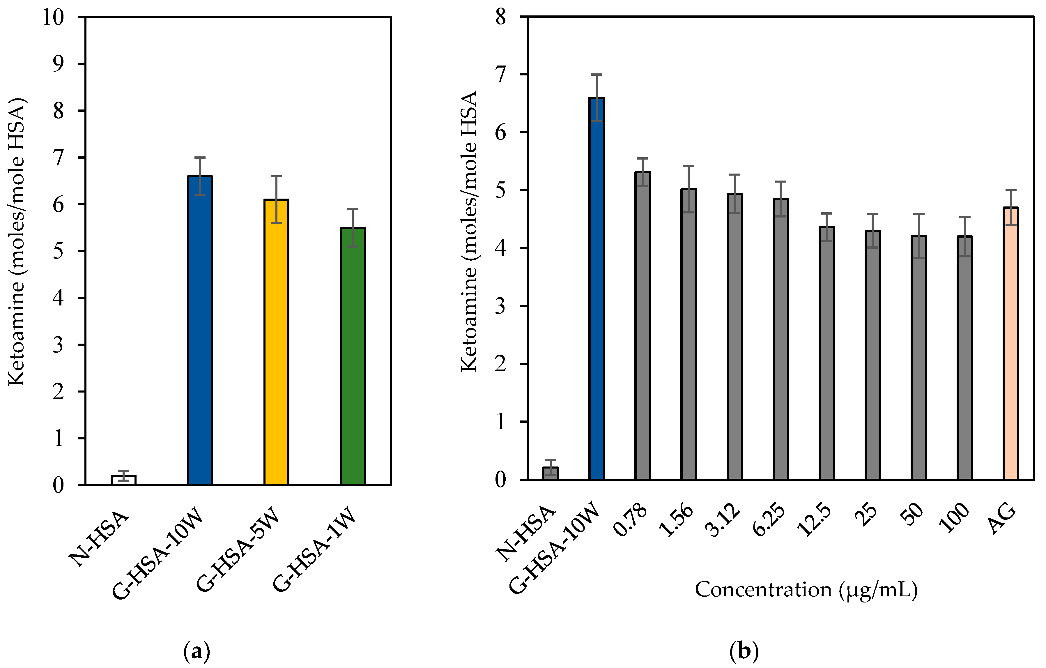
Early reactions between free amino groups and D-glucose, which may also include a small proportion of lysyl residues, can lead to the formation of ketoamine, which is an early glycation product. Glycation induced ketoamine formation in our reaction samples was estimated colorimetrically using NBT method [36]. HSA samples were incubated with D-glucose (0.05 M) for different time durations, and the subsequent generation of ketoamines was detected. High amounts of ketoimines were found in all glycated samples of HSA [G-HSA-10W (6.6 ± 0.4), G-HSA-5W (6.1 ± 0.5), and G-HSA-1W (5.5 ± 0.4) moles/mol HSA] (Figure 2a). Moreover, a significant (p < 0.001) difference in the amount of ketoamine moieties found in glycated samples vs. N-HSA was established (0.21 ± 0.10 moles/mol HSA).
It has been studied that glycation causes alteration of blood proteins (albumin). These proteins inherently have important functions which may be compromised due to the substantial changes in their conformation. We conducted different sets of experiments to detect the anti-glycation role of garlic extract. Different concentrations of garlic extracts were mixed with glycation reactions. Aminoguanidine is a well-known anti-glycation agent used as a control in all the analyses.
N-HSA, G-HSA-10W, and G-HSA-10W samples with garlic extracts were analyzed for the formation of ketoamines. No significant differences were observed between G-HSA-10W samples alone or in presence of garlic extract (Figure 2b). Ketoamines formed at an early stage of glycation and the formation of these cannot be inhibited significantly by garlic extracts alone as well in presence of aminoguanidine. Aminoguanidine is one of the best known synthetic anti-glycating agents that has the ability to inhibit the last stage of glycation [2]. Therefore, aminoguanidine was used as control in this result as well as in all other analyses.

3.1.4. Estimation of Protein Bound Carbonyl Compounds

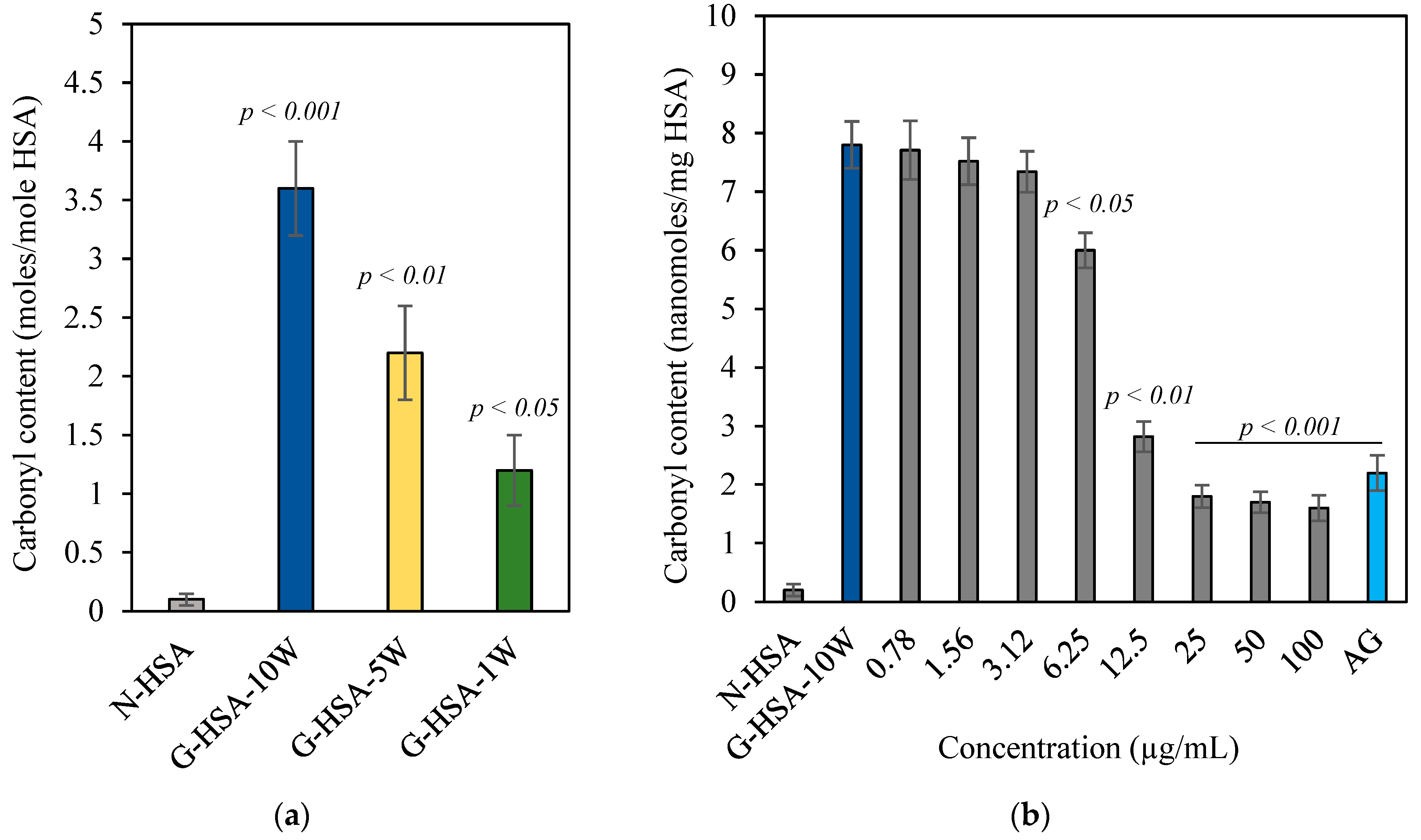
Carbonyl compounds bound to the HSA molecules were estimated and served as overall protein oxidation markers. Significantly high (p < 0.001) amounts of protein-bound carbonyl groups were detected in G-HSA-10W (3.6 ± 0.44 nanomoles/mg of HSA) sample followed by G-HSA-5W (2.2 ± 0.33 nanomoles/mg of HSA) and G-HSA-1W (1.2 ± 0.31 nanomoles/mg of HSA) as compared to the N-HSA sample (Figure 3a). Negligible amounts of carbonyl groups bound to protein were found in non-glycated samples of HSA (0.02 ± 0.01 nanomoles/mg of HSA).
Formation of carbonyl content in G-HSA-10W showed significant reduction (p < 0.001) when varying garlic extract concentrations 25, 50, and 100 µg/mL were added (Figure 3b). The concentration of 25 µg/mL of garlic extract was found to be most effective in decreasing the carbonyl content formation in glycation reactions. Moreover, no further appreciable change in carbonyl content was detected with further increase in extract concentrations. The addition of lower concentration of garlic extract (6.25 and 12.5 µg/mL) also showed a significant difference compared to G-HSA-10W sample (Figure 3b).

3.1.5. Toxicity of Garlic Extract

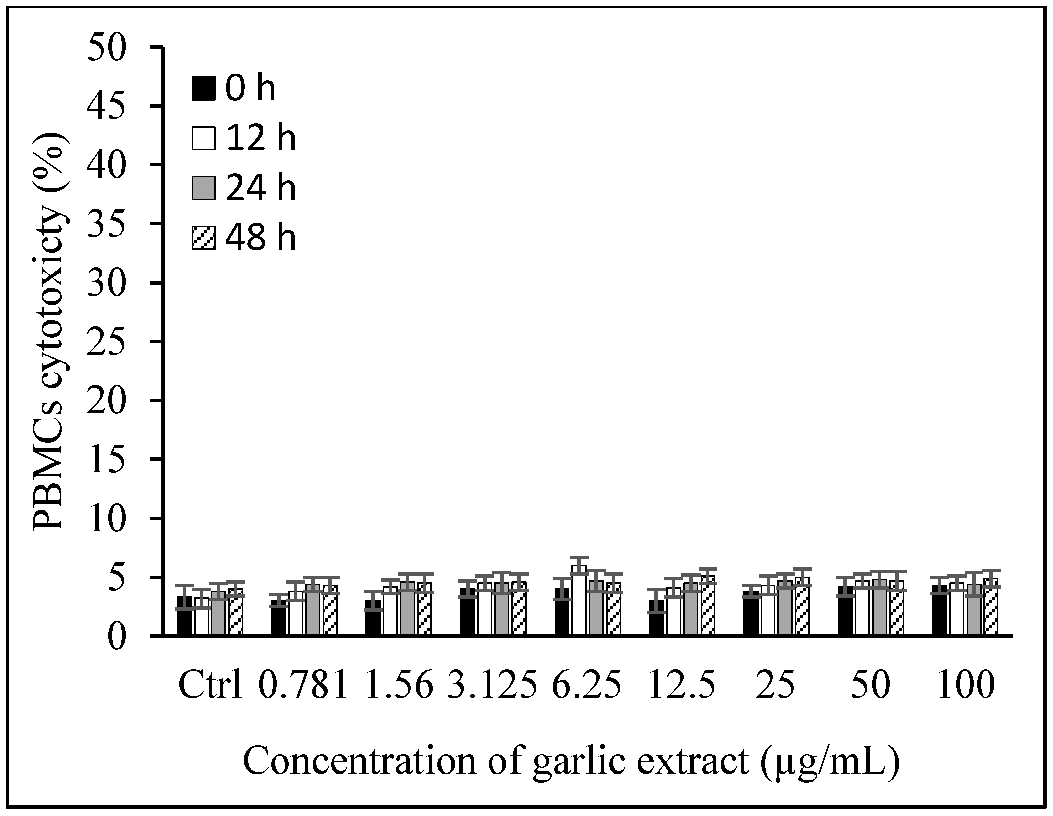
To analyze the toxicity of the garlic extract, freshly isolated PBMCs were used (Figure 4) in MTT assay. Varying concentrations (0.781–100 μg/mL) of garlic extract all showed very low cytotoxicities (3–5.1%) (Figure 4). Even using a higher concentration of the extract (100 µg/mL), no significant PBMCs death was observed. Control samples including PBMCs were incubated for similar durations (0, 12, 24, and 48 h) without garlic extracts. This result showed that garlic extract is not toxic towards blood cells.

3.2. Biophysical Analyses

3.2.1. AGE Pentosidine Formation

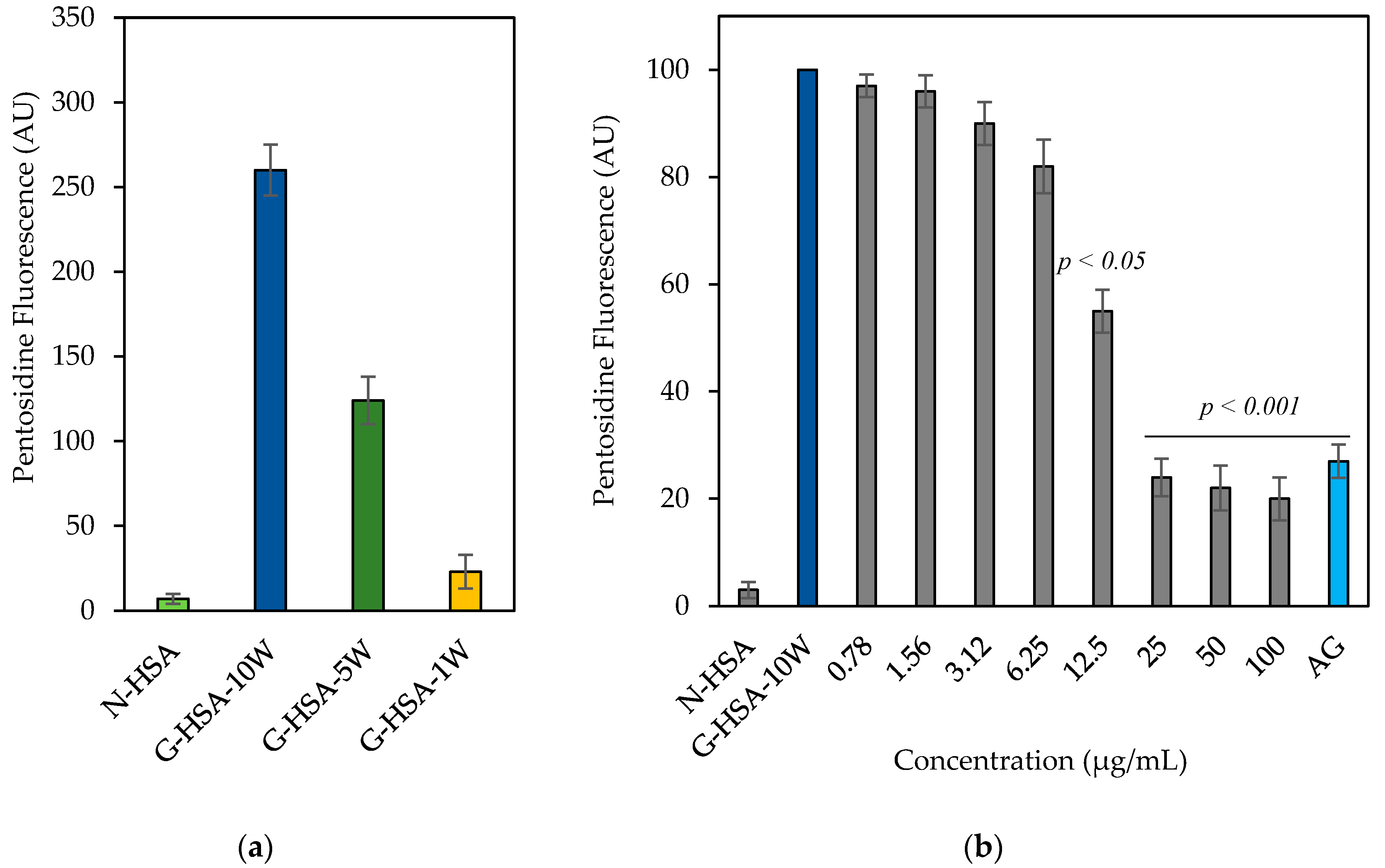
Modified and non-modified HSA samples were analyzed for the fluorophore pentosidine, which is an important AGE compound. A significant (p < 0.001) amount of pentosidine specific fluorescence (260 AU) was observed in G-HSA-10W as compared to N-HSA (Figure 5a). HSA sample glycated for 5 weeks also showed high pentosidine fluorescence (p < 0.01). However, HSA sample glycated for only 1 week showed less pentosidine fluorescence.
Significant (p < 0.01) decrease in AGE pentosidine formation in the reaction mixture was observed at the concentration of 12.5 µg/mL of garlic extract. Furthermore, higher concentrations (25, 50, and 100 µg/mL) of the extract exhibited highly significant (p < 0.001) inhibition in the formation of pentosidine (Figure 5b).

3.2.2. UV and Tryptophan Spectral Analyses

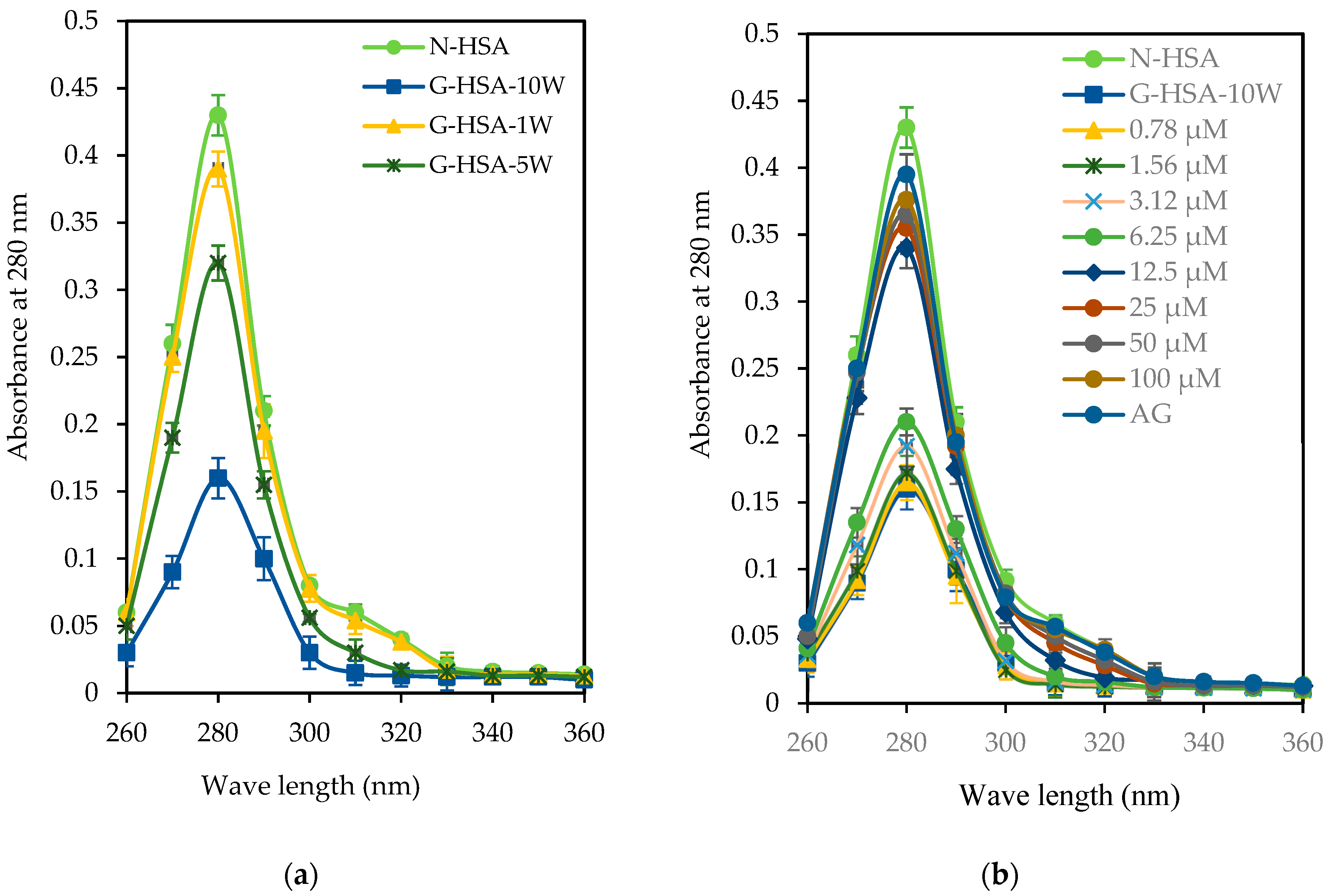
Overall spectral analysis was completed for glycated and non-glycated protein molecules by measuring all the samples at 280 nm. Significant differences in the UV intensities were observed between non-glycated (0.43 OD) vs. G-HSA-10W (0.16 OD; p < 0.001) samples followed by G-HSA-5W (0.32 OD; p < 0.05) (Figure 6a). However, G-HSA-1W did not show a significant change compared to N-HSA. The hypochromicity in UV intensities after 5 and 10 weeks of incubation of the glycation reaction indicated protein folding.
The UV intensity of G-HSA-10W increased to near UV intensity of N-HSA, when garlic extracts were introduced into the reaction mixture. However, significant increase in the UV intensity was observed mainly at 12.5 µg/mL of garlic extract. This shift further increased with the increase in extract concentrations (25, 50, and 100 µg/mL) (Figure 6b). Hence, garlic extract (12.5 µg/mL or more) reduces the protein folding of HSA molecule and helps in retaining the normal protein structure, which was altered by the glycation.
HSA protein molecule has a single tryptophan residue. Fluorescence analysis of tryptophan residue indicates site-specific changes in the molecule due to glycation. All the samples were analyzed for fluorescence emission spectra over 290–430 nm, with an excitation wavelength of 285 nm. Emission spectra showed maximum intensity at 330 nm for N-HSA (25.3 AU), G-HSA-1W (23.0 AU), and G-HSA-5W (20.1 AU). However, the G-HSA-10W exhibited 8.7 AU intensity at 320 nm (Figure 7a). This change in the emission spectra, showing a 10 nm blue shift, and a significant fluorescence intensity (N-HSA vs. G-HSA-10W), was due to glycation. Glycation may induce tryptophan microenvironment protein folding, leading the tryptophan residue away from polar to a more nonpolar environment.
The above result showed significant tryptophan microenvironment alterations in HSA molecules due to glycation. In this result, we examined the inhibition in tryptophan microenvironment changes exerted by the different concentrations of the extract. Extract concentration of 12.5 µg/mL showed a respectable increase in tryptophan fluorescence (14.0 AU) in the glycation mixture vs. glycation reaction without extract (8.7 AU) (Figure 7b). Moreover, the 25 µg/mL concentration of extract exhibited both significant increase in the fluorescence intensity (20.1 AU) towards the native protein and blueshift of ∼10 nm occurs. This result shows higher concentrations (12.5 µg/mL or more) of garlic extract inhibit changes in the tryptophan microenvironment and induce the exposure of tryptophan residue to a polar environment.

4. Discussion

Glucose and other sugars can chemically react with macromolecules (proteins, phospholipids, fatty acids, and nucleic acids) in vivo; an inevitable metabolic process. However, these changes are substantially low under normal physiological conditions. Under adverse and abnormal conditions, such as hyperglycemia, under extreme stress, aging and other chronic diseases, glucose and other sugars can react with the biomolecules in an accelerated manner [1,4,7]. These reactions can produce many other chemical products and intermediates such as ketoamines, carbonyl compounds, and AGEs. It has been observed in previous studies that glycation of protein induces structural alterations, which may potentially decrease the functional efficiency of these protein molecules [41].
Natural products and their compounds are usually harmless, affordable, and have traditionally been utilized to treat various human illnesses since antiquity. According to a World Health Organization (WHO) survey, more than 80% of people use traditional plant-based medicines for their primary health care [42]. Natural products and secondary metabolites have previously been mentioned as promising sources of therapeutically relevant biomolecules for drug development [43].
Synthetic medications like ibuprofen, warfarin, and diclofenac have grown in popularity over time, despite the fact that they have a variety of adverse effects. Many medicinal plants have a wide range of medical uses, and are widely utilized due to their low toxicity, minimum side effects, and potent dynamic healing effects. Garlic is a herbaceous perennial belonging to the Amaryllidaceae plant family. It is among the most potent medicinal plants and has traditionally been used in herbal medicine to prevent and cure a wide variety of illnesses, including cardiovascular disease, atherosclerosis, hyperlipidemia, thrombosis, hypertension, and diabetes [25]. Garlic is considered one of the more potent antioxidant natural products, a staple in our diets for centuries. In this study, garlic extract showed high contents of phenolic as well as flavonoid compounds.
Detectable forms of chemical damage induced by glycation include AGEs, lipid peroxidation products, ROS modified amino acids, chlorine and nitrogen, and racemized amino acids [24]. Several serious diseases, including aging, inflammation, cancer, diabetes, cardiovascular disease, arthritis, cataracts, muscular degeneration, and impaired wound healing, are linked to oxidative stress and ROS. Moreover, oxidative stress and high glucose levels can damage blood proteins concomitantly with the formation of TAGE. Total phenolic content and flavonoid content of garlic extract was found to be in accordance with a previous report [25]. This investigation shows that garlic extract (100 µg/mL) shows 31.63% H2O2 reducing activity which was comparable to the previously published reducing ability of garlic extract at a concentration of 100 µg/mL [25].
Our data shows that incubation of HSA with D-glucose leads to the formation of ketoimine, as high amounts of ketoimines were found in all glycated samples of HSA. The formation of ketoamines was highest in G-HSA-10W, indicating that incubation time greatly impacts the degree of glycation. This is in accordance with previous studies where the high degree of glycation is related to the duration of incubation [1,44]. Although ketoimines decreased in the presence of garlic extract, however, the difference was not significant.
The carbonyl content of the protein is considered a hallmark of protein oxidation. α-dicarbonyl compounds are intermediary products of a glycation reaction, which undergo a multistep reaction to produce AGEs [4]. A high content of carbonyl compounds was detected in the glycated HSA sample, which significantly decreased in the presence of garlic extract with a concentration of 6.25 µg/mL or more. This finding also supports the antioxidant properties of garlic extract compounds. Our results of decreased carbonyl content of glycated protein in the presence of garlic extract are in accordance with the results of Garmia et al. [45]. Moreover, an even higher extract concentration (100 µg/mL) did not show any toxicity towards freshly isolated PBMCs.
Pentosidine is one of the well-known AGE molecules. Glycated samples exhibited high fluorescence intensities indicative of pentosidine. G-HSA-10W had the highest fluorescent intensity compared to native HSA. However, garlic extract showed protection against the pentosidine formation, and this protection increased with an increase in the concentration of garlic extract. Thus, our data demonstrates that garlic extract significantly protects HSA against gluco-oxidation and cross-linking, caused by glycation.
Glycation of HSA contributes to alteration in the structure of HSA, resulting in a decrease in absorbance at 280 nm or hypochromicity. These changes markedly reduced when garlic extract was added to the reaction mixture, in a dose-dependent manner. Hence, there is an increase in hyperchromicity in the presence of garlic extract. This data is in accordance with a previous study involving the protecting ability of a garlic compound (i.e., alliin,) against structural changes in superoxide dismutase caused by glycation [46]. AGEs are aggregated high molecular weight compounds and have specific fluorescence spectra [1]. In this study, autofluorescence was used for AGEs formation analysis. HSA incubated with glucose showed characteristic AGEs specific fluorescence spectra. This may be due to tryptophan microenvironment alterations in HSA molecules due to glycation. This study shows that higher concentrations of garlic extract inhibit changes in the tryptophan microenvironment and induced the exposure of tryptophan residue to a polar environment. Hence, garlic extract showed protection from AGEs formation. Our biophysical analysis for protection of glycated sample by garlic extract is in accordance with the previous reports of Anwar and Younus who showed that alliin of garlic extract protected HSA from glycation induced structural changes and formation of AGEs [46].
Our data demonstrates that garlic extract was observed to suppress the formation of ketoamine, carbonyl compounds, and AGEs, produced due to the glycation reaction in a dose-dependent manner. Furthermore, prevention of structural alterations in glycated HSA samples were observed on addition of garlic extract. The presence of polyphenolic components may be responsible for the anti-glycating and anti-AGEs production actions of garlic extract. As a result, our findings show that antioxidant activity, polyphenolic content, and suppression of AGEs formation activity of garlic extract might be responsible for the therapeutic potential and benefits of garlic extract, including prevention of diabetes and its associated complications.

5. Conclusions

Glycation reaction with biomolecules produces excessive biochemical compounds, which may directly or indirectly exert negative effects on human health. Several benefits have been shown due to the consumption of natural products. Garlic and its active components have been shown to play a vital role in illness management. Garlic extract showed good antioxidant, anti-glycating, AGEs generation inhibition activity. Garlic extract has the capacity to decrease the glycation burden on blood proteins. As a result of our findings, we believe that garlic extract might be employed as prospective medication candidates with broad therapeutic potential in various medical disciplines such as in diabetic patients.

Author Contributions

Conceptualization, M.W.A.K., S.S.; methodology, M.W.A.K., A.A.O., A.F.M.A., A.Y.M.A.; validation, A.F.M.A., A.K.D.A., A.K.A., A.Y.M.A.; formal analysis, M.W.A.K., A.A.O., A.F.M.A., A.K.A.; investigation, M.W.A.K., A.A.O., A.F.M.A., A.K.A., S.S.; resources, E.M.A., S.A.A.-Z., A.K.D.A.; data curation, M.W.A.K. and A.A.O.; writing—original draft preparation, M.W.A.K., S.S.; writing—review and editing, E.M.A., S.A.A.-Z., A.K.D.A.; supervision, M.W.A.K.; funding acquisition, M.W.A.K. All authors have read and agreed to the published version of the manuscript.

Funding

This research has been funded by Scientific Research Deanship at the University of Ha’il–Saudi Arabia through project number RG-20 120.

Institutional Review Board Statement

This study has been reviewed and approved by the Research Ethics Committee (REC) at the University of Hail dated: 27/11/2020 and approved by university president letter number Nr. 20455/5/42 dated 16/04/1442 H.

Informed Consent Statement

Not applicable.

Data Availability Statement

The data presented in this study are available on request from the corresponding author.

Conflicts of Interest

The authors declare no conflict of interest.

References

  1. Khan, M.W.A.; Rasheed, Z.; Khan, W.A.; Ali, R. Biochemical, biophysical, and thermodynamic analysis of in vitro glycated human serum albumin. Biochemistry (Moscow) 2007, 72, 146–152. [Google Scholar] [CrossRef]
  2. Khan, M.A.; Anwar, S.; Aljarbou, A.N.; Al-Orainy, M.; Aldebasi, Y.H.; Islam, S.; Younus, H. Protective effect of thymoquinone on glucose or methylglyoxal-induced glycation of superoxide dismutase. Int. J. Biol. Macromol. 2014, 65, 16–20. [Google Scholar] [CrossRef] [PubMed]
  3. Anwar, S.; Alam Khan, M.; Sadaf, A.; Younus, H. A structural study on the protection of glycation of superoxide dismutase by thymoquinone. Int. J. Biol. Macromol. 2014, 69, 476–481. [Google Scholar] [CrossRef]
  4. Khan, M.; Al Otaibi, A.; Sherwani, S.; Khan, W.; Alshammari, E.; Al-Zahrani, S.; Saleem, M.; Khan, S.; Alouffi, S. Glycation and Oxidative Stress Increase Autoantibodies in the Elderly. Molecules 2020, 25, 3675. [Google Scholar] [CrossRef] [PubMed]
  5. Nimbalkar, V.V.; Mandlik, R.V.; Naik, S.R.; Maseeh, A. Nonenzymatic glycosylation: A biochemical link between chronic hyperglycemia and pathophysiologic processes associated with diabetic complications and aging related debilities. Biomed. Aging Pathol. 2012, 2, 133–142. [Google Scholar] [CrossRef]
  6. Khan, M.W.A.; Qadrie, Z.L.; Khan, W.A. Antibodies against Gluco-Oxidatively Modified Human Serum Albumin Detected in Diabetes-Associated Complications. Int. Arch. Allergy Immunol. 2010, 153, 207–214. [Google Scholar] [CrossRef]
  7. Alouffi, S.; Sherwani, S.; Al-Mogbel, M.S.; Sherwani, M.K.A.; Khan, M.W.A. Depression and Smoking Augment the Production of Circulating Autoantibodies against Glycated HSA in Rheumatoid Arthritis Patients. Int. Arch. Allergy Immunol. 2018, 177, 170–180. [Google Scholar] [CrossRef] [PubMed]
  8. Schröter, D.; Höhn, A. Role of Advanced Glycation End Products in Carcinogenesis and their Therapeutic Implications. Curr. Pharm. Des. 2018, 24, 5245–5251. [Google Scholar] [CrossRef]
  9. Semba, R.D.; Nicklett, E.J.; Ferrucci, L. Does Accumulation of Advanced Glycation End Products Contribute to the Aging Phenotype? J. Gerontol. Ser. A Boil. Sci. Med. Sci. 2010, 65A, 963–975. [Google Scholar] [CrossRef] [Green Version]
  10. Simm, A.; Wagner, J.; Gursinsky, T.; Nass, N.; Friedrich, I.; Schinzel, R.; Czeslik, E.; Silber, R.; Scheubel, R. Advanced glycation endproducts: A biomarker for age as an outcome predictor after cardiac surgery? Exp. Gerontol. 2007, 42, 668–675. [Google Scholar] [CrossRef]
  11. Nowotny, K.; Jung, T.; Grune, T.; Höhn, A. Accumulation of modified proteins and aggregate formation in aging. Exp. Gerontol. 2014, 57, 122–131. [Google Scholar] [CrossRef]
  12. Takata, T.; Sakasai-Sakai, A.; Ueda, T.; Takeuchi, M. Intracellular toxic advanced glycation end-products in cardiomyocytes may cause cardiovascular disease. Sci. Rep. 2019, 9, 2121. [Google Scholar] [CrossRef] [Green Version]
  13. Takeuchi, M.; Takino, J.; Sakasai-Sakai, A.; Takata, T.; Ueda, T.; Tsutsumi, M.; Hyogo, H.; Yamagishi, S. Involvement of the TAGE-RAGE system in non-alcoholic steatohepatitis: Novel treatment strategies. World J. Hepatol. 2014, 6, 880–893. [Google Scholar] [CrossRef] [PubMed]
  14. Takeuchi, M.; Yamagishi, S. TAGE (toxic AGEs) hypothesis in various chronic diseases. Med. Hypotheses 2004, 63, 449–452. [Google Scholar] [CrossRef]
  15. Koriyama, Y.; Furukawa, A.; Muramatsu, M.; Takino, J.-I.; Takeuchi, M. Glyceraldehyde caused Alzheimer’s disease-like alterations in diagnostic marker levels in SH-SY5Y human neuroblastoma cells. Sci. Rep. 2015, 5, 13313. [Google Scholar] [CrossRef] [Green Version]
  16. Takeuchi, M. Serum Levels of Toxic AGEs (TAGE) May Be a Promising Novel Biomarker for the Onset/Progression of Lifestyle-Related Diseases. Diagnostics 2016, 6, 23. [Google Scholar] [CrossRef] [PubMed]
  17. Takino, J.; Nagamine, K.; Hori, T.; Sakasai-Sakai, A.; Takeuchi, M. Contribution of the toxic advanced glycation end-products-receptor axis in nonalcoholic steatohepatitis-related hepatocellular carcinoma. World J. Hepatol. 2015, 7, 2459–2469. [Google Scholar] [CrossRef]
  18. Takeuchi, M.; Sakasai-Sakai, A.; Takino, J.; Takata, T.; Ueda, T.; Tsutsumi, M. Toxic AGEs (TAGE) theory in the pathogenesis of NAFLD and ALD. Int. J. Diabetes Clin. Res. 2015, 2, 036. [Google Scholar] [CrossRef]
  19. Takeuchi, M.; Takino, J.-I.; Sakasai-Sakai, A.; Takata, T.; Tsutsumi, M. Toxic AGE (TAGE) Theory for the Pathophysiology of the Onset/Progression of NAFLD and ALD. Nutrients 2017, 9, 634. [Google Scholar] [CrossRef] [Green Version]
  20. Rahbar, S.; Figarola, L.J. Novel Inhibitors of Advanced Glycation Endproducts. Arch. Biochem. Biophys. 2003, 419, 63–79. [Google Scholar] [CrossRef] [PubMed]
  21. Indyk, D.; Bronowicka-Szydełko, A.; Gamian, A.; Kuzan, A. Advanced glycation end products and their receptors in serum of patients with type 2 diabetes. Sci. Rep. 2021, 11, 13264. [Google Scholar] [CrossRef]
  22. Singh, V.P.; Bali, A.; Singh, N.; Jaggi, A.S. Advanced Glycation End Products and Diabetic Complications. Korean J. Physiol. Pharmacol. 2014, 18, 1–14. [Google Scholar] [CrossRef] [Green Version]
  23. Alsahli, M.A.; Anwar, S.; Alzahrani, F.M.; Almatroudi, A.; Alfheeaid, H.; Khan, A.A.; Allemailem, K.S.; Almatroodi, S.A.; Rahmani, A.H. Health Promoting Effect of Phyllanthus emblica and Azadiractha indica against Advanced Glycation End Products Formation. Appl. Sci. 2021, 11, 8819. [Google Scholar] [CrossRef]
  24. Anwar, S.; Khan, S.; Almatroudi, A.; Khan, A.A.; Alsahli, M.A.; Almatroodi, S.A.; Rahmani, A.H. A review on mechanism of inhibition of advanced glycation end products formation by plant derived polyphenolic compounds. Mol. Biol. Rep. 2021, 48, 787–805. [Google Scholar] [CrossRef] [PubMed]
  25. Almatroodi, S.A.; Anwar, S.; Almatroudi, A.; Khan, A.A.; AlRumaihi, F.; Alsahli, M.A.; Rahmani, A.H. Hepatoprotective Effects of Garlic Extract against Carbon Tetrachloride (CCl4)-Induced Liver Injury via Modulation of Antioxidant, Anti-Inflammatory Activities and Hepatocyte Architecture. Appl. Sci. 2020, 10, 6200. [Google Scholar] [CrossRef]
  26. Thomson, M.; Al-Qattan, K.K.; Js, D.; Ali, M. Anti-diabetic and anti-oxidant potential of aged garlic extract (AGE) in streptozotocin-induced diabetic rats. BMC Complement. Altern. Med. 2015, 16, 17. [Google Scholar] [CrossRef] [PubMed] [Green Version]
  27. Sun, Y.E.; Wang, W.; Qin, J. Anti-hyperlipidemia of garlic by reducing the level of total cholesterol and low-density lipoprotein: A meta-analysis. Medicine (Baltimore) 2018, 97, e0255. [Google Scholar] [CrossRef]
  28. Sobenin, I.A.; Myasoedova, V.A.; Iltchuk, M.I.; Zhang, D.-W.; Orekhov, A.N. Therapeutic effects of garlic in cardiovascular atherosclerotic disease. Chin. J. Nat. Med. 2019, 17, 721–728. [Google Scholar] [CrossRef]
  29. Moutia, M.; Habti, N.; Badou, A. In Vitro and In Vivo Immunomodulator Activities of Allium sativum L. Evid.-Based Complement. Altern. Med. 2018, 2018, 4984659. [Google Scholar] [CrossRef] [Green Version]
  30. Abu Almaaty, A.; Rashed, H.; Soliman, M.; Fayad, E.; Althobaiti, F.; El-Shenawy, N. Parasitological and Biochemical Efficacy of the Active Ingredients of Allium sativum and Curcuma longa in Schistosoma mansoni Infected Mice. Molecules 2021, 26, 4542. [Google Scholar] [CrossRef]
  31. Elosta, A.; Slevin, M.; Rahman, K.; Ahmed, N. Aged garlic has more potent antiglycation and antioxidant properties compared to fresh garlic extract in vitro. Sci. Rep. 2017, 7, 39613. [Google Scholar] [CrossRef] [PubMed] [Green Version]
  32. Anwar, S.; Almatroudi, A.; Allemailem, K.S.; Joseph, R.J.; Khan, A.A.; Rahmani, A.H. Protective Effects of Ginger Extract against Glycation and Oxidative Stress-Induced Health Complications: An In Vitro Study. Processes 2020, 8, 468. [Google Scholar] [CrossRef]
  33. Ordonez, A.A.L.; Gomez, J.D.; Vattuone, M.A.; Lsla, M.I. Antioxidant activities of Sechium edule (Jacq.) Swartz extracts. Food Chem. 2006, 97, 452–458. [Google Scholar] [CrossRef]
  34. Taie, H.A.A.; Salama, Z.A.-E.R.; Radwan, S. Potential activity of basil plants as a source of antioxidants and anticancer agents as affected by organic and bio-organic fertilization. Not. Bot. Horti Agrobot. 2010, 38, 119–127. [Google Scholar]
  35. Ruch, R.J.; Cheng, S.-J.; Klaunig, J.E. Prevention of cytotoxicity and inhibition of intercellular communication by antioxidant catechins isolated from Chinese green tea. Carcinogenesis 1989, 10, 1003–1008. [Google Scholar] [CrossRef]
  36. Mashiba, S.; Uchida, K.; Okuda, S.; Tomita, S. Measurement of glycated albumin by the nitroblue tetrazolium colorimetric method. Clin. Chim. Acta 1992, 212, 3–15. [Google Scholar] [CrossRef]
  37. Ahmed, N.; Furth, J.A. A microassay for protein glycation based on the periodate method. Analyt. Biochem. 1991, 192, 109–111. [Google Scholar] [CrossRef]
  38. Levine, R.L.; Williams, J.A.; Stadtman, E.P.; Shacter, E. Carbonyl assays for determination of oxidatively modified proteins. Methods Enzymol. 1994, 233, 346–357. [Google Scholar] [CrossRef]
  39. Otaibi, A.A.; Sherwani, S.; Al-Zahrani, S.A.; Alshammari, E.M.; Khan, W.A.; Alsukaibi, A.K.D.; Khan, S.N.; Khan, M.W.A. Biologically Active α-Amino Amide Analogs and γδ T Cells-A Unique Anticancer Approach for Leukemia. Front. Oncol. 2021, 11, 706586. [Google Scholar] [CrossRef]
  40. Liggins, J.; Furth, J.A. Role of protein-bound carbonyl groups in the formation of advanced glycation end products. Biochim. Biophys. Acta 1997, 1361, 123–130. [Google Scholar] [CrossRef] [Green Version]
  41. Khan, M.W.; Al Otaibi, A.; Al-Zahrani, S.A.; Alshammari, E.M.; Haque, A.; Alouffi, S.; Khan, W.A.; Khan, S.N. Experimental and theoretical insight into resistance to glycation of bovine serum albumin. J. Mol. Struct. 2021, 1230, 129645. [Google Scholar] [CrossRef]
  42. Anwar, S.; Almatroudi, A.; Alsahli, M.A.; Khan, M.A.; Khan, A.A.; Rahmani, A.H. Natural Products: Implication in Cancer Prevention and Treatment through Modulating Various Biological Activities. Anti-Cancer Agents Med. Chem. 2020, 20, 2025–2040. [Google Scholar] [CrossRef]
  43. Atanasov, A.G.; Zotchev, S.B.; Dirsch, V.M.; The International Natural Product Sciences Taskforce; Supuran, C.T. Natural products in drug discovery: Advances and opportunities. Nat. Rev. Drug Discov. 2021, 20, 200–216. [Google Scholar] [CrossRef] [PubMed]
  44. Ahmad, K.; Khan, S.; Adil, M.; Saeed, M.; Srivastava, A.K. Structure based molecular inhibition of Caspase-8 for treatment of multi-neurodegenerative diseases using known natural compounds. Bioinformation 2014, 10, 191–195. [Google Scholar] [CrossRef] [Green Version]
  45. Garima, G.; Moin, S.; Aggarwal, N.K.; Gupta, P.K.; Dwivedi, S.; Gupta, U. a study on the role of dietary agent aged garlic extract on protein glycation. World J. Pharm. Res. 2015, 4, 468–476. [Google Scholar]
  46. Anwar, S.; Younus, H. Inhibitory effect of alliin from Allium sativum on the glycation of superoxide dismutase. Int. J. Biol. Macromol. 2017, 103, 182–193. [Google Scholar] [CrossRef]
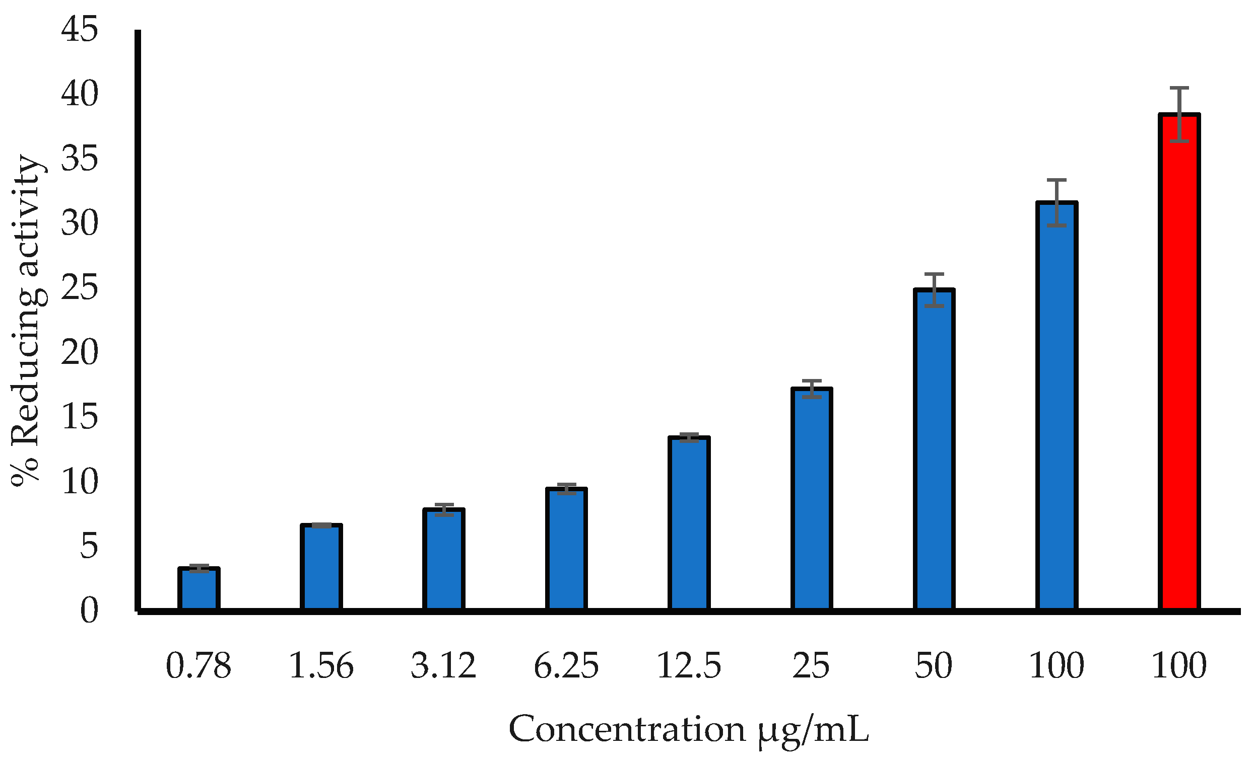
Figure 1. Percentage H2O2 scavenging activity of garlic extract. Various concentrations of garlic extract (0.78, 1.56, 3.12, 6.25, 12.5, 25, 50, and 100 µg/mL) (blue column). The red column belongs to 100 µg/mL of standard antioxidant “Ascorbic acid”.
Applsci 11 11028 g001
Figure 2. Formation of ketoamines in the process of glycation with different incubation times (1, 5, and 10 weeks) (a). Effect of garlic extract on the formation of ketoamines in the process of HSA glycation (b), different concentrations of garlic extract (0.78–100 µg/mL) were incubated with glycation reactions for 10 weeks. All the reactions were incubated under similar conditions. Data is presented as mean ± SD values. t test was adopted for the comparison between the non-glycated and glycated samples as well as between G-HSA-10W alone or with garlic extracts.
Applsci 11 11028 g002
Figure 3. Formation of protein bound carbonyl content in the non-glycated and glycated samples of HSA (a). Effect of garlic extract on the formation of protein bound carbonyl content in the process of HSA glycation (10 weeks) with different concentrations (0.78–100 µg/mL) (b). All the reactions were carried out under similar experimental conditions. Data is presented as mean ± SD values. t test was adopted for comparison between non-glycated and glycated samples of HSA, and significance was calculated.
Applsci 11 11028 g003
Figure 4. Freshly isolated PBMCs were incubated with different concentrations of garlic extract (0.781–100 µg/mL) and durations (0–48 h) of incubations.
Applsci 11 11028 g004
Figure 5. AGE molecule pentosidine specific fluorescence was recorded for non-glycated and glycated HSA samples (a). Effect of garlic extract on the formation of AGEs (pentosidine) during glycation of HSA (10 weeks) with different concentrations (0.78–100 µg/mL) of garlic (b). Data presented as mean ± SD values. t test was adopted for comparison between non-glycated vs. glycated HSA samples and significance was calculated.
Applsci 11 11028 g005
Figure 6. UV-spectra were analyzed for N-HSA, G-HSA-1W, G-HSA-5W, and G-HSA-10W (a). Effect of garlic extract on the overall protein structure changes during glycation of HSA (10 weeks) with different concentrations (0.78–100 µg/mL) (b). All the sample readings were recorded in the range of 260–360 nm. Data is presented as mean ± SD values. t test was adopted for the comparison between N-HSA vs. G-HSA-1W, G-HSA-5W, and G-HSA-10W.
Applsci 11 11028 g006
Figure 7. Tryptophan fluorescence emission spectra were estimated for N-HSA, G-HSA-1W, G-HSA-5W, and G-HSA-10W (a). Effect of garlic extract on the protein microenvironment structure alteration during glycation of HSA (10 weeks) with different concentrations (0.78–100 µg/mL) (b). All the samples were excited with a wavelength of 285 nm. Data is presented as mean ± SD values. t test was adopted for the comparison between N-HSA vs. G-HSA-1W, G-HSA-5W, and G-HSA-10W.
Applsci 11 11028 g007
Table 1. Secondary metabolites in garlic.
Preliminary ScreeningGarlic Extract
Weight of dry powder 50 g
Yield5.19%
ExtractAqueous
Flavonoid+
Polyphenolic compounds+
Total phenolic compounds21.45 ± 0.02 mg gallic acid equivalent/g dry weight of extract
Total flavonoid content16.58 ± 0.03 mg quercetin
equivalent/g dry weight of the extract
Publisher’s Note: MDPI stays neutral with regard to jurisdictional claims in published maps and institutional affiliations.