Article

In Vitro Evaluation of the Antibacterial and Antioxidant Activities of Extracts of Gracilaria gracilis with a View into Its Potential Use as an Additive in Fish Feed

MARE—IPLeiria—Marine and Environmental Sciences Centre, Polytechnic of Leiria, Edifício CETEMARES, Avenida Porto de Pesca, 2520-641 Peniche, Portugal
*
Author to whom correspondence should be addressed.
Appl. Sci. 2021, 11(14), 6642; https://doi.org/10.3390/app11146642
Submission received: 23 June 2021 / Revised: 13 July 2021 / Accepted: 16 July 2021 / Published: 20 July 2021

Abstract

:

Featured Application

To obtain extracts of the red seaweed Gracilaria gracilis that have antimicrobial and antioxidant properties for incorporation as an additive in fish feed.

Abstract

Fish in aquaculture systems are subject to several stressors that inhibit the immune response and potentiate the development of disease and increased mortality. The inclusion of additives in the fish diet, namely seaweeds or their extracts, that are natural sources of bioactive compounds can be an important tool for promoting the health and well-being of these animals. The present study aims at the development of sustainable and effective methodologies for the extraction of bioactive compounds of the red seaweed Gracilaria gracilis, exploring its antibacterial and antioxidant potential and considering its potential use as an additive for functional fish feeds. The yield of the extraction methods was evaluated upon the use of sequential solid–liquid extraction techniques with ethanol and water as solvents, different extraction temperatures (room temperature: 40 °C and 70 °C), and extraction time. The results demonstrated that the adoption extraction times of 30 min at 40 °C provided higher yields. We also evaluated the antioxidant capacity and the antibacterial properties of the obtained extracts against different strains that cause fish diseases by disk diffusion and broth microdilution methods. The antioxidant activity was determined by the DPPH reducing capacity method and quantification of total polyphenols content (TPC). With these results, we can establish extraction procedures that allow the future use of G. gracilis extracts, with antibacterial and antioxidant effects in a safe and effective way.

1. Introduction

Marine seaweeds are renewable natural resources that have high potential for application in the pharmaceutical, food and nutritional, nutraceutical, animal feed, packaging, and cosmetic industries, among others [1,2,3,4]. These organisms are generally rich in proteins, minerals, vitamins, and fibers, and also reveal the presence of highly diverse bioactive compounds [5,6,7,8,9,10,11]. Red seaweeds (Rhodophyta) are macroscopic, benthic, and multicellular organisms that are commonly used in human nutrition, mainly in Asian countries. They are also important sources of thickening and gelling food additives due to their richness in phycocolloids such as agar or carrageenan [4,8,12,13]. These organisms often thrive in environments where light, salinity, and temperatures can reach extremes [13,14,15]. To adapt to such extreme conditions, seaweeds produce a wide variety of secondary metabolites that exhibit powerful biological activities and are therefore highly valued as active and functional ingredients not only for the food and feedstock industry, but also in health care and cosmetics [10,11,16,17,18]. The production of active compounds in algae can be adjusted by manipulating the appropriate growing conditions; thus, they are natural bioreactors [19]. Seaweed extracts commonly present different bioactivities related to the presence of compounds such as polysaccharides, pigments, compounds with antioxidant activity, and substances similar to plant growth regulators [6,7,20]. The antibacterial activity present in macroalgae extracts is usually attributed to compounds such as proteins but also to polyphenols, polysaccharides, pigments, and PUFAs [6,21,22]. Antioxidant activity, conversely, seems to be frequently associated with glutathione, mycosporine-like amino acids, ascorbate, pigments, and polyphenols, among other compounds, and is widely reported in red seaweeds [6,23,24].
In this sense, it is also interesting to establish adequate, profitable, and ecologically acceptable extraction procedures that allow not only the meeting of the legal requirements regarding the use of solvents on food and feed production processes but also to ensure of all the properties of interest that seaweeds possess [24,25]. Traditional extraction techniques such as Soxhlet, solid–liquid extraction (SLE), or liquid-liquid extraction (LLE) can be adjusted, minimizing the cost of solvents and the times involved [26]. The use of new extraction techniques such as extraction by supercritical fluids (SFE), ultrasound, and microwave assisted extraction (MAE), among others, provide an effective alternative [5,27,28] but require high investment in equipment. When the intention is to incorporate algae extracts in food or animal feed, the objectives go beyond obtaining a product with nutritional value. These goals are usually related to the bioactive properties of these extracts and the potential to obtain food/feed with desirable functional properties; therefore, extraction procedures must be carefully evaluated.
Gracilaria gracilis (Stackhouse) Steentoft, L. M. Irvine & Farnham 1995, is a red seaweed (Rhodophyta) considered an agarophyte due to its high agar content [12,14]. It is widely distributed throughout the world and lives in temperate waters up to a maximum of 30 °C and tolerating a wide range of salinity values. This highly valuable seaweed can also be a source of different organic compounds such as R-phycoerythrin, arachidonic acid, proteins, and phenols in addition to their antioxidant and radical scavenging potential [8,29]. Antimicrobial activity has also been reported in this genus [30,31,32,33] related to the presence of compounds such as polyphenols and fatty acids [30]. In these seaweeds, as in many other living organisms, reactive oxygen species (ROS) are commonly produced during metabolism but there are enzymatic and non-enzymatic cellular defense mechanisms [34] that allow their elimination. Antioxidant compounds are interesting as supplements as they play a role in preventing diseases associated with oxidative stress that occur due to the reduction in the presence of antioxidant compounds or excess ROS.
The objective of the present study was (i) to establish efficient and low-cost extraction methodologies aimed at the incorporation of bioactive extracts in aquaculture fish feed and (ii) to explore the antibacterial and antioxidant potential of G. gracilis extracts. Based on solid–liquid extraction techniques, several extraction parameters were tested, seeking to evaluate the influence of solvent, temperature, and extraction time on the extraction yield. The different extracts obtained under different conditions were later tested for their antioxidant and antimicrobial capacity. Thus, we intend to provide a base that supports the use of extracts of G. gracilis as a functional ingredient in food/feed with antibacterial and antioxidant effects.

2. Materials and Methods

2.1. Sample Collection and Preparation

The samples of G. gracilis were collected at Lagoa de Óbidos, Braço do Bom Sucesso (39°24′1″ N, 9°13′11″ W) in Portugal. Sampling took place during low tide and the transportation of the thalli to the laboratory was managed in dark cooler boxes seeking to ensure the preservation of the biomass. In the laboratory, the biomass was thoroughly washed in seawater and the necrotic parts and epiphytes were removed. Healthy portions were selected, packaged, and stored at −20 °C. Before the extraction procedures, the thalli were dried at 25 °C in a ventilated laboratory oven (Binder, FD115) during the course of 48 h until reaching a constant weight, were reduced into powder in a blender, and then were sieved (particles smaller than 200 µm) [35]. Seaweed powder was stored at −20 °C.

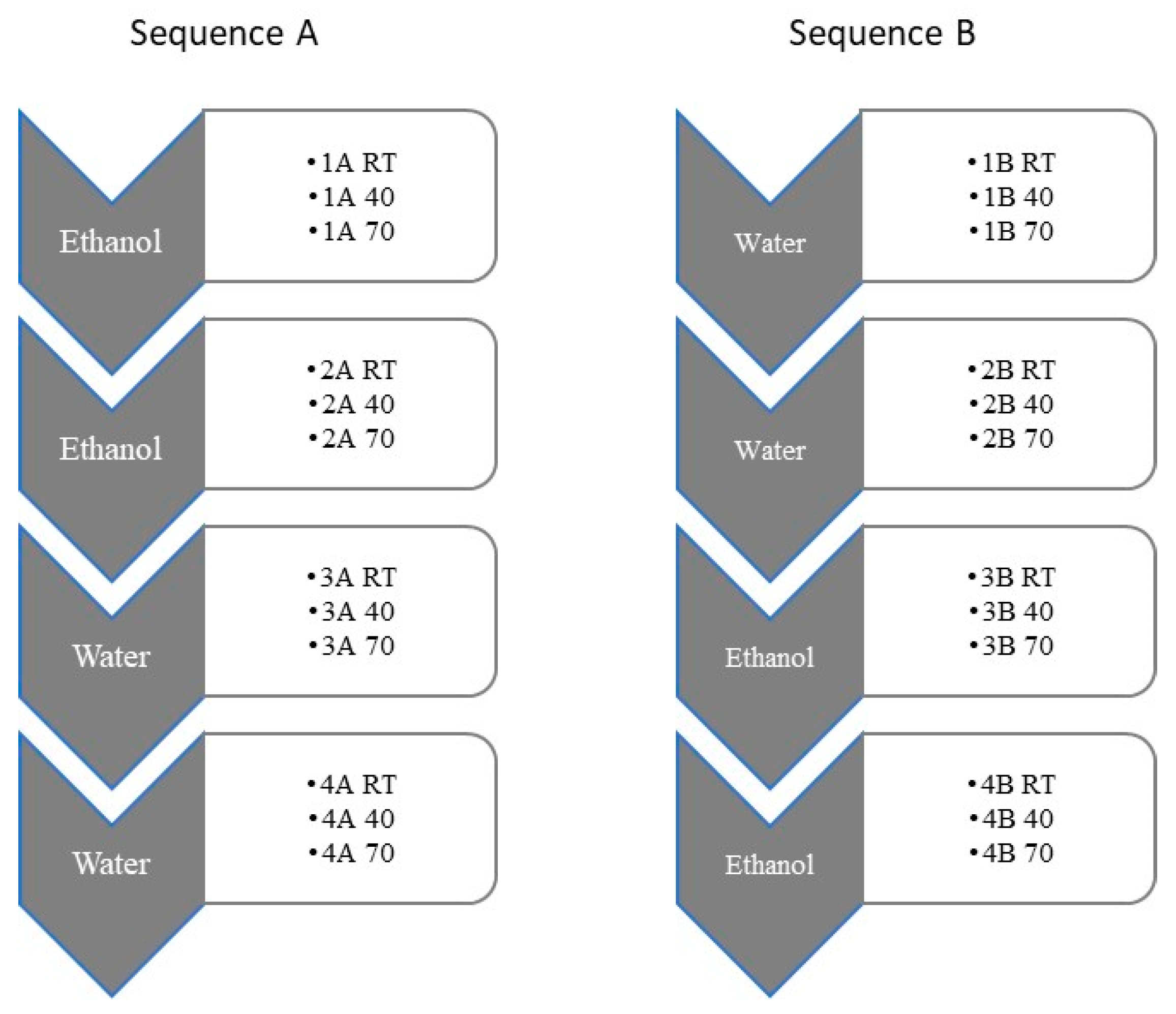
2.2. Aqueous and Ethanolic Liquid Extractions

The extraction procedures were performed considering three different parameters: solvent type, extraction temperature, and extraction time. In brief, 10 g of powered dried biomass were mixed with 100 mL of solvent (ethanol or distilled water). The mixtures were stirred at room temperature (RT) and protected from light for 5, 10, 30 min, 1 h, or 24 h (n = 4). As a result of these preliminary tests, the extraction time was set to 30 min and different temperatures (room temperature (RT); 40 °C; 70 °C) were subsequently tested. The extractions with different solvents were done sequentially at the same temperature as indicated in Figure 1 [36,37]. Briefly, in the samples of sequence A, the biomass is first extracted with ethanol. Then, we proceeded with a second extraction from the same biomass again with ethanol, collecting the compounds that are soluble in this solvent and that may not have yet been removed in the 1st pass of this solvent. Then, the same biomass was extracted twice with water. This process is repeated using the three different temperatures under test, separately, always for 30 min. The same applies to samples of sequence B but in this case the biomass is extracted first with water. Afterwards, the biomass is extracted again with water, followed by two more extractions with ethanol obtained after drying the four extracts. Again, the full sequence is repeated with the samples under testing using different extraction temperatures for 30 min. This allowed us to examine the properties of extracts that were sequentially obtained with the same solvent and realize the existence of compounds that despite being soluble were not removed with the first pass of the solvent. The extracts were then filtered through filter paper (Whatman No.1) and centrifuged at 8000× g for 10 min at RT. The ethanol extracts were dried in a rotary evaporator at 40 °C (Heidolph, Laborota 4000) and the aqueous extracts were freeze-dried. The dried extracts were stored at 4 °C and dissolved in the respective solvents at a concentration of 50 mg mL−1 (antimicrobial analysis) or 10 mg mL−1 (antioxidant analysis). In total, four independent extractions sequences were performed both for sequence A and B and for the three different temperatures tested.

2.3. Antioxidant Activity

The antioxidant activity of the extracts was assessed through the free radical scavenging activity measured by 2,2-Diphenyl-1-picryhydrazyl radical (DPPH) based on the procedure described in Duan et al. [38]. This activity is obtained through a system that generates a stable free radical as it is useful to determine the antioxidant potential at a preliminary stage. Briefly, in a 96-well microplate protected from light, 2 µL of each sample was added to 198 µL DPPH in ethanol (0.1 mM) (Sigma, Steinheim, Germany). The reaction was determined after 0.5 h at room temperature in the dark. The absorbance was measured spectrophotometrically (Evolution 201, Thermo Scientific, Madison, WI, USA) at 517 nm. Control was obtained with 2 µL of absolute ethanol/distilled water and 198 µL of DPPH solution, and a blank measurement was made with 2 µL of extract and 198 µL of absolute ethanol. The results were expressed in percentage of inhibition of DPPH using Equation (1).
DPPH   i n h i b i t i o n   ( % ) = [ [ 1 ( A s / A b ) ] ÷ A c ] × 100
where As is the absorbance of the algae extract, Ab is the absorbance of blank samples, and Ac is the absorbance of the control.
The quantification of TPC was made through the Folin-Ciocalteu method [39] and adapted to the microscale. In a 96-well microplate, protected from light, it was added to each well 158 µL of ultrapure water, 2 µL of the sample (or gallic acid (Merck) to calibration curve or 2 µL of ultrapure water using as control), and 10 µL of the Folin-Ciocalteu reagent (VWR Chemicals). After 2 min, 30 µL Na2CO3 (20%) (Chem-Lab) were added. After incubation in the dark at RT for 1 h, the samples were measured spectrophotometrically (Evolution 201, Thermo Scientific, Madison, WI, USA) at 755 nm. The phenolic content results are expressed as Gallic acid equivalents (mg GAE g extract −1).
All measurements for the four independent extraction procedures were performed in triplicates (n = 12).

2.4. Antimicrobial Susceptibility Test

The antimicrobial susceptibility tests are performed in vitro and are adapted to determine if an extract is effective as a potential antimicrobial agent. These methods are based on standardized procedures, allowing for high reproducibility and consequently a high degree of confidence in the results obtained [40].

2.4.1. Bacterial Strains and Growth Conditions

The ethanolic and aqueous extracts were individually tested against Aeromonas hydrophila subsp. hydrophila DSM 30187, Aeromonas salmonicida subsp. salmonicida DSM 19634, Edwardsiella piscicida DSM 104083, Photobacterium damselae subsp. damselae DSM 7482, Vibrio anguillarum DSM 21597, and Yersinia ruckeri DSM 18506. All cultures were obtained from the German Collection of Microorganisms and Cell Cultures (DSMZ). Marine strains were grown in Tryptic Soy Broth (TSB) or Tryptic Soy Agar (TSA) (VWR Chemicals) supplemented with 1% sodium chloride (NaCl) (Normax Chem). Cultures were incubated at the optimal temperature for each strain according to the information provided by the supplier.

2.4.2. Disk Diffusion Method

The disk diffusion method was performed according to the recommendations of the National Committee for Clinical Laboratory Standards (NCCLS) [40] as they are widely used to screen for antimicrobial activity in algae extracts [22,25]. For disk diffusion susceptibility tests, overnight cultures were suspended in saline solution (1.5 % NaCl) to give a final density equivalent to 1 × 10 8 cfu/mL (equivalent to 0.5 McFarland standard) and spread in Muller Hinton Agar (MHA) (VWR Chemicals) plates supplemented with 1.5% NaCl with a sterile cotton swab [40]. Sterile filter paper discs (6 mm diameter) with 20 µL of the extract (50 mg mL−1) were placed on the previously inoculated MHA plates. Chloramphenicol discs (Oxoid) (30 µg per disk) were used as a positive control and sterile discs with 20 µL each of absolute ethanol/sterile water were used as a negative control. The plates were incubated 24–48 h at the optimal temperature for each strain and the inhibition zone was measured (mm). All tests were performed in triplicates for the four independent extraction procedures (n = 12).

2.4.3. Microdilution Method

The broth dilution method was performed according to the indications of the National Committee for Clinical Laboratory Standards (NCCLS) [41], a frequent method used to study the antimicrobial activity in seaweed extracts [22,42,43]. For the susceptibility tests, non-treated and round-bottom 96-well microplates (Fisher Scientific) with 170 µL of Muller Hinton Broth (MHB) supplemented with 1.5 % NaCl were inoculated with 10 µL of standardized inoculum and 20 µL of each extract (50 mg mL−1) (n = 8). Plates were incubated at the optimal temperature of each strain for 18 h. The antimicrobial activity was detected by the reduction of the visible turbidity measured by recording of the optical density (OD600) in a plate reader (Spectrophotometer Thermo Scientific, Evolution 201). Results were expressed as a percentage of inhibition using Equation (2).
%   i n h i b i t i o n = [ 1 A b s   e x t A b s ] × 100
where Abs ext is the absorbance measured in the wells that contain bacterial strain growing in the presence of the extract and Abs refers to the same measure in wells that contain the bacterial strain.

2.5. Statistical Analysis

Calculations were performed with SPSS Statistics 27 (IBM Corporation, New York, NY, USA, EUA). All statistical analyses were considered significant when at a level of 5% (p-value < 0.05). To test normality, the Kolmogorov–Smirnov test was used in addition to the analysis of the Q-Q plots. As the data did not fulfil the normality assumption, the nonparametric Kruskal–Wallis tests were used. Whenever applicable, the Bonferroni multiple-comparison test was performed after the Kruskal–Wallis test to detect differences between extracts.

3. Results

3.1. Aqueous and Ethanolic Sequential Extracts

The extracts of G. gracilis were made sequentially using water and ethanol as described in Section 2.2. These solvents are environmentally sustainable and food grade, thus allowing the use of extracts for food/feed purposes. Preliminary studies allowed for the setting of 30 min as the ideal extraction time. This time in addition to that of 60 min gave rise to greater efficiency of extraction; thus, the time of 30 min was chosen, notably the shortest time. The extraction yields obtained from the use of different solvents (ethanol or water) at different temperatures (room temperature (RT); 40 °C; 70 °C) are shown in Table 1. The sequential extractions are numbered from one to four according to the extraction order. The results suggest that ethanol extracts exhibited lower extraction yields (0.41–3.68 g 100 dw−1) than the aqueous extracts that demonstrated results that range from 6.62 to 18.49 g 100 dw−1. The highest yield (18.49 g 100 dw−1) was obtained at 70 °C in the first aqueous extraction. As expected, the second extraction using the same solvent consistently presenting values lower than the first one. However, in some cases, curiously the values of the second and fourth extractions present values that are quite significant, particularly when using water as a solvent.

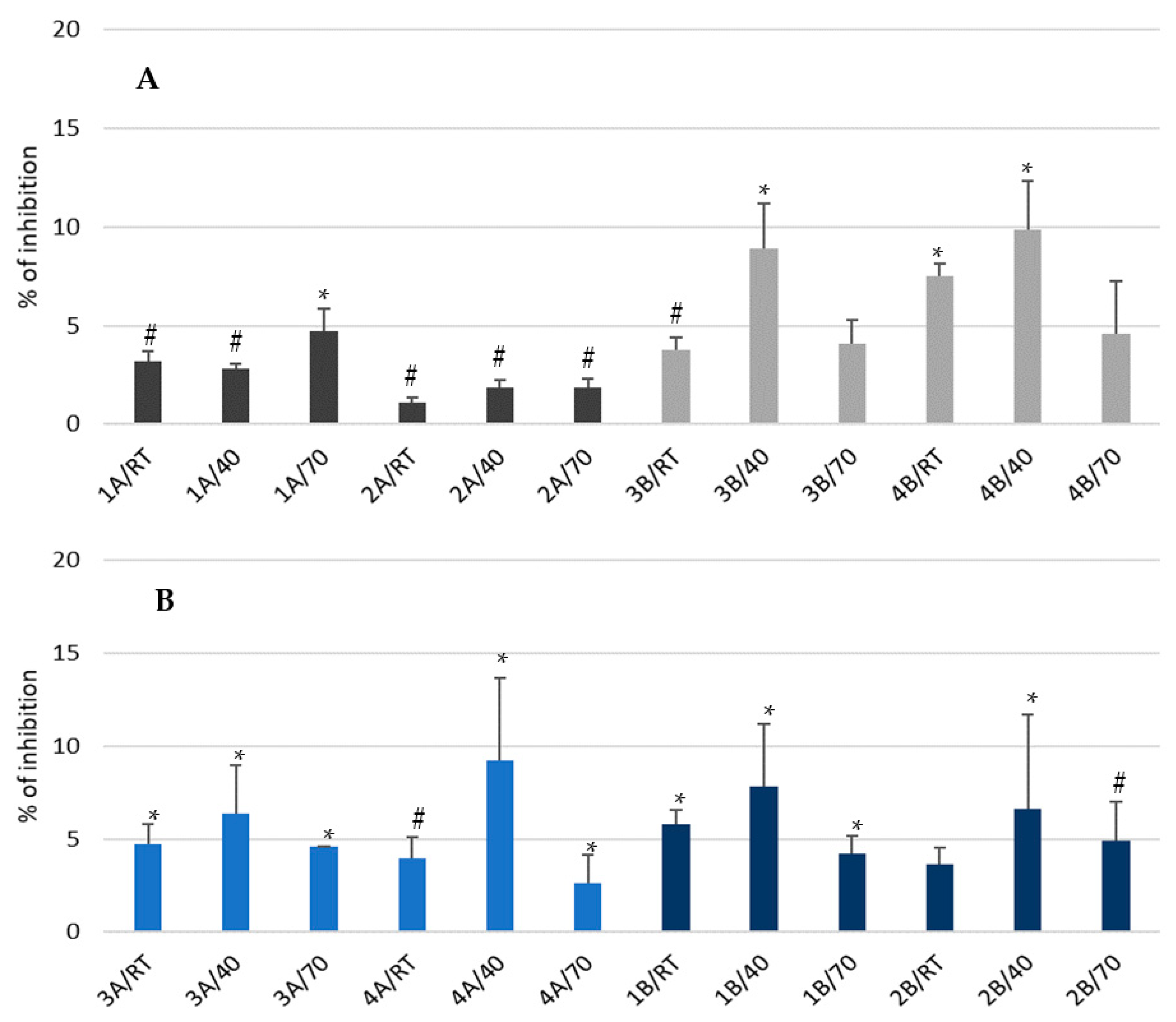
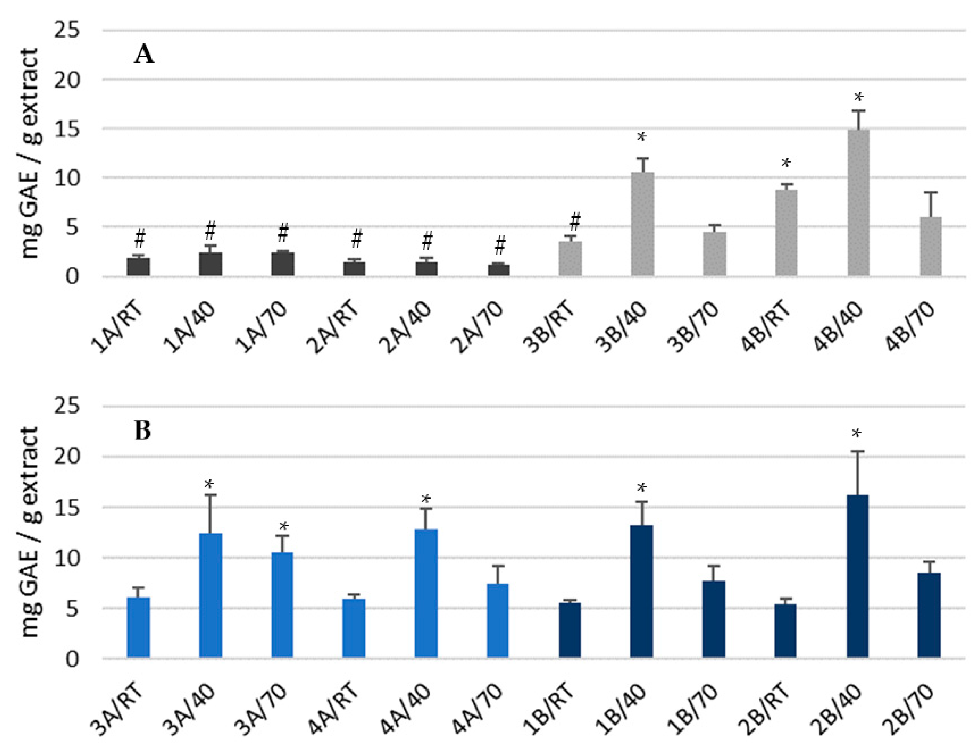
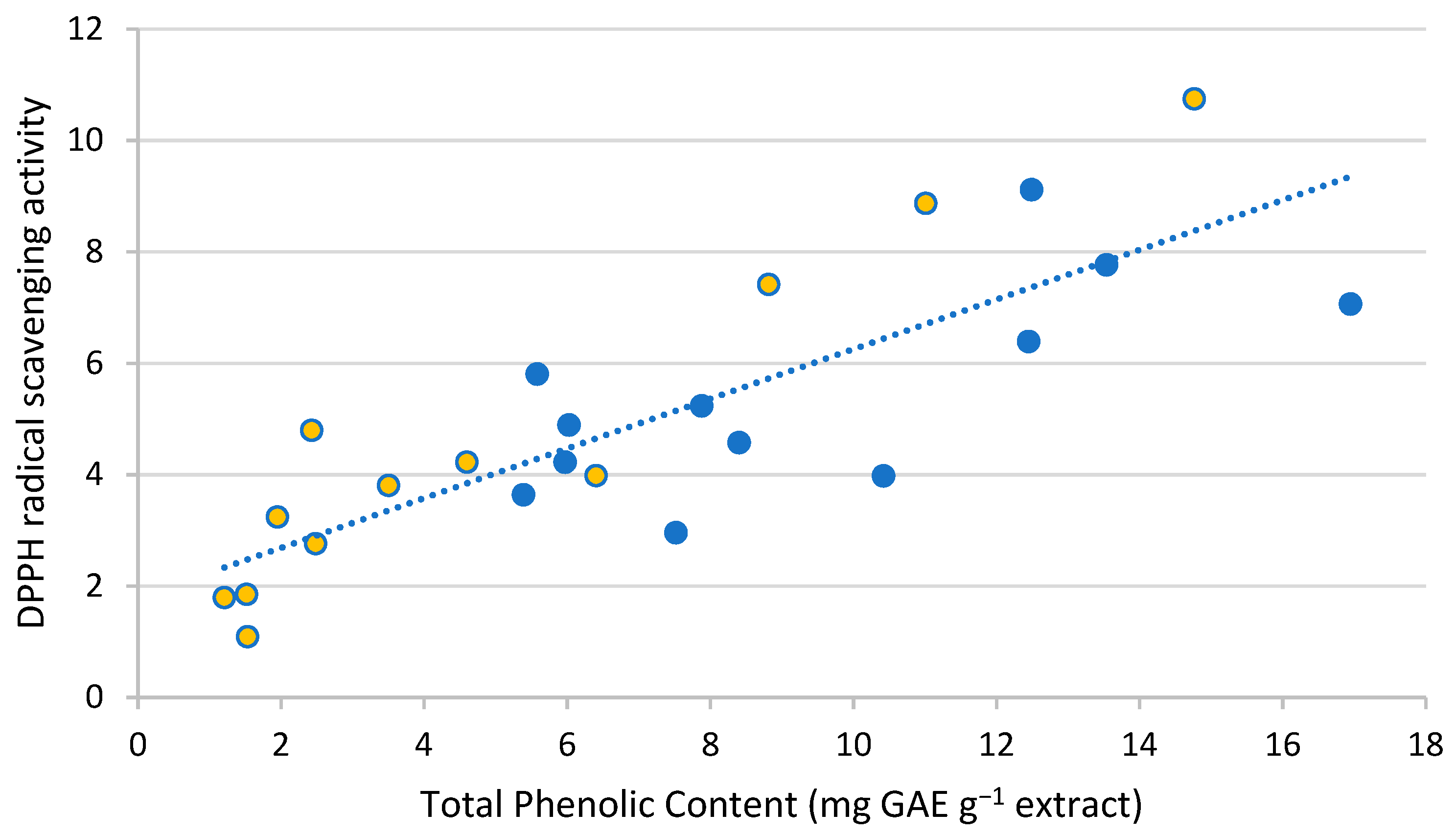
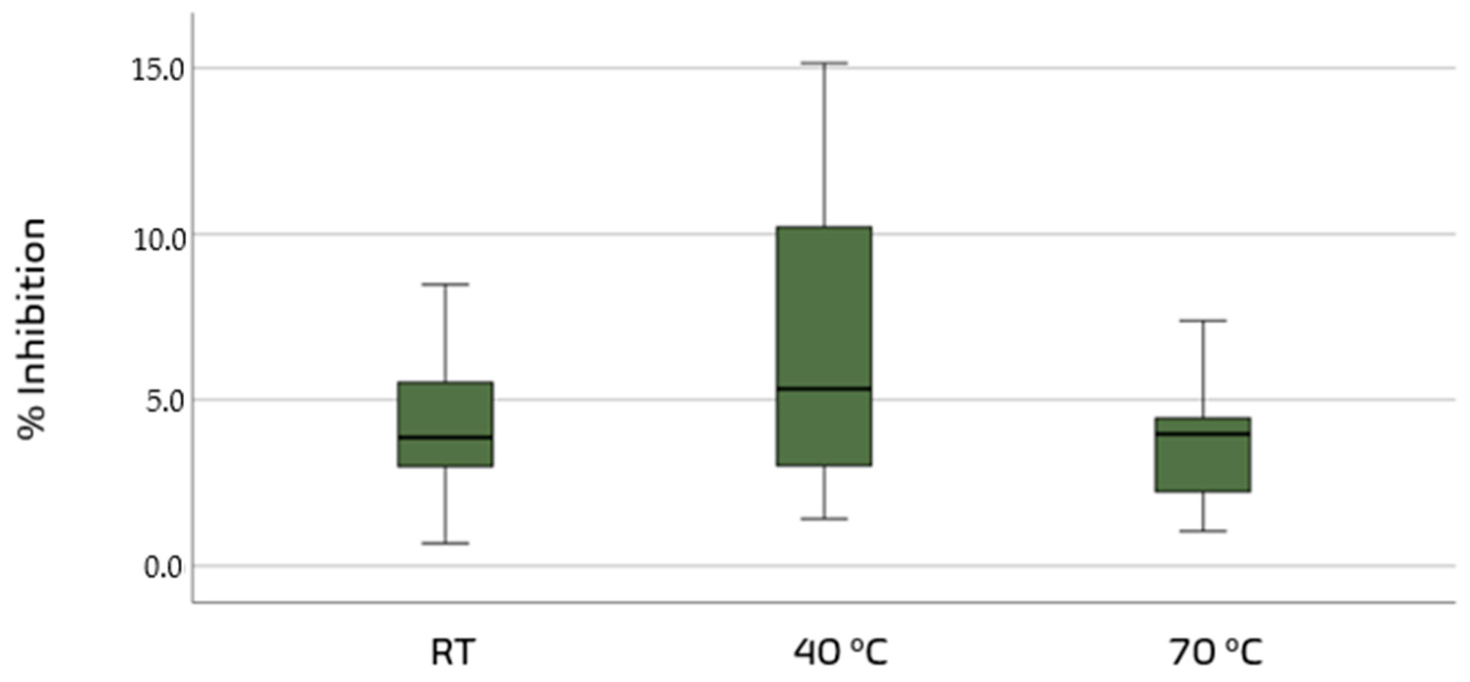
3.2. Antioxidant Activities of Ethanolic and Aqueous Extracts

The antioxidant activities were measured through the DPPH system for the 24 different extraction conditions. In general terms, as the data did not fulfil the normality assumption, the nonparametric Kruskal–Wallis test was used. In the first approach, the results were analyzed according to the solvent type (ethanol or water). The results suggest that there are significant differences among the ethanolic and the aqueous extracts (Kruskal–Wallis, Bonferroni test, F = 3.849, p = 0.050) with average values of 4.63% for the ethanol extracts and 5.42% for the aqueous extracts. Additionally, when we analyzed the data according to the extraction temperature factor, higher values occurred at 40 °C (average of 6.68%) with statistically different values from the samples extracted at room temperature or 70 °C (Kruskal–Wallis, Bonferroni test, F = 7.579, p = 0.023). There was strong evidence (p < 0.003, adjusted using the Bonferroni correction) of a difference between the extracts at 40 °C that produced higher values of inhibition and the group of samples obtained at room temperature (4.42%) or at 70 °C (3.84%). The box plot for the median values of DPPH inhibition activity (%) for extractions at different temperatures is presented in Figure 2.
Overall, the global comparison and statistical analysis of DPPH inhibition values (%) indicates that there are significant differences among the various samples analyzed (Kruskal–Wallis, F = 61.067, p-value < 0.05) (Figure 3). The sample with the lowest DPPH inhibition (2A/RT) is not significantly different from the other second ethanolic extractions regardless of the extraction temperature. The highest inhibition value sample (4B/40) is not significantly different from most samples extracted at 40 °C except for the first and second extractions using ethanol that overall exhibit the lowest values.
In relation to the quantification of the total phenolic content (TPC) and when comparing the different solvents used (ethanol or water), we observed that there are significant differences (Kruskal–Wallis, F = 70.48, p-value < 0.05) between samples (Figure 4).
Globally, aqueous extracts have higher mean TPC values (9.14 mg GAE g−1 extract) than ethanol extracts (4.76 mg GAE g−1 extract). For the TPC values at different temperatures, significant differences can also be observed (Kruskal–Wallis, F = 36.52, p-value < 0.05) and we can state that extraction temperature of 40 °C is globally the temperature that reaches higher TPC values (10.14 mg GAE g−1 extract) when compared to 70 °C or room temperature (respectively 6.03 and 4.85 mg GAE g−1 extract). The comparison of TPC values from all extracts also indicates that we have significant differences between them (Kruskal–Wallis, F = 227.72, p-value < 0.05). The pairwise comparison of the extracts demonstrated that the samples 1A and 2A, regardless of the temperature of extraction and in addition to sample 3B/RT, demonstrated reduced values for TPC. The sample with the highest TPC value (2B/40) is also not significantly different from samples 3B (ethanol) or 3A, 4A and 1B (water), all extracted at 40 °C. The second (2B/40) water extraction and the fourth (4B/40) ethanol extraction both obtained at 40 °C, similarly to the DPPH inhibition results, revealed higher values for TPC (Figure 4) of 16.95 and 14.77 mg GAE g−1 extract, respectively. Regarding the aqueous fractions, the results suggest differences between the various extracts (Kruskal–Wallis, Bonferroni test, F = 87.988, p-value < 0.05) as the extract 2B/40 revealed higher activity among the aqueous extracts, although it is not clear whether there is also a correlation with the temperature of the extraction. In ethanolic extracts, the higher mean values for TPC were found in the fourth extraction (14.77 mg GAE g−1 extract), obtained after two water extractions and a first ethanol extraction. Additionally, we also found a clear positive correlation between the DPPH radical scavenging activity and the TPC in our samples (Figure 5) with values of r = 0.826.

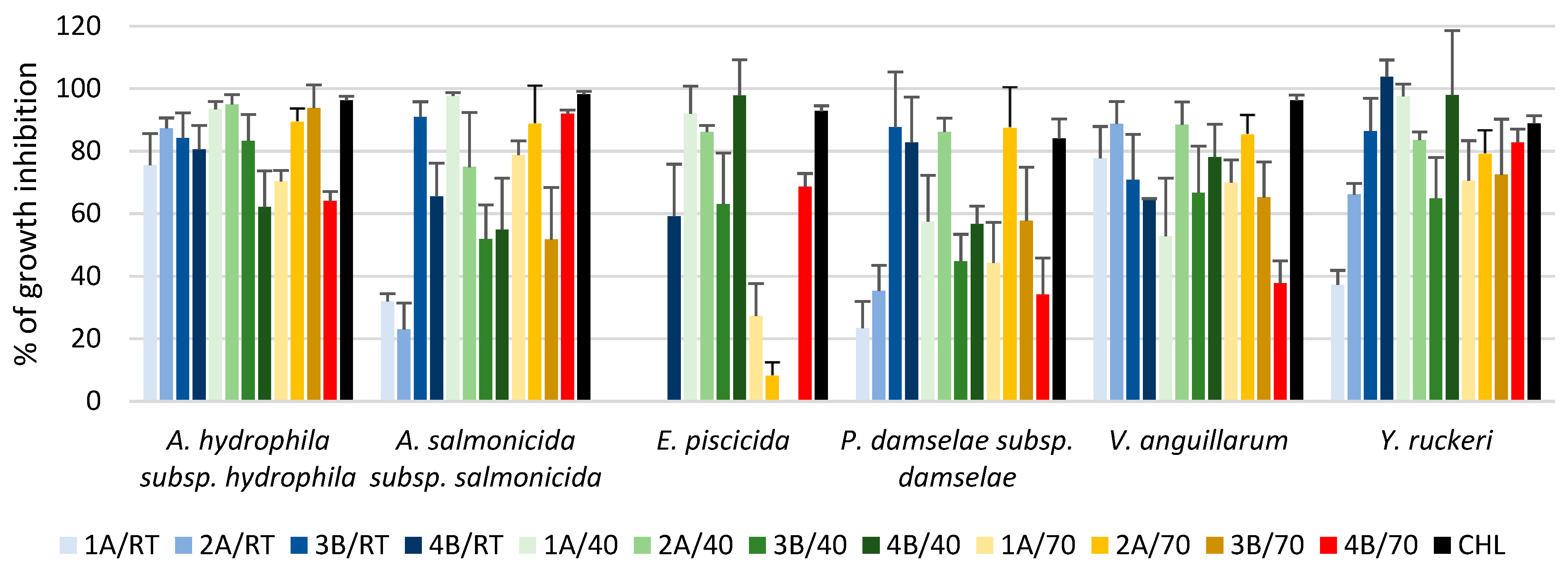
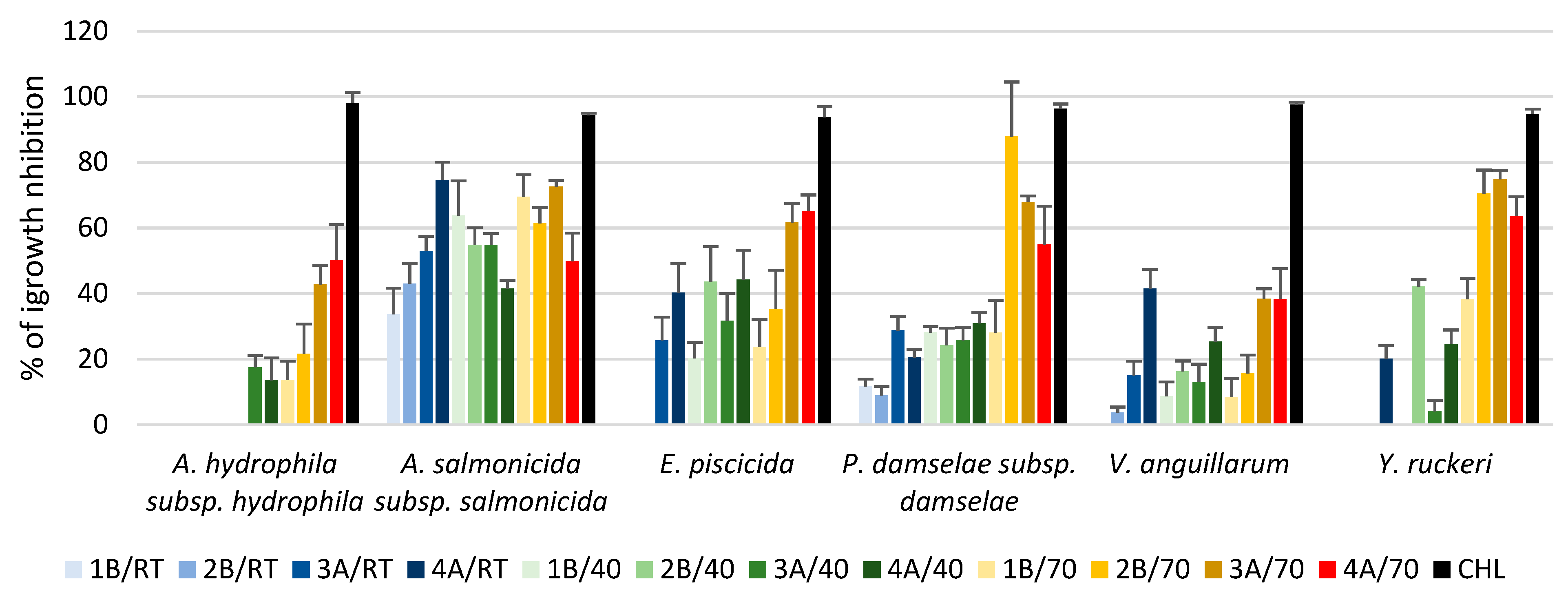
3.3. Antibacterial Activities of Ethanolic and Aqueous Extracts

The analysis of the results of the inhibition tests on the agar plate was conducted considering all the different extracts prepared (i.e., four ethanolic and four aqueous). As a first step, the results obtained with different extraction times (5 min, 10 min, 30 min, 1 h, and 24 h) were recorded. These demonstrated that with extraction times of 5 min, 10 min, and 24 h, the inhibition values are almost undetectable. Larger inhibition halos appear with an extraction time of 30 min. In all cases, noticeable halos are detected only in the ethanol extracts, indicating that the compounds that exhibit activity against the bacterial strains tested must be mainly insoluble in water and of a polar nature. Table 2 presents the inhibition halos for the 30 min extractions using ethanol as solvent. All null results were excluded.
The results of the growth inhibition tests on liquid media for both for ethanolic and aqueous extracts are shown in Figure 6 and Figure 7, respectively. The percentage of growth inhibition of fish pathogenic bacteria in the presence of ethanolic extracts demonstrated higher mean values (66.2%) than the results in the presence of aqueous extracts (30.6%). Therefore, it is possible that the antimicrobial bioactive metabolites present are mostly extracted by ethanol. The highest percentages of inhibition were recorded in ethanol extracts against A. hydrophila subsp. hydrophila (81.6%) and in Y. ruckeri (78.41%), thus the least interesting values registered in the aqueous extracts against A. hydrophila subsp. hydrophila (13.28%). In general, the growth inhibition in the presence of aqueous extracts occurs mainly in extracts obtained at higher temperatures (Figure 7, bars in shades of yellow/red), as the lowest temperatures (RT or 40 °C) are less effective in extracting compounds with antimicrobial activity against the strains in study.

4. Discussion

The use of macroalgae, namely the red seaweed G. gracilis, in animal feed and food has been widely sought after. In nutritional terms, these algae can be used, for example, in protein replacement or for nutritional enrichment [44,45,46,47,48,49,50,51]. Additionally, the bioactive properties of this species allow us to foresee a series of benefits associated with its use in animal feed products with clear benefits for animal health and welfare [52,53,54,55]. The sustainability associated with the collection of these algae in their natural environment or their production in aquaculture systems can play an important role in their biotechnological use. There are already studies that indicate both the optimal conditions for the cultivation of this species and the conditions that allow for the maximization of the production of some nutritional or bioactive compounds [30,56,57,58,59].
Regarding the inclusion of bioactive components, the presence of antioxidant and/or antimicrobial compounds in animal feed can lead to additional levels of protection against cellular oxidation mechanisms and pathogenic diseases, contributing to the improvement of the animal health and well-being, especially in fish from aquaculture [60,61] that are exposed to several stress factors. These bioactive compounds can positively impact a farm’s profitability and the welfare of animals in captivity that would translate into a better-quality product. The use of macroalgae as an additive in feed can be achieved in the form of biomass or an extract, the latter being obtained with non-toxic and appropriate grade solvents with subsequent incorporation during the feed manufacturing process. Several studies demonstrate that the activities exhibited by crude extracts are different depending on the temperature, extraction time, and solvents used [26,62,63,64], the most common including water, ethanol, hexane, methanol, and mixtures of different solvents. The different extraction solvents according to their polarity allow the fractionation and separation of different compounds such as pigments (chlorophyll a and b and carotenoids), alkaloids, and different classes of phenolic compounds, among others. The use of water, a highly polar solvent (polarity index of 10.2), allows for the obtaining of extracts not only enriched in soluble proteins and sugars but also pigments, starch, and minerals, in addition to the use of other water-soluble compounds [65]. Ethanol allows for the extraction of slightly polar compounds (polarity index of 5.2) such as polar lipids and fatty acids, some phenolic compounds, and carotenoids, among others [62,66,67,68]. Generally, the phenolic compounds are better extracted with increased solvent polarity [64] and thus it is expected to find them in higher concentrations in highly polar solvents.
In this study the use of conventional solid–liquid extraction processes with ethanol or water as solvents sought to obtain extracts with different compositions and bioactivities. Extraction yields were higher with water as reported in other studies with red seaweeds including Agarophyton vermiculophyllum (formerly Gracilaria vermiculophylla) [69,70,71]. However, the results obtained in the ethanolic fractions in terms of average yield are lower than those obtained by Chan et al. [36] regarding Crassiphycus changii (formerly Gracilaria changii, 13.06 ± 1.14%). In A. vermiculophyllum, the average yield for the ethanolic and aqueous extract were 6.5 ± 0.1% and 33.4 ± 1.5%, respectively [71], higher than the values we found. Extraction efficiency is a highly variable parameter depending on cell rupture efficiency, solvent, pH, temperature, and extraction time, among other factors.
The antioxidant activity present in several macroalgal extracts indicates a correlation with the total phenolic content [43,72], justifying why many studies focus on the quantification of these compounds. Often, extracts that have a lower content of pigments and phenolic compounds demonstrate reduced antioxidant activity. The use of effective and rigorous methods of measuring TPC is essential as they allow us to better understand the effect that these compounds can have on animal health and well-being, considering they are one of the most important natural bioactive compounds. The quantification of TPC is considered an indicator of antioxidant capacity; however, it is a parameter that can be assessed with slightly different methodologies (with different reference substances, extract concentrations, reaction times, and temperatures, among other factors) and the comparison of results is somehow complex. In the red seaweeds Pterocladiella capillacea and Osmundaria obtusiloba, the use of 70% ethanol was effective as a solvent for the extraction of phenolic compounds [22] especially those that were of a polar nature such as the polyphenols bond to proteins or sugars, saponins, phlorotannin’s, and organic acids, among others [73]. In these two species, the authors found a TPC of 15.23 and 30.54 mg GAE g−1 extract, respectively [22]. Also, the studies of Farvin and Jacobsen [71] that studied the TPC in six red seaweed species found high values in Polysiphonia fucoides (19.2 ± 0.61 mg GAE g−1 dw) and Porphyra purpurea (2.99 ± 0.05 mg GAE g−1 dw). In the red algae Hypnea musciformis, Hypnea valentiae, and Jania rubens, the TPC reached values of 9.84, 6.91, and 4.95 mg GAE g−1 dw in methanolic extracts [74]. In this study, the fractioning of crude extracts demonstrated that the fractions of the polar solvent (specially ethyl acetate) had greater antioxidant potential, particularly in H. musciformis. In Porphyra tenera, the TPC varied from 10.81 to 32.14 mg GAE g−1 in the extract depending on the temperature and the solvent used in the extraction [75]. Additionally, in Kappaphycus alvarezii, some compounds that exhibited remarkable antioxidant activity were already identified and characterized [76,77,78]. In Chondracanthus chamissoi and Laurencia chilensis, the estimation of TPC indicated a relatively low level with the higher concentrations obtained with dichloromethane and ethyl acetate extracts of L. chilensis (2.15 ± 0.14 and 2.27 ± 0.35 mg GAE g−1 dw, respectively) [79]. In Crassiphycus birdiae (formerly Gracilaria birdiae), the hydrocolloid fraction obtained with water at 90 °C exhibited interesting antioxidant properties (IC50 = 1.62 mg mL−1) in addition to a high extraction yield (27.2 % dw) [80], demonstrating it to be an interesting source of antioxidant compounds. The TPC obtained by these authors in the ethanol extracts of C. birdiae and Crassiphycys corneus (formerly G. cornea) were 1.13 and 0.88 mg GAE g−1 extract, respectively. Furthermore, in these species, the ethanol extracts have a high antioxidant performance although lower than the commercial antioxidant BHT. The IC50 values for the ethanolic extracts on C. birdiae and C. corneus (formerly Gracilaria cornea) were, respectively, of 0.76 and 0.77 mg mL−1 (BHT, 0.48 mg mL−1) [80]. Our results demonstrate that the maximum TPC is found in aqueous extracts obtained at 40 °C (16.95 mg GAE g−1 extract), in line with previous studies in red algae, although different species and solvents may give rise to quite different results. Thus, these values make this alga an interesting source of polyphenolic compounds. In addition, the choice of solvent must also consider that these extracts are intended for the feed industry and that less toxic, sustainable, low cost, and high availability solvents should be chosen; that is, effective industrial use should be accessible and able to arouse interest.
Regarding the DPPH free-radical activity, this has been widely used to determine the antioxidant potential of seaweed extracts [80]. Using a sequential extraction method with ethanol as solvent, the free radical scavenging activity by the DPPH assay was 59.2% and 39.2% in Gracilaria foliifera and Anthophycus longifolius, respectively (formerly Sargassum longifolium) [61]. The authors found greater activity in the ethanol extract when compared to other solvents. In C. birdiae and C. corneus, the IC50 values for the ethanol extracts (0.77 and 0.76 mg mL−1, respectively) were quite low considering that the same value for BHT is 0.48 mg mL−1. Other authors found greater free radical scavenging activity in the aqueous extracts of A. vermiculophyllum, Devaleraea mollis (formerly Palmaria palmata), Chondrus crispus, Porphyra purpurea, and Asparagopsis taxiformis when compared to ethanolic extracts [70,71], similar to the results we found for G. gracilis. Aqueous extracts also gave a higher extraction yield, thus the most promising extract in this regard as found in previous studies [70]. Extracts with high levels of TPC had greater activity as DPPH radical scavengers as demonstrated by the coefficient of determination found (Figure 5, R2 = 0.7327) and according to other authors [17,36,74]. This suggests that seaweed polyphenols may be responsible for the antioxidant properties noticed particularly in aqueous extracts.
Bacterial diseases are one of the main causes of mortality and losses in the aquaculture sector with recurrent problems due to the emergence of antibiotic resistance and the presence of the residues of these compounds and their derivatives in animal products. Seaweeds have drawn attention as natural sources of metabolites with antimicrobial activity. Simultaneously, vibriosis is a common disease in aquaculture systems caused by bacteria of the genus Vibrio that can result in high mortality rates. Other bacteria such as Aeromonas hydrophila, Aeromonas salmonicida, Edwardsiella piscicida, Photobacterium damselae subsp. Damselae, or Yersinia ruckeri can cause an outbreak of disease with serious consequences. Studies report that the use of lyophilized Gracilaria chilensis (10%) as a feed additive resulted in a significant increase in the growth rate of Salmo salar [55,81] but also exhibited a marked anti-viral activity (68.02 ± 18.34) against salmon anemia virus. Lipid extracts of Gracilariopsis longissima (formerly Gracilaria longissima) exhibited a broad spectrum of activity against Vibrio sp. [33]. Additionally, studies on the antimicrobial activity of chloroform/methanol extracts from marine algae of the Gracilaria genus (Gracilaria dura, G. gracilis, and G. longissima) and their effects against six species of fish pathogenic Vibrio sp. indicated that G. longissimi exhibits high activity against Vibrio ordalii, Vibrio salmonicida, Vibrio alginolyticus, and Vibrio vulnificus [33]. In general, the authors emphasize the antibacterial potential of the extracts of this species of seaweed as a supplement for health promotion in aquaculture, although G. gracilis extracts were active only against V. salmonicida. The bioactivity ethyl acetate extracts from Gracilaria arcuata against Aeromonas hydrophila and Vibrio sp. have also been reported [82] and fractioning of the extracts revealed that hexadecanoid acid and sterol are the main antibacterial compounds. Ethanol extracts from Asparagopsis taxiformis, a red alga, have been shown to inhibit the fish pathogenic bacteria Aeromonas salmonicida subsp. salmonicida, Vibrio alginolyticus, and V. vulnificus by the standard disk diffusion method in a significant manner [42]. Moderate activity was registered for Photobacterium damselae subsp. damselae, P. damselae subsp. piscicida, V. harveyi, and V. parahaemolyticus. Our results regarding the in vitro antimicrobial activity of the ethanolic and aqueous extracts of G. gracilis in the disk diffusion method produced poorly expressed results with minor inhibition halos against the six strains of pathogenic bacteria tested (Aeromonas hydrophila subsp. hydrophila, Aeromonas salmonicida subsp. salmonicida, Edwardsiella piscicida, Photobacterium damselae subsp. damselae, Vibrio anguillarum, and Yersinia ruckeri). In terms of the results of the growth inhibition tests in liquid media, we can note that the ethanol extracts exhibited greater antibacterial activity than the aqueous extracts. In the liquid medium, the highest mean percent of growth inhibition was reported for the ethanol extracts against Aeromonas hydrophila subsp. hydrophila with 81.57 ± 10.89% and Yersinia ruckeri with 78.51 ± 17.38%. Interestingly, the ethanolic extracts demonstrated more expressive inhibition results when the extraction temperature was 40 °C, in contrast to the aqueous extracts with the highest mean inhibition values obtained were with the extracts at 70 °C.

5. Conclusions

The results obtained in the present study demonstrate that the macroalgae G. gracilis exhibits antioxidant and antimicrobial activity against fish pathogenic bacteria and can therefore be used as a potential additive in fish feed. The aqueous extracts obtained a higher extraction yield and exhibited greater antioxidant activity; however, ethanolic extracts exhibited higher antimicrobial activity particularly against A. hydrophila subsp. hydrophila and Y. ruckeri but also against A. salmonicida subsp. salmonicida, E. piscicida, P. damselae subsp. Damselae, and V. anguillarum. In similar studies conducted by other authors [26,63,64], it was found that the effectiveness of extraction is strongly conditioned by the different extraction conditions that in turn affects the type of compounds extracted. In the present study, we demonstrated that a temperature of 40 °C is generally the most effective when associated to an extraction time of 30 min. Any of the solvents used are safe and accessible, not generating toxic residues from the extraction which is desirable when considering these extracts as feed additives. The potential application of these extracts should be confirmed in the future, namely for in vivo systems.

Author Contributions

Conceptualization, C.A., A.P.C., M.V.F., T.M. and T.B.; methodology, C.A., A.P.C. and T.B.; validation, C.A. and A.P.C.; formal analysis, C.A. and A.P.C.; investigation, C.A., A.P.C., M.V.F., T.M. and T.B.; data curation, C.A.; writing—original draft preparation, C.A. and A.P.C.; writing—review and editing, C.A., T.M. and T.B.; supervision, C.A.; project administration, T.B.; funding acquisition, T.B. All authors have read and agreed to the published version of the manuscript.

Funding

This study had the support of Fundação para a Ciência e Tecnologia (FCT) through the strategic project UIDB/04292/2020 granted to MARE and the project Operational Programme MAR2020 through the project 16-02-01-FMP-84–Seaweedfeeds.

Data Availability Statement

The data presented in this study are available on request from the corresponding author C.A. upon reasonable request.

Conflicts of Interest

The authors declare no conflict of interest. The funders had no role in the design of the study, in the collection, analyses, or interpretation of data, in the writing of the manuscript, or in the decision to publish the results.

References

  1. Araújo, R.; Vázquez Calderón, F.; Sánchez López, J.; Azevedo, I.C.; Bruhn, A.; Fluch, S.; Garcia Tasende, M.; Ghaderiardakani, F.; Ilmjärv, T.; Laurans, M.; et al. Current Status of the Algae Production Industry in Europe: An Emerging Sector of the Blue Bioeconomy. Front. Mar. Sci. 2021, 7, 1–24. [Google Scholar] [CrossRef]
  2. Leandro, A.; Pereira, L.; Gonçalves, A.M.M. Diverse Applications of Marine Macroalgae. Mar. Drugs 2020, 18, 17. [Google Scholar] [CrossRef] [Green Version]
  3. Morais, T.; Cotas, J.; Pacheco, D.; Pereira, L. Seaweeds Compounds: An Ecosustainable Source of Cosmetic Ingredients? Cosmetics 2021, 8, 8. [Google Scholar] [CrossRef]
  4. Abdul Khalil, H.P.S.; Saurabh, C.K.; Tye, Y.Y.; Lai, T.K.; Easa, A.M.; Rosamah, E.; Fazita, M.R.N.; Syakir, M.I.; Adnan, A.S.; Fizree, H.M.; et al. Seaweed Based Sustainable Films and Composites for Food and Pharmaceutical Applications: A Review. Renew. Sustain. Energy Rev. 2017, 77, 353–362. [Google Scholar] [CrossRef]
  5. Ibañez, E.; Herrero, M.; Mendiola, J.; Castro-Puyana, M. Marine bioactive compounds: Sources, characterization and applications. In Marine Bioactive Compounds: Sources, Characterization and Applications; Hayes, M., Ed.; Springer Science|Business Media: New York, NY, USA, 2012; Volume 9781461412, pp. 1–229. ISBN 9781461412472. [Google Scholar]
  6. Michalak, I.; Chojnacka, K. Algae as Production Systems of Bioactive Compounds. Eng. Life Sci. 2015, 15, 160–176. [Google Scholar] [CrossRef]
  7. Misurcová, L. Chemical Composition of Seaweeds. In Handbook of Marine Macroalgae: Biotechnology and Applied Phycology; Kim, S.-K., Ed.; John Wiley & Sons, Ltd.: Chichester, UK, 2011; ISBN 9780470979181. [Google Scholar]
  8. Francavilla, M.; Franchi, M.; Monteleone, M.; Caroppo, C. The Red Seaweed Gracilaria gracilis as a Multi Products Source. Mar. Drugs 2013, 11, 3754–3776. [Google Scholar] [CrossRef] [Green Version]
  9. MaCartain, P.; Gill, C.I.R.; Brooks, M.; Campbell, R.; Rowland, I.R. Nutritional Value of Edible Seaweeds. Nutr. Rev. 2007, 65, 535–543. [Google Scholar] [CrossRef]
  10. Biris-Dorhoi, E.S.; Michiu, D.; Pop, C.R.; Rotar, A.M.; Tofana, M.; Pop, O.L.; Socaci, S.A.; Farcas, A.C. Macroalgae—A Sustainable Source of Chemical Compounds with Biological Activities. Nutrients 2020, 12, 3085. [Google Scholar] [CrossRef]
  11. Holdt, S.L.; Kraan, S. Bioactive Compounds in Seaweed: Functional Food Applications and Legislation. J. Appl. Phycol. 2011, 23, 543–597. [Google Scholar] [CrossRef]
  12. Capillo, G.; Sanfilippo, M.; Aliko, V.; Spanò, N.; Gioele, C.; Marilena, S.; Valbona, A.; Nunziacarla, S.; Andrea, S.; Antonio, M. Gracilaria gracilis, Source of Agar: A Short Review. Curr. Org. Chem. 2017, 21, 380–386. [Google Scholar] [CrossRef]
  13. Martín, L.A.; Rodríguez, M.C.; Matulewicz, M.C.; Fissore, E.N.; Gerschenson, L.N.; Leonardi, P.I. Seasonal Variation in Agar Composition and Properties from Gracilaria gracilis (Gracilariales, Rhodophyta) of the Patagonian Coast of Argentina. Phycol. Res. 2013, 61, 163–171. [Google Scholar] [CrossRef]
  14. Marinho-Soriano, E.; Bourret, E. Effects of Season on the Yield and Quality of Agar from Gracilaria Species (Gracilariaceae, Rhodophyta). Bioresour. Technol. 2003, 90, 329–333. [Google Scholar] [CrossRef]
  15. Paiva, L.; Lima, E.; Neto, A.I.; Baptista, J. Seasonal Variability of the Biochemical Composition and Antioxidant Properties of Fucus spiralis at Two Azorean Islands. Mar. Drugs 2018, 16, 248. [Google Scholar] [CrossRef] [Green Version]
  16. Lourenço-Lopes, C.; Fraga-Corral, M.; Jimenez-Lopez, C.; Pereira, A.G.; Garcia-Oliveira, P.; Carpena, M.; Prieto, M.A.; Simal-Gandara, J. Metabolites from Macroalgae and Its Applications in the Cosmetic Industry: A Circular Economy Approach. Resources 2020, 9, 101. [Google Scholar] [CrossRef]
  17. Marinho, G.S. Antioxidant Content and Activity of the Seaweed Saccharina latissima: A Seasonal Perspective. Antioxidant 2018, 31, 1343–1354. [Google Scholar] [CrossRef] [Green Version]
  18. Gomez-Zavaglia, A.; Prieto Lage, M.A.; Jimenez-Lopez, C.; Mejuto, J.C.; Simal-Gandara, J. The Potential of Seaweeds as a Source of Functional Ingredients of Prebiotic and Antioxidant Value. Antioxidants 2019, 8, 406. [Google Scholar] [CrossRef] [Green Version]
  19. Hafting, J.T.; Craigie, J.S.; Stengel, D.B.; Loureiro, R.R.; Buschmann, A.H.; Yarish, C.; Edwards, M.D.; Critchley, A.T. Prospects, and Challenges for Industrial Production of Seaweed Bioactives. J. Phycol. 2015, 51, 821–837. [Google Scholar] [CrossRef]
  20. Stengel, D.B.; Walker, J.M.; Connan, S. Natural Products from Marine Algae IN Series Editor, 1st ed.; Stengel, G.B., Connan, S., Eds.; Humana Press: Hertfordshire, UK, 2015; ISBN 9781493926831. [Google Scholar]
  21. Pérez, M.J.; Falqué, E.; Domínguez, H. Antimicrobial Action of Compounds from Marine Seaweed. Mar. Drugs 2016, 14, 52. [Google Scholar] [CrossRef] [Green Version]
  22. De Alencar, D.B.; de Carvalho, F.C.T.; Rebouças, R.H.; dos Santos, D.R.; dos Santos Pires-Cavalcante, K.M.; de Lima, R.L.; Baracho, B.M.; Bezerra, R.M.; Viana, F.A.; dos Fernandes Vieira, R.H.S.; et al. Bioactive Extracts of Red Seaweeds Pterocladiella capillacea and Osmundaria obtusiloba (Floridophyceae: Rhodophyta) with Antioxidant and Bacterial Agglutination Potential. Asian Pac. J. Trop. Med. 2016, 9, 372–379. [Google Scholar] [CrossRef]
  23. Peixoto, M.J.; Salas-Leitón, E.; Pereira, L.F.; Queiroz, A.; Magalhães, F.; Pereira, R.; Abreu, H.; Reis, P.A.; Gonçalves, J.F.M.; de Almeida Ozório, R.O. Role of Dietary Seaweed Supplementation on Growth Performance, Digestive Capacity and Immune and Stress Responsiveness in European Seabass (Dicentrarchus labrax). Aquac. Rep. 2016, 3, 189–197. [Google Scholar] [CrossRef] [Green Version]
  24. Guaratini, T.; Lopes, N.P.; Marinho-Soriano, E.; Colepicolo, P.; Pinto, E. Antioxidant Activity and Chemical Composition of the Non Polar Fraction of Gracilaria domingensis (Kützing) Sonder Ex Dickie and Gracilaria birdiae (Plastino & Oliveira). Rev. Bras. Farmacogn. 2012, 22, 724–729. [Google Scholar] [CrossRef] [Green Version]
  25. El Shafay, S.M.; Ali, S.S.; El-Sheekh, M.M. Antimicrobial Activity of Some Seaweeds Species from Red Sea, against Multidrug Resistant Bacteria. Egypt. J. Aquat. Res. 2016, 42, 65–74. [Google Scholar] [CrossRef] [Green Version]
  26. Alassali, A.; Cybulska, I. Methods for Upstream Extraction and Chemical Characterization of Secondary Metabolites from Algae Biomass. Adv. Tech. Biol. Med. 2015, 4, 1–16. [Google Scholar] [CrossRef] [Green Version]
  27. Grosso, C.; Valentão, P.; Ferreres, F.; Andrade, P.B. Alternative and Efficient Extraction Methods for Marine-Derived Compounds. Mar. Drugs 2015, 13, 3182–3230. [Google Scholar] [CrossRef] [PubMed] [Green Version]
  28. Cikos, A.M.; Jokić, S.; Šubarić, D.; Jerković, I. Overview on the Application of Modern Methods for the Extraction of Bioactive Compounds from Marine Macroalgae. Mar. Drugs 2018, 16, 348. [Google Scholar] [CrossRef] [PubMed] [Green Version]
  29. De Almeida, C.L.F.; De S. Falcão, H.; De M. Lima, G.R.; De A. Montenegro, C.; Lira, N.S.; De Athayde-Filho, P.F.; Rodrigues, L.C.; De Souza, M.d.F.V.; Barbosa-Filho, J.M.; Batista, L.M. Bioactivities from Marine Algae of the Genus Gracilaria. Int. J. Mol. Sci. 2011, 12, 4550–4573. [Google Scholar] [CrossRef] [PubMed]
  30. Capillo, G.; Savoca, S.; Costa, R.; Sanfilippo, M.; Rizzo, C.; Lo Giudice, A.; Albergamo, A.; Rando, R.; Bartolomeo, G.; Spanò, N.; et al. New Insights into the Culture Method and Antibacterial Potential of Gracilaria gracilis. Mar. Drugs 2018, 16, 492. [Google Scholar] [CrossRef] [PubMed] [Green Version]
  31. Kasanah, N.; Amelia, W.; Mukminin, A.; Triyanto; Isnansetyo, A. Antibacterial Activity of Indonesian Red Algae Gracilaria edulis against Bacterial Fish Pathogens and Characterization of Active Fractions. Nat. Prod. Res. 2018, 33, 3303–3307. [Google Scholar] [CrossRef] [PubMed]
  32. Kanjana, K.; Radtanatip, T.; Asuvapongpatana, S.; Withyachumnarnkul, B.; Wongprasert, K. Solvent Extracts of the Red Seaweed Gracilaria fisheri Prevent Vibrio harveyi Infections in the Black Tiger Shrimp Penaeus monodon. Fish Shellfish Immunol. 2011, 30, 389–396. [Google Scholar] [CrossRef]
  33. Cavallo, R.A.; Acquaviva, M.I.; Stabili, L.; Cecere, E.; Petrocelli, A.; Narracci, M. Antibacterial Activity of Marine Macroalgae against Fish Pathogenic Vibrio Species. Cent. Eur. J. Biol. 2013, 8, 646–653. [Google Scholar] [CrossRef]
  34. Cavas, L.; Yurdakoc, K. A Comparative Study: Assessment of the Antioxidant System in the Invasive Green Alga Caulerpa racemosa and Some Macrophytes from the Mediterranean. J. Exp. Mar. Biol. Ecol. 2005, 321, 35–41. [Google Scholar] [CrossRef]
  35. Afonso, C.; Correia, A.P.; Freitas, M.V.; Baptista, T.; Neves, M.; Mouga, T. Seasonal Changes in the Nutritional Composition of Agarophyton vermiculophyllum (Rhodophyta, Gracilariales) from the Center of Portugal. Foods 2021, 10, 1145. [Google Scholar] [CrossRef] [PubMed]
  36. Chan, P.T.; Matanjun, P.; Yasir, S.M.; Tan, T.S. Antioxidant Activities and Polyphenolics of Various Solvent Extracts of Red Seaweed, Gracilaria changii. J. Appl. Phycol. 2015, 27, 2377–2386. [Google Scholar] [CrossRef]
  37. Vlachos, V.; Critchley, A.T.; Von Holy, A. Establishment of a Protocol for Testing Antimicrobial Activity in Southern African Macroalgae. Microbios 1996, 88, 115–123. [Google Scholar]
  38. Duan, X.J.; Zhang, W.W.; Li, X.M.; Wang, B.G. Evaluation of Antioxidant Property of Extract and Fractions Obtained from a Red Alga, Polysiphonia urceolata. Food Chem. 2006, 95, 37–43. [Google Scholar] [CrossRef]
  39. Singleton, V.L.; Rossi, J.A.J. Colorimetry to Total Phenolics with Phosphomolybdic Acid Reagents. Am. J. Enol. Vitic. 1965, 16, 144–158. [Google Scholar]
  40. Clinical and Laboratory Standards Institute. Performance Standards for Antimicrobial Disk Susceptibility Tests: Approved Standard, 11th ed.; CLSI document M02-A11: Wayne, PA, USA, 2012; Volume 32, ISBN 1562384856. [Google Scholar]
  41. Clinical and Laboratory Standards Institute. Methods for Determining Bactericidal Activity of Antimicrobial Agents: Approved Guideline M26-A; CLSI: Wayne, PA, USA, 1999; Volume 19, ISBN 1562383841. [Google Scholar]
  42. Marino, F.; Di Caro, G.; Gugliandolo, C.; Spanò, A.; Faggio, C.; Genovese, G.; Morabito, M.; Russo, A.; Barreca, D.; Fazio, F.; et al. Preliminary Study on the in vitro and in vivo Effects of Asparagopsis taxiformis Bioactive Phycoderivates on Teleosts. Front. Physiol. 2016, 7, 1–11. [Google Scholar] [CrossRef] [Green Version]
  43. Kosanić, M.; Ranković, B.; Stanojković, T. Biological Activities of Two Macroalgae from Adriatic Coast of Montenegro. Saudi J. Biol. Sci. 2015, 22, 390–397. [Google Scholar] [CrossRef] [PubMed] [Green Version]
  44. Morais, T.; Inácio, A.; Coutinho, T.; Ministro, M.; Cotas, J.; Pereira, L.; Bahcevandziev, K. Seaweed Potential in the Animal Feed: A Review. J. Mar. Sci. Eng. 2020, 8, 559. [Google Scholar] [CrossRef]
  45. Garcia-Vaquero, M.; Hayes, M. Red and Green Macroalgae for Fish and Animal Feed and Human Functional Food Development. Food Rev. Int. 2016, 32, 15–45. [Google Scholar] [CrossRef]
  46. Miranda, M.; Lopez-Alonso, M.; Garcia-Vaquero, M. Macroalgae for functional feed development: Applications in aquaculture, ruminant and swine feed industries. In Seaweeds: Biodiversity, Environmental Chemistry and Ecological Impacts; Newton, P., Ed.; NOVA Science Publishers: Hauppauge, NY, USA, 2017. [Google Scholar]
  47. Younis, E.S.M.; Al-Quffail, A.S.; Al-Asgah, N.A.; Abdel-Warith, A.W.A.; Al-Hafedh, Y.S. Effect of Dietary Fish Meal Replacement by Red Algae, Gracilaria arcuata, on Growth Performance and Body Composition of Nile Tilapia Oreochromis Niloticus. Saudi J. Biol. Sci. 2018, 25, 198–203. [Google Scholar] [CrossRef]
  48. Niu, J.; Chen, X.; Lu, X.; Jiang, S.-G.; Lin, H.-Z.; Liu, Y.-J.; Huang, Z.; Wang, J.; Wang, Y.; Tian, L.-X. Effects of Different Levels of Dietary Wakame (Undaria pinnatifida) on Growth, Immunity and Intestinal Structure of Juvenile Penaeus monodon. Aquaculture 2015, 435, 78–85. [Google Scholar] [CrossRef]
  49. Wan, A.H.L.; Davies, S.J.; Soler-Vila, A.; Fitzgerald, R.; Johnson, M.P. Macroalgae as a Sustainable Aquafeed Ingredient. Rev. Aquac. 2019, 11, 458–492. [Google Scholar] [CrossRef]
  50. Sotoudeh, E.; Jafari, M. Effects of Dietary Supplementation with Red Seaweed, Gracilaria pygmaea, on Growth, Carcass Composition and Hematology of Juvenile Rainbow Trout, Oncorhynchus mykiss. Aquac. Int. 2017, 25, 1857–1867. [Google Scholar] [CrossRef]
  51. Al-Asgah, N.A.; Younis, E.S.M.; Abdel-Warith, A.W.A.; Shamlol, F.S. Evaluation of Red Seaweed Gracilaria arcuata as Dietary Ingredient in African Catfish, Clarias gariepinus. Saudi J. Biol. Sci. 2016, 23, 205–210. [Google Scholar] [CrossRef] [PubMed] [Green Version]
  52. Afonso, C.; Mouga, T. Seaweeds as Fish Feed Additives. In Seaweeds as Plant Fertilizer, Agricultural Biostimulants and Animal Fodder; Pereira, L., Bahcevandziev, K., Joshi, N., Eds.; CRC Press: Boca Raton, FL, USA, 2019. [Google Scholar]
  53. EL-Sayed Hussein, E.M.; Ebtehal EL-Sayed Hussein, C.M. Effect of Seaweed Supplemented Diets on Nile Tilapia, Oreochromis niloticus Performance. Int. J. Fish. Aquat. Stud. 2017, 5, 205–210. [Google Scholar]
  54. Passos, R.; Correia, A.P.; Ferreira, I.; Pires, P.; Pires, D.; Gomes, E.; do Carmo, B.; Santos, P.; Simões, M.; Afonso, C.; et al. Effect on Health Status and Pathogen Resistance of Gilthead Seabream (Sparus aurata) Fed with Diets Supplemented with Gracilaria gracilis. Aquaculture 2021, 531, 735888. [Google Scholar] [CrossRef]
  55. Lozano Muñoz, I.; Wacyk, J.; Perez, C.; Carrasco, J.; Cortez-San Martin, M. Diets Enriched in Red Seaweed (Pyropia columbina and Gracilaria chilensis) Cryo Concentrates Modulate the Immune-Relevant Gene Encoding the Mx Antiviral Protein in Salmon (Salmo Salar) White Blood Cells. J. Appl. Phycol. 2018, 31, 1415–1424. [Google Scholar] [CrossRef]
  56. Freitas, M.V.; Mouga, T.; Correia, A.P.; Afonso, C.; Baptista, T. New Insights on the Sporulation, Germination, and Nutritional Profile of Gracilaria gracilis (Rhodophyta) Grown under Controlled Conditions. J. Mar. Sci. Eng. 2021, 9, 562. [Google Scholar] [CrossRef]
  57. Said, R.B.; Mensi, F.; Majdoub, H.; Said, A.B.; Said, B.B.; Bouraoui, A. Effects of Depth and Initial Fragment Weights of Gracilaria gracilis on the Growth, Agar Yield, Quality, and Biochemical Composition. J. Appl. Phycol. 2018, 30, 2499–2512. [Google Scholar] [CrossRef]
  58. Mensi, F.; Nasraoui, S.; Bouguerra, S.; Ben Ghedifa, A.; Chalghaf, M. Effect of Lagoon and Sea Water Depth on Gracilaria gracilis Growth and Biochemical Composition in the Northeast of Tunisia. Sci. Rep. 2020, 10, 1–12. [Google Scholar] [CrossRef]
  59. Inácio, A.C.; Morais, T.; Cotas, J.; Pereira, L.; Bahcevandziev, K. Cultivation of Gracilaria gracilis in an Aquaculture System at Mondego River (Portugal) Estuary Adjacent Terrain. In Proceedings of the 1st International Conference on Water Energy Food and Sustainability (ICoWEFS 2021), Leiria, Portugal, 10–12 May 2021; Springer: Berlin/Heidelberg, Germany, 2021. [Google Scholar]
  60. Thanigaivel, S.; Chandrasekaran, N.; Mukherjee, A.; Thomas, J. Investigation of Seaweed Extracts as a Source of Treatment against Bacterial Fish Pathogen. Aquaculture 2015, 448, 82–86. [Google Scholar] [CrossRef]
  61. Thanigaivel, S.; Vidhya Hindu, S.; Vijayakumar, S.; Mukherjee, A.; Chandrasekaran, N.; Thomas, J. Differential Solvent Extraction of Two Seaweeds and Their Efficacy in Controlling Aeromonas salmonicida Infection in Oreochromis mossambicus: A Novel Therapeutic Approach. Aquaculture 2015, 443, 56–64. [Google Scholar] [CrossRef]
  62. Njiru, C.W.; Makonde, H.M.; Kahindo, J.M.; Matoka, E.O.; Matoka, C.M. Bio-Prospecting of Macro-Algae for Potential Industrial Dyes. Afr. J. Biotechnol. 2018, 17, 804–810. [Google Scholar] [CrossRef]
  63. Godlewska, K.; Michalak, I.; Tuhy, Ł.; Chojnacka, K. The Influence of Ph of Extracting Water on the Composition of Seaweed Extracts and Their Beneficial Properties on Lepidium sativum. BioMed Res. Int. 2017, 2017, 7248634. [Google Scholar] [CrossRef] [PubMed] [Green Version]
  64. Santos, S.A.O.; Félix, R.; Pais, A.C.S.; Rocha, S.M.; Silvestre, A.J.D. The Quest for Phenolic Compounds from Macroalgae: A Review of Extraction and Identification Methodologies. Biomolecules 2019, 9, 847. [Google Scholar] [CrossRef] [PubMed] [Green Version]
  65. Trivedi, N.; Baghel, R.S.; Bothwell, J.; Gupta, V.; Reddy, C.R.K.; Lali, A.M.; Jha, B. An Integrated Process for the Extraction of Fuel and Chemicals from Marine Macroalgal Biomass. Sci. Rep. 2016, 6, 1–8. [Google Scholar] [CrossRef] [Green Version]
  66. Callejón, M.J.J.; Medina, A.R.; Guillén, S.O.; Grima, E.M.; Moreno, P.A.G.; Cerdán, L.E. Simultaneous Extraction and Fractionation of Lipids from the Microalga Nannochloropsis sp. for the Production of EPA-Rich Polar Lipid Concentrates. J. Appl. Phycol. 2020, 32, 1117–1128. [Google Scholar] [CrossRef] [Green Version]
  67. Vieira, F.A.; Guilherme, R.J.R.; Neves, M.C.; Abreu, H.; Rodrigues, E.R.O.; Maraschin, M.; Coutinho, J.A.P.; Ventura, S.P.M. Single-Step Extraction of Carotenoids from Brown Macroalgae Using Non-Ionic Surfactants. Sep. Purif. Technol. 2017, 172, 268–276. [Google Scholar] [CrossRef]
  68. Wahlström, N.; Harrysson, H.; Undeland, I.; Edlund, U. A Strategy for the Sequential Recovery of Biomacromolecules from Red Macroalgae Porphyra umbilicalis Kützing. Ind. Eng. Chem. Res. 2018, 57, 42–53. [Google Scholar] [CrossRef]
  69. El-Sheekh, M.M.; El-Shenody, R.A.E.K.; Bases, E.A.; El Shafay, S.M. Comparative Assessment of Antioxidant Activity and Biochemical Composition of Four Seaweeds, Rocky Bay of Abu Qir in Alexandria, Egypt. Food Sci. Technol. 2021, 41, 29–40. [Google Scholar] [CrossRef]
  70. Nunes, N.; Valente, S.; Ferraz, S.; Barreto, M.C.; Pinheiro de Carvalho, M.A.A. Nutraceutical Potential of Asparagopsis taxiformis (Delile) Trevisan Extracts and Assessment of a Downstream Purification Strategy. Heliyon 2018, 4, e00957. [Google Scholar] [CrossRef] [PubMed] [Green Version]
  71. Farvin, K.H.S.; Jacobsen, C. Phenolic Compounds and Antioxidant Activities of Selected Species of Seaweeds from Danish Coast. Food Chem. 2013, 138, 1670–1681. [Google Scholar] [CrossRef]
  72. Han, D.; Loukianoff, S.; McLaughlin, L. Oxidative stress indices: Analytical aspects and significance. In Handbook of Oxidants and Antioxidants in Exercise; Sen, C.K., Packer, L., Hänninen, O.O.P., Eds.; Elsevier Science: Amsterdam, The Netherlands, 2000; pp. 433–483. [Google Scholar]
  73. Cho, M.; Lee, H.S.; Kang, I.J.; Won, M.H.; You, S. Antioxidant Properties of Extract and Fractions from Enteromorpha prolifera, a Type of Green Seaweed. Food Chem. 2011, 127, 999–1006. [Google Scholar] [CrossRef] [PubMed]
  74. Chakraborty, K.; Joseph, D.; Praveen, N.K. Antioxidant Activities and Phenolic Contents of Three Red Seaweeds (Division: Rhodophyta) Harvested from the Gulf of Mannar of Peninsular India. J. Food Sci. Technol. 2015, 52, 1924–1935. [Google Scholar] [CrossRef] [Green Version]
  75. Hwang, E.S.; Thi, N. Do Effects of Extraction and Processing Methods on Antioxidant Compound Contents and Radical Scavenging Activities of Laver (Porphyra tenera). Prev. Nutr. Food Sci. 2014, 19, 40–48. [Google Scholar] [CrossRef]
  76. Makkar, F.; Chakraborty, K. First Report of Dual Cyclooxygenase-2 and 5-Lipoxygenase Inhibitory Halogen Derivatives from the Thallus of Intertidal Seaweed Kappaphycus alvarezii. Med. Chem. Res. 2018, 27, 2331–2340. [Google Scholar] [CrossRef]
  77. Makkar, F.; Chakraborty, K. Unprecedented Antioxidative Cyclic Ether from the Red Seaweed Kappaphycus alvarezii with Anti-Cyclooxygenase and Lipoxidase Activities. Nat. Prod. Res. 2017, 31, 1131–1141. [Google Scholar] [CrossRef] [PubMed]
  78. Makkar, F.; Chakraborty, K. Antioxidant and Anti-Inflammatory Oxygenated Meroterpenoids from the Thalli of Red Seaweed Kappaphycus alvarezii. Med. Chem. Res. 2018, 27, 2016–2026. [Google Scholar] [CrossRef]
  79. Miranda-Delgado, A.; Montoya, M.J.; Paz-Araos, M.; Mellado, M.; Villena, J.; Arancibia, P.; Madrid, A.; Jara-Gutiérrez, C. Antioxidant and Anti-Cancer Activities of Brown and Red Seaweed Extracts from Chilean Coasts. Lat. Am. J. Aquat. Res. 2018, 46, 301–313. [Google Scholar] [CrossRef]
  80. Souza, B.W.S.; Cerqueira, M.A.; Bourbon, A.I.; Pinheiro, A.C.; Martins, J.T.; Teixeira, J.A.; Coimbra, M.A.; Vicente, A.A. Chemical Characterization and Antioxidant Activity of Sulfated Polysaccharide from the Red Seaweed Gracilaria birdiae. Food Hydrocoll. 2012, 27, 287–292. [Google Scholar] [CrossRef] [Green Version]
  81. Lozano, I.; Wacyk, J.M.; Carrasco, J.; Cortez-San Martín, M.A. Red Macroalgae Pyropia columbina and Gracilaria chilensis: Sustainable Feed Additive in the Salmo Salar Diet and the Evaluation of Potential Antiviral Activity against Infectious Salmon Anemia Virus. J. Appl. Phycol. 2016, 28, 1343–1351. [Google Scholar] [CrossRef]
  82. Agra Wijnana, A.P.; Kasanah, N. Triyanto Bioactivity of Red Seaweed Gracilaria arcuata against Aeromonas hydrophila and Vibrio sp. Nat. Prod. J. 2018, 8, 147. [Google Scholar] [CrossRef]
Figure 1. Representative scheme of the two sequences of extraction (A and B) with different solvents (ethanol or water), temperatures (room temperature (RT); 40 °C; 70 °C), and the respective extracts obtained.
Applsci 11 06642 g001
Figure 2. Boxplots for the median values of the inhibition of the DPPH radical (%) in the presence of extracts obtained at different temperatures.
Applsci 11 06642 g002
Figure 3. Inhibition of DPPH radical (%) in the presence of extracts obtained sequentially at different temperatures and using ethanol (A) or water (B) as a solvent. Values are presented as mean ± SD (n = 4). If the values are not significantly different at 0.05 significance compared to the ones obtained from the highest value sample (4B/40), they are signaled with *; if the values are not significantly different at 0.05 significance compared to the ones obtained from the lowest value sample 2A/RT, they are signaled with #. Darker colored bars refer to the first and second extractions, with the lighter colored bars referring to the third and fourth extractions.
Applsci 11 06642 g003
Figure 4. Quantification of total phenolic content (TPC, mg of GAE g−1 extract) of the extracts obtained sequentially at different temperatures and using ethanol (A) or water (B) as a solvent. Values are presented as mean ± SD (n = 4). If the values are not significantly different at 0.05 significance compared to the ones obtained from the highest value sample 4B/40, they are signaled with *; if the values are not significantly different at 0.05 significance compared to the ones obtained from the lowest value sample 2A/RT, they are signaled with #. Darker colored bars refer to the first and second extractions, with the lighter colored bars referring to the third and fourth extractions.
Applsci 11 06642 g004
Figure 5. Linear correlation between the total polyphenolic content (mg of GAE g−1 extract) and DPPH antioxidant activity. Scatter plot diagram illustrating the correlation using ethanol (orange) or water (blue) as solvent.
Applsci 11 06642 g005
Figure 6. Growth inhibition (%) of fish pathogenic bacteria in the presence of ethanol extracts of G. gracilis in liquid media after 18 h of growth. The extracts were obtained at room temperature (RT, bars in shades of blue), 40 °C (40, bars in shades of green), or 70 °C (70, bars in shades of yellow/red) for 30 min, and chloramphenicol (CHL, black bars) was used as a positive control. Results are expressed as average values ± SD (n = 4).
Applsci 11 06642 g006
Figure 7. Growth inhibition (%) of fish pathogenic bacteria in the presence of aqueous extract of G. gracilis in liquid media after 18 h of growth. The extracts were obtained at room temperature (RT, bars in shades of blue), 40 °C (40, bars in shades of green), or 70 °C (70, bars in shades of yellow/red) for 30 min, and chloramphenicol (CHL, black bars) was used as a positive control. Results are expressed as average values ± SD (n = 4).
Applsci 11 06642 g007
Table 1. Extraction yield as g of extract per 100 g dw for the different fractions obtained. Results are shown as the mean ± standard deviation (n = 4). RT is defined as room temperature. A and B represent the different solvent order.
Extraction OrderSolventRT40 °C70 °C
A1stEthanol0.78 ± 0.141.18 ± 0.251.20 ± 0.21
2ndEthanol0.41 ± 0.090.46 ± 0.070.57 ± 0.07
3rdWater15.91 ± 1.4616.63 ± 0.7415.65 ± 1.52
4thWater6.62 ± 1.038.94 ± 1.2510.99 ± 1.71
B1stWater15.86 ± 1.1417.03 ± 0.3718.49 ± 1.25
2ndWater7.75 ± 1.4010.72 ± 0.6610.36 ± 0.40
3rdEthanol2.76 ± 0.553.46 ± 0.463.68 ± 1.03
4thEthanol0.68 ± 0.200.68 ± 0.360.79 ± 0.19
Table 2. Antimicrobial activity of G. gracilis ethanolic fractions obtained after 30 min extraction at room temperature and chloramphenicol (CHL) against several fish pathogenic bacteria strains. The results are expressed as mean diameter of inhibition halos (mm) ± standard deviation (n = 4).
Strain2A3B4BCHL
Aeromonas hydrophila-8 ± 0.1-35 ± 0.1
Aeromonas salmonicida8 ± 0.19 ± 0.5-40 ± 0.1
Edwardsiella piscicida-9 ± 0.1-40 ± 0.1
Photobacterium damselae9 ± 0.19 ± 0.18 ± 0.130 ± 0.1
Vibrio anguillarum8 ± 0.18 ± 0.1-35 ± 0.1
Yersinia ruckeri8 ± 0.19 ± 0.1-35 ± 0.1
Publisher’s Note: MDPI stays neutral with regard to jurisdictional claims in published maps and institutional affiliations.

