Article

Unveiling Biological Activities of Marine Fungi: The Effect of Sea Salt

by
Micael F. M. Gonçalves
1,
Ana Paço
2,
Luís F. Escada
1,
Manuela S. F. Albuquerque
1,
Carlos A. Pinto
3,
Jorge A. Saraiva
3,
Ana Sofia Duarte
4,
Teresa A. P. Rocha-Santos
2,
Ana Cristina Esteves
1,* and
Artur Alves
1
1
CESAM, Department of Biology, University of Aveiro, 3810-193 Aveiro, Portugal
2
CESAM, Department of Chemistry, University of Aveiro, 3810-193 Aveiro, Portugal
3
LAQV-REQUIMTE, Department of Chemistry, University of Aveiro, 3810-193 Aveiro, Portugal
4
Center for Interdisciplinary Investigation (CIIS), Faculty of Dental Medicine, Universidade Católica Portuguesa, 3504-505 Viseu, Portugal
*
Author to whom correspondence should be addressed.
Appl. Sci. 2021, 11(13), 6008; https://doi.org/10.3390/app11136008
Submission received: 13 May 2021 / Revised: 20 June 2021 / Accepted: 24 June 2021 / Published: 28 June 2021

Abstract

:
There is an urgent need for new substances to overcome current challenges in the health sciences. Marine fungi are known producers of numerous compounds, but the manipulation of growth conditions for optimal compound production can be laborious and time-consuming. In Portugal, despite its very long coastline, there are only a few studies on marine fungi. From a collection of Portuguese marine fungi, we screened for antimicrobial, antioxidant, enzymatic, and cytotoxic activities. Mycelia aqueous extracts, obtained by high pressure-assisted extraction, and methanolic extracts of culture media showed high antioxidant, antimicrobial, and cytotoxic activities. The mycelium extracts of Cladosporium rubrum showed higher antioxidant potential compared to extracts from other fungi. Mycelia and culture media extracts of Aspergillus affinis and Penicillium lusitanum inhibited the growth of Staphylococcus aureus, Kocuria rhizophila, Enterococcus faecalis, Escherichia coli, Klebsiella pneumoniae, and Pseudomonas aeruginosa, including multiresistant strains. Penicillium lusitanum and Trichoderma aestuarinum inhibited the growth of clinical strains of Candida albicans, C. glabrata, C. parapsilosis, and C. tropicalis. All extracts from culture media were cytotoxic to Vero cells. Sea salt induced alterations in the mycelium’s chemical composition, leading to different activity profiles.

1. Introduction

Marine environments represent the last frontier of biodiversity. Even though marine habitats account for more than 70% of the Earth’s surface, supporting large numbers of living organisms, they remain biologically unexplored. Marine fungi have been largely overlooked in comparison to terrestrial fungi. They represent less than 1% of the known fungal species [1,2], remaining a poorly characterized and poorly understood group. In this respect, marine fungi only recently have attracted attention and are yet to find a prominent place in biotechnology [3,4,5].
Under extreme conditions, such as high salinity, ultraviolet light exposition, low temperature, limited access to nutrients and substrates for growth, and even extreme hydrostatic pressure [6], marine fungi have developed unique physiological and chemical capabilities to thrive and survive [6,7]. They have been isolated from a variety of habitats including mangrove plants, macroalgae, seagrass, other marine plants or macrophytes, coral reefs, drift- and intertidal wood, vertebrate and invertebrate marine animals, phytoplankton, sea ice, sea garbage, and coastal and open-ocean water columns [8,9].
Studies have shown that marine fungi are a biochemically diverse group of organisms which provide a plentiful and diverse source of unique novel bioactive natural compounds with a wide range of industrial, agricultural, cosmetic, and pharmaceutical applications [4,10,11,12,13]. Recently, many studies focused on the identification of compounds produced by marine fungi [9]. Marine fungi produce a wide diversity of enzymes with biotechnological relevance, such as amylase, chitinase, fucoidanase, galactosidase, glucosidase, inulinase, keratinase, laccase, lipase, lignin peroxidase, protease, polygalacturonase, and xylanase [14]. Additionally, they are known to produce secondary metabolites including terpenes, steroids, polyketides, peptides, and alkaloids [3,4]. These secondary metabolites are associated with a wide range of activities, such as antibacterial [15,16], antifungal [12], antiviral [13,17], anticancer [18,19], cytotoxic [20], and antioxidant activities [21]. In addition, recent studies recognized marine fungi as being useful in the biodegradation of polyethylene microplastics [22,23].
While searching for biotechnologically relevant activities produced by marine fungi, we screened fungal species isolated from marine and estuarine environments for enzymatic, antimicrobial, antioxidant, and cytotoxic activities. We also investigated whether sea salt can have pronounced effects on bioactivities from marine fungi.

2. Materials and Methods

2.1. Fungal Strains

Twenty-five fungal strains isolated from different substrates from marine and estuarine environments in Portugal were used in this study (Table 1). These strains were previously identified by Crous et al. [24,25], Gonçalves et al. [26,27,28,29,30,31,32] and Vicente et al. [33]. All strains were maintained at 25 °C in potato dextrose agar (PDA) containing 3% sea salt (Sigma-Aldrich, Darmstadt, Germany) prior to testing.

2.2. Detection of Extracellular Enzymes

Ten different types of culture medium were used to detect extracellular activity of the following enzymes: amylase, caseinase, cellulase, chitinase, gelatinase, laccase, pectin lyase, pectinase, urease, and xylanase. Culture media were prepared as described by Esteves et al. [34], with the exception of the medium for chitinase detection. Briefly, the substrates (0.2% (w/v) starch, 1% (w/v) skimmed milk, 0.5% (w/v) carboxymethylcellulose, 1% (w/v) gelatin, 1% (w/v) tannic acid, 0.5% (w/v) pectin, 2% (w/v) urea, and 0.5% (w/v) xylan, respectively) were independently added to a solution of 1.5% (w/v) agar. The activities were detected by the formation of a halo around the mycelium (caseinases, gelatinases, laccases, and ureases) or after the addition of Lugol solution (amylases), Congo Red (cellulases and xylanases) or cetyltrimethyl ammonium bromide (pectinases). Chitinase activity was measured using a medium containing (g/L) 4.5 g of colloidal chitin, 0.3 g of magnesium sulfate heptahydrate (MgSO4.7H2O), 3 g of ammonium sulfate (NH4SO4), 2 g of monopotassium phosphate (KH2PO4), 1 g of citric acid monohydrate (C6H8O7.H2O), 15 g of bacto-agar, and 0.15 g of bromocresol purple (C21H16Br2O5S). The pH was set to 4.7. Activity was detected as a visible purple halo around the mycelium [35]. The preparation of colloidal chitin was adapted from Agrawal and Kotasthane [36]. Briefly, 1 g of chitin was dissolved in 20 mL of concentrated HCl (37%) and left on a magnetic stirrer overnight at 4 °C. After this, 500 mL of ice-cold 96% ethanol was added and stirred for 2 h and then centrifuged at 3000 rpm for 10 min at 4 °C. The precipitate was washed with sterile water repeatedly, until the pH of the solution was 7. Colloidal chitin was finally dried with absorbent filter paper. Two conditions of each culture medium in triplicate were used, with and without 3% sea salt (Sigma-Aldrich, Darmstadt, Germany). All agar media were inoculated with a 5 mm diameter agar plug from an actively 14 days growing culture and incubated at 25 °C in the dark until the mycelium reached about 2 cm in diameter.

2.3. Detection of Antibacterial Activity Based on Dual Culture Test-Variant

Fungi were inoculated on one side of 90 mm plates with PDA (with and without 3% sea salt) in triplicate and incubated at 25 °C until the mycelium reached the middle of the plate. Three Gram-positive bacteria, Staphylococcus aureus (ATCC 6538), Kocuria rhizophila (ATCC 9341) and Enterococcus faecalis (ATCC 29212), and three Gram-negative bacteria, Escherichia coli (ATCC 25922), multiresistant Klebsiella pneumoniae (P9.5) and Pseudomonas aeruginosa (4P6.2), obtained from MicroLab from Department of Biology, University of Aveiro, Aveiro, Portugal, were used. These two multiresistant strains were isolated from the Lis River (Central Portugal) and were resistant to β-lactams and other classes of antibiotics [37]. Cultivation was done on Plate Count Agar (VWR Chemicals, Alfragide, Portugal) overnight at 37 °C. Cell suspensions were prepared using fresh bacterial colonies and turbidity adjusted to 0.5 McFarland. Each bacterium was streaked horizontally on each fungal plate and incubated overnight at 37 °C.

2.4. Production of Crude Extracts and Extraction

Eight of the marine fungal strains were selected for further characterization. Two plugs of mycelia were inoculated into 1 L Erlenmeyer flasks containing 250 mL of potato dextrose broth (PDB) in two conditions: with and without 3% sea salt and incubated at 25 °C for 14 days under stationary conditions (4 replicates for each condition). Afterwards, the mycelium was removed through sterile filter paper, and then, the culture medium was filtrated with 0.45 μm cellulose membranes (GN-6 Metricel, Pall Corporation, New York, NY, USA) followed by 0.2 μm nitrate cellulose membranes (Sartorius Stedim Biotech, Gottingen, Germany) in a vacuum system. The mycelia and culture media were stored at −80 °C until lyophilization.
Dried mycelia were weighed, and 40 mg of each lyophilized mycelium was used for evaluation of the antioxidant activity and 10 mg to be analyzed by FTIR-ATR (Fourier Transform Infrared Spectroscopy—Attenuated Total Reflectance). For antioxidant activity determination, lyophilized mycelium samples were extracted with 70% (v/v) methanol in water. Briefly, 1.5 mL of 70% methanol was added to the samples and kept in an orbital shaker at 700 rpm, 25 °C for 1 h. Then, the samples were centrifuged at 10,000× g at 4 °C for 15 min and the supernatant was collected. The extraction process was repeated once, and the mycelial extract solutions were stored at −80 °C. Then, the remaining lyophilized mycelia were transferred to polyamide-polyethylene bags (Albipack-Packaging Solutions, Águeda, Portugal). For each 1 g of lyophilized mycelium, 80 mL of 22% ethanol was added. The bags were heat sealed manually and subjected to high pressure-assisted extraction (HPE). HPE is an emergent extraction methodology that uses the application of high pressure (100–600 MPa) to provide an efficient extraction process, preserving the bioactivity of heat labile bioactive compounds [38]. HPE was performed at 200 MPa for 1 min at room temperature in a hydrostatic press (Hiperbaric 55, Hiperbaric, Burgos, Spain), with a pressure vessel of 200 mm inner diameter and 2000 mm length. After extraction, the aqueous extracts were removed from the bags and filtered through a glass filter funnel using glass microfiber filter 0.47 mm (Prat Dumas, Couze-St-Front, France) and the mycelium was discarded. Ethanol was evaporated under vacuum using a rotary evaporator and the extracts were frozen at −80 °C until lyophilization.
Two grams of the dried culture medium were transferred to falcon tubes. Methanol (MeOH) for HPLC (Frilabo, Maia, Portugal) was used for extraction. Briefly, 20 mL of cold 80% MeOH in (−80 °C) was added to each falcon tube and vortex for 5 min. Each mixture was centrifuged at 14,000× g for 10 min at 4 °C to remove precipitated proteins. The supernatant was collected, and the extraction process was repeated once. After extraction, the methanolic extracts were filtered using a glass microfiber filter 0.47 mm (Prat Dumas, Couze-St-Front, France) and evaporated in vacuo using a rotary evaporator. The extracts were stored at −80 °C until lyophilization.

2.5. Antibacterial Activity of Fungal Extracts

The dried crude extracts from the mycelia and culture media were tested for their antimicrobial activity against the same six bacteria mentioned above using the disk diffusion method described by the National Committee for Clinical Laboratory Standards (NCCLS) [39]. Bacterial suspensions were prepared from 24 h-old fresh bacterial cultures and adjusted to turbidity of 0.5 McFarland. Mueller–Hinton agar (Oxoid, Hampshire, England) plates were inoculated with a sterile swab and the plates were allowed to dry. The dried crude extracts were dissolved in sterile water to a concentration of 500 mg/mL. Then, 10 μL of the dissolved extract was pipetted to 6-mm diameter sterile filter circular paper disks (Liofilchem, Roseto degli Abruzzi, Italy). Discs with sterile water were used as negative controls and commercial discs containing amoxicillin (10 mg/disc) and tigecycline (15 mg/disc) (Oxoid, England) were used as positive controls. After 15 min at room temperature, the plates were incubated at 37 °C for 24 h. The diameter of the inhibition zone was measured in millimeters and the assay was carried out three times for each strain.

2.6. Anti-Fungal Activity of Fungal Extracts

Six clinical strains of Candida species, C. albicans (3), C. glabrata (1), C. parapsilosis (1) and C. tropicalis (1), obtained from patients with clinical signs of oral candidiasis at the University Dental Clinic, Faculty of Dental Medicine, Universidade Católica Portuguesa, Viseu, Portugal, were tested. The ethics committee of the same faculty approved the consent form and research protocol. These strains were cryopreserved in 20% glycerol at −80 °C and, during experiments, maintained on Sabouraud Dextrose Agar (SDA) at 4 °C. Suspensions were prepared from 24 h-old fresh yeast cultures and adjusted to a turbidity of 0.5 McFarland. SDA plates were inoculated with the aid of a sterile swab and allowed to dry. Then, 10 μL of the dissolved fungal extracts (500 mg/mL) was pipetted onto 6 mm diameter sterile filter circular paper disks (Liofilchem, Roseto degli Abruzzi, Italy). Discs with sterile water were used as negative controls and cycloheximide (20 mg/disc) was used as a positive control. Three replicates were used for each extract. After 15 min at room temperature, the plates were incubated at 37 °C for 24 h. The diameter of the inhibition zone was measured in millimeters.

2.7. Cytotoxic Activity of Culture Media Extracts

In vitro cytotoxicity assessment was performed as previously reported by Duarte et al. [40]. A Vero cell line (ECACC 88020401, African green monkey kidney cells, GMK clone) was grown and maintained according to Ammerman et al. [41]. The multiwell plates were incubated at 37 °C in 5% carbon dioxide (CO2) for 24 h. Vero cells were treated with 10 μL of the media extracts for 18 h. Two different concentrations were analyzed (500 and 250 mg/mL in sterile water) and the assay was carried out five times for each concentration. After the incubation period, the medium was removed by aspiration and 50 μL of DMEM with 10% resazurin (0.1 mg/mL in PBS) was added to each well to assess cell viability. The multiwell plates were incubated at 37 °C in 5% CO2 for 3 h. The absorbance was read at 570 and 600 nm wavelengths in a multiwell plate spectrophotometer (Varioskan LUX, Thermo Scientific, Waltham, MA, USA). Sterile water was used as the control.

2.8. Antioxidant Activity of Mycelium

2.8.1. DPPH-Free Radical Scavenging Activity

Total antioxidant capacity was measured based on the DPPH (2,2-diphenyl-1-picrylhydrazyl)-free radical scavenging method adapted from Xu et al. [42]. A total of 22 μL of mycelial extract and 200 μL of 120 μM DPPH solution dissolved in 70% methanol were added to a 96-well microplate and left in the dark at room temperature for 30 min. The absorbance was measured at a wavelength of 517 nm (A1). The absorbance of the solvent was measured as A0. Each sample was determined in quintuplicate. Trolox was used as standard to determine the calibration curve (0–1 mg/mL). The percentage of scavenging activity was also determined using Equation (1):
s c a v e n i n g   % = ( A 0 A 1 A 0 ) × 100 .

2.8.2. Phenolic Compounds Quantification

Total phenolic content was estimated by Folin–Ciocalteu’s method adapted from Singleton et al. [43]. A total of 20 μL of mycelial extract, 90 μL of distilled water and 10 μL of Folin–Ciocalteau reagent solution were added to a 96-well microplate and left in the dark at room temperature for 6 min. Then, 80 μL of 7% sodium carbonate solution were added to each well and incubated in the dark at room temperature for 2 h. The absorbance was measured at a wavelength of 750 nm. Each sample was determined in quintuplicate. Gallic acid was used as standard to build a calibration curve (0–1 mg/mL).

2.8.3. Ortho-Phenols Quantification

Total ortho-phenols were quantified using sodium molybdate colorimetric assay adapted from Singleton et al. [43]. A total of 160 μL of mycelial extract and 40 μL of 5% sodium molybdate solution were added to a 96-well microplate and left in the dark at room temperature for 15 min. The absorbance was measured at a wavelength of 370 nm. Each sample was determined in quintuplicate. Gallic acid was used as standard to calculate calibration curve (0–1 mg/mL).

2.8.4. Flavonoid’s Quantification

Total flavonoid content was measured following the aluminum chloride colorimetric assay adapted from Chang et al. [44]. A total of 60 μL of mycelial extract and 28 μL of 5% sodium nitrite solution were added to a 96-well microplate and left in the dark at room temperature for 6 min. Then, 28 μL of 10% aluminum chloride solution was added to each well and incubated again for 6 min in the dark. After that, 120 μL of 4% sodium hydroxide solution was added to each well and gently shook. The absorbance was measured at 370 nm. Each sample was determined in quintuplicate. Catechin was used as standard to calculate calibration curve (0–0.5 mg/mL).

2.9. FTIR-ATR Analysis of Mycelium

FTIR analyses provide a spectrum that allows for differentiating the functional groups of molecules, including those from fungi [45]. Lyophilized mycelia grown with and without 3% sea salt were analyzed by FTIR-ATR using a Perkin Elmer (Norwalk, CT, USA) Spectrum BX FTIR instrument. The mycelia were analyzed at a 4 cm−1 resolution within the 4000–400 nm range. Air was used for the background spectrum.

3. Results

3.1. Screening of Enzymatic and Antibacterial Activity

The marine strains assayed exhibited a broad spectrum of different extracellular enzymatic activities (Table S1 in Supplementary Materials). For example, amylase was detected in E. enteromorphae in the presence of salt but not in the absence of salt in culture medium. On the other hand, Nep. aureus revealed amylase activity when cultivated only without salt. Most of the strains expressed amylase, caseinase, cellulase, pectin lyase, pectinase, and xylanase, while the least detected enzymes were laccases. Chitinase activity was detected in ten out of the 25 strains in this study.
Fungi exhibited also different antibacterial activity when cultivated in media with and without 3% sea salt (Table S2 in Supplementary Materials). Twenty-one strains (out of the 25 tested) inhibited the growth at least one bacterium. Overall, the strains tested were more effective against Gram-positive than Gram-negative bacteria. Twelve strains were able to inhibit the growth of E. coli, while the multiresistant K. pneumoniae and P. aeruginosa strains were the least inhibited (by six and nine strains, respectively).
The eight strains with higher enzymatic and antimicrobial activities (A. affinis, A. protuberus, C. rubrum, E. cladophorae, Paral. halima, Paras. aestuarinum, Pen. lusitanum, and T. aestuarinum) were selected for further characterization: mycelium chemical composition, antibacterial, anti-yeast, antioxidant, and cytotoxic activities.

3.2. FTIR-ATR Analysis of Mycelium

The FTIR spectra (Figure 1) of the dried mycelium of the selected strains show two main regions. The region 3600–3000 cm1 includes the signals from the vibrations of O–H and N–H bonds by carboxyl, hydroxyl and phenol groups with H–bonds, amines, and amides. The second region (3000–2800 cm1) is mainly attributed to C–H bonds in alkane backbone and aldehydes [46]. The region 1800–800 cm1 contains more specific information on the chemical composition of the mycelium, where it was possible to observe differences in its spectra in both conditions and in all species analyzed, with the exception of A. affinis. The region 1800–1200 cm1 includes five main peaks at ~ 1720, ~ 1630, ~ 1530, ~ 1370, and ~ 1230 cm1. The peak at ~ 1720 cm1 is attributed to the bond vibrations caused by the C=O bonds, typically from lipids [23]. The peak at ~ 1630 cm1 is commonly attributed to amides, aromatic rings, and conjugated alkenes. The peak at ~ 1530 cm1 is mainly due to the aromatic rings conjugated with C=C, N–H from secondary amines and amides and C–O bending. As mentioned in other studies, this region is also important to observe chitin and ergosterol [46,47]. The region between the peaks of ~ 1370 and ~ 1230 cm1 is affected by polysaccharides from C–O bonds from different sources, amines, and amides, C–N from chitin, and also O–H bonds. Absorbance reaches its highest values in the region between 1200 and 800 cm1. This region joins the signals from the vibrations of C–O (C–OH included) found in carbohydrates, C–N and C–C [46]. Lastly, the region 800–400 cm1 is less informative, with hardly distinguishable bands.

3.3. Antibacterial Activity of Fungal Extracts

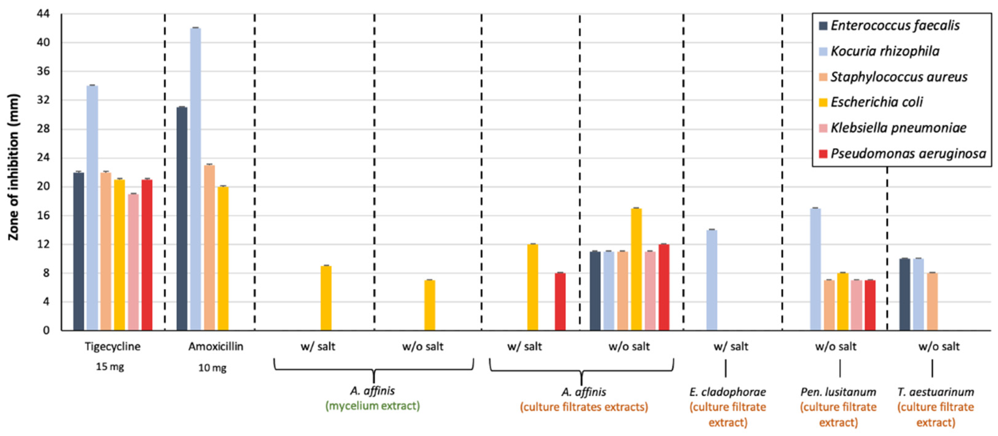
At a concentration of 500 mg/mL, only the mycelium extract of A. affinis (with and without sea salt) inhibited the growth of E. coli (Figure 2). Regarding culture media extracts, A. affinis, E. cladophorae, Pen. lusitanum, and T. aestuarinum showed antibacterial activity. Differences were also observed in culture media extracts with and without sea salt. The culture medium extract of A. affinis grown without sea salt inhibited the growth of all bacteria tested. In addition, Pen. lusitanum, regardless of the presence of sea salt in the growth medium, inhibited the growth of all Gram-positive and Gram-negative bacteria tested, except for E. faecalis, but with a smaller zone of inhibition when compared with A. affinis. The culture medium extract of A. affinis grown in the presence of sea salt inhibited the growth of E. coli and P. aeruginosa. On the other hand, the culture medium extract of T. aestuarinum grown in the presence of sea salt exhibited antibacterial activity only against Gram-positive bacteria. The culture medium extract of E. cladophorae grown in the presence of sea salt inhibited the growth of K. rhizophila.

3.4. Anti-Fungal Activity of Fungal Extracts

Differences in the inhibitory activity of Candida growth were observed in the mycelium and culture medium extracts of fungi grown with and without sea salt.
From all the mycelium extracts, only that from Pen. lusitanum inhibited Candida strains (Figure 3). When grown without sea salt, Pen. lusitanum mycelium extract inhibited only the growth of C. albicans strains. Nonetheless, the presence of sea salt led to larger C. albicans inhibition halos than of other Candida species tested. Candida glabrata and C. parapsilosis were not inhibited by salt- and non-salt mycelia extracts from Pen. lusitanum. The C. albicans strains tested were resistant to cycloheximide.
Regarding culture media extracts, only extracts of Pen. lusitanum and T. aestuarinum grown without sea salt showed anti-Candida activity. The culture medium extract of Pen. lusitanum inhibited the growth of all Candida strains tested, while the culture medium extract of T. aestuarinum inhibited C. albicans (strain 61C) and C. glabrata.

3.5. Cytotoxic Activity of Culture Media Extracts

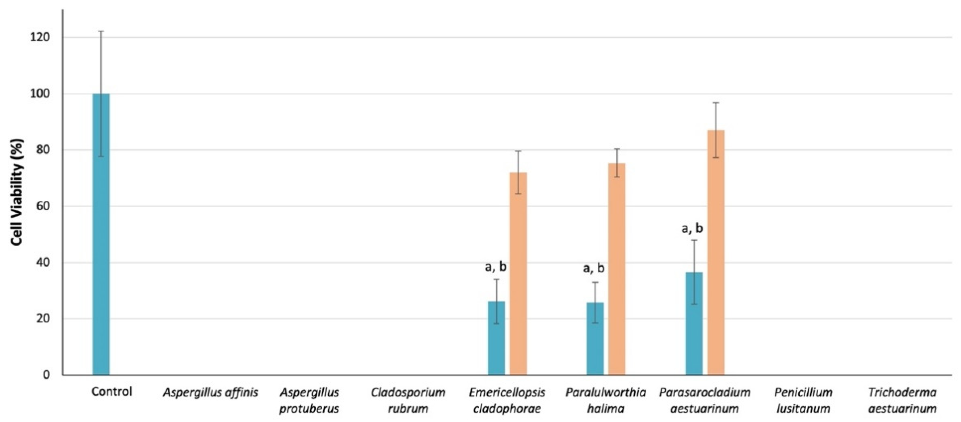
At 500 mg/mL, all culture medium extracts induced 100% of Vero cell mortality (data not shown). At 250 mg/mL (Figure 4), culture medium extracts of E. cladophorae, Paral. halima, and Paras. aestuarinum grown without 3% sea salt led to 72%, 75%, and 87% cell viability, respectively. Cell viability was significantly impaired by saline culture medium extracts.

3.6. Antioxidant Activity of Mycelium

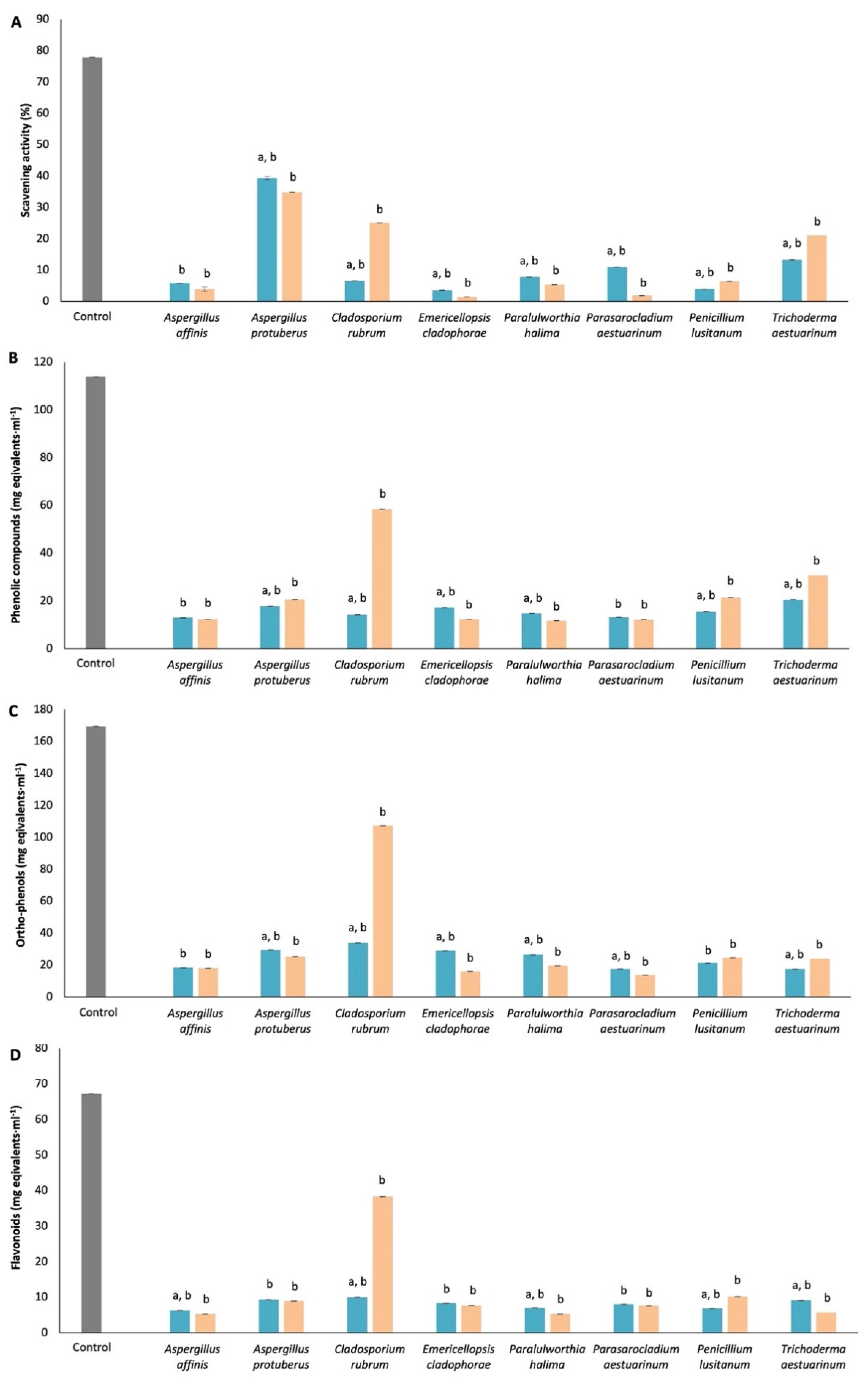
The data show that the mycelium extracts of C. rubrum, Pen. lusitanum and T. aestuarinum had higher antioxidant activity when cultivated in media without 3% sea salt than in the presence of sea salt, while for A. affinis, A. protuberus, E. cladophorae, Paral. halima and Paras. aestuarinum, the opposite was observed (Figure 5). In general, the mycelium extract of C. rubrum, when grown without sea salt, showed a high antioxidant content (58.42, 107.36, and 38.33 mg equivalent.mL1 in phenolic compounds, ortho-phenols and flavonoids, respectively). The exception was A. protuberus in saline and non-saline samples, which showed 39.33 and 34.86 mg equivalent.mL1, respectively, in DPPH-free radical scavenging activity.

4. Discussion

Metabolites from marine fungi are diverse and are described as expressing various biotechnological relevant activities. Our results confirm this with an emphasis on enzymatic, antibacterial, anti-yeast and antioxidant activities, revealing significant bioresources for outstanding opportunities to study natural product drug discovery.
Fungi express a spectrum of enzymes crucial for host colonization [48] and for the degradation of organic matter to obtain nutrients [34]. A wide range of activities have been identified from marine fungi, including antibacterial, antidiabetic, antifungal, antitumor, and cytotoxic activities, many of which can be attributed to specific enzymes [10]. Eighty-eight percent of the strains we assayed express cellulolytic activity, 68% express xylanases, and 92% and 80% express pectinases and pectin lyases, respectively.
Xylanases are glycoside hydrolases able to catalyze the hydrolysis of xylan (one of the most abundant polysaccharides in plants and algae) to xylose [49]. The most well-known examples of fungal species expressing xylanases belong to the genera Aspergillus, Penicillium and Trichoderma, these being the main commercial source of this type of enzyme [49]. Here, we demonstrate that A. affinis, A. protuberus, Pen. lusitanum, and T. aestuarinum are also able of produce xylanases. These enzymes can be used in the pulp and paper industry, as a food supplement to poultry, in bread and various drinks, in textiles, in bleaching processes, and in the production of other industrial reagents [50]. Bucher et al. [51] described a group of marine fungi—commonly occurring in mangroves expressing xylanase and cellulase activities. Additionally, Raghukumar et al. [52] showed that Lophiostoma mangrovei and Hypoxylon oceanicum can produce hemicellulases such as xylanases. Our strains of Neocamarosporium, a typical genus found in association with halophytes (salt tolerant plants) in marine and estuarine habitats, also showed xylanolytic activity. Other lignicolous fungi isolated from submerged wood such as Remispora submersa, Z. maritima, Z. pseudomaritima, S. taiwanensis, Paralulworthia halima, and Lulworthia cf. purpurea also showed cellulolytic activity. Lignicolous mangrove fungi such as Halorosellinia oceanica, Lignincola laevis, and Trematosphaeria mangrovei are capable of producing extracellular cellulases, namely endoglucanase, cellobiohydrolase, and β-glucosidase [53].
Previous studies have shown that marine fungi are also capable of expressing amylases [54,55]. Seventeen (out of 25) strains expressed amylolytic activity. This group of enzymes can catalyze the hydrolysis of starch molecules and have many industrial applications, such as in detergent and fuel alcohol production, in textiles, in the paper industry, and in bakeries [49,56]. Some genera including some marine ones are reported as a source of amylases, such as Alternaria, Aspergillus, Ceriosporopsis, Corollospora, Dendryphiella, Lindra, Lulworthia, Microthelia, Penicillium, Trichoderma, and Zalerion [57].
Urea represents one of the sources of nitrogen that support growth in marine fungi [58]. Metalloenzymes such as urease catalyze the hydrolysis of urea to carbon dioxide and ammonia [59]. Ureolytic microorganisms are able to precipitate carbonates and could be used for biorecovery of valuable metals disseminated in the environment [60]. From the strains assayed, 44% have ureolytic activity (A. protuberus, R. submersa, Neodevriesia aestuarina, Neptunomyces aureus, and S. taiwanensis).
Chitinolytic activity was detected in A. affinis, A. protuberus, C. rubrum, E. cladophorae, R. submersa, Neo. aestuarina, Nep. aureus, Para. fusiforme, Pen. lusitanum, T. aestuarinum, and Verrucoconiothyrium ambiguum. Chitin is a biopolymer of N-acetylglucosamine and is a structural component of fungal cell walls, as well as of the exoskeletons of several marine invertebrates, and can be degraded by chitinase into monomers [49]. Chitinases can be used as antifungal agents and biopesticides [61]. They are also useful in the manufacture of pharmaceutical products, as tools in cell engineering, and in medical diagnosis [62]. Some marine fungi capable of synthesizing chitinases are already known, such as species belonging to Aspergillus, Rhizopus, Penicillium, Beauveria [9], and Clonostachys rosea isolated from seagrass [63].
Another relevant group of enzymes for the lignin degradative process with bleaching applications in the wood, paper, and cellulose industries are laccases [52], which are a family of multicopper oxidases distributed across bacteria, fungi, and plants [64]. These enzymes are constitutively secreted and are stable in the extracellular environment contributing to their attractiveness for biotechnological applications [65]. Several marine fungal strains isolated from seagrasses, algae, and decaying wood have been reported to produce laccases, such as Penicillium spp., Trichoderma spp., Lulworthia spp., Z. maritima, Aspergillus sclerotiorum, Cladosporium cladosporioides, and Mucor racemosus [52,66,67]. In this study, only T. aestuarinum and V. ambiguum expressed extracellular laccases.
Proteases also have a relevant role in the biotechnology industry, including the pharmaceutical industry. As reviewed earlier [68], proteases, namely collagenases, have important applications in the health industry, such as the use of collagenases in the enzymatic debridement of wounds and of other injuries where the removal of devitalized tissue is necessary.
Some studies have explored the influence of salt in the enzymatic activity of marine fungi. For example, Chamekh et al. [69] evaluated different enzymatic activities of halotolerant and halophilic fungi and Arfi et al. [70] reported the presence of more than 400 lignocellulolytic enzymes from the mangrove fungus Pestalotiopsis sp. The authors concluded that the presence of salt modifies lignocellulolytic enzyme composition, with an increase in the secretion of xylanases and cellulases and a decrease in the production of oxidases, as we also observed in this study. Additionally, Chen et al. [71] and Bonugli-Santos et al. [72] achieved similar results in lignin enzymes when fungi were grown in saline conditions. In this study, sea salt induced the alteration of the enzymatic profiles detected.
Eight strains were selected for a deeper characterization of their biotechnological potential. These strains belong to genera widely known to produce active extracellular metabolites with antibacterial activity, such as Penicillium, Aspergillus, Trichoderma, and Cladosporium [73], or even Emericellopsis [74]. In the present work, 80% of the 25 strains tested in the preliminary screening were able to inhibit the growth of at least one bacterium, mainly Gram-positive bacteria, during the primary screening. However, only four strains (A. affinis, E. cladophorae, Pen. Lusitanum, and T. aestuarinum) of the eight fungi selected for the fermentation assay retained their antibacterial activity. Similar observations were reported in other studies, in which fungi showed activity in solid media but not when subjected to fermentation. This apparent loss of activity may be due to the type of medium, culture conditions, or to the extraction process [75,76,77]. In general, crude extracts of extracellular media showed higher activity than mycelia extracts. Aspergillus affinis led to the largest inhibition halos, inhibiting the growth of the three-Gram negative bacteria, including the multiresistant strains K. pneumoniae and P. aeruginosa. These two multiresistant strains were isolated from the Lis River (Central Portugal) [37]. These authors highlighted that the presence of antibiotic-resistant bacteria and resistance genes in this particular environment is a major public health risk. The escalation of antibiotic-resistant infections globally during the last few decades imposes the need to search for new antimicrobial compounds and measures to contain the dispersion of this type of resistance. Therefore, the antibacterial activity exhibited by A. affinis (CMG 70) and Pen. lusitanum (MUM 18.49/CMG 8) against K. pneumoniae and P. aeruginosa suggests that these strains can be potential sources of antibacterial molecules. Additionally, as observed in enzymatic assays, variations between saline and non-saline culture conditions in antibacterial activity were detected, i.e., the presence or absence of salt affected the production of active metabolites.
Penicillium species are important producers of antibacterial compounds, but only a few are reported to produce antifungal substances. In this study, both mycelium and culture medium extracts of Pen. lusitanum showed a higher inhibitory effect on the C. albicans strains tested than the cycloheximide control. These results suggest that Pen. lusitanum (MUM 18.49/CMG 8) could be a source of compounds useful in anti-Candida therapy, but more studies are necessary to confirm this. Moreover, Komai et al. [78] reported the presence of two metabolites of Pen. simplicissimum active against other filamentous fungi and yeasts, such as C. albicans and Cryptococcus neoformans. Kaleem et al. [79,80] also isolated metabolites from a marine Penicillium sp. able to inhibit the growth of a methicillin-resistant S. aureus, E. coli, and of C. albicans. Currently, candidiasis represents the fourth leading cause of nosocomial infections, and mortality remains highly dependent on the infecting Candida species [81] and on the immune state of the patient [82]. Over the last two decades, the number of infections caused by non-C. albicans species (including C. glabrata, C. parapsilosis, and C. tropicalis, that we used in this study) has increased considerably [83]. Clinical manifestations of candidiasis in humans are diverse and the patients can show several symptoms, such as stomatitis, gastritis, pneumonia, lung empyema, cystitis, meningitis, otitis, vulvovaginitis, and others [81]. Some Candida strains are resistant to some topical and systemic antifungals used in the management of candidiasis, such as fluconazole, benomyl, or cycloheximide [84,85]. We also observed the resistance of C. albicans to cycloheximide, but little is known about the factors involved in the mechanisms of resistance. Resistance to this antimycotic has already been reported in C. maltosa involving the CYHR gene, which confers this resistance, and in Saccharomyces cerevisiae linked to mutations in ribosomal proteins [85].
All crude culture medium extracts of the fungal species tested induced 100% cytotoxicity at 500 mg/mL against a mammalian cell line (Vero cells). We showed that cytotoxicity was concentration dependent, and cell viability was observed only after fungal extract dilution. The data suggest that the fungal extracts were more cytotoxic when species were cultured in the presence of sea salt. Aspergillus affinis and Pen. lusitanum, which exhibited effective antibacterial activity were highly cytotoxic. Some studies have already reported the cytotoxicity evaluation in cancer cell lines of compounds produced by fungal marine species, including Aspergillus, Cladosporium, Penicillium, and Trichoderma [86,87,88]. In fact, marine fungi with the ability to synthesize cytotoxic compounds showed the potential for the treatment of cancer diseases [89]. Further studies using the extracts that did not induce Vero cells mortality are required to evaluate the cytotoxicity on cancer cell lines for cancer research purposes.
In the last few years, natural antioxidants have generated considerable interest in medicine and in the food industry. In consequence, attention has been focused on the characterization of the antioxidant properties of medicinal plant extracts, spices, and mushrooms [90]. Although filamentous fungi are also known to produce antioxidant activity, they are less explored. Some studies of filamentous fungi, such as of Aspergillus, Chaetomium, Cladosporium, Colletotrichum, Epicoccum, Lecanicillium, and Penicillium, revealed that they are also sources of antioxidants in the form of their secondary metabolites [21,91].
All major naturally occurring antioxidant classes, including DPPH, phenols, and flavonoids, were found in the methanolic extracts of each fungal species studied. The highest antioxidant activity, including phenols, ortho-phenols, and flavonoids, was observed for the extract of C. rubrum (MUM 19.39/CMG 28) when cultivated without sea salt, which may be a strain-specific characteristic. Phenolic compounds represent the largest category of natural products and are the most widely reported to exhibit antioxidant activity [44]. To deal with different stress situations, fungi produce phenolic compounds that act as natural lipophilic radical scavengers [92]. With flavonoids, these compounds provide fungi with natural defenses and protection from oxidative stress-caused damage against the free radicals, metal chelators and pro-oxidants [93]. Some studies revealed that antioxidant capacity in filamentous fungi was attributed mainly to their phenolic content [94]. Even though methanolic extracts of all fungal species studied were shown to possess antioxidant activities, extraction methods, solvents, time, temperature, and pH are crucial factors in obtaining natural antioxidants from different sources [92]. However, there are other mechanisms through which antioxidants can show their activity than by direct reaction mechanism [92]. We showed that these extracts from fungal marine species prove to also be a rich source of antioxidants components.
According to FTIR analyses, the main differences found in functional groups between saline and non-saline culture conditions were chitin and ergosterol, lipids, and carbohydrates. Recently, Danilova et al. [95] showed changes in the membrane lipid composition of Emericellopsis alkalina under different concentrations of NaCl. The present study suggests that sea salt applies a selective pressure on the activity profiles of the fungi assayed, and induces an alteration in the chemical composition of fungal cell walls.

5. Conclusions

Filamentous fungi are the preferred source of industrial enzymes due to their excellent capacity for extracellular enzyme production. Marine fungi harbor promising compounds with relevant applications in biotechnology. One major advantage of the utilization of marine fungi in drug discovery is that their diversity offers an almost endless source of novel bioactive compounds. In this study, we focused on extract activities rather than in pure metabolites: extracts are easier and cheaper to obtain than pure metabolites. Furthermore, the activity profiles from the extracts of the marine fungi studied were demonstrated to be responsive to sea salt.

Supplementary Materials

The following are available online at https://www.mdpi.com/article/10.3390/app11136008/s1, Table S1: Detection of extracellular activity, Table S2: Screening of antibacterial activity.

Author Contributions

M.F.M.G.; conceptualization, formal analysis, investigation, methodology, writing—original draft, A.P.; investigation, writing—review and editing, L.F.E.; investigation, writing—review and editing, M.S.F.A.; investigation, writing—review and editing, C.A.P.; investigation, writing—review and editing, J.A.S.; writing—review and editing, A.S.D.; investigation, writing—review and editing, T.A.P.R.-S.; writing—review and editing, A.C.E.; conceptualization, methodology, supervision, writing—review and editing, A.A.; conceptualization, funding acquisition, methodology, project administration, resources, supervision, writing—review and editing. All authors have read and agreed to the published version of the manuscript.

Funding

This research was supported by the Portuguese Foundation for Science and Technology (FCT) to CESAM (UIDB/50017/2020+UIDP/50017/2020), to Center for Interdisciplinary Research in Health (UIDB/04279/2020 and UIDP/04279/2020) and to LAQV-REQUIMTE research Unit (UIDB/50006/2020) and the PhD grants of M. Gonçalves (SFRH/BD/129020/2017), A. Paço (SFRH/BD/146930/2019) and Carlos A. Pinto (SFRH/BD/137036/2018).

Institutional Review Board Statement

Not applicable.

Informed Consent Statement

Informed consent was obtained from all subjects involved in the study.

Data Availability Statement

The authors declare to honor the Principles of Transparency and Best Practice in Scholarly Publishing about Data.

Conflicts of Interest

The authors declare no conflict of interest.

References

  1. Jones, E.B.G. Are there more marine fungi to be described? Bot. Mar. 2011, 54, 343–354. [Google Scholar] [CrossRef]
  2. Richards, T.A.; Jones, M.D.; Leonard, G.; Bass, D. Marine fungi: Their ecology and molecular diversity. Annu. Rev. Mar. Sci. 2012, 4, 495–522. [Google Scholar] [CrossRef] [PubMed]
  3. Bugni, T.S.; Ireland, C.M. Marine-derived fungi: A chemically and biologically diverse group of microorganisms. Nad. Prod. Rep. 2004, 21, 143–163. [Google Scholar] [CrossRef]
  4. Imhoff, J.F.; Labes, A.; Wiese, J. Bio-mining the microbial treasures of the ocean: New natural products. Biotechnol. Adv. 2011, 29, 468–482. [Google Scholar] [CrossRef]
  5. Overy, D.P.; Rämä, T.; Oosterhuis, R.; Walker, A.K.; Pang, K.-L. The neglected marine fungi, sensu stricto, and their isolation for natural products’ discovery. Mar. Drugs 2019, 17, 42. [Google Scholar] [CrossRef] [Green Version]
  6. Gladfelter, A.S.; James, T.Y.; Amend, A.S. Marine fungi. Curr. Biol. 2019, 29, R191–R195. [Google Scholar] [CrossRef]
  7. Sakayaroj, J.; Pang, K.L.; Jones, E.G. Multi-gene phylogeny of the Halosphaeriaceae: Its ordinal status, relationships between genera and morphological character evolution. Fungal Divers. 2011, 46, 87–109. [Google Scholar] [CrossRef]
  8. Pang, K.L.; Overy, D.P.; Jones, E.B.G.; Calado, M.; Burgaud, G.; Walker, A.K.; Johnson, J.A.; Kerr, R.G.; Cha, H.-J.; Bills, J.F. ‘Marine fungi’ and ‘marine-derived fungi’ in natural product chemistry research: Toward a new consensual definition. Fungal Biol. Rev. 2016, 30, 163–175. [Google Scholar] [CrossRef]
  9. Raghukumar, S. Fungi in Coastal and Oceanic Marine Ecosystems; Springer: New York, NY, USA, 2017; 364p. [Google Scholar]
  10. Mayer, A.M.S.; Rodríguez, A.D.; Taglialatela-Scafati, O.; Fusetani, N. Marine pharmacology in 2009–2011: Marine compounds with antibacterial, antidiabetic, antifungal, anti-inflammatory, antiprotozoal, antituberculosis, and antiviral activities; affecting the immune and nervous systems, and other miscellaneous mechanisms of action. Mar. Drugs 2013, 11, 2510–2573. [Google Scholar]
  11. Li, X.; Li, X.M.; Xu, G.M.; Li, C.S.; Wang, B.G. Antioxidant metabolites from marine alga-derived fungus Aspergillus wentii EN-48. Phytochem. Lett. 2014, 7, 120–123. [Google Scholar] [CrossRef]
  12. Chen, C.J.; Zhou, Y.Q.; Liu, X.X.; Zhang, W.J.; Hu, S.S.; Lin, L.P.; Huo, G.M.; Jiao, R.H.; Tan, R.X.; Ge, H.M. Antimicrobial and anti-inflammatory compounds from a marine fungus Pleosporales sp. Tetrahedron Lett. 2015, 56, 6183–6189. [Google Scholar] [CrossRef]
  13. Moghadamtousi, S.Z.; Nikzad, S.; Kadir, H.A.; Abubakar, S.; Zandi, K. Potential antiviral agents from marine fungi: An overview. Mar. Drugs 2015, 13, 4520–4538. [Google Scholar] [CrossRef]
  14. Bonugli-Santos, R.C.; dos Santos Vasconcelos, M.R.; Passarini, M.R.; Vieira, G.A.; Lopes, V.C.; Mainardi, P.H.; dos Santos, J.A.; Duarte, L.A.; Otero, I.V.R.; Yoshida, A.M.S.; et al. Marine-derived fungi: Diversity of enzymes and biotechnological applications. Front. Microbiol. 2015, 6, 269. [Google Scholar] [CrossRef] [PubMed]
  15. Xu, L.; Meng, W.; Cao, C.; Wang, J.; Shan, W.; Wang, Q. Antibacterial and antifungal compounds from marine fungi. Mar. Drugs 2015, 13, 3479–3513. [Google Scholar] [CrossRef]
  16. Han, J.; Liu, M.; Jenkins, I.D.; Liu, X.; Zhang, L.; Quinn, R.J.; Feng, Y. Genome-inspired Chemical exploration of marine fungus Aspergillus fumigatus MF071. Mar. Drugs 2020, 18, 352. [Google Scholar] [CrossRef]
  17. Yasuhara-Bell, J.; Lu, Y. Marine compounds and their antiviral activities. Antivir. Res. 2010, 86, 231–240. [Google Scholar] [CrossRef]
  18. Kumar, A.; Henrissat, B.; Arvas, M.; Syed, M.F.; Thieme, N.; Benz, J.P.; Sørensen, J.L.; Record, E.; Pöggeler, S.; Kempken, F. De novo assembly and genome analyses of the marine-derived Scopulariopsis brevicaulis strain LF580 unravels life-style traits and anticancerous scopularide biosynthetic gene cluster. PLoS ONE 2015, 10, e0140398. [Google Scholar] [CrossRef]
  19. Deshmukh, S.K.; Prakash, V.; Ranjan, N. Marine fungi: A source of potential anticancer compounds. Front. Microbiol. 2018, 8, 2536. [Google Scholar] [CrossRef] [PubMed]
  20. Jans, P.E.; Mfuh, A.M.; Arman, H.D.; Shaffer, C.V.; Larionov, O.V.; Mooberry, S.L. Cytotoxicity and mechanism of action of the marine-derived fungal metabolite trichodermamide B and synthetic analogues. J. Nat. Prod. 2017, 80, 676–683. [Google Scholar] [CrossRef] [Green Version]
  21. Sun, H.H.; Mao, W.J.; Chen, Y.; Guo, S.D.; Li, H.Y.; Qi, X.H.; Chen, Y.-L.; Xu, J. Isolation, chemical characteristics and antioxidant properties of the polysaccharides from marine fungus Penicillium sp. F23-2. Carbohydr. Polym. 2009, 78, 117–124. [Google Scholar] [CrossRef]
  22. Russell, J.R.; Huang, J.; Anand, P.; Kucera, K.; Sandoval, A.G.; Dantzler, K.W.; Hickman, D.; Jee, J.; Kimovec, F.M.; Koppstein, D.; et al. Biodegradation of polyester polyurethane by endophytic fungi. Appl. Environ. Microbiol. 2011, 77, 6076–6084. [Google Scholar] [CrossRef] [PubMed] [Green Version]
  23. Paço, A.; Duarte, K.; da Costa, J.P.; Santos, P.S.; Pereira, R.; Pereira, M.E.; Freitas, A.C.; Duarte, A.C.; Rocha-Santos, T.A.P. Biodegradation of polyethylene microplastics by the marine fungus Zalerion maritimum. Sci. Total Environ. 2017, 586, 10–15. [Google Scholar] [CrossRef]
  24. Crous, P.W.; Wingfield, M.J.; Lombard, L.; Roets, F.; Swart, W.J.; Alvarado, P.; Carnegie, A.J.; Moreno, G.; Luangsa-Ard, J.; Thangavel, R.; et al. Fungal Planet description sheets: 951–1041. Persoonia 2019, 43, 223–425. [Google Scholar] [CrossRef]
  25. Crous, P.W.; Wingfield, M.J.; Chooi, Y.H.; Gilchrist, C.L.M.; Lacey, E.; Pitt, J.I.; Roets, F.; Swart, W.J.; Cano-Lira, J.F.; Valenzuela-Lopez, N.; et al. Fungal Planet description sheets: 1042–1111. Persoonia 2020, 44, 301–459. [Google Scholar] [CrossRef] [PubMed]
  26. Gonçalves, M.F.M.; Aleixo, A.; Vicente, T.F.L.; Esteves, A.C.; Alves, A. Three new species of Neocamarosporium isolated from saline environments: N. aestuarinum sp. nov., N. endophyticum sp. nov. and N. halimiones sp. nov. Mycosphere 2019, 10, 608–621. [Google Scholar] [CrossRef]
  27. Gonçalves, M.F.M.; Vicente, T.F.L.; Esteves, A.C.; Alves, A. Neptunomyces aureus gen. et sp. nov. (Didymosphaeriaceae, Pleosporales) isolated from algae in Ria de Aveiro, Portugal. MycoKeys 2019, 60, 31–44. [Google Scholar] [CrossRef] [PubMed]
  28. Gonçalves, M.F.M.; Santos, L.; Silva, B.M.V.; Abreu, A.C.; Vicente, T.F.L.; Esteves, A.C.; Alves, A. Biodiversity of Penicillium species from marine environments in Portugal and description of Penicillium lusitanum sp. nov., a novel species isolated from sea water. Int. J. Syst. Evol. Microbiol. 2019, 69, 3014–3021. [Google Scholar] [CrossRef] [PubMed]
  29. Gonçalves, M.F.M.; Silva, B.M.V.; Esteves, A.C.; Alves, A. Verrucoconiothyrium ambiguum sp. nov., a novel species isolated from sea water, and affiliation of the genus Verrucoconiothyrium to the family Didymellaceae. Int. J. Syst. Evol. Microbiol. 2019, 69, 3769–3776. [Google Scholar] [CrossRef]
  30. Gonçalves, M.F.M.; Esteves, A.C.; Alves, A. Revealing the hidden diversity of marine fungi in Portugal with the description of two novel species, Neoascochyta fuci sp. nov. and Paraconiothyrium salinum sp. nov. Int. J. Syst. Evol. Microbiol. 2020, 70, 5337–5354. [Google Scholar] [CrossRef]
  31. Gonçalves, M.F.M.; Vicente, T.F.L.; Esteves, A.C.; Alves, A. Novel halotolerant species of Emericellopsis and Parasarocladium associated with macroalgae in an estuarine environment. Mycologia 2020, 112, 154–171. [Google Scholar] [CrossRef]
  32. Gonçalves, M.F.M.; Abreu, A.C.; Hilário, S.; Alves, A. Diversity of marine fungi associated with wood baits in the estuary Ria de Aveiro with descriptions of Paralulworthia halima comb. nov., Remispora submersa sp. nov. and Zalerion pseudomaritima sp. nov. Mycologia 2021, 113, 664–683. [Google Scholar] [CrossRef] [PubMed]
  33. Vicente, T.F.L.; Gonçalves, M.F.M.; Brandão, C.; Fidalgo, C.; Alves, A. Diversity of fungi associated with macroalgae from an estuarine environment and description of Cladosporium rubrum sp. nov. and Hypoxylon aveirense sp. nov. Int. J. Syst. Evol. Microbiol. 2021, 71, 004630. [Google Scholar] [CrossRef] [PubMed]
  34. Esteves, A.C.; Saraiva, M.; Correia, A.; Alves, A. Botryosphaeriales fungi produce extracellular enzymes with biotechnological potential. Can. J. Microbiol. 2014, 60, 332–342. [Google Scholar] [CrossRef] [PubMed]
  35. Fenice, M.; Selbmann, L.; Di Giambattista, R.; Federici, F. Chitinolytic activity at low temperature of an Antarctic strain (A3) of Verticillium lecanii. Res. Microbiol. 1998, 149, 289–300. [Google Scholar] [CrossRef]
  36. Agrawal, T.; Kotasthane, A.S. Chitinolytic assay of indigenous Trichoderma isolates collected from different geographical locations of Chhattisgarh in Central India. SpringerPlus 2012, 1, 73. [Google Scholar] [CrossRef] [Green Version]
  37. Teixeira, P.; Tacão, M.; Pureza, L.; Gonçalves, J.; Silva, A.; Cruz-Schneider, M.P.; Henriques, I. Occurrence of carbapenemase-producing Enterobacteriaceae in a Portuguese river: blaNDM, blaKPC and blaGES among the detected genes. Environ. Pollut. 2020, 260, 113913. [Google Scholar] [CrossRef]
  38. Azmir, J.; Zaidul, I.S.M.; Rahman, M.M.; Sharif, K.M.; Mohamed, A.; Sahena, F.; Jahurul, M.H.A.; Ghafoor, K.; Norulaini, N.A.N.; Omar, A.K.M. Techniques for extraction of bioactive compounds from plant materials: A review. J. Food Eng. 2013, 117, 426–436. [Google Scholar] [CrossRef]
  39. National Committee for Clinical Laboratory Standards (NCCLS). Performance Standards for Antimicrobial Disk and Dilution Susceptibility Testing; Fourteenth Informational Supplement NCCLS documents M100-S14; Clinical and Laboratory Standards Institute: Wayne, PA, USA, 2004.
  40. Duarte, A.S.; Cavaleiro, E.; Pereira, C.; Merino, S.; Esteves, A.C.; Duarte, E.P.; Tomás, J.M.; Correia, A.C. Aeromonas piscicola AH-3 expresses an extracellular collagenase with cytotoxic properties. Lett. Appl. Microbiol. 2015, 60, 288–297. [Google Scholar] [CrossRef]
  41. Ammerman, N.C.; Beier-Sexton, M.; Azad, A.F. Growth and maintenance of Vero cell lines. Curr. Protoc. Microbiol. 2009. [Google Scholar] [CrossRef] [Green Version]
  42. Xu, B.J.; Chang, S.K.C. A comparative study on phenolic profiles and antioxidant activities of legumes as affected by extraction solvents. J. Food Sci. 2007, 72, S159–S166. [Google Scholar] [CrossRef] [PubMed]
  43. Singleton, V.L.; Orthofer, R.; Lamuela-Raventós, R.M. Analysis of total phenols and other oxidation substrates and antioxidants by means of folin-ciocalteu reagent. Methods Enzymol. 1999, 299, 152–178. [Google Scholar]
  44. Chang, C.-C.; Yang, M.-H.; Wen, H.-M.; Chern, J.-C. Estimation of total flavonoid content in propolis by two complementary colorimetric methods. J. Food Drug Anal. 2002, 10, 178–182. [Google Scholar]
  45. Chhetri, D.R.; Chhetri, A.; Shahi, N.; Tiwari, S.; Karna, S.K.L.; Lama, D.; Pokharel, Y.R. Isaria tenuipes Peck, an entomopathogenic fungus from Darjeeling Himalaya: Evaluation of in-vitro antiproliferative and antioxidant potential of its mycelium extract. BMC Complement. Med. Ther. 2020, 20, 1–14. [Google Scholar] [CrossRef]
  46. Girometta, C.; Dondi, D.; Baiguera, R.M.; Bracco, F.; Branciforti, D.S.; Buratti, S.; Lazzaroni, S.; Savino, E. Characterization of mycelia from wood-decay species by TGA and IR spectroscopy. Cellulose 2020, 27, 6133–6148. [Google Scholar] [CrossRef]
  47. Di Mario, F.; Rapana, P.; Tomati, U.; Galli, E. Chitin and chitosan from Basidiomycetes. Int. J. Biol. Macromol. 2008, 43, 8–12. [Google Scholar] [CrossRef] [PubMed]
  48. Hyde, K.D.; Jones, E.G.; Leaño, E.; Pointing, S.B.; Poonyth, A.D.; Vrijmoed, L.L. Role of fungi in marine ecosystems. Biodivers. Conserv. 1998, 7, 1147–1161. [Google Scholar] [CrossRef]
  49. Liu, X.; Kokare, C. Microbial enzymes of use in industry. In Biotechnology of Microbial Enzymes; Academic Press: Cambridge, MA, USA, 2017; pp. 267–298. [Google Scholar]
  50. Bajpai, P. Xylanolytic Enzymes; Academic Press: Cambridge, MA, USA, 2014. [Google Scholar]
  51. Bucher, V.V.C.; Hyde, K.D.; Pointing, S.B.; Reddy, C.A. Production of wood decay enzymes, mass loss and lignin solubilization in wood by marine ascomycetes and their anamorphs. Fungal Divers. 2004, 15, 1–14. [Google Scholar]
  52. Raghukumar, C.; Raghukumar, S.; Chinnaraj, A.; Chandramohan, D.; D’Souza, T.M.; Reddy, C.A. Laccase and other lignocellulose modifying enzymes of marine fungi isolated from the coast of India. Bot. Mar. 1994, 37, 515–523. [Google Scholar] [CrossRef]
  53. Pointing, S.B.; Hyde, K.D. Lignocellulose-degrading marine fungi. Biofouling 2000, 15, 221–229. [Google Scholar] [CrossRef]
  54. Sarkar, S.; Pramanik, A.; Mitra, A.; Mukherjee, J. Bioprocessing data for the production of marine enzymes. Mar. Drugs 2010, 8, 1323–1372. [Google Scholar] [CrossRef] [Green Version]
  55. Homaei, A.; Ghanbarzadeh, M.; Monsef, F. Biochemical features and kinetic properties of α-amylases from marine organisms. Int. J. Biol. Macromol. 2016, 83, 306–314. [Google Scholar] [CrossRef]
  56. Sundarram, A.; Murthy, T.P.K. α-amylase production and applications: A review. J. Appl. Environ. Microbiol. 2014, 2, 166–175. [Google Scholar]
  57. Sahoo, K.; Dhal, N.K.; Das, R. Production of amylase enzyme from mangrove fungal isolates. Afr. J. Biotechnol. 2014, 13, 4338–4346. [Google Scholar]
  58. Sguros, P.L.; Rodrigues, J.; Simms, J. Role of marine fungi in the biochemistry of the oceans. V. Patterns of constitutive nutritional growth responses. Mycologia 1973, 65, 161–174. [Google Scholar] [CrossRef]
  59. Kumari, D.; Qian, X.Y.; Pan, X.; Achal, V.; Li, Q.; Gadd, G.M. Microbially-induced carbonate precipitation for immobilization of toxic metals. Adv. Appl. Microbiol. 2016, 94, 79–108. [Google Scholar] [PubMed] [Green Version]
  60. Li, Q.; Gadd, G.M. Biosynthesis of copper carbonate nanoparticles by ureolytic fungi. Appl. Microbiol. Biotechnol. 2017, 101, 7397–7407. [Google Scholar] [CrossRef] [Green Version]
  61. Berini, F.; Casartelli, M.; Montali, A.; Reguzzoni, M.; Tettamanti, G.; Marinelli, F. Metagenome-sourced microbial chitinases as potential insecticide proteins. Front. Microbiol. 2019, 10, 1358. [Google Scholar] [CrossRef] [PubMed]
  62. Dahiya, N.; Tewari, R.; Hoondal, G.S. Biotechnological aspects of chitinolytic enzymes: A review. Appl. Microbiol. Biotechnol. 2006, 71, 773–782. [Google Scholar] [CrossRef]
  63. Pasqualetti, M.; Barghini, P.; Giovannini, V.; Fenice, M. High production of chitinolytic activity in halophilic conditions by a new marine strain of Clonostachys rosea. Molecules 2019, 24, 1880. [Google Scholar] [CrossRef] [PubMed] [Green Version]
  64. Reiss, R.; Ihssen, J.; Richter, M.; Eichhorn, E.; Schilling, B.; Thöny-Meyer, L. Laccase versus laccase-like multi-copper oxidase: A comparative study of similar enzymes with diverse substrate spectra. PLoS ONE 2013, 8, e65633. [Google Scholar]
  65. Couto, S.R.; Herrera, J.L.T. Laccases in pollution control. Ter. Aquat. Environ. Tox. 2007, 1, 34–45. [Google Scholar]
  66. Bonugli-Santos, R.C.; Durrant, L.R.; da Silva, M.; Sette, L.D. Production of laccase, manganese peroxidase and lignin peroxidase by Brazilian marine-derived fungi. Enzyme Microb. Technol. 2010, 46, 32–37. [Google Scholar] [CrossRef]
  67. Ben Ali, W.; Chaduli, D.; Navarro, D.; Lechat, C.; Turbé-Doan, A.; Bertrand, E.; Faulds, C.B.; Sciara, G.; Lesage-Meessen, L.; Record, E.; et al. Screening of five marine-derived fungal strains for their potential to produce oxidases with laccase activities suitable for biotechnological applications. BMC Biotechnol. 2020, 20, 1–13. [Google Scholar] [CrossRef] [PubMed]
  68. Duarte, A.S.; Correia, A.; Esteves, A.C. Bacterial collagenases—A review. Crit. Rev. Microbiol. 2016, 42, 106–126. [Google Scholar] [CrossRef]
  69. Chamekh, R.; Deniel, F.; Donot, C.; Jany, J.L.; Nodet, P.; Belabid, L. Isolation, identification and enzymatic activity of halotolerant and halophilic fungi from the Great Sebkha of Oran in Northwestern of Algeria. Mycobiology 2019, 47, 230–241. [Google Scholar] [CrossRef] [PubMed]
  70. Arfi, Y.; Chevret, D.; Henrissat, B.; Berrin, J.G.; Levasseur, A.; Record, E. Characterization of salt-adapted secreted lignocellulolytic enzymes from the mangrove fungus Pestalotiopsis sp. Nat. Commun. 2013, 4, 1–9. [Google Scholar] [CrossRef] [Green Version]
  71. Chen, H.-Y.; Xue, D.-S.; Feng, X.-Y.; Yao, S.-J. Screening and production of ligninolytic enzyme by a marine-derived fungal Pestalotiopsis sp. J63. Appl. Biochem. Biotechnol. 2011, 165, 1754–1769. [Google Scholar] [CrossRef] [PubMed]
  72. Bonugli-Santos, R.C.; Durrant, L.R.; Sette, L.D. The production of ligninolytic enzymes by marine-derived basidiomycetes and their biotechnological potential in the biodegradation of recalcitrant pollutants and the treatment of textile effluents. Water Air Soil Pollut. 2012, 223, 2333–2345. [Google Scholar] [CrossRef]
  73. Kong, F.D.; Zhou, L.M.; Ma, Q.Y.; Huang, S.Z.; Wang, P.; Dai, H.F.; Zhao, Y.X. Metabolites with Gram-negative bacteria quorum sensing inhibitory activity from the marine animal endogenic fungus Penicillium sp. SCS-KFD08. Arch. Pharm. Res. 2017, 40, 25–31. [Google Scholar] [CrossRef]
  74. Inostroza, A.; Lara, L.; Paz, C.; Perez, A.; Galleguillos, F.; Hernandez, V.; Becerra, J.; González-Rocha, G.; Silva, M. Antibiotic activity of Emerimicin IV isolated from Emericellopsis minima from Talcahuano Bay, Chile. Nat. Prod. Res. 2018, 32, 1361–1364. [Google Scholar] [CrossRef]
  75. De Siqueira, V.M.; Conti, R.; De Araújo, J.M.; Souza-Motta, C.M. Endophytic fungi from the medicinal plant Lippia sidoides Cham. and their antimicrobial activity. Symbiosis 2011, 53, 89–95. [Google Scholar] [CrossRef]
  76. dos Santos, I.P.; da Silva, L.C.; da Silva, M.V.; de Araújo, J.M.; Cavalcanti, M.D.S.; Lima, V.L. Antibacterial activity of endophytic fungi from leaves of Indigofera suffruticosa Miller (Fabaceae). Front. Microbiol. 2015, 6, 350. [Google Scholar] [CrossRef] [PubMed]
  77. Marcellano, J.P.; Collanto, A.S.; Fuentes, R.G. Antibacterial activity of endophytic fungi isolated from the bark of Cinnamomum mercadoi. Pharmacogn. J. 2017, 9, 405–409. [Google Scholar] [CrossRef] [Green Version]
  78. Komai, S.-I.; Hosoe, T.; Itabashi, T.; Nozawa, K.; Yaguchi, T.; Fukushima, K.; Kawai, K. New penicillide derivatives isolated from Penicillium simplicissimum. J. Nat. Med. 2006, 60, 185–190. [Google Scholar] [CrossRef]
  79. Kaleem, S.; Ge, H.; Yi, W.; Zhang, Z.; Wu, B. Isolation, structural elucidation, and antimicrobial evaluation of the metabolites from a marine-derived fungus Penicillium sp. ZZ1283. Nat. Prod. Res. 2019, 1–9. [Google Scholar] [CrossRef] [PubMed]
  80. Kaleem, S.; Qin, L.; Yi, W.; Lian, X.Y.; Zhang, Z. Bioactive metabolites from the Mariana Trench sediment-derived fungus Penicillium sp. SY2107. Mar. Drugs 2020, 18, 258. [Google Scholar] [CrossRef] [PubMed]
  81. Pal, M. Candidiasis: An important opportunistic mycosis of global public health concern. Res. Infect. Dis. Trop. Med. 2020, 2, 49–51. [Google Scholar]
  82. Rosa, N.; Campos, B.; Esteves, A.C.; Duarte, A.S.; Correia, M.J.; Silva, R.M.; Barros, M. Tracking the functional meaning of the human oral-microbiome protein-protein interactions. In Advances in Protein Chemistry and Structural Biology; Academic Press: Cambridge, MA, USA, 2020; Volume 121, pp. 199–235. [Google Scholar]
  83. Pal, M. Aspergillosis: A highly infectious global mycosis of human and animal. Clin. Biotechnol. Microbiol. 2017, 1, 46–49. [Google Scholar]
  84. Ben-Yaacov, R.; Knoller, S.; Caldwell, G.A.; Becker, J.M.; Koltin, Y. Candida albicans gene encoding resistance to benomyl and methotrexate is a multidrug resistance gene. Antimicrob. Agents Chemother. 1994, 38, 648–652. [Google Scholar] [CrossRef] [Green Version]
  85. Sasnauskas, K.; Jomantienė, R.; Lebedienė, E.; Lebedys, J.; Januska, A.; Janulaitis, A. Cloning and sequence analysis of a Candida maltosa gene which confers resistance to cycloheximide. Gene 1992, 116, 105–108. [Google Scholar] [CrossRef]
  86. El-Kashef, D.H.; Youssef, F.S.; Reimche, I.; Teusch, N.; Müller, W.E.; Lin, W.; Frank, M.; Liu, Z.; Proksch, P. Polyketides from the marine-derived fungus Aspergillus falconensis: In silico and in vitro cytotoxicity studies. Bioorg. Med. Chem. 2020, 29, 115883. [Google Scholar] [CrossRef] [PubMed]
  87. Tang, R.; Kimishima, A.; Ishida, R.; Setiawan, A.; Arai, M. Selective cytotoxicity of epidithiodiketopiperazine DC1149B, produced by marine-derived Trichoderma lixii on the cancer cells adapted to glucose starvation. J. Nat. Med. 2020, 74, 153–158. [Google Scholar] [CrossRef] [PubMed]
  88. Wang, C.N.; Lu, H.M.; Gao, C.H.; Guo, L.; Zhan, Z.Y.; Wang, J.J.; Liu, Y.-H.; Xiang, S.-T.; Wang, J.; Luo, X.W. Cytotoxic benzopyranone and xanthone derivatives from a coral symbiotic fungus Cladosporium halotolerans GXIMD 02502. Nat. Prod. Res. 2020, 1–8. [Google Scholar] [CrossRef]
  89. Marchese, P.; Garzoli, L.; Gnavi, G.; O’Connell, E.; Bouraoui, A.; Mehiri, M.; Murphy, J.M.; Varese, G.C. Diversity and bioactivity of fungi associated with the marine sea cucumber Holothuria poli: Disclosing the strains potential for biomedical applications. J. Appl. Microbiol. 2020, 129, 612–625. [Google Scholar] [CrossRef]
  90. Bhanja, T.; Kumari, A.; Banerjee, R. Enrichment of phenolics and free radical scavenging property of wheat koji prepared with two filamentous fungi. Bioresour. Technol. 2009, 100, 2861–2866. [Google Scholar] [CrossRef] [PubMed]
  91. Tavares, D.G.; Barbosa, B.V.L.; Ferreira, R.L.; Duarte, W.F.; Cardoso, P.G. Antioxidant activity and phenolic compounds of the extract from pigment-producing fungi isolated from Brazilian caves. Biocatal. Agric. Biotechnol. 2018, 16, 148–154. [Google Scholar] [CrossRef]
  92. Hameed, A.; Hussain, S.A.; Yang, J.; Ijaz, M.U.; Liu, Q.; Suleria, H.A.R.; Song, Y. Antioxidants potential of the filamentous fungi (Mucor circinelloides). Nutrients 2017, 9, 1101. [Google Scholar] [CrossRef] [Green Version]
  93. Belozerskaya, T.A.; Gessler, N.N. Reactive Oxygen Species and the strategy of antioxidant defense in fungi: A Review. Appl. Biochem. Microbiol. 2007, 43, 506–515. [Google Scholar] [CrossRef]
  94. Smith, H.; Doyle, S.; Murphy, R. Filamentous fungi as a source of natural antioxidants. Food Chem. 2015, 185, 389–397. [Google Scholar] [CrossRef]
  95. Danilova, O.A.; Ianutsevich, E.A.; Bondarenko, S.A.; Georgieva, M.L.; Vikchizhanina, D.A.; Groza, N.V.; Bilanenko, E.N.; Tereshina, V.M. Osmolytes and membrane lipids in the adaptation of micromycete Emericellopsis alkalina to ambient pH and sodium chloride. Fungal Biol. 2020, 124, 884–891. [Google Scholar] [CrossRef]
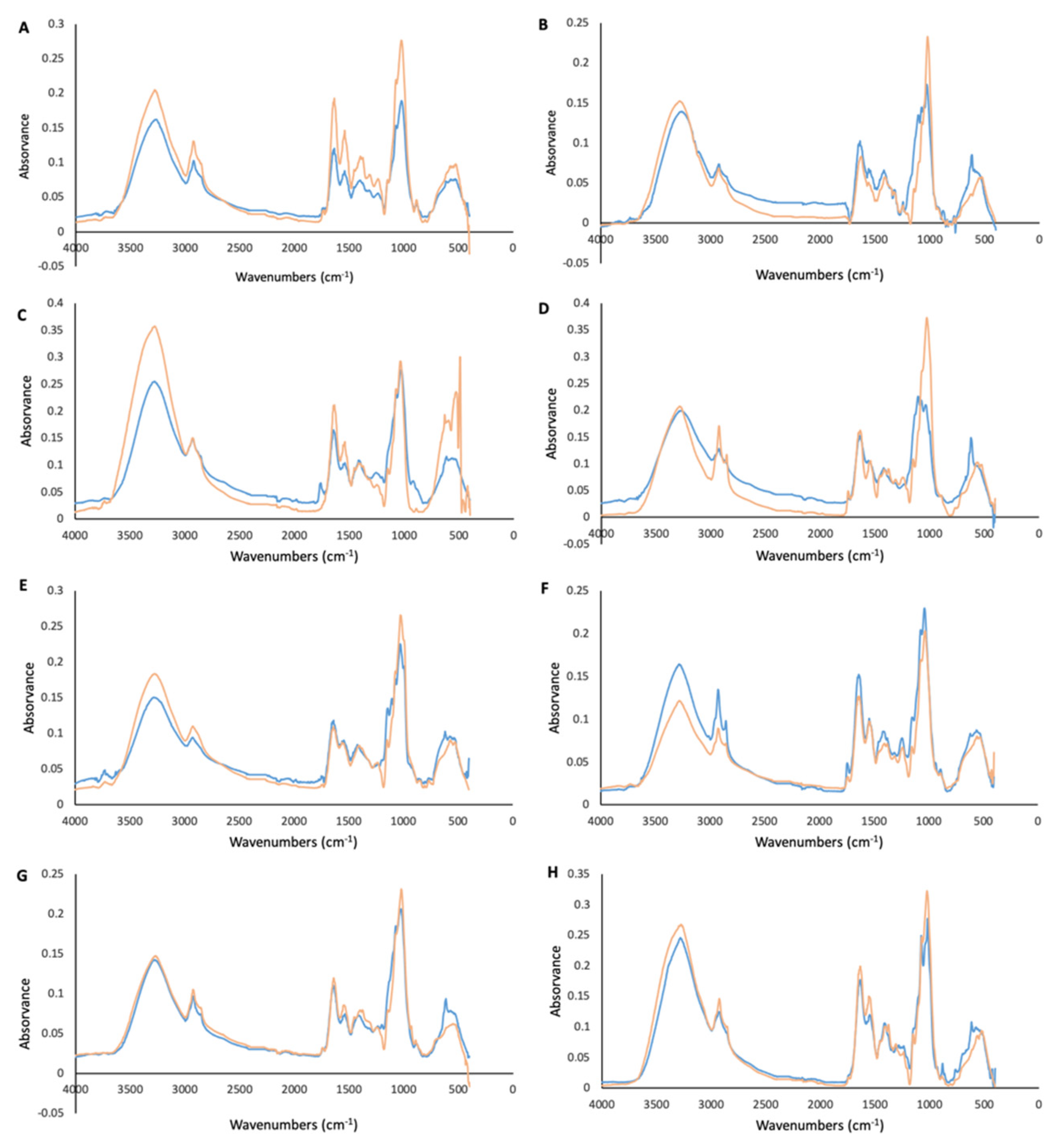
Figure 1. FTIR-ATR spectra of fungal mycelia grown with (blue) and without 3% sea salt (orange). (A) A. affinis (CMG 70); (B) A. protuberus (CMG 71); (C) C. rubrum (MUM 19.39); (D) E. cladophorae (MUM 19.33); (E) Paral. halima (MUM 20.56); (F) Paras. aestuarinum (MUM 19.35); (G) Pen. lusitanum (MUM 18.49); (H) T. aestuarinum (MUM 19.05).
Applsci 11 06008 g001
Figure 2. Antibacterial activity of the mycelium and culture media extracts of A. affinis (CMG 70), E. cladophora (MUM 19.33), Pen. lusitanum (MUM 18.49) and T. aestuarinum (MUM 19.05) using the disk diffusion assay. Data are shown as average (n = 3) ± standard error. Extracts not inducing inhibition halos were not included.
Applsci 11 06008 g002
Figure 3. Anti-fungal activity of the mycelium and culture media extracts of Pen. lusitanum (MUM 18.49) and T. aestuarinum (MUM 19.05) using the disk diffusion assay. Data are shown as average (n = 3) ± standard error. Extracts not inducing inhibition halos were not included.
Applsci 11 06008 g003
Figure 4. Cytotoxicity of the culture media extracts (250 mg/mL) with (blue) and without 3% sea salt (orange) of E. cladophorae (MUM 19.33), Paral. halima (MUM 20.56) and Paras. aestuarinum (MUM 19.35) using Vero cells. Data are shown as average (n = 5) ± standard error. (a) indicates a significantly different t-test, p < 0.05, in relation to the correspondent non-saline sample, and (b) indicates a significantly different t-test, p < 0.05, in relation to the control.
Applsci 11 06008 g004
Figure 5. Antioxidant activity of fungal mycelium with (blue) and without 3% sea salt (orange) using methanolic extracts. (A) Percentage of DPPH scavenging activity; (B) phenolic compounds’ content; (C) ortho-phenols’ content; (D) flavonoids’ content. Data are presented as average (n = 5) ± standard deviation. (a) indicates a significantly different t-test, p < 0.05, in relation to the correspondent non-saline sample, and (b) indicates a significantly different t-test, p < 0.05, in relation to the control.
Applsci 11 06008 g005
Table 1. List of fungal strains used in this study.
SpeciesStrainHost/SubstrateReference
Aspergillus affinisCMG 70Sea water[30]
Aspergillus protuberusCMG 71Sea water[30]
Cladosporium rubrumMUM 19.39/CMG 28Enteromorpha sp.[33]
Emericellopsis cladophoraeMUM 19.33/CMG 25Cladophora sp.[31]
Emericellopsis enteromorphaeMUM 19.34/CMG 26Enteromorpha sp.[31]
Emericellopsis phycophilaMUM 19.32/CMG 15Filamentous green alga[31]
Lulworthia cf. purpureaMUM 20.50/CMG 55Submerged wood[32]
Lulworthia cf. purpureaMUM 20.56/CMG 57Submerged wood[32]
Neocamarosporium aestuarinumMUM 18.55/CMG 4Sea water[26]
Neocamarosporium endophyticumMUM 18.56/CAA 808Halimione portulacoides[26]
Neocamarosporium halinionesMUM 18.54/CAA 807Halimione portulacoides[26]
Neodevriesia aestuarinaMUM 19.27/CMG 9Saline water[25]
Neptunomyces aureusMUM 19.38/CMG 10AGracilaria gracilis[27]
Paraconiothyrium salinumMUM 19.91/CMG 49Sponge[30]
Paralulworthia halimaMUM 20.56/CMG 69Submerged wood[32]
Parasarocladium aestuarinumMUM 19.35/CMG 30Fucus sp.[31]
Parasarocladium alavarienseMUM 19.36/CMG 32Rhodophyta[31]
Parasarocladium fusiformeMUM 19.37/CMG 36Ulva sp.[31]
Penicillium lusitanumMUM 18.49/CMG 8Sea water[28]
Remispora submersaMUM 20.48/CMG 53Submerged wood[32]
Sedecimiella taiwanensisCMG 51Submerged wood[32]
Trichoderma aestuarinumMUM 19.05/CMG 1Saline water[24]
Verrucoconiothyrium ambiguumMUM 18.57/CMG 5Sea water[29]
Zalerion maritimaMUM 20.138/CMG 67Submerged wood[32]
Zalerion pseudomaritimaMUM 20.49/CMG 65Submerged wood[32]
CAA: culture collection of Artur Alves, housed at Department of Biology, University of Aveiro, Aveiro, Portugal; CMG: culture collection of Micael Gonçalves, housed at Department of Biology, University of Aveiro, Aveiro, Portugal; MUM: culture collection hosted at Center for Biological Engineering of University of Minho, Braga, Portugal.
Publisher’s Note: MDPI stays neutral with regard to jurisdictional claims in published maps and institutional affiliations.