Article

Effect of Photosensitization Mediated by Curcumin on Carotenoid and Aflatoxin Content in Different Maize Varieties

1
School of Agriculture and Food Sciences, The University of Queensland, St. Lucia, QLD 4072, Australia
2
Faculdade de Agronomia e Engenharia Florestal, Universidade Eduardo Mondlane, Av. Julius Nyerere 3453, Maputo 1102, Mozambique
3
ARC Industrial Transformation Training Centre for Uniquely Australian Foods, Queensland Alliance for Agriculture and Food Innovation, The University of Queensland, Coopers Plains, QLD 4108, Australia
4
Centre for Nutrition and Food Sciences, Queensland Alliance for Agriculture and Food Innovation, The University of Queensland, St. Lucia, QLD 4072, Australia
*
Author to whom correspondence should be addressed.
Appl. Sci. 2021, 11(13), 5902; https://doi.org/10.3390/app11135902
Submission received: 13 May 2021 / Revised: 18 June 2021 / Accepted: 23 June 2021 / Published: 25 June 2021

Abstract

:
Mycotoxins are naturally occurring toxins produced by certain types of fungi that contaminate food and feed, posing serious health risks to human and livestock. This study evaluated the combination of blue light with curcumin to inactivate Aspergillus flavus spores, its effect on aflatoxin B1 (AFB1) production and maintaining carotenoid content in three maize varieties. The study was first conducted in vitro, and the spore suspensions (104 CFU·mL−1) were treated with four curcumin concentrations (25 and 50 µM in ethanol, 1000 and 1250 µM in propylene glycol) and illuminated at different light doses from 0 to 130.3 J·cm−2. The photoinactivation efficiency was light-dose dependent with the highest photoinactivation of 2.3 log CFU·mL−1 achieved using 1000 µM curcumin at 104.2 J·cm−2. Scanning electron microscopy revealed cell wall deformations as well as less density in photosensitized cells. Photosensitization of maize kernels gave rise to a complete reduction in the viability of A. flavus and therefore inhibition of AFB1 production, while no significant (p > 0.05) effect was observed using either light or curcumin. Moreover, photosensitization did not affect the carotenoids in all the studied maize varieties. The results suggest that photosensitization is a green alternative preservation technique to decontaminate maize kernels and reduce consumer exposure to AFB1 without any effect on carotenoid content.

1. Introduction

Maize (Zea mays L.) is one of the most important cereal crops in the world, coming third after wheat and rice. This crop can be used for several purposes such as animal feed, raw material in the industry and human consumption. Nevertheless, maize is highly susceptible to contamination by fungi. Depending on the environmental and storage conditions, fungi may produce mycotoxins, which are toxic secondary metabolites contaminating foods throughout the value chain [1]. Contamination of maize by mycotoxin is of serious concern, especially in developing countries where maize is a staple food. Owing to their genotoxic and carcinogenic effect, aflatoxins produced by Aspergillus flavus Link and Aspergillus parasiticus Speare are classified as the most poisonous mycotoxins [2]. Normal food processing techniques such as heat treatment and freezing are not sufficient to destroy mycotoxins [3]. Thus, there is a need for research in novel preservation techniques to control fungal growth in food and feed and prevent mycotoxin accumulation, particularly green alternatives to the current conventional preservation techniques.
Photosensitization, a technique that uses a photoactive compound (photosensitizer) to eradicate microorganisms by the generation of reactive oxygen species (ROS), was recently introduced as an alternative approach to decontaminate food products and food contact surfaces [4]. This technique has shown promising inhibitory effects against yeasts, mould, viruses, parasites, Gram-negative and Gram-positive bacteria both in vitro and in vivo [5], mainly depositing on the surface of fruits and fresh-cut vegetables [6].
Curcumin is a predominant polyphenolic compound in turmeric (Curcuma longa L.) rhizomes that has been extensively used for several years as a natural food colorant and dietary supplement [7]. Recent studies have demonstrated the phototoxic activity of curcumin when combined with blue light in some food materials such as maize, fresh date fruit and oysters [8,9]. Curcumin presents a maxima absorption intensity in the region between 414 and 434 nm, depending on the solvent [10]. In the food matrix, curcumin absorption range can overlap with naturally occurring substances, such as yellow carotenoids which are known to absorb light in a broad range from 400 to 500 nm, with three maxima and shoulders [11,12].
Carotenoids are naturally occurring pigments that are abundant in plants. Consumption of carotenoid-rich food is associated with a low incidence of several diseases such as cardiovascular diseases, cancers, age-related macular degeneration, and cataracts [13]. Additionally, there is substantial evidence suggesting a positive effect of high carotenoid concentration on inhibiting aflatoxin biosynthesis [14,15,16]. Nevertheless, carotenoids are light-sensitive and can degrade into other compounds or isomerise into different conformation via photo-isomerisation [17], which could lead to the loss of their biological function. Moreover, carotenoids are singlet oxygen quencher [18,19], and previous studies have shown that β-carotene could prevent photosensitized oxidation [20,21]. Therefore, high concentration of carotenoids in food could potentially affect the efficiency of singlet oxygen generation during photosensitization, resulting in lower microbial photoinactivation.
Information regarding the effect of curcumin-mediated photosensitization on the nutritional profile and sensory properties of food is limited, especially for food grains including maize. Temba et al. [22] evaluated the effect of curcumin-mediated photoinactivation on A. flavus spores in maize kernels and flour, achieving 2 log CFU·g−1 reduction. In another study, curcumin-mediated photosensitization could reduce 73% AFB1 concentration in maize kernels after 10 days of storage at 26 °C [23]. Glueck et al. [4] reported up to 4 log CFU·g−1 reduction of Escherichia coli on mung beans and fenugreek seeds, using 435-nm LED array in combination with SACUR-3, a curcumin derivative. Nevertheless, none of these studies evaluated the effect of photosensitization on the nutritional profile and sensory properties of the studied food material. Therefore, this study aimed to evaluate the effect of photosensitization using LEDs and curcumin on inactivation of A. flavus growth and aflatoxin B1 accumulation on three maize varieties, as well as its influence on maize kernel colour, individual carotenoid compounds and total carotenoid content.

2. Materials and Methods

2.1. Inoculum Preparation

An aflatoxin producing reference strain of A. flavus ATCC 26944 (American Type Culture Collection, In Vitro Technologies Pty. Ltd., Noble Park, VIC, Australia) was grown on malt extract agar (MEA) (Thermo Fisher Scientific Pty. Ltd., Scoresby, VIC, Australia) for seven days at 25 °C. Spore harvest was done by flooding the culture surface with 10 mL of phosphate buffer saline (PBS) solution containing 0.1% Tween 80 (polyoxyethylene (20) sorbitan monooleate), followed by gentle swirling. The suspension was then collected and vortexed for one minute, followed by centrifuging (Eppendorf Centrifuge 5415 D, Hamburg-Eppendorf, Germany) at 10,000 rpm for another minute to sediment the spores. The supernatant was removed, the pellet was resuspended in sterile PBS containing 0.1% Tween 80. Then, 15% (v/v) of glycerol was added to the suspension and the spore solution was stored at −20 °C until further experiments.

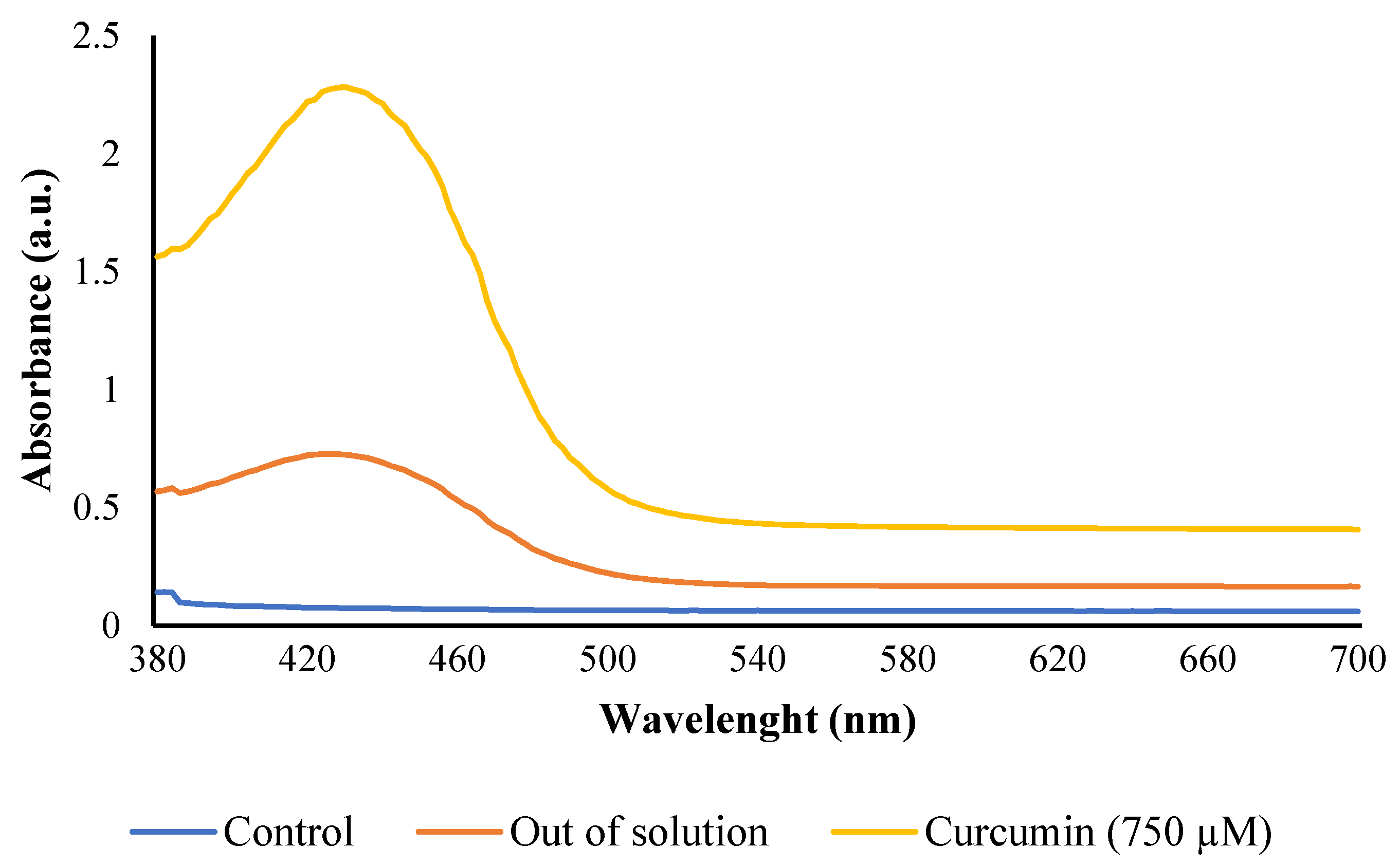
2.2. Photosensitizer Solutions

Curcumin (Sigma Aldrich, St. Louis, MO, USA) stock solution (2000 µM) was prepared using absolute ethanol (≥99.8%; Sigma Aldrich) or 50% (v/v) aqueous propylene glycol (≥99.5%; Sigma Aldrich). The working solutions were freshly prepared prior to the photosensitization by diluting the curcumin stock solutions with distilled water to different concentrations at 25 and 50 µM for ethanol, and 1000 and 1250 µM for propylene glycol. Curcumin concentrations were defined based on previous experiments using a xenon arc lamp, which has shown that when dissolved in ethanol curcumin is effective at concentrations below 100 µM, while when dissolved in propylene glycol it is effective at concentrations above 750 µM [22,24].

2.3. Light Source

High-intensity 430 ± 3 nm LED was used for photosensitization treatment. Temperature and relative humidity inside the light-box were monitored using Inkbird Bluetooth mini smart sensor (IBS-TH1 mini, Inkbird, Regents Park, NSW, Australia) (±0.3 °C temp., ±3% RH). The LED was accommodated in a box impermeable to light, with built in cooling fans to prevent the heat accumulation during illumination. The photosynthetic photon flux density (PPFD) was monitored during illumination using a spectrometer (HR-450, Hipoint, Taichung City, Taiwan). The PPFD (μmol·m−2·s−1) was converted to irradiance (W·cm−2) to calculate the light dose delivered to the sample after a particular time of illumination using Equation (1) [25]:
E = Pt
where E is the light dose (J·cm−2), P is the irradiance (W·cm−2), and t is the illumination time (s).

2.4. Maize Kernel Samples

Three maize varieties including yellow (Pioneer® hybrid P1756), white (Pioneer® hybrid P1477W), and popcorn maize accession were used for in vivo trials. White and yellow maize kernels were bought from Pioneer® Seeds Australia (Brisbane, QLD, Australia), and the popcorn maize accession was obtained from the University of Queensland’s breeding program. All samples were kept at room temperature and used within 3 months.

2.5. In Vitro Trials-Effect of Photosensitization on A. flavus Spores

2.5.1. Study Design

Four concentrations of curcumin solutions (stated in Section 2.2) were combined with eight different light doses, ranging from 0 to 130.3 J/cm2. Four applied treatments included photosensitization (the combination of light and curcumin-P+L+), the negative control (no curcumin, no light-PoLo), light treatment (light, no curcumin-PoL+), and curcumin treatment (curcumin, no light-P+Lo). Ethanol and propylene glycol at concentrations up to 2.5% (v/v) and 25% (v/v), respectively, were also included to test any effects from the solvents on A. flavus growth using an agar well diffusion method [26]. Three sets of the experiments were conducted, and all treatments were conducted in triplicate in each set.

2.5.2. Evaluation of Photosensitization Effect on A. flavus Spores

For photosensitization trials, 1 mL of A. flavus spore suspension (equivalent to 104 CFU·mL−1) was mixed with 1 mL of curcumin solution in a small Petri dish (35 mm × 10 mm), followed by incubation for 10 min at room temperature in dark before illumination. Peptone water (0.1%; w/v) was used instead of photosensitizer in the negative control (PoLo) and light treatment (PoL+). Then, the Petri dish (without lid) was placed under the light source with 10 cm distance and illuminated at 430 ± 3 nm. After treatment, 100 µL aliquots of the photosensitized mixtures were plated onto MEA and incubated at 25 °C for 72 h to determine the Colony-Forming Unit (CFU) using a colony counter (Stuart Scientific, Stone, UK). The fungal survival was calculated by log reduction (LR) of CFU following Equation (2):
LR = Log10 (A) − Log10 (B)
where: A: CFU·mL−1 of the negative control; B: CFU·mL−1 of photosensitized spores.

2.5.3. Determination of Reactive Oxygen Species (ROS) Formation

The generation of singlet oxygen was monitored using 1,3-diphenylisobenzofuran (DPBF; Arcos Organics, Thermo Fisher Scientific Pty. Ltd., Scoresby, VIC, Australia) as a specific reactant [27,28]. Briefly, equal aliquots (100 µL) of photosensitizer and DPBF solutions (50 µg/mL prepared in absolute ethanol) were mixed and illuminated at the investigated light doses. DPBF absorbance was monitored at 410 nm using a micro-plate reader (Infinite® M200, Tecan, Port Melbourne, VIC, Australia). The absorbance of DPBF was monitored every 10 min for a total duration of 30 min, to obtain an estimate about the time-dependent production of ROS after the photosensitizer was subjected to light illumination. The rate of ROS production is proportional to the rate of decrease of DPBF absorbance at 410 nm as a function of illumination time, represented by the slope of the equation.

2.5.4. Morphological Changes on A. flavus Spores

The scanning electron microscopy (SEM) was used to evaluate the morphology of A. flavus spores subjected to different treatment conditions. Sample preparation was carried out according to Smijs et al. [29] with some modifications. Inoculum of both photosensitized spores (~105 CFU·mL−1) and control (untreated spores) were firstly centrifuged at 10,000 rpm for 15 min to remove the media, then washed three times with 0.1 M PBS, followed by centrifugation to remove the unbound photosensitizer. The pellet was mixed with 3% glutaraldehyde (prepared in 0.1 M sodium cacodylate buffer at pH 7.2) and incubated at room temperature for 1 h, then at 4 °C overnight. Next, the samples were dried overnight at room temperature before observation by SEM (Neoscope JCM-5000 SEM, JEOL Ltd., Tokyo, Japan).

2.6. In Vivo Trials-Effect of Photosensitization on Maize Kernels

2.6.1. Inoculation of Kernels

Dry maize kernels were sterilized by autoclaving (121 °C, 15 psi, 15 min). Three replicates of 1.5 g of sterile maize were immersed in 5 mL of sterile PBS, followed by mixing at 1400 rpm for 30 s using a vortex. From the resulting suspension, 100 µL aliquot was plated on MEA to determine CFU of negative control. For positive control, the remaining sterilized maize was inoculated by immersing in 500 mL spore’s suspension (105 CFU·mL−1) for 30 s, the liquid was decanted, and the maize kernels were dried at 25 °C overnight, followed by CFU determination as described for negative control.

2.6.2. Photosensitization Treatment

The inoculated maize kernels were immersed in 1000 µM curcumin (dissolved in propylene glycol) for 5 min under dim light/dark. The kernels were then removed from curcumin solution and illuminated at 430 nm using a light dose of 104.2 J/cm2. In the control treatment, curcumin solution was replaced by PBS. After illumination, the kernels were dried at 25 °C overnight, and ca. 1.5 g of dried kernels were mixed with PBS (5 mL), vortexed at 1400 rpm for 30 s, and plated on MEA to determine the CFU.

2.7. Extraction and Quantification of Aflatoxin B1

After the photosensitization treatment, Petri dishes (3 cm diameter) containing maize samples were placed inside 6 cm-diameter Petri dishes filled with water to provide favourable growth conditions for the mould, and the maize samples were incubated for 14 days at 25 ± 0.5 °C. AFB1 was determined at 7-day intervals. AFB1 content was determined as previously reported by Suylok et al. [30], with some modifications. Briefly, extraction solvent (2 mL) of acetonitrile/Milli-Q water/formic acid at a ratio of 79/20/1 (v/v/v) was added to 0.5 g of maize kernels. The mixture was mechanically shaken for 90 min using an orbital shaker (Ratek OM1, Ratek Instruments Pty Ltd., Boronia, VIC, Australia), and subsequently centrifuged (Eppendorf Centrifuge 5810 R) at 3000 rpm for 2 min at room temperature. The supernatant (1 mL) was diluted with an equal volume of the extraction solvent and filtered through a 0.22 µm hydrophilic PTFE syringe filter into a HPLC vial for AFB1 analysis by a Shimadzu LC-ESI-MS/MS (Shimadzu, Kyoto, Japan).

2.8. Effect of Photosensitization on Maize Kernel Colour

Potential changes in the colour of photosensitized maize kernels were determined using a handheld colorimeter (Konica Minolta CR-400, Thermo Fisher Scientific Pty Ltd., Scoresby, VIC, Australia). The colour parameters L*, a*, and b* were measured in three replicates (four kernels per replication). The total colour change was calculated using Equation (3). Non-treated kernels were used as a reference.
∆E = (∆L*2 + ∆a*2 + ∆b*2)1/2
Chroma and hue angle were calculated using Equations (4) and (5):
Chroma = [(a*)2 + (b*)2]1/2
Hue (°) = tan−1 (b*/a*)
The presence of residual curcumin inside the maize kernel was evaluated using a spectrophotometer (GenesysTM 20, Thermo Fisher Scientific Pty Ltd., Scoresby, VIC, Australia). Five grams of the kernels were milled into a fine powder using a coffee grinder and the powder samples were extracted with 4 mL of 50% propylene glycol (v/v), followed by centrifugation at 10,000 rpm for 5 min. Curcumin solution in propylene glycol (750 µM) was included as a reference, and maize kernels that were not soaked in curcumin were also used as a blank. The absorption intensity of the extract was monitored from 390 nm to 700 nm using a micro-plate reader.

2.9. Carotenoid Extraction and Quantification

The extraction and quantification of carotenoids were conducted following the procedures of Saini and Keum [31] with some modifications. Briefly, maize kernels were first ground using a coffee grinder and then milled with a ball mill (Retsch MM 301, Metrohm, Brendale, QLD, Australia) to obtain a fine powder. Powdered maize samples (0.5 g) were mixed with 6 mL of ethanol containing 0.1% butylated hydroxytoluene (BHT) (w/v) and vortexed for 30 s. Ten millilitres (mL) solvent mixture of DCM and hexane (30:70; v/v) containing 0.1% BHT was added to the mixture to extract carotenoid compounds into the upper layer. NaCl (3 mL, 5%) was added to the mixture to facilitate phase separation, followed by centrifugation (Eppendorf Centrifuge 5810 R) at 3900 rpm for 10 min. Then, the supernatant was collected, and the pellet was re-extracted twice with DCM/hexane mixture with sonication for 10 min at 25 ± 2 °C. All extraction steps were performed under dim light to prevent the isomerization of carotenoids. The supernatants were combined and evaporated until dryness using nitrogen gas. The dried extract was redissolved in methyl tert-butyl ether (MTBE) and methanol (1:1, v/v) solution containing 0.1% BHT and filtered through a 0.22 µm hydrophilic PTFE syringe filter before UPLC-PDA analysis.

2.10. Data Analysis

The results are reported as mean values with standard errors (SE). Data analysis was conducted using Microsoft Excel 2018 and IBM SPSS Statistic version 20.0 software (SPSS Inc., Chicago, IL, USA). Significant differences were evaluated using a one-way ANOVA analysis. Tukey’s HSD test was selected, and tests were conducted under 95% of confidence interval.

3. Results and Discussion

3.1. LED Light Dose and Temperature Profile

Figure 1 depicts a typical temperature profile recorded upon illumination at three different irradiances (10.4, 28.9 and 36.2 mW·cm−2). An increase in the irradiance caused a rise in the temperature inside the light-box. The highest irradiance resulted in an increase of temperature by 12.8 and 19.67 °C after 20 and 30 min of illumination, respectively. On the other hand, the temperature only increased by 2.34 °C when the lowest irradiance was used. An increase in temperature has also been reported in several studies on photosensitization using LED [32]. Although small fans were installed in the light chamber, this did not prevent the temperature increase during photosensitization.

3.2. In Vitro Experiments

3.2.1. Effect of Photosensitization on A. flavus Spores

The efficiency of A. flavus spores’ inactivation by photosensitization was dependent on the light dose applied for the treatment. Whereas, using the same solvent (ethanol or propylene glycol), the tested concentrations of curcumin did not show a significant difference (p > 0.05) in their inactivation efficiency. Photosensitization (P+L+) significantly (p < 0.05) reduced the viability of A. flavus spores using light doses above 34.7 J·cm−2 (Figure 2). Compared to the negative control (PoLo), no significant change was observed on the population of spores that were either treated with the photosensitizer (P+Lo) or light (PoL+) (Figure 2), indicating a synergic effect from the combination of light and curcumin in reducing the multiplication of A. flavus. The efficiency of inactivation was not compared between the solvents as different concentrations of curcumin were used for each solvent.
An increase in the light dose to 86.9 J·cm−2 enhanced photosensitization efficiency. However, further increase of light dose to 130.3 J·cm−2 did not significantly increase (p > 0.05) the efficiency (Figure 2). The presence of curcumin in ethanolic solution resulted in the highest inactivation with 1.9 log CFU·mL−1 reduction at 104.2 J·cm−2 using 25 µM curcumin. However, the same light dose combined with 40-fold higher curcumin concentration (1000 µM prepared in propylene glycol) resulted in 2.3 log CFU·mL−1 reduction. Since curcumin is partially soluble in propylene glycol, higher concentrations of the photosensitizer are required to achieve a significant fungal inactivation as previously reported [24]. Treatments with light doses below 34.7 J·cm−2 did not show any significant effects on the viability of A. flavus spores (p > 0.05) compared to the negative control PoLo (data not shown). Furthermore, the spores did not exhibit any susceptibility to ethanol or propylene glycol solutions, as evaluated by the inhibition zone through well-diffusion assay, suggesting that the solvents used are non-toxic to the A. flavus spores at the concentrations applied in this study.
Although an increase in temperature inside the box was recorded during illumination, the microbial inactivation was achieved as a result of a photodynamic effect of curcumin in the presence of light, but not the heating effect. This was ascertained by submitting the spores to the light alone, which did not show a significant effect on spores’ viability even though a temperature as high as 42.27 °C was achieved for 130.4 J·cm−2 light dose. Aspergillus flavus can grow in temperatures ranging from 12 to 48 °C, although the optimum growth occurs at 37 °C [33]. Moreover, variation of fluid temperature from 15 to 45 °C has been reported to have no significant impact on A. flavus viability [23]. Therefore, the temperatures achieved upon LED illumination were not high enough to inactivate the spores.
The phototoxicity of curcumin has been demonstrated against fungi, bacteria and virus [8,9,24]. Although the exact mechanism of microbial inactivation through photosensitization is still not well understood, it has been proposed as a result of ROS formation when the photosensitizer is illuminated at an appropriate wavelength [34,35]. In turn, ROS react with cellular components such as lipids, proteins and nucleic acids, ultimately causing microbial cell death [5]. Results obtained from the present study suggest that 430-nm LEDs are capable of exciting curcumin to the triplet state, and when returning to the ground state, the photosensitizer combines with oxygen resulting in the generation of ROS.

3.2.2. Reactive Oxygen Species (ROS) Generation

To determine ROS formation, 50 µM and 250 μM curcumin prepared in ethanol and propylene glycol solution, respectively, were selected. The former was the optimum working concentration in vitro, whereas the latter, however, was selected since, although the 1000 μM curcumin in propylene glycol resulted in the highest photoinactivation of A. flavus, the absorbance was above the maximum detection limit of the spectrophotometer used (absorbance over 2.5). Therefore, a lower concentration of curcumin in propylene glycol was used in the experiment.
The ROS generation was indirectly monitored using DPBF, a reactive oxygen species quencher [36]. The absorption intensity of DPBF at 410 nm provides a qualitative estimation of ROS generated during photosensitization, and its decrease indicates the generation of ROS. Light intensity of 36.2 mW·cm−2 led to a higher and more rapid reduction in DPBF absorption intensity, which in turn represents a swift generation of ROS (Figure 3). Additionally, illumination at 36.2 mW·cm−2 (k36.2 = 2.2277) gave rise to 1.4 times faster ROS generation in ethanolic curcumin solution than, illumination at 28.9 mW/cm−2 (k28.9 = 1.622). For curcumin dissolved in propylene glycol, the ROS were generated 1.6 times faster at 36.2 mW·cm−2, than at 28.9 mW·cm−2 (k36.2 = −0.7561 and k28.9 = −0.4841, respectively). The DPBF decay in ethanolic curcumin solution followed second-order kinetics (Figure 3c). However, first-order kinetics was observed when using propylene glycol as the solvent (Figure 3d).
The use of ethanolic curcumin solution ensures a generation of a higher amount of ROS than curcumin in propylene glycol, though higher concentration was used in the latter solvent. These results correlate with the observed photoinactivation efficacy of curcumin in ethanol and propylene glycol against A. flavus spores. To achieve an effective microbial inactivation, higher curcumin concentrations were required using propylene glycol (>500 µM). Whereas curcumin concentrations as low as 25 µM were effective against the spores using ethanol as solvent. No significant change (p > 0.05) was observed in the absorption intensity of DPBF through dark incubation with the photosensitizer.
Curcumin is highly soluble in ethanol but showed partial solubility in propylene glycol. Therefore, it is reasonable to assume that all curcumin dissolved in the ethanolic solution is readily available to absorb light and produce ROS, resulting in a high efficiency at a low concentration. Nevertheless, using propylene glycol, a large amount of curcumin is needed to produce enough quantum yield to photoinactivate the spores, as it was previously reported. Using a propylene glycol solution of curcumin to inactivate several fungi species, including Aspergillus spp., Al-Asmari et al. [24] observed that dye concentrations over 600 µM were required to inactivate roughly 90% of fungal spores. However, concentrations between 100 and 400 µM inactivated only up to 60% of the spores. The same authors evaluated the efficacy of curcumin in propylene glycol to inactivate the naturally occurring microflora in date fruits and reported 1400 µM of curcumin as the most effective concentration against various microorganisms [8]. On the other hand, Temba, Fletcher, Fox, Harvey and Sultanbawa [22] observed that 15–50 µM of curcumin dissolved in ethanol were effective in reducing the colony-forming ability of A. flavus. A reduction of up to 3 log units could be observed. However, a further increase of the curcumin concentration to 100 µM did not cause any additional inactivation of the spores.
DPBF is the most used quencher to estimate the concentration of ROS in media. The decrease of DPBF intensity is due to its oxidation with ROS generated upon illumination of the photosensitizer solution. On the other hand, incubation in the dark showed no reduction in the DPBF absorption intensity since the generation of ROS to interact with the probe does not occur when the photosensitizer is incubated in the absence of light [37,38]. The results show that ROS are efficiently generated upon the irradiation of curcumin with 430-nm LED, and the higher the concentration of curcumin, the more DPBF is consumed which suggests a high generation of ROS.
Differences in ROS yielded by curcumin upon illumination has also been reported in previous studies [39]. A hypothesis explaining the differences in ROS yield of a photosensitizer based on the solvent used was proposed by Geroge and Kishen [28] who studied the ROS generation by Methylene Blue (MB) dissolved in different solvents such as 70% polyethylene glycol (PEG), 70% glycerol, and a mixture of glycerol: ethanol: water (30:20:50, v/v/v). The authors observed that the photobleaching of DPBF was lower in PEG and glycerol than in the formulation containing ethanol. It was suggested that the difference could be due to the type of interaction between the solvent molecules and the excited state of the photosensitizer. Nevertheless, understanding the effect of the solvent on the ROS generation yield by the photosensitizer is a subject that merits a thorough evaluation in future studies.

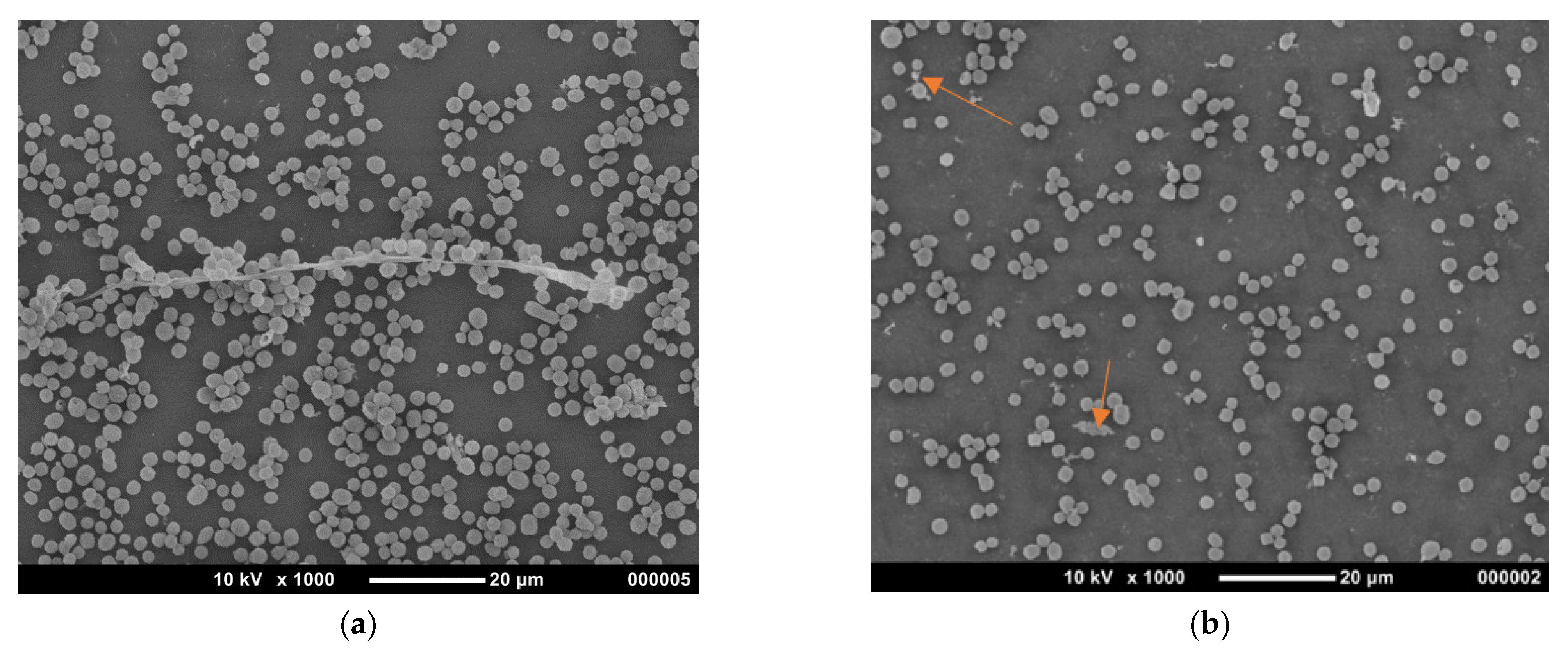
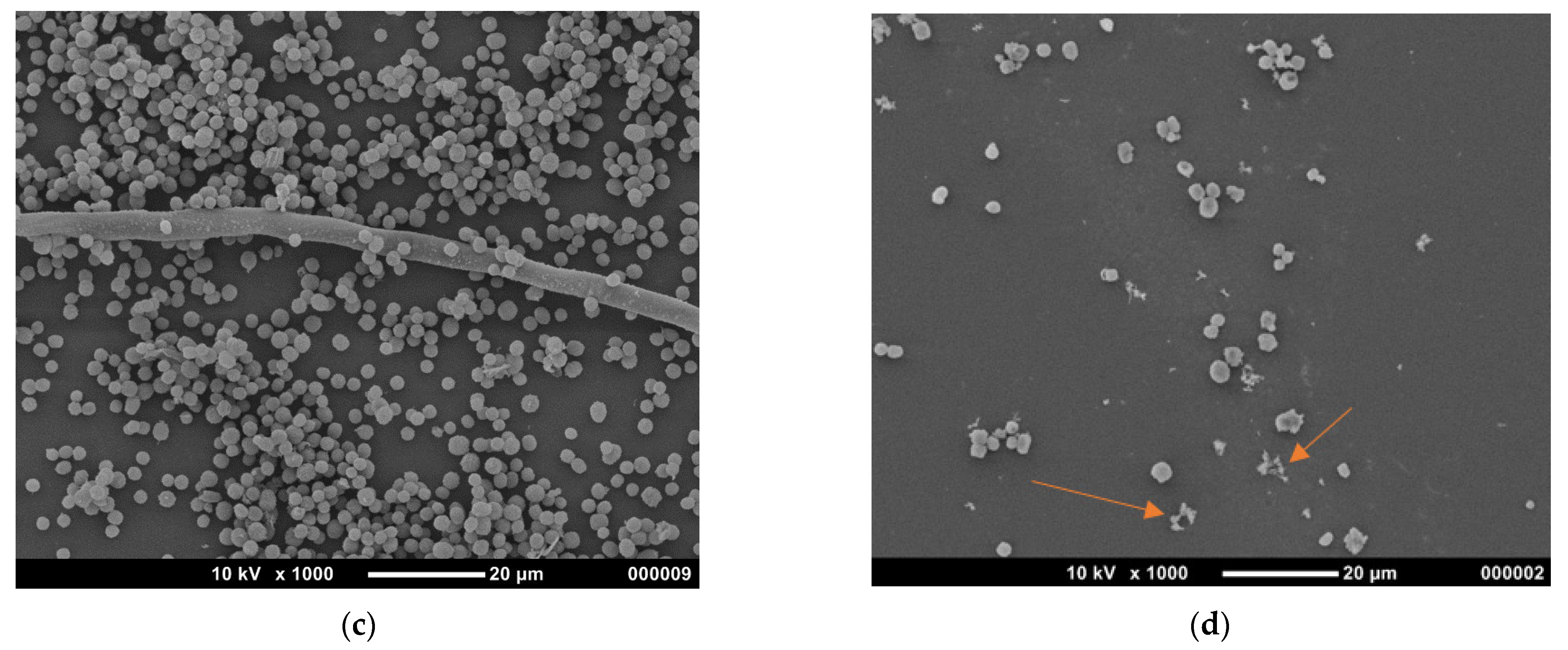
3.2.3. Morphological Changes of A. flavus Spores

Figure 4 presents representative SEM micrographs of A. flavus spores subjected to different treatments. The results show that spores treated with light had lower density and some cell wall deformations. On the other hand, non-treated (control) and curcumin-treated cells showed higher density and hyphal filament with no apparent deformations with the normal globular shape. In contrast, photosensitized spores showed the least cell density with some distortions in cellular shape, where the extent of damage was enough to cause cell death. Some cells in the photosensitization treatment still showed normal shape which substantiates the fact that some of the cells were still viable in vitro. The reduction in cell density due to photosensitization has been previously reported in Candida spp. [40,41].
The cell wall is dynamic and undergoes changes in response to oxidative stress. ROS generated during photosensitization react with macromolecules such as proteins, polysaccharides, lipids and nucleic acids causing molecular changes that can negatively affect cellular physiology and morphology [42]. Deformations of the cell wall may result in intracellular content leakage, causing the cell shrinking and ultimately cell death [43] which has been reported for several microorganisms such as Trichophyton rubrum [29], Escherichia coli [43], and Candida albicans [44].
It was observed that light treatment alone was able to cause deformations of the cell wall in some spores, which is in agreement with previous reports [29]. Nevertheless, Smijs et al. [29] reported that changes caused by light treatment on T. rubrum spores were reversible, and the spores exhibited normal growth after eight days of incubation. This could explain the observed normal growth of light treated spores.

3.3. In Vivo Experiment

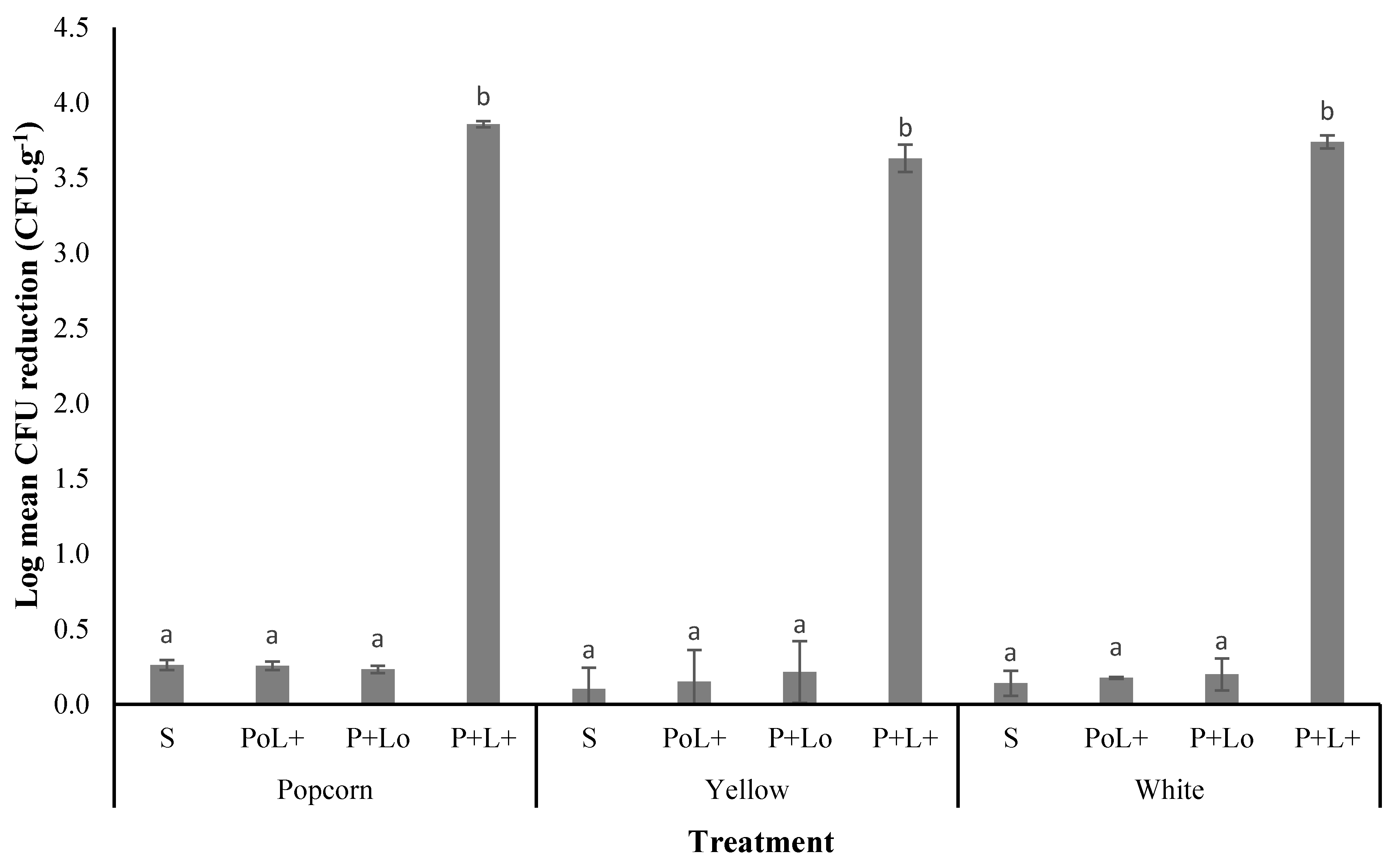
3.3.1. Photodecontamination of A. flavus on Maize

The combination of 1000 µM of curcumin in propylene glycol and 102.4 J·cm−2 was selected according to the in vitro results which had showed to inactivate A. flavus spores (Section 3.2). The average initial spore’s load in popcorn, yellow and white maize were 5.78, 5.44 and 5.61 log CFU·g−1, respectively. The viability of spores was not affected (p > 0.05) by curcumin or light treatment. Moreover, up to 0.39 ± 0.06 log CFU·g−1 reduction was obtained through 25% propylene glycol application on maize kernels (Figure 5), which corresponds to the concentration of the solvent in 1000 µM of curcumin. On the other hand, A. flavus colonies were not observed in the experiments with photosensitized maize kernel extract, suggesting that photosensitization completely removed the spores from the kernel surface, which was in contrast to the in vitro results, where only 2.3 log CFU·mL−1 reduction was achieved. The immersion of kernel in curcumin solution for 5 min before illumination might have washed the spores on its surface, reducing the microbial load before illumination. Consequently, the initial concentration of spores on the maize surface may have been lower than that used in vitro. Complete decontamination of food through curcumin-mediated photosensitization has also been reported in oysters inoculated with Vibrio parahaemolyticus [45].
Contrary to our results, several studies have reported lower photoinactivation efficiency on food surface than in vitro [46,47], which have been attributed to the irregularity and different light-reflecting properties of the food under illumination [48]. In a study reported by Temba et al. [22], nearly 2 log CFU·g−1 reduction was achieved on maize kernel and flour through the combination of curcumin and light, against 3 log CFU·g−1 reduction achieved in vitro. This divergence of the results by Temba et al. [22] and those reported in the present study might be related to the use of different light doses, 60 J·cm−2 (Temba et al.) vs. 102.4 J·cm−2 (present study). In addition, the illumination source used by Temba and co-workers [22] was unidimensional, and though the kernels were constantly turned over during illumination, the light may have been distributed irregularly, reducing the effectiveness of the treatment. Nevertheless, these are mere assumptions, and further studies are necessary to explore the effect of the maize matrix on the efficiency of photosensitization.

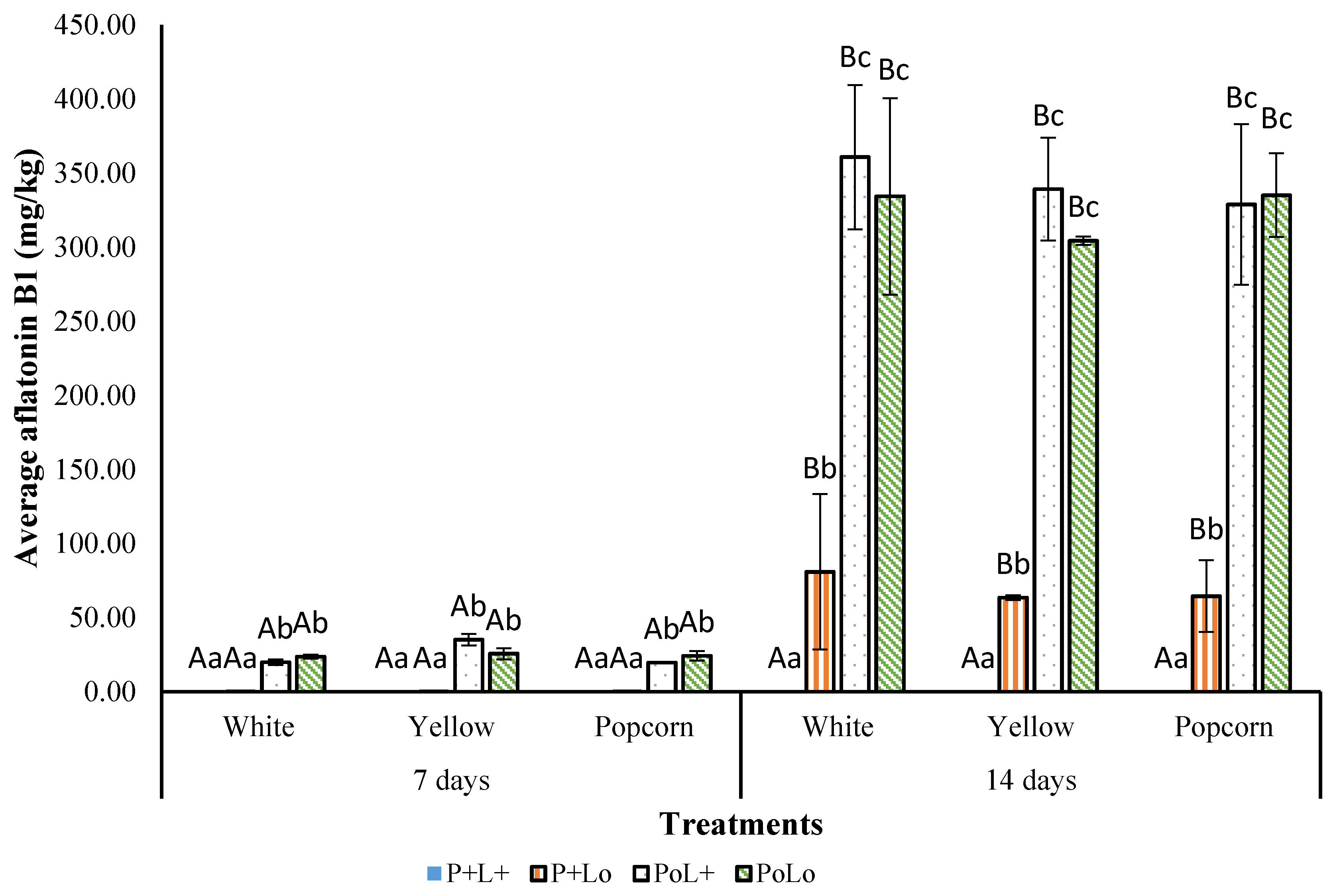
3.3.2. Effect of Photosensitization on AFB1 Production

The initial concentration of AFB1 was below the limit of quantification (LOQ = 0.01 µg·L−1) in all treatments and maize kernels. After seven days of incubation, AFB1 was still undetectable (below the LOQ) both in photosensitized and curcumin-treated maize kernels, whereas the control and the light treated ones showed AFB1 accumulation over incubation time. Furthermore, there were no differences (p > 0.05) in AFB1 accumulation amongst the investigated maize varieties (Figure 6).
From seven to fourteen days of incubation, a significant (p < 0.05) increase in AFB1 concentration was observed in all treatments, except for the photosensitized kernels where AFB1 was still undetectable. The curcumin-treated kernels showed an increase by over 60-fold after 14 days of incubation, reaching more than 63 mg·kg−1 AFB1 in white, yellow and popcorn maize. On the other hand, the light-treated white kernels showed a 18-fold increase in AFB1 concentration after 14 days. Yellow and popcorn varieties showed a significant (p < 0.05) increase to over 335.08 mg·kg−1 AFB1. The control was not different (p > 0.05) from the light treatment in all three varieties.
Findings from this study substantiate the potential of photosensitization as an effective clean green technology to decontaminate food and reduce mycotoxin exposure to consumers, which is in agreement with previous studies [4,49]. Curcumin-mediated photosensitization has been reported to reduce the accumulation of AFB1 by 73% on maize kernel after 10 days of incubation at moist conditions [23]. In another study, the combination of blue light and toluidine blue-O, a synthetic photosensitizer, reduced the formation of alternariol by Alternaria alternata by over 90% for isolates from squash, tomato, pepper and cucumber [49].
Our results on AFB1 formation corroborates with the visual observations of mould growth on maize kernel. In photosensitized kernels, the colony-forming ability of A. flavus was completely suppressed, and no fungal growth was visually observed on the maize samples even after 14 days of incubation at moist conditions. On the other hand, AFB1 was not detectable in curcumin-treated kernels after seven days of incubation, which is consistent with no fungal growth observation in the same period. It could be suggested that propylene glycol in curcumin solution is acting as a coating layer on the maize surface, encapsulating curcumin and releasing it throughout the seven days of incubation, which increases the contact time between curcumin and spores, reducing their viability. The antimicrobial properties of curcumin against different fungal pathogens, including A. flavus, have been documented in previous reports [50,51,52]. Additionally, soaking the kernels in curcumin solution might have partially washed the spores from the kernel surface reducing their concentration and delaying germination during the incubation period.

3.3.3. Effect of Photosensitization on Maize Colour

To determine whether photosensitization had any effect on the colour of maize, the kernels were analysed immediately after treatment. Although photosensitization did not have a significant effect (p > 0.05) on the colour parameters, i.e., lightness index (L*), greenness index (a*) and yellowness index (b*), and chroma, the mean CIELAB Delta-E (∆E) value was over 3.0 (Table 1), implying that the difference between the control and photosensitized maize kernels were perceptive to an untrained eye, as indicated by Pathare et al. [53]. Photosensitization of the kernels faded the curcumin owing to its light susceptibility, especially in neutral-basic conditions [54]. Similarly, a loss in yellow-orange colour of riboflavin was observed after photosensitization of smoked salmon, where riboflavin was used as a photosensitizer [55].
The obtained extracts from photosensitized maize kernels showed a peak in the region of curcumin maxima absorption, i.e., 410–430 nm (Figure 7). The existence of a peak indicates that 5-min soaking of maize resulted in curcumin absorption to the inner parts of the kernel, which was unlikely photobleached upon illumination. The blue LED light (400–470 nm) is a superficial decontamination technique, with a depth penetration below 1 mm [56]. Therefore, the LED light did not penetrate the maize matrix, and only the curcumin existing on the kernel surface was degraded or excited for ROS production.

3.4. Effect of Blue Light on Carotenoid Content in Maize Kernels

The illumination of maize kernels was conducted at 130.3 J·cm−2 (i.e., 36.2 mW·cm−2, 30 min), the most extreme studied conditions for A. flavus photoinactivation. The white variety presented the lowest content of total carotenoids among the three varieties studied, with an initial content of 1.74 µg·g−1 dry weight (DW), followed by the yellow variety with the medium content of 12.90 µg·g−1 DW. The initial content of carotenoids in popcorn maize was about 19-fold and 2.6-fold of that of white and yellow maize, respectively, with a total content of 33.44 µg·g−1 DW (Table 2). Zeaxanthin was the predominant carotenoid compound in all varieties, and its highest concentration was found in the popcorn accession of up to 24.04 µg·g−1 DW.
The use of LED light did not affect (p > 0.05) the content of carotenoids in the investigated maize varieties. Light exposure might cause carotenoid degradation resulting in the formation of carotenoid radical cations [57]. However, the wavelengths mostly responsible for carotenoid degradation are within the UV range [58]. The light used in this study was within the visible range, and the average illuminance for a light dose of 130.3 J·cm−2 was 2843 ± 16.15 lux. Therefore, although effective on reducing the viability of A. flavus spores, the irradiance and illumination time used were not able to cause a significant degradation or isomerization of carotenoids in the three maize varieties.
Similarly, previous studies in fruits have also reported a non-significant effect of LED illumination on the nutritional content, although an increase in some compounds was observed. Kim et al. [59] evaluated the effect of LED illumination on antioxidant capacity and the content of beta-carotene, flavonoids, lycopene, and ascorbic acid in fresh-cut mango. It was shown that illumination did not affect the antioxidant capacity of fruits and the content of β-carotene, ascorbic acid, and lycopene, while the content of flavonoids increased 1.9 times. More than 95% of β-carotene could be retained in tomato juice by using a constant illuminance of 2476 lux for 24 h at 4 ⁰C [60]. These results substantiate the potential of using LEDs in photosensitization to reduce the microbial load on the maize surface without affecting carotenoids.
Previous studies have reported a reduction in mycotoxin accumulation in the presence of carotenoids [15,16]. However, no significant differences were observed in AFB1 accumulation between white maize having a relatively low carotenoid content of 1.74 µg·g−1 DW and popcorn maize with a relatively high content of 33.44 µg·g−1 DW. In addition, the inhibition of mycotoxin accumulation by carotenoids is strain-dependent [14,15]. Furthermore, it has also been reported that the reduction in mycotoxin production depends on the individual carotenoids present, particularly β-carotene and β-cryptoxanthin have a significant effect on the aflatoxin synthesis [16]. In the present study, only the popcorn maize had detectable levels of β-cryptoxanthin and β-carotene, while zeaxanthin was the predominant carotenoid in all varieties. Furthermore, technical challenges such as developing the most efficient procedure to disperse the curcumin solution, need to be addressed, when upscaling this new green preservation technique to farm/industry-scale.

4. Conclusions

Curcumin in combination with 430 nm LEDs reduced the viability of A. flavus spores in vitro and on the surface of maize kernels, resulting in an inhibition of the synthesis of aflatoxin B1. At a given curcumin concentration, the inactivation efficiency was light-dose dependent. The highest fungal inactivation was achieved using 1000 µM curcumin and a light dose of 81.5 J/cm2. The use of photosensitization did not cause significant changes in the carotenoid content as well as the colour attributes of the maize kernels. The use of novel and green technologies such as photosensitization could be a valuable alternative to tackle the challenges posed by aflatoxins and their toxicity for humans. The reduction of fungal contamination on maize kernels could present a positive step towards the mitigation of the current level of postharvest losses in maize, and also enhance the nutritional value and quality of this important crop. Optimization of the curcumin concentration to minimize its effect on the sensory quality of food products is crucial and will benefit not only the food industry but also consumers.

Author Contributions

Conceptualization, Y.S., M.S.D. and R.N.; methodology, Y.S., M.S.D., R.N., A.D.T.P., H.T.H., M.C., M.E.N. and T.J.O.; formal analysis, R.N.; investigation, R.N. and M.S.D.; data curation, and writing—original draft preparation, R.N.; writing—review and editing, Y.S., M.S.D., A.D.T.P., H.T.H., M.E.N. and R.N.; funding acquisition and supervision, Y.S. All authors have read and agreed to the published version of the manuscript.

Funding

This research was supported by the Australian Government through an Australia Awards Scholarship (AUSAID-DFAT) for Master of Science studies, the University of Queensland, Horticulture Innovation Australia Ltd. (Naturally Nutritious project HN15001) and the Department of Agriculture and Fisheries.

Institutional Review Board Statement

Not applicable.

Informed Consent Statement

Not applicable.

Data Availability Statement

The data presented in this study are available on request from the corresponding author.

Acknowledgments

The authors would like to thank Andrew Cusack and Margaret Currie for their technical assistance.

Conflicts of Interest

The authors declare no conflict of interest.

References

  1. Fink-Gremmels, J.; van der Merwe, D. Mycotoxins in the food chain: Contamination of foods of animal origin. In Chemical Hazards in Foods of Animal Origin; Smulders, F.J.M., Rietjens, I.M.C.M., Rose, M., Eds.; Wageningen Academic Publishers: Wageningen, The Netherlands, 2019; Volume 7, pp. 1190–1198. [Google Scholar]
  2. Sarma, U.P.; Bhetaria, P.J.; Devi, P.; Varma, A. Aflatoxins: Implications on health. Indian J. Clin. Biochem. 2017, 32, 124–133. [Google Scholar] [CrossRef]
  3. Al-Jaal, B.; Salama, S.; Al-Qasmi, N.; Jaganjac, M. Mycotoxin contamination of food and feed in the Gulf Cooperation Council countries and its detection. Toxicon 2019, 171, 43–50. [Google Scholar] [CrossRef] [PubMed]
  4. Glueck, M.; Schamberger, B.; Eckl, P.; Plaetzer, K. New horizons in microbiological food safety: Photodynamic Decontamination based on a curcumin derivative. Photochem. Photobiol. Sci. 2017, 16, 1784–1791. [Google Scholar] [CrossRef] [PubMed] [Green Version]
  5. Hu, J.; Lin, S.; Tan, B.; Hamzah, S.; Lin, Y.; Kong, Z.; Zhang, Y.; Zheng, B.; Zeng, S. Photodynamic inactivation of Burkholderia cepacia by curcumin in combination with EDTA. Food Res. Int. 2018, 111, 265–271. [Google Scholar] [CrossRef] [PubMed]
  6. Ghate, V.S.; Zhou, W.; Yuk, H.G. Perspectives and trends in the application of photodynamic inactivation for microbiological food safety. Compr. Rev. Food Sci. Food Saf. 2019, 18, 402–424. [Google Scholar] [CrossRef] [PubMed] [Green Version]
  7. de Oliveira, E.; Tosati, J.; Tikekar, R.; Monteiro, A.; Nitin, N. Antimicrobial activity of curcumin in combination with light against Escherichia coli O157: H7 and Listeria innocua: Applications for fresh produce sanitation. Postharvest Biol. Technol. 2018, 137, 86–94. [Google Scholar] [CrossRef]
  8. Al-Asmari, F.; Mereddy, R.; Sultanbawa, Y. The effect of photosensitization mediated by curcumin on storage life of fresh date (Phoenix dactylifera L.) fruit. Food Control. 2018, 93, 305–309. [Google Scholar] [CrossRef] [Green Version]
  9. Liu, F.; Li, Z.; Cao, B.; Wu, J.; Wang, Y.; Xue, Y.; Xu, J.; Xue, C.; Tang, Q.J. The effect of a novel photodynamic activation method mediated by curcumin on oyster shelf life and quality. Food Res. Int. 2016, 87, 204–210. [Google Scholar] [CrossRef]
  10. Priyadarsini, K.I. Photophysics, photochemistry and photobiology of curcumin: Studies from organic solutions, bio-mimetics and living cells. J. Photochem. Photobiol. C 2009, 10, 81–95. [Google Scholar] [CrossRef]
  11. Thrane, J.-E.; Kyle, M.; Striebel, M.; Haande, S.; Grung, M.; Rohrlack, T.; Andersen, T. Spectrophotometric analysis of pigments: A critical assessment of a high-throughput method for analysis of algal pigment mixtures by spectral deconvolution. PLoS ONE 2015, 10, e0137645. [Google Scholar] [CrossRef] [Green Version]
  12. Lichtenthaler, H.K.; Buschmann, C. Chlorophylls and carotenoids: Measurement and characterization by UV-VIS spectroscopy. Curr. Protoc. Food Anal. Chem. 2001, 1, F4. 3.1–F4. 3.8. [Google Scholar] [CrossRef]
  13. Meléndez-Martínez, A.J.; Stinco, C.M.; Mapelli-Brahm, P. Skin carotenoids in public health and nutricosmetics: The emerging roles and applications of the UV radiation-absorbing colourless carotenoids phytoene and phytofluene. Nutrients 2019, 11, 1093. [Google Scholar] [CrossRef] [PubMed] [Green Version]
  14. Norton, R.A. Effect of carotenoids on aflatoxin B1 synthesis by Aspergillus flavus. Phytopathology 1997, 87, 814–821. [Google Scholar] [CrossRef] [PubMed] [Green Version]
  15. Wicklow, D.; Norton, R.; McAlpin, C. β-Carotene inhibition of aflatoxin biosynthesis among Aspergillus flavus genotypes from Illinois corn. Mycoscience 1998, 39, 167–172. [Google Scholar] [CrossRef]
  16. Suwarno, W.; Hannok, P.; Palacios-Rojas, N.; Windham, G.; Crossa, J.; Pixley, K. Provitamin A carotenoids in grain reduce aflatoxin contamination of maize while combating vitamin A deficiency. Front. Plant Sci. 2019, 10, 30. [Google Scholar] [CrossRef] [Green Version]
  17. Aman, R.; Schieber, A.; Carle, R. Effects of heating and illumination on trans− cis isomerization and degradation of β-carotene and lutein in isolated spinach chloroplasts. J. Agric. Food Chem. 2005, 53, 9512–9518. [Google Scholar] [CrossRef] [PubMed]
  18. Ramel, F.; Birtic, S.; Cuiné, S.; Triantaphylidès, C.; Ravanat, J.-L.; Havaux, M. Chemical quenching of singlet oxygen by carotenoids in plants. Plant Physiol. 2012, 158, 1267–1278. [Google Scholar] [CrossRef] [Green Version]
  19. Jung, M.; Min, D. Effects of quenching mechanisms of carotenoids on the photosensitized oxidation of soybean oil. J. Am. Oil Chem. Soc. 1991, 68, 653–658. [Google Scholar] [CrossRef]
  20. Dutta, D.; Chaudhuri, U.; Chakraborty, R. Structure, health benefits, antioxidant property and processing and storage of carotenoids. Afr. J. Biotechnol. 2005, 4, 1510–1520. [Google Scholar] [CrossRef]
  21. Oshima, S.; Ojima, F.; Sakamoto, H.; Ishiguro, Y.; Terao, J. Inhibitory effect of β-carotene and astaxanthin on photosensitized oxidation of phospholipid bilayers. J. Nutr. Sci. Vitaminol. 1993, 39, 607–615. [Google Scholar] [CrossRef] [Green Version]
  22. Temba, B.A.; Fletcher, M.T.; Fox, G.P.; Harvey, J.J.W.; Sultanbawa, Y. Inactivation of Aspergillus flavus spores by curcumin-mediated photosensitization. Food Control 2016, 59, 708–713. [Google Scholar] [CrossRef]
  23. Temba, B.A.; Fletcher, M.T.; Fox, G.P.; Harvey, J.; Okoth, S.A.; Sultanbawa, Y. Curcumin-based photosensitization inactivates Aspergillus flavus and reduces aflatoxin B1 in maize kernels. Food Microbiol. 2019, 82, 82–88. [Google Scholar] [CrossRef] [PubMed]
  24. Al-Asmari, F.; Mereddy, R.; Sultanbawa, Y. A novel photosensitization treatment for the inactivation of fungal spores and cells mediated by curcumin. J. Photochem. Photobiol. B 2017, 173, 301–306. [Google Scholar] [CrossRef] [PubMed]
  25. Maclean, M.; MacGregor, S.J.; Anderson, J.G.; Woolsey, G. Inactivation of bacterial pathogens following exposure to light from a 405-nanometer light-emitting diode array. Appl. Environ. Microbiol. 2009, 75, 1932–1937. [Google Scholar] [CrossRef] [PubMed] [Green Version]
  26. Gonelimali, F.D.; Lin, J.; Miao, W.; Xuan, J.; Charles, F.; Chen, M.; Hatab, S.R. Antimicrobial properties and mechanism of action of some plant extracts against food pathogens and spoilage microorganisms. Front. Microbiol. 2018, 9, 1639. [Google Scholar] [CrossRef]
  27. Rout, B.; Liu, C.-H.; Wu, W.-C. Photosensitizer in lipid nanoparticle: A nano-scaled approach to antibacterial function. Sci. Rep. 2017, 7, 1–14. [Google Scholar] [CrossRef] [Green Version]
  28. George, S.; Kishen, A. Photophysical, photochemical, and photobiological characterization of methylene blue formulations for light-activated root canal disinfection. J. Biomed. Opt. 2007, 12, 034029. [Google Scholar] [CrossRef]
  29. Smijs, T.; Mulder, A.; Pavel, S.; Onderwater, J.; Koerten, H.; Bouwstra, J. Morphological changes of the dermatophyte Trichophyton rubrum after photodynamic treatment: A scanning electron microscopy study. Med. Mycol. 2008, 46, 315–325. [Google Scholar] [CrossRef] [Green Version]
  30. Sulyok, M.; Berthiller, F.; Krska, R.; Schuhmacher, R. Development and validation of a liquid chromatography/tandem mass spectrometric method for the determination of 39 mycotoxins in wheat and maize. Rapid Commun. Mass Spectrom. 2006, 20, 2649–2659. [Google Scholar] [CrossRef]
  31. Saini, R.K.; Keum, Y.-S. Carotenoid extraction methods: A review of recent developments. Food Chem. 2018, 240, 90–103. [Google Scholar] [CrossRef]
  32. Ghate, V.; Kumar, A.; Kim, M.-J.; Bang, W.-S.; Zhou, W.; Yuk, H.-G. Effect of 460 nm light emitting diode illumination on survival of Salmonella spp. on fresh-cut pineapples at different irradiances and temperatures. J. Food Eng. 2017, 196, 130–138. [Google Scholar] [CrossRef]
  33. Yu, J.; Fedorova, N.D.; Montalbano, B.G.; Bhatnagar, D.; Cleveland, T.E.; Bennett, J.W.; Nierman, W.C. Tight control of mycotoxin biosynthesis gene expression in Aspergillus flavus by temperature as revealed by RNA-Seq. FEMS Microbiol. Lett. 2011, 322, 145–149. [Google Scholar] [CrossRef] [Green Version]
  34. Ghorbani, J.; Rahban, D.; Aghamiri, S.; Teymouri, A.; Bahador, A. Photosensitizers in antibacterial photodynamic therapy: An overview. Laser Ther. 2018, 27, 293–302. [Google Scholar] [CrossRef] [Green Version]
  35. Seidi Damyeh, M.; Mereddy, R.; Netzel, M.E.; Sultanbawa, Y. An insight into curcumin-based photosensitization as a promising and green food preservation technology. Compr. Rev. Food Sci. Food Saf. 2020, 19, 1727–1759. [Google Scholar] [CrossRef] [PubMed]
  36. Nsubuga, A.; Mandl, G.A.; Capobianco, J.A. Investigating the reactive oxygen species production of Rose Bengal and Merocyanine 540-loaded radioluminescent nanoparticles. Nanoscale Adv. 2021, 3, 1375–1381. [Google Scholar] [CrossRef]
  37. Diaz-Diestra, D.; Beltran-Huarac, J.; Bracho-Rincon, D.P.; González-Feliciano, J.A.; González, C.I.; Weiner, B.R.; Morell, G. Biocompatible ZnS: Mn quantum dots for reactive oxygen generation and detection in aqueous media. J. Nanopart. Res. 2015, 17, 1–14. [Google Scholar] [CrossRef] [PubMed] [Green Version]
  38. Hong, S.H.; Koo, M.-A.; Lee, M.H.; Seon, G.M.; Park, Y.J.; Jeong, H.; Kim, D.; Park, J.-C. An effective method to generate controllable levels of ROS for the enhancement of HUVEC proliferation using a chlorin e6-immobilized PET film as a photo-functional biomaterial. Regen. Biomater. 2021, 8, rbab005. [Google Scholar] [CrossRef] [PubMed]
  39. Chignell, C.; Bilskj, P.; Reszka, K.; Motten, A.; Sik, R.; Dahl, T. Spectral and photochemical properties of curcumin. Photochem. Photobiol. 1994, 59, 295–302. [Google Scholar] [CrossRef]
  40. Ahmed, H.; Mekawey, A.; Morsy, M. Cellular structural changes in two candida species caused by photodynamic therapy. Photodiagn. Photodyn. Ther. 2016, 4, 39–54. [Google Scholar]
  41. Sakita, K.M.; Conrado, P.C.; Faria, D.R.; Arita, G.S.; Capoci, I.R.; Rodrigues-Vendramini, F.A.; Pieralisi, N.; Cesar, G.B.; Gonçalves, R.S.; Caetano, W. Copolymeric micelles as efficient inert nanocarrier for hypericin in the photodynamic inactivation of Candida species. Future Microbiol. 2019, 14, 519–531. [Google Scholar] [CrossRef] [PubMed]
  42. Baptista, A.; Sabino, C.; Núñez, S.; Miyakawa, W.; Martin, A.; Ribeiro, M. Photodynamic damage predominates on different targets depending on cell growth phase of Candida albicans. J. Photochem. Photobiol. B 2017, 177, 76–84. [Google Scholar] [CrossRef]
  43. Pudziuvyte, B.; Bakiene, E.; Bonnett, R.; Shatunov, P.; Magaraggia, M.; Jori, G. Alterations of Escherichia coli envelope as a consequence of photosensitization with tetrakis (N-ethylpyridinium-4-yl) porphyrin tetratosylate. Photochem. Photobiol. Sci. 2011, 10, 1046–1055. [Google Scholar] [CrossRef]
  44. Dong, S.; Shi, H.; Zhang, X.; Chen, X.; Cao, D.; Mao, C.; Gao, X.; Wang, L. Difunctional bacteriophage conjugated with photosensitizers for Candida albicans-targeting photodynamic inactivation. Int. J. Nanomed. 2018, 13, 2199. [Google Scholar] [CrossRef] [PubMed] [Green Version]
  45. Wu, J.; Mou, H.; Xue, C.; Leung, A.; Xu, C.; Tang, Q.-J. Photodynamic effect of curcumin on Vibrio parahaemolyticus. Photodiagn. Photodyn. Ther. 2016, 15, 34–39. [Google Scholar] [CrossRef] [PubMed]
  46. Luksiene, Z.; Buchovec, I.; Paskeviciute, E. Inactivation of food pathogen Bacillus cereus by photosensitization in vitro and on the surface of packaging material. J. Appl. Microbiol. 2009, 107, 2037–2046. [Google Scholar] [CrossRef] [PubMed]
  47. Tortik, N.; Spaeth, A.; Plaetzer, K. Photodynamic decontamination of foodstuff from Staphylococcus aureus based on novel formulations of curcumin. Photochem. Photobiol. Sci. 2014, 13, 1402–1409. [Google Scholar] [CrossRef]
  48. Luksiene, Z.; Paskeviciute, E. Novel approach to the microbial decontamination of strawberries: Chlorophyllin-based photosensitization. J. Appl. Microbiol. 2011, 110, 1274–1283. [Google Scholar] [CrossRef]
  49. Alananbeh, K.; BouQellah, N.; Al Harbi, M.; Ouf, S. The Efficacy of Photosensitizers on Mycelium Growth, Mycotoxin and Enzyme Activity of Alternaria spp. Jordan J. Biol. Sci. 2018, 11, 43–50. [Google Scholar]
  50. Shlar, I.; Droby, S.; Choudhary, R.; Rodov, V. The mode of antimicrobial action of curcumin depends on the delivery system: Monolithic nanoparticles vs. supramolecular inclusion complex. RSC Adv. 2017, 7, 42559–42569. [Google Scholar] [CrossRef] [Green Version]
  51. Gitika, A.; Mishra, R.; Panda, S.K.; Mishra, C.; Sahoo, P.R. Evaluation of antifungal activity of Curcumin against Aspergillus flavus. Int. J. Curr. Microbiol. Appl. Sci. 2019, 8, 2323–2329. [Google Scholar] [CrossRef]
  52. Pasrija, R.; Kundu, D. Interaction of curcumin with azoles and polyenes against aspergillus infections. Res. J. Life Sci. Bioinform. Pharm. Chem. Sci. 2018, 4, 271–279. [Google Scholar]
  53. Pathare, P.B.; Opara, U.L.; Al-Said, F.A.-J. Colour measurement and analysis in fresh and processed foods: A review. Food Bioproc. Technol. 2013, 6, 36–60. [Google Scholar] [CrossRef]
  54. Mondal, S.; Ghosh, S.; Moulik, S.P. Stability of curcumin in different solvent and solution media: UV–visible and steady-state fluorescence spectral study. J. Photochem. Photobiol. B 2016, 158, 212–218. [Google Scholar] [CrossRef] [PubMed]
  55. Josewin, S.; Ghate, V.; Kim, M.-J.; Yuk, H.-G. Antibacterial effect of 460 nm light-emitting diode in combination with riboflavin against Listeria monocytogenes on smoked salmon. Food Control 2018, 84, 354–361. [Google Scholar] [CrossRef]
  56. Ash, C.; Dubec, M.; Donne, K.; Bashford, T. Effect of wavelength and beam width on penetration in light-tissue interaction using computational methods. Lasers Med. Sci. 2017, 32, 1909–1918. [Google Scholar] [CrossRef]
  57. da Silva, M.; Paese, K.; Guterres, S.; Pohlmann, A.; Rutz, J.; Cantillano, R.; Nora, L.; de Oliveira Rios, A. Thermal and ultraviolet–visible light stability kinetics of co-nanoencapsulated carotenoids. Food Bioprod. Process. 2017, 105, 86–94. [Google Scholar] [CrossRef] [Green Version]
  58. Limbo, S.; Torri, L.; Piergiovanni, L. Light-induced changes in an aqueous β-carotene system stored under halogen and fluorescent lamps, affected by two oxygen partial pressures. J. Agric. Food Chem. 2007, 55, 5238–5245. [Google Scholar] [CrossRef]
  59. Kim, M.-J.; Bang, W.S.; Yuk, H.-G. 405 ± 5 nm light emitting diode illumination causes photodynamic inactivation of Salmonella spp. on fresh-cut papaya without deterioration. Food Microbiol. 2017, 62, 124–132. [Google Scholar] [CrossRef] [PubMed]
  60. Pesek, C.; Warthesen, J. Photodegradation of carotenoids in a vegetable juice system. J. Food Sci. 1987, 52, 744–746. [Google Scholar] [CrossRef]
Figure 1. Temperature profile inside the light chamber upon blue LED illumination at three different irradiances (10.4, 28.9 and 36.2 mW·cm−2).
Applsci 11 05902 g001
Figure 2. Effect of curcumin-based photosensitization at different curcumin concentrations, prepared in ethanol and propylene glycol, and light doses on reduction of Aspergillus flavus spores in vitro. Data are means ± SE (n = 9); means with different lowercase letters indicate significant (p < 0.05) differences at different light doses and means with uppercase letters indicate significant (p < 0.05) differences at the same light dose.
Applsci 11 05902 g002
Figure 3. (a) Decay of DPBF absorbance upon illumination (EtOH-CUR = curcumin in ethanol); (b) Decay of DPBF absorbance upon illumination (CUR-PG = curcumin in 50% propylene glycol); (c) Kinetic decay of DPBF absorbance in ethanolic curcumin solution (second-order kinetics); (d) Kinetic decay of DPBF absorbance in curcumin-propylene glycol solution (first-order kinetics). Data are means ± SE (n = 3).
Applsci 11 05902 g003aApplsci 11 05902 g003b
Figure 4. Scanning electron micrographs of Aspergillus flavus spores after different treatments (×1000; scale bar = 20 µm). (a) PoLo = control; (b) PoL+ = light; (c) P+Lo = photosensitizer; (d) P+L+ = photosensitization. Arrows in the SEM micrographs indicate cell wall deformation.
Applsci 11 05902 g004aApplsci 11 05902 g004b
Figure 5. Photodecontamination of Aspergillus flavus spores on three maize varieties (popcorn, yellow and white). S = solvent (propylene glycol); PoL+ = light; P+Lo = photosensitizer; P+L+ = photosensitization. Data are means ± SE (n = 3); means with different letters in the same column are significantly different (p < 0.05).
Applsci 11 05902 g005
Figure 6. Concentration of aflatoxin B1 on maize kernel after photosensitization (P+L+), photosensitizer (P+L+), light (PoL+) treatments and negative control (PoLo). Data are means ± SE (n = 3); means followed by different letters (lowercase letter for treatments in the same variety and uppercase letter for different periods in the same variety) are significantly different (p < 0.05).
Applsci 11 05902 g006
Figure 7. Maize extract absorption spectra after soaking in curcumin solution and illumination with blue light at 104.2 J·cm−2. Control maize kernels dipped in the solvent before illumination (propylene glycol); out of solution maize kernels dipped in 1000 µM of curcumin in propylene glycol before illumination.
Applsci 11 05902 g007
Table 1. Colour parameters in maize kernels submitted to different treatments.
TreatmentL*a*b*C∆E
PoLo white81.9 ± 0.5 a−0.7 ± 0.3 a29.4 ± 0.9 b29.4 ± 0.9 b-
P+Lo white81.7 ± 0.8 a−6.5 ± 0.4 b50.6 ± 1.8 a51.0 ± 1.7 a21.9 a
P+L+ white79.8 ± 0.5 a0.1 ± 0.4 a26.8 ± 0.7 b26.8 ± 0.7 b3.5 b
PoLo yellow74.8 ± 0.5 a6.8 ± 0.7 ab39.8 ± 2.5 b40.3 ± 2.5 b-
P+Lo yellow77.6 ± 1.3 a4.4 ± 0.9 b54.1 ± 1.9 a54.2 ± 1.9 a14.7 a
P+L+ yellow77.2 ± 0.5 a7.5 ± 0.6 a37.6 ± 2.4 b38.3 ± 2.3 b3.3 b
PoLo popcorn74.1 ± 0.6 a9.8 ± 0.9 a28.5 ± 1.3 a30.2 ± 1.4 b-
P+Lo popcorn77.4 ± 0.9 a5.8 ± 1.0 b34.9 ± 1.8 a35.3 ± 1.7 a8.7 a
P+L+ popcorn76.0 ± 0.7 a8.7 ± 0.7 a25.4 ± 1.9 a26.9 ± 1.8 b3.8 b
PoLo = control; P+Lo = curcumin treatment; P+L+ = photosensitization; C = chroma; data are means ± SE (n = 3); data with different letters in the same column and maize variety are significantly different (p < 0.05). The comparison of means was performed within the same variety for different treatments, as the aim was to assess if different treatments result (or not) in colour changes on the surface of the maize kernel.
Table 2. Carotenoid content in the studied maize varieties (white, yellow and popcorn) before and after illumination using LED blue light at 130.3 J·cm2.
TreatmentIndividual
Carotenoids
Carotenoid Content (µg·g−1 DW)
WhiteYellowPopcorn
ControlLutein0.22 ± 0.02 a5.69 ± 0.19 b5.67 ± 0.07 b
Zeaxanthin1.54 ± 0.01 a7.20 ± 0.14 b24.04 ± 0.26 c
β-cryptoxanthinn.d.n.d.1.86 ± 0.07 b
β-carotenen.d.n.d.1.84 ± 0.09 b
Total1.74 ± 0.00 Aa12.90 ± 0.32 Ab33.44 ± 0.48 Ac
IlluminationLutein0.17 ± 0.01 a6.07 ± 0.48 b5.76 ± 0.24 b
Zeaxanthin1.45 ± 0.02 a7.29 ± 0.42 b24.67 ± 0.70 c
β-cryptoxanthinn.d.n.d.2.29 ± 0.07 a
β-carotenen.d.n.d.3.30 ± 0.076 b
Total1.62 ± 0.01 Aa13.36 ± 0.91 Ab36.01 ± 1.05 Ac
Data are means ± SE (n = 3); data with different letters in the same row are significantly different (p < 0.05); n.d.: not detectable.
Publisher’s Note: MDPI stays neutral with regard to jurisdictional claims in published maps and institutional affiliations.

