1. Introduction
Complete dentures have been manufactured using a conventional workflow, which requires multiple visits of the patients to a dental clinic and complex laboratory procedures [
1,
2]. With recent advances in computer-aided design and computer-aided manufacturing (CAD-CAM) technology, the digital design and fabrication of complete dentures have become an active area of research. The advantages of this workflow include the elimination of cumbersome denture tooth arrangement and subsequent resin injection or packing and reproducibility of the dentures, which can be attributed to the use of a digital workup; the designed data file can be stored in a backup device and reused later to manufacture the same denture [
3].
Moreover, the performance of intraoral scanners has also dramatically increased; several studies and clinical reports have described the feasibility of intraoral scanners for obtaining digital scans in edentulous arches [
4,
5]. This direct digitization reduces chair time for impression making and enables real-time verification of the accuracy of the obtained impression through a monitor connected to the intraoral scanner. Nevertheless, a barrier for complete digitization is the inability to establish a three-dimensional (3D) interarch relationship using intraoral scanners. When using intraoral scanners, the interarch relationship is established by registering the bilateral buccal surfaces of the teeth; however, in cases of complete edentulism, this approach is not applicable since there are no teeth present to be registered [
6].
The transformation of a negative imprint into a positive object is a fundamental concept of dentistry; for example, a definitive cast is fabricated by pouring dental stone into a negative dental impression. Commercial reverse engineering software programs have a similar function in a digital world; they enable free transformation of the 3D scan data between positive and negative shapes. This paper presents a digital workflow to register the interarch relationship by using a reversing and superimposing (RAS) technique.
2. Materials and Methods
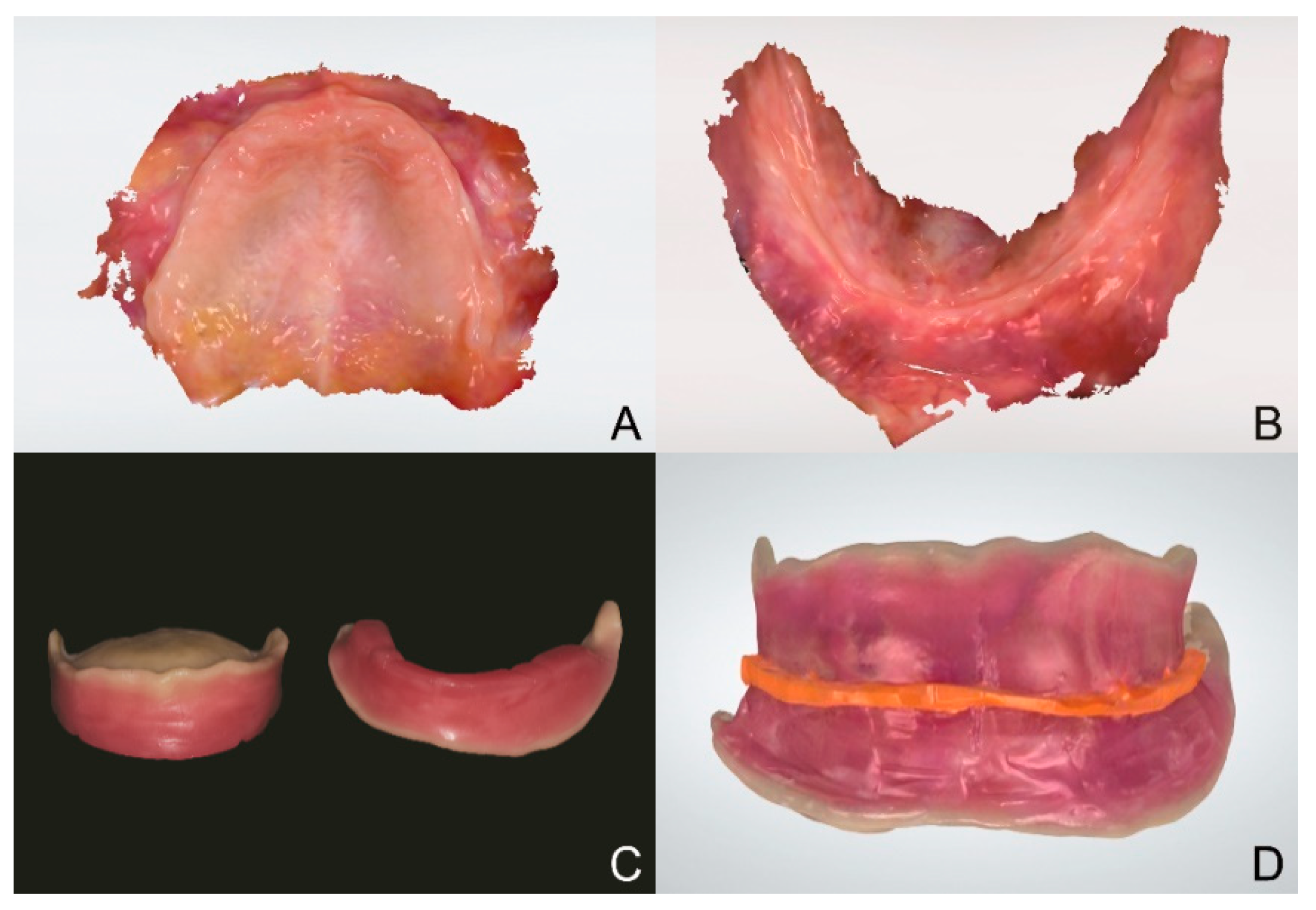
The digital intraoral scan data of both the maxillary and mandibular arches were obtained using an intraoral scanner (TRIOS 3; 3Shape A/S, Copenhagen, Denmark) (
Figure 1A,B). The scanned data were saved in the standard tessellation language (STL) file format. Modeless trial denture bases were designed using a CAD software program (Meshmixer; Autodesk, San Rafael, CA, USA) and additively manufactured using a 3D printing material (NextDent Try-In TI2; 3D Systems, NextDent B.V., Soesterberg, The Netherlands) and a 3D printer (NextDent 5100; 3D Systems, NextDent B.V., Soesterberg, The Netherlands). A wax rim was subsequently placed on each of the trial denture bases (
Figure 1C). The trial denture base and wax rim assemblies were placed in the patient’s mouth. They could be adjusted, if needed, to achieve better esthetic and functional outcomes. The interarch relationship was recorded using an occlusal registration material (O-Bite; DMG, Hamburg, Germany). The obtained, united assembly was scanned using the same intraoral scanner (TRIOS 3; 3Shape A/S, Copenhagen, Denmark) (
Figure 1D).
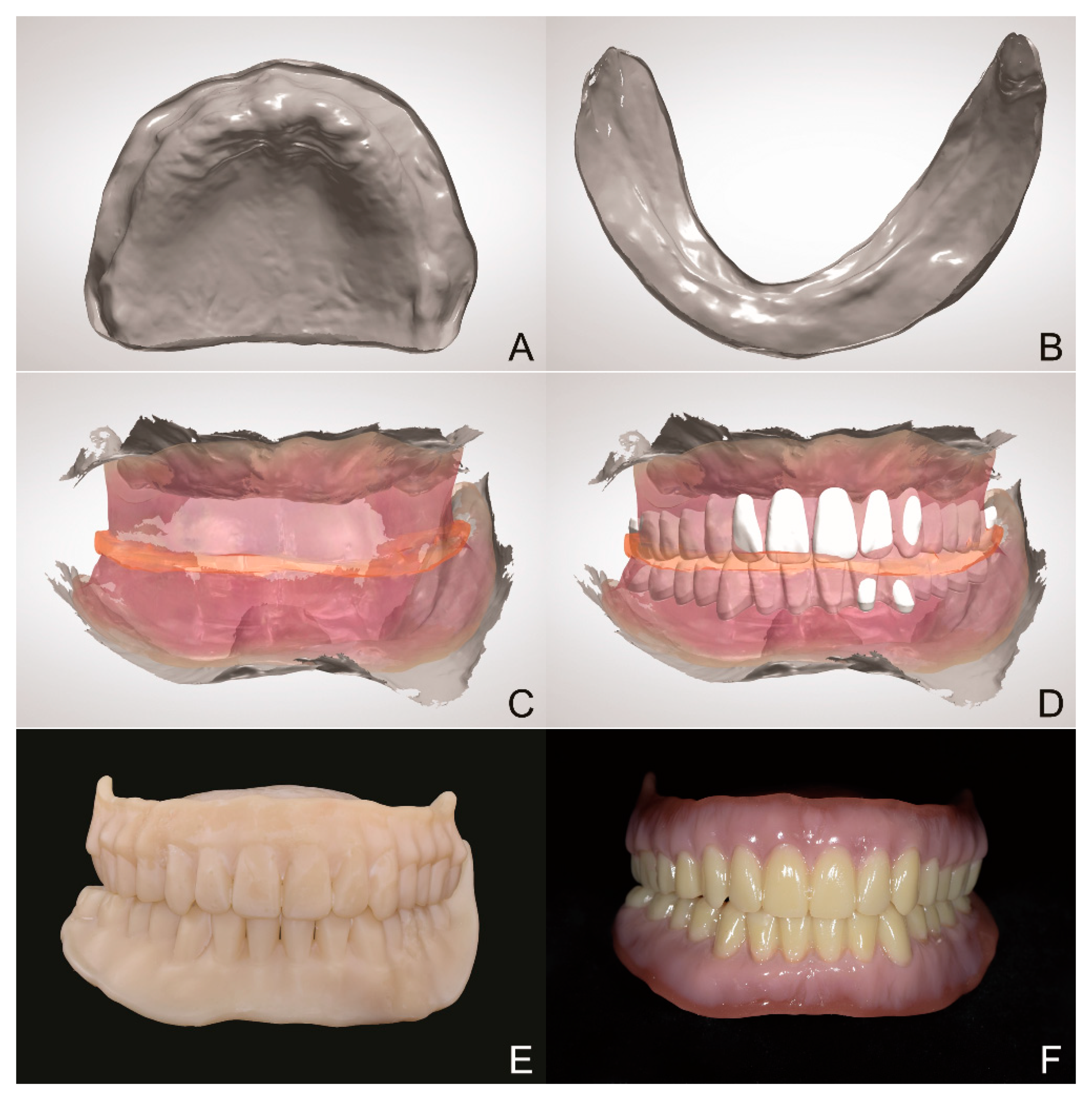
The STL file of the scan data of the united assembly with a registered interarch relationship was imported into a metrology/inspection software program (Geomagic Control X version 2018.0.1, 3D Systems, Rock Hill, SC, USA) to reverse the maxillary and mandibular portions individually. Subsequently, each portion was saved as an individual STL file (
Figure 2A,B). The maxillary STL file obtained using the intraoral scanner was superimposed onto the reversed maxillary STL file. The same procedure was performed with the mandibular STL file, thus, establishing the interarch relationship for the original STL files (those obtained using the intraoral scanner) (
Figure 2C).
The artificial teeth were arranged using the transformed maxillary and mandibular STL files as well as the STL file (or the color format file (.dcm)) of the united assembly. The occlusal plane of the assembly could be viewed by adjusting the translucency of the .dcm file (
Figure 2D). Try-in dentures were additively manufactured with a 3D printing material (NextDent Try-In TI0; 3D Systems, NextDent B.V., Soesterberg, The Netherlands) and the 3D printer (NextDent 5100; 3D Systems, NextDent B.V., Soesterberg, The Netherlands) to evaluate esthetics and function (
Figure 2E). Thereafter, the gingival and tooth sections were additively manufactured individually and characterized (
Figure 2F). The digitally fabricated complete dentures were placed in the patient’s mouth and evaluated for esthetics and function.
3. Discussion
Since the 1980s, fabrication of dental prostheses using CAD-CAM technology has become faster and more convenient than conventional methods, with the development of various scanning devices, software, and manufacturing machines [
3]. Recently, an increasing number of studies have focused on reporting the methodologies and clinical cases for the fabrication of complete dentures using intraoral scanners and CAD-CAM systems. Presently, the edentulous area can be scanned using intraoral scanners without the aid of artificial indicators. Nevertheless, it was impossible to obtain the interarch relationship solely using intraoral scanners owing to the absence of distinct characteristics on the buccal side of the edentulous gingivae. By using this technique, a 4-step completely digital workflow for complete denture fabrication in edentulous patients was established: direct intraoral scanning, interarch relationship registration by the RAS technique, computer-aided designing, and additive manufacturing.
Russo et al. proposed a digital occlusal registration method, which overlaps the data obtained from facial scans with those obtained from occlusal rim scans using conventional methods [
7]. However, errors may exist when using this method as the occlusal scan data are superimposed on the narrow, exposed surfaces when the patient smiles. The method proposed in the present study, i.e., the RAS technique, may be another effective option since it uses intaglio surfaces of each trial denture base for superimposition.
The present workflow is advantageous for the following reasons: First, a fully digital workflow is established, eliminating the need for making impressions that may cause discomfort to elderly edentulous patients, and the number of patient visits is reduced. Second, the technique can be expanded to the fabrication of new complete dentures using old complete dentures that exhibit decreased vertical dimension of occlusion (VDO). Old complete dentures can be relined to re-establish proper VDO, and the RAS technique can be applied by reversing the old complete dentures. This eliminates the step of remaking impressions of edentulous arches.
4. Conclusions
The present article describes a completely digital workflow for complete denture fabrication in edentulous patients. The workflow begins with data acquisition using an intraoral scanner, followed by data processing using reverse engineering and CAD software programs, and ends with additive manufacturing.
Author Contributions
Conceptualization, K.C.O.; methodology, H.S.M. and K.C.O.; software, J.J.; validation, K.C.O.; formal analysis, K.C.O.; investigation, H.-J.L. and J.J.; resources, K.C.O.; data curation, K.C.O.; writing—original draft preparation, H.-J.L. and J.J.; writing—review and editing, H.S.M. and K.C.O.; visualization, H.-J.L. and J.J.; supervision, H.S.M. and K.C.O.; project administration, K.C.O.; funding acquisition, K.C.O. All authors have read and agreed to the published version of the manuscript.
Funding
This research was funded by the National Research Foundation of Korea (NRF) grant funded by the Korea government (MSIT) (No. 2018R1C1B6005989).
Institutional Review Board Statement
Not applicable.
Informed Consent Statement
Not applicable.
Data Availability Statement
Not applicable.
Conflicts of Interest
The authors declare no conflict of interest.
References
- Jacob, R.F. The traditional therapeutic paradigm: Complete denture therapy. J. Prosthet. Dent. 1998, 79, 6–13. [Google Scholar] [CrossRef]
- Walmsley, A.; Pinsent, R.; Laird, W. Complete dentures: 1. Treatment planning and preliminary care. Dent. Update 1991, 18, 255, 257–260. [Google Scholar] [PubMed]
- Beuer, F.; Schweiger, J.; Edelhoff, D. Digital dentistry: An overview of recent developments for CAD/CAM generated restorations. Br. Dent. J. 2008, 204, 505–511. [Google Scholar] [CrossRef] [PubMed]
- Goodacre, B.J.; Goodacre, C.J. Using Intraoral Scanning to Fabricate Complete Dentures: First Experiences. Int. J. Prosthodont. 2018, 31, 166–170. [Google Scholar] [CrossRef] [PubMed]
- Russo, L.L.; Salamini, A. Single-arch digital removable complete denture: A workflow that starts from the intraoral scan. J. Prosthet. Dent. 2018, 120, 20–24. [Google Scholar] [CrossRef] [PubMed]
- Fang, Y.; Fang, J.H.; Jeong, S.M.; Choi, B.H. A technique for digital impression and bite registration for a single edentulous arch. J. Prosthodont. 2019, 28, e519–e523. [Google Scholar] [CrossRef] [PubMed]
- Russo, L.L.; Salamini, A.; Troiano, G.; Guida, L. Digital dentures: A protocol based on intraoral scans. J. Prosthet. Dent. 2021, 125, 597–602. [Google Scholar] [CrossRef] [PubMed]
| Publisher’s Note: MDPI stays neutral with regard to jurisdictional claims in published maps and institutional affiliations. |
© 2021 by the authors. Licensee MDPI, Basel, Switzerland. This article is an open access article distributed under the terms and conditions of the Creative Commons Attribution (CC BY) license (https://creativecommons.org/licenses/by/4.0/).


