Article

Postextraction Ridge Width Alterations Following Socket Seal Surgery—A Retrospective Study

1
Private Practice, Tel Aviv 6439414, Israel
2
Department of Oral Biology, The Goldschleger School of Dental Medicine, Sackler Faculty of Medicine, Tel Aviv University, Tel Aviv 6997801, Israel
3
Department of Endodontology, The Goldschleger School of Dental Medicine, Sackler Faculty of Medicine, Tel Aviv University, Tel Aviv 6997801, Israel
4
Department of Periodontology, Faculty of Dental Medicine, Hebrew University of Jerusalem, Hadassah Medical Center, Jerusalem 90000, Israel
5
Department of Oral Rehabilitation, The Goldschleger School of Dental Medicine, Sackler Faculty of Medicine, Tel Aviv University, Tel Aviv 6997801, Israel
6
Department of Periodontology and Oral Implantology, The Goldschleger School of Dental Medicine, Sackler Faculty of Medicine, Tel Aviv University, Tel Aviv 6997801, Israel
*
Author to whom correspondence should be addressed.
Appl. Sci. 2021, 11(1), 324; https://doi.org/10.3390/app11010324
Submission received: 5 December 2020 / Revised: 27 December 2020 / Accepted: 28 December 2020 / Published: 31 December 2020
(This article belongs to the Special Issue Predictable Restorative Dentistry)

Abstract

:
Background and objectives: Preservation of alveolar ridge contour following tooth extraction is important to allow for restoration with prosthetics and implants. Socket seal surgery was introduced more than two decades ago for preservation of the form, volume and bone quality of the postextraction ridge. The aim of this retrospective study was to assess the changes in alveolar ridge contour following socket seal surgery and to evaluate the survival of the soft tissue grafts sealing the extraction site. Materials and Methods: Digital images of the extraction sites treated with the socket seal surgery were obtained immediately after extraction and after a healing time of 6 months to measure the changes in the alveolar width. In addition, the sites were photographed 1 week postsurgery to evaluate the vitality of the soft tissue grafts. Results: The overall mean loss of buccal width following socket seal surgery was 5.3% (SD = 13.4%). The mean change in width at the incisor area (1.5% ± 13.1) was significantly lesser than those in the canine area (−17.0% ± 2.1, p < 0.01) or premolar area (−10.5% ± 11.2, p < 0.01). One-week post-surgery 69% of the grafts were vital, and 31% were partially vital. Complete necrosis of the grafts did not occur. Conclusions: Our results suggest that socket seal surgery has a beneficial effect on alveolar dimensional changes 6 months following tooth extraction and that the teeth that benefit mostly from socket seal surgery are incisors.

1. Introduction

Tooth extraction is usually followed by significant dimensional changes of the alveolar ridge contour [1,2,3,4,5]. Resorption of the alveolar bone following tooth extraction and consequent invagination of the mucosa affect mainly the buccal aspect of the residual alveolar ridge [6,7]. The mean resorption in the height and width of the alveolar ridge may reach approximately 50% and occurs predominantly within the first 3–6 months [3,8].
Postextraction ridge deformation, especially in the anterior region of the maxilla, may cause functional, phonetic, and aesthetic problems. It is therefore of great importance to minimize negative soft and hard tissue changes after tooth removal to preserve the natural tissue contours, for which diverse soft and hard tissue procedures have been developed [9,10].
Different treatment modalities have been proposed to prevent postextraction ridge deformation and reduce the need for additional ridge augmentation procedures. These include Guided Bone Regeneration (GBR) techniques [11,12], grafting bone-substitute materials [13,14], overbuilding of the facial bony wall [15], immediate implant placement [16], the use of platelet concentrate [17], partial extraction protocols [18] and forced orthodontic extraction [19]. Most of the methods proposed to prevent postextraction ridge deformation were primarily aimed at preserving bone tissue [20]. However, techniques to achieve primary soft tissue wound closure have also been developed, mainly related to immediate implant placement, such as coronally repositioned flap [21] and rotated palatal flap [22].
It is still not clear which of the proposed materials for socket preservation are the most suitable: autogenous [23], allogenic [24,25] or alloplastic [26]. Moreover, the relative importance of bone grafts, membrane or soft tissue graft in the success of socket preservation still needs to be determined [27]. Other important factors influencing the choice of a grafting material include cost-effectiveness, as well as the influence of the biomaterial on the final implant outcomes [28].
Socket seal surgery (SSS), a simplified, minimally invasive regenerative approach, was introduced more than two decades ago as a tool for optimizing the preservation of the hard and soft tissue components of the alveolar ridge immediately following tooth extraction [29]. This approach involves the placement of free gingival graft to cover the augmented alveolar socket and may be suitable for immediate or delayed implant placement, as well as for the achievement of an esthetically pleasing pontic site [29,30].
It was proposed that stabilizing the soft tissue architecture with SSS minimizes soft tissue shrinkage [31]. Studies in dogs demonstrated that placing a deproteinized bovine bone material (DBBM) into the extraction socket and sealing the socket with a free gingival graft was beneficial in limiting the volumetric shrinkage [20,32]. However, due to the limited blood supply there is a risk of incomplete wound healing and necrosis of the soft tissue graft [33].
A retrospective study in humans demonstrated that SSS is effective in minimizing the loss of soft tissue and alveolar bone after tooth removal [34], however, another study showed that the extraction itself induced alveolar bone resorption regardless of whether the socket was treated with a free gingival graft [35].
In general, there are limited available data in the dental literature on residual alveolar ridge volume changes after tooth extraction using the SSS technique. Therefore, the aim of this study was to examine if the negative postextraction alveolar ridge contour changes in the buccal aspect could be reduced or prevented by using SSS and to evaluate the survival of the free autogenous soft tissue grafts sealing the extraction sites.

2. Materials and Methods

Although this is a retrospective study, its design was approved by the Institutional Ethics Committee of Tel Aviv University (IEC No. 0001834-1). Data from SSS cases treated by one clinician (CL) between January 2010 and December 2019 were retrospectively collected and analyzed.

2.1. Case Selection

Patients were included in the study if they had a comprehensive medical and dental history. Sockets prepared either for a delayed implant placement or as a pontic site were included in the study if they met the following inclusion criteria: The bony walls of the extraction socket were intact, the socket was filled with particulate deproteinized bovine bone mineral (DBBM) and covered with a soft tissue graft. All included cases contained records required for analysis.
Exclusion criteria consisted of smoking and a history of diabetes, chemotherapy, radiation therapy or bisphosphonate treatment. Patients suffering from periodontal disease were also excluded from this study.

2.2. Photographic Documentation

Images of the SSS sites were obtained by standardized photography with a DSLR camera. The camera was positioned perpendicular to the occlusal/incisal surface of the teeth adjacent to the extraction site. All the pictures were taken by the same operator (CL). Only photographs with near ideal ortho-radial positioning were included in the study. This explains the relatively limited number of cases (36) included in the study, reflecting the scarcity of perfectly photographed clinical cases. The pictures were taken immediately after extraction (T0), 1week post-surgery (T1) and after 6 months of healing (T6).

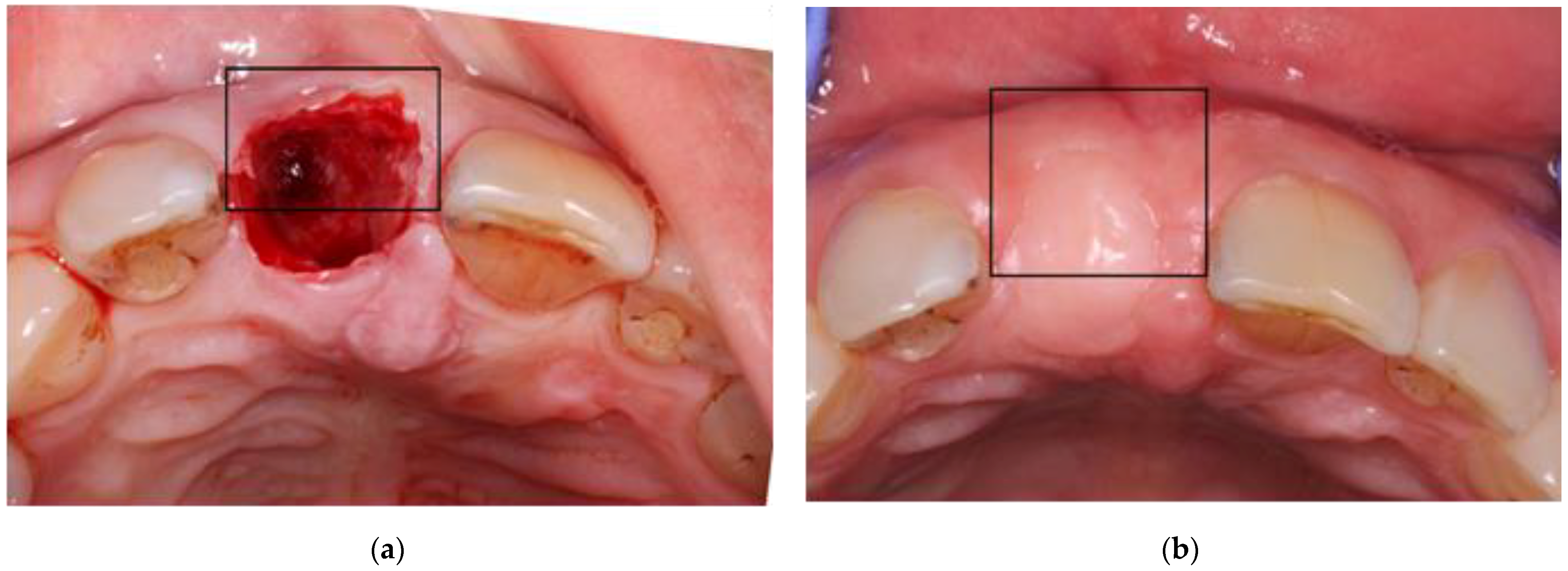
2.3. Surgical Technique

The failing tooth was removed from the socket as gently as possible, avoiding pressure on the bony walls or surrounding soft tissue, without flap elevation. The bony socket walls were curetted thoroughly to remove all granulation tissue. The gingival socket walls were de-epithelialized with a round bur in their inner aspect and the particulate deproteinized bovine bone mineral (Bio-Oss, Geistlich Biomaterials, Wolhusen, Switzerland) was placed into the socket until it reached the coronal bony margins of the socket. A free gingival graft of 2- to 3-mm thickness was harvested from masticatory mucosa palatal to the second premolar, and a hemostatic collagen agent was placed at the donor site and attached to it with mattress sutures. The soft tissue graft was placed on top of the particulate bone graft and was stabilized by simple interrupted sutures to the extraction socket soft tissue margins. At 1-week post-surgery, sutures were removed and the sites were gently cleaned with gauze soaked in saline solution.

2.4. Measurements of the Alveolar Ridge Contour Changes

T0 images (taken immediately after extraction) and T6 images (taken after 6 months of healing) were imported into the PowerPoint software (Microsoft corp., Redmond, Washington, WA, USA). A rectangle, the width of which corresponds to the edentulous gap, was superimposed on the T0 photograph, which was rotated until the rectangle was perpendicular to a line connecting the mid bucco-lingual point on the 2 neighboring teeth (Figure 1a). The upper border of the rectangle was moved to correspond to the most buccal edge of the extraction site. The vertical/horizontal ratio of the rectangle was registered so that the buccal dimension of the extraction site was defined as percent of the gap between the neighboring teeth. The rectangle was copied onto the T6 photograph, which was adapted to it while keeping the vertical-to-horizontal proportions intact and the same procedure was repeated (Figure 1b). With this technique, the dimensions of the 2 photographs were equalized before measurements were made. Notably, the measurements reflected only the outer contour of the alveolar socket, without separating the bony and soft tissue components. The buccal width of the site of the T6 photograph was multiplied by 100 and divided by that of the T0 photograph. Consequently, a value greater than 100 means that the site gained buccal width at 6 months and a value less than 100 means that its buccal dimensions diminished.

2.5. Soft Tissue Contour Analysis at 6 Months

The status of the soft tissues of the treated site was assessed by visual inspection of the T6 photographs. Any concavities in the crestal area were considered as invaginations [36].

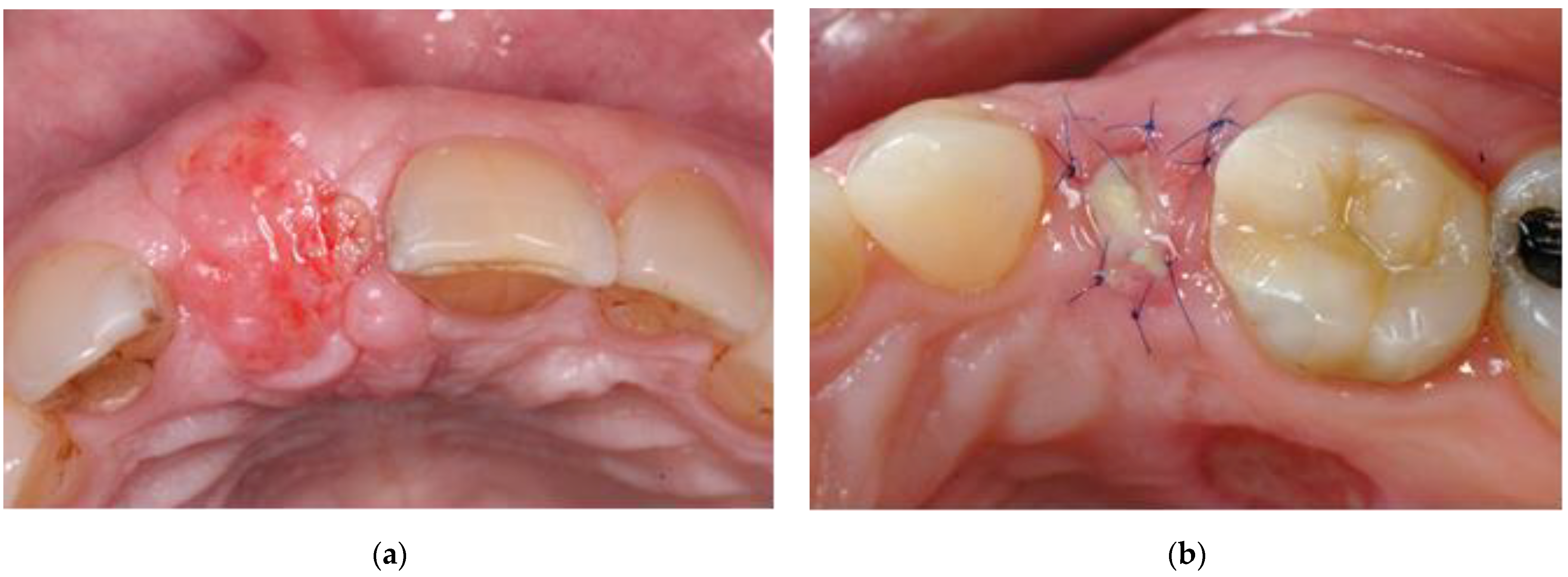
2.6. Evaluation of the Soft Tissue Grafts at 1 week

The status of the soft tissue grafts was assessed by visual inspection of the T1 photographs and was divided into 3 groups:
Group 1.
Vital: reddish tissue color indicating the graft has integration of blood supply (Figure 2a).
Group 2.
Partially vital: part of the graft presented signs of vitality (as mentioned above) and another part showed signs of necrosis, i.e., amorphous white material (Figure 2b).
Group 3.
Necrotic: the graft was exfoliating or had a yellow-grayish color indicating lack of blood supply.

2.7. Statistical Analysis

The effect of the tooth location on the T6/T0 ratio (%) was tested with one-way ANOVA, followed by post-hoc inter-group comparisons using the LSD correction. The distribution of invaginations among locations was analyzed with the Chi-square test. Analyses were conducted with the SPSS software and a value of p < 0.05 was considered significant.

3. Results

A total of 36 cases were included in the study. All the teeth were located in the maxilla, 14 of them were premolars, 18 incisors and 4 canines (Table 1). Of the 36 cases examined, 6 served as pontic site and 30 cases were sites for future implant placement.

3.1. Postextraction Ridge Width Alterations at 6 Months

The final ridge width varied from 74% to 121% of the original value. Although considerable inter-site variation was seen in the degree of ridge width alterations, the overall mean loss of width was 5.3% (SD = 13.4%) (Table 1). Site location had a significant effect on socket width change (p < 0.005). Incisor sites did not lose width (1.5% ± 13.1 change), while loss of width was present in the canine area (17.0% ± 2.1, p < 0.01 vs. incisors) and premolar area (10.5% ± 11.2, p < 0.01 vs. incisors).

3.2. Assessment of Soft Tissue Contour at 6 Months

At 6 months postextraction, an invagination of the alveolar ridge was recorded in 5 (14%) of all cases examined (Table 2). The differences in distribution of the invaginations among the different sites did not reach statistical significance.

3.3. Evaluation of the Soft Tissue Grafts at 1 Week

One-week post-surgery, 25 grafts (69%) were vital, and 11 (31%) were partially vital. Complete necrosis of the grafts did not occur in any of the described cases. No correlation was found between partial necrosis of the socket seal and occurrence of invagination.

4. Discussion

Numerous studies have examined the spontaneous post-extraction healing of alveolar bone [1,2,3,4,5]. According to these studies, the resorption of the ridge is more pronounced in the buccal aspect of the socket than in its lingual aspect [2,4]. Therefore, this retrospective analysis focused on the dimensional changes of the buccal aspect of the healing sockets. In this study, the average loss of buccal width at 6 months postextraction was about 5%. However, different areas showed different magnitudes of change in buccal dimensions. Incisor sites treated with SSS did not lose any buccal width, while the loss of width amounted to about 10% at the premolar sites and around 17% at canine sites. The results of the present study should be firstly compared with a healing process without external influences, i.e., untreated extraction alveoli. When the natural course of healing was monitored, an overall reduction in width of the ridge ranges from 25% after 6 months for incisors [37] to 50% in premolars and molars [3]. Moreover, ridge alterations in the esthetic zone may be more pronounced in thin-walled phenotypes [38]. We found an overall mean reduction of 5% in buccal width when SSS was applied, suggesting this technique has a beneficial effect on the dimensional changes 6 months following tooth extraction. This may be explained by: a. presence of bone graft material in the healing socket; b. superior stabilization of soft tissue margins of the socket at the initial healing phase and prevention of the soft tissue collapse; and c. efficient containment and protection of the bone graft particles resulting in minimal volumetric changes, minimizing the need for further soft and hard tissue augmentation procedures. Our results also suggest that the teeth that gain most from SSS are incisors.
The slighter reduction in width of the ridge after extraction of incisors may be attributed to the lower buccolingual diameter and cross sectional area of the root of incisors close to alveolar crest compared to canines, pre-molars and molars [39]. Thus the total area and the buccolingual diameter of the post-extraction alveolar socket orifice is lesser in the incisors compared to other types of teeth and the reduction in width of the ridge is less pronounced.
Many clinicians have used immediate implant placement as a technique for alveolar ridge preservation [16,40,41]. While immediate implant placement reduces the number of surgeries and saves time, it fails to prevent the horizontal and vertical ridge alterations that follow tooth extraction [8,42]. This may cause soft tissue recessions and should not be recommended when aesthetics are important [43]. Immediate implant placement should also be avoided at extraction sites in which achievement of primary implant stability requires positioning of the implant in a prosthetically incorrect position [44]. In such cases, SSS may be strongly considered.
According to a recent systematic review [45], xenografts are the most suitable grafting material for ridge preservation and prevention of alveolar bone resorption, when compared to allografts or alloplastic materials or to natural healing. However, these materials have also disadvantages, such as poor vascularization and long-term residual non-vital particles [46]. New biologically active materials have been developed to overcome the disadvantages of conventional bone substitutes, such as leukocyte-platelet-rich fibrin (L-PRF) and Platelet-rich fibrin (PRF) [47]. However, recent findings [48,49] have suggested that PRF may not significantly enhance bone formation after tooth extraction, as compared to spontaneous healing.
Tal et al. reported that 26% of SSS-covering grafts were non-vital one week post-surgery [33]. In the present study, no complete necrosis of the transplanted graft was observed. These results are in agreement with other studies [31,50]. Vital grafts may result in superior stabilization of soft tissue margins of the socket and efficient containment and protection of the bone graft particles, which may result in minimal volumetric changes in the long term, minimizing the need for further soft and hard tissue augmentation procedures.
Furthermore, while approximately 90% of spontaneously healing postextraction sockets present invaginations in the crestal area [36], we found invaginations only in 14% of the SSS cases, mostly in the premolar region.
The limitations of this study include the relatively small number of cases available to us for this analysis and the inability of clinical photographs to separate the changes in the alveolar bone from those in the overlying mucosa. Although only photographs with near ideal ortho-radial positioning were included in the study, unavoidably the angulation of the T0/T6 photographs may be slightly different, which may result in some inaccuracy in the absolute measurements but not in the comparison among different sites. Nevertheless, clinical photography is a simple and non-invasive technique which enables easy and straightforward clinical evaluation of the different socket preservation treatment modalities in the eyes of the clinician and patient. Other, more sophisticated techniques, like computed tomography or sounding and histological evaluation are necessary for comprehensive assessment of the efficiency of the particular therapeutic modality while addressing bone and mucosa separately.
Within the limitations of this retrospective study, socket seal surgery seems effective in minimizing the postextraction dimensional changes of the alveolar ridge contour in maxillary teeth, with a more pronounced effect at incisor sites. Additional studies comparing SSS to other ridge preservation techniques are needed to confirm the results observed in the current study.

5. Conclusions

This study found that using Socket Seal Surgery in maxillary extraction sites decreased significantly the loss of buccal alveolar width after tooth removal, with a most prominent effect in incisors, possibly due to the lesser overall reduction in width of the ridge after extraction compared to other teeth.

Author Contributions

Conceptualization, C.L. and E.W.; methodology, M.W.; formal analysis, O.B., R.W., T.C. and S.M.; investigation, O.B. and T.C.; resources, C.L.; data curation, O.B., R.W., T.C. and S.M.; writing—original draft preparation, E.W.; writing—review and editing, M.W. and C.L. All authors have read and agreed to the published version of the manuscript.

Funding

This research received no external funding.

Institutional Review Board Statement

The study was approved by the Institutional Ethics Committee of Tel Aviv University (IEC No. 0001834-1) and was conducted according to the guidelines of the Declaration of Helsinki.

Informed Consent Statement

Informed consent was obtained from all subjects involved in the study.

Data Availability Statement

Data sharing not applicable.

Conflicts of Interest

The authors declare no conflict of interest.

References

  1. Amler, M.H.; Johnson, P.L.; Salman, I. Histological and histochemical investigation of human alveolar socket healing in undisturbed extraction wounds. J. Am. Dent. Assoc. 1960, 61, 32–44. [Google Scholar] [CrossRef] [PubMed]
  2. Pietrokovski, J.; Massler, M. Alveolar ridge resorption following tooth extraction. J. Prosthet. Dent. 1967, 17, 21–27. [Google Scholar] [CrossRef]
  3. Schropp, L.; Wenzel, A.; Kostopoulos, L.; Karring, T. Bone healing and soft tissue contour changes following single-tooth extraction: A clinical and radiographic 12-month prospective study. Int. J. Periodontics Restor. Dent. 2003, 23, 313–323. [Google Scholar]
  4. Araujo, M.G.; Lindhe, J. Dimensional ridge alterations following tooth extraction. An experimental study in the dog. J. Clin. Periodontol. 2005, 32, 212–218. [Google Scholar] [CrossRef]
  5. Fickl, S.; Zuhr, O.; Wachtel, H.; Kebschull, M.; Hürzeler, M.B. Hard tissue alterations after socket preservation with additional buccal overbuilding: A study in the beagle dog. J. Clin. Periodontol. 2009, 36, 898–904. [Google Scholar] [CrossRef]
  6. Tan, W.L.; Wong, T.L.T.; Wong, M.C.M.; Lang, N.P. A systematic review of post-extractional alveolar hard and soft tissue dimensional changes in humans. Clin. Oral Implant. Res. 2012, 23, 1–21. [Google Scholar] [CrossRef] [PubMed]
  7. Van Der Weijden, F.; Dell’Acqua, F.; Slot, D.E. Alveolar bone dimensional changes of post-extraction sockets in humans: A systematic review. J. Clin. Periodontol. 2009, 36, 1048–1058. [Google Scholar] [CrossRef]
  8. Araujo, M.G.; Sukekava, F.; Wennstrom, J.L.; Lindhe, J. Ridge alterations following implant placement in fresh extraction sockets: An experimental study in the dog. J. Clin. Periodontol. 2005, 32, 645–652. [Google Scholar] [CrossRef]
  9. Avila-Ortiz, G.; Chambrone, L.; Vignoletti, F. Effect of alveolar ridge preservation interventions following tooth extraction: A systematic review and meta-analysis. J. Clin. Periodontol. 2019, 46, 195–223. [Google Scholar] [CrossRef] [Green Version]
  10. Jung, R.E.; Ioannidis, A.; Hämmerle, C.H.F.; Thoma, D.S. Alveolar ridge preservation in the esthetic zone. Periodontol. 2000 2018, 77, 165–175. [Google Scholar] [CrossRef]
  11. Lekovic, V.; Kenney, E.B.; Weinlaender, M.; Han, T.; Klokkevold, P.; Nedic, M.; Orsini, M. A Bone Regenerative Approach to Alveolar Ridge Maintenance Following Tooth Extraction. Rep. 10 Cases 1997, 68, 563–570. [Google Scholar] [CrossRef]
  12. Iasella, J.M.; Greenwell, H.; Miller, R.L.; Hill, M.; Drisko, C.; Bohra, A.A.; Scheetz, J.P. Ridge Preservation with Freeze-Dried Bone Allograft and a Collagen Membrane Compared to Extraction Alone for Implant Site Development: A Clinical and Histologic Study in Humans. J. Periodontol. 2003, 74, 990–999. [Google Scholar] [CrossRef] [PubMed]
  13. Artzi, Z.; Nemcovsky, C.E. The Application of Deproteinized Bovine Bone Mineral for Ridge Preservation Prior to Implantation. Clinical and Histological Observations in a Case Report. J. Periodontol. 1998, 69, 1062–1067. [Google Scholar] [CrossRef] [PubMed]
  14. Carmagnola, D.; Adriaens, P.; Berglundh, T. Healing of human extraction sockets filled with Bio-OssR. Clin. Oral Implant. Res. 2003, 14, 137–143. [Google Scholar] [CrossRef] [PubMed]
  15. Brugnami, F.; Caiazzo, A. Efficacy evaluation of a new buccal bone plate preservation technique: A pilot study. Int. J. Periodontics Restor. Dent. 2011, 31, 67–73. [Google Scholar]
  16. Clementini, M.; Agostinelli, A.; Castelluzzo, W.; Cugnata, F.; Vignoletti, F.; De Sanctis, M. The effect of immediate implant placement on alveolar ridge preservation compared to spontaneous healing after tooth extraction: Radiographic results of a randomized controlled clinical trial. J. Clin. Periodontol. 2019. [Google Scholar] [CrossRef]
  17. Canellas, J.; da Costa, R.C.; Breves, R.C.; de Oliveira, G.P.; Figueredo, C.; Fischer, R.G.; Thole, A.A.; Medeiros, P.J.D.; Ritto, F.G. Tomographic and histomorphometric evaluation of socket healing after tooth extraction using leukocyte- and platelet-rich fibrin: A randomized, single-blind, controlled clinical trial. J. Craniomaxillofac Surg 2020, 48, 24–32. [Google Scholar] [CrossRef]
  18. Bäumer, D.; Zuhr, O.; Rebele, S.; Hürzeler, M. Socket Shield Technique for immediate implant placement—Clinical, radiographic and volumetric data after 5 years. Clin. Oral Implant. Res. 2017, 28, 1450–1458. [Google Scholar] [CrossRef]
  19. Salama, H.; Salama, M. The role of orthodontic extrusive remodeling in the enhancement of soft and hard tissue profiles prior to implant placement: A systematic approach to the management of extraction site defects. Int. J. Periodontics Restor. Dent. 1993, 13, 312–333. [Google Scholar]
  20. Fickl, S.; Zuhr, O.; Wachtel, H.; Stappert, C.F.J.; Stein, J.M.; Hürzeler, M.B. Dimensional changes of the alveolar ridge contour after different socket preservation techniques. J. Clin. Periodontol. 2008, 35, 906–913. [Google Scholar] [CrossRef]
  21. Becker, W.; Becker, B.E. Guided tissue regeneration for implants placed into extraction sockets and for implant dehiscences: Surgical techniques and case report. Int. J. Periodontics Restor. Dent. 1990, 10, 376–391. [Google Scholar]
  22. Nemcovsky, C.E.; Artzi, Z.; Moses, O. Rotated palatal flap in immediate implant procedures. Clin. Oral Implant. Res. 2000, 11, 83–90. [Google Scholar] [CrossRef] [PubMed]
  23. Becker, W.; Becker, B.E.; Caffesse, R. A Comparison of Demineralized Freeze-Dried Bone and Autologous Bone to Induce Bone Formation in Human Extraction Sockets. J. Periodontol. 1994, 65, 1128–1133. [Google Scholar] [CrossRef] [PubMed]
  24. Artzi, Z.; Tal, H.; Dayan, D. Porous Bovine Bone Mineral in Healing of Human Extraction Sockets. Part 1: Histomorphometric Evaluations at 9 Months. J. Periodontol. 2000, 71, 1015–1023. [Google Scholar] [CrossRef]
  25. Froum, S.; Cho, S.-C.; Rosenberg, E.; Rohrer, M.; Tarnow, D. Histological Comparison of Healing Extraction Sockets Implanted With Bioactive Glass or Demineralized Freeze-Dried Bone Allograft: A Pilot Study. J. Periodontol. 2002, 73, 94–102. [Google Scholar] [CrossRef]
  26. Serino, G.; Biancu, S.; Iezzi, G.; Piattelli, A. Ridge preservation following tooth extraction using a polylactide and polyglycolide sponge as space filler: A clinical and histological study in humans. Clin. Oral Implant. Res. 2003, 14, 651–658. [Google Scholar] [CrossRef] [Green Version]
  27. Faria-Almeida, R.; Astramskaite-Januseviciene, I.; Puisys, A.; Correia, F. Extraction Socket Preservation with or without Membranes, Soft Tissue Influence on Post Extraction Alveolar Ridge Preservation: A Systematic Review. J. Oral Maxillofacial Res. 2019, 10. [Google Scholar] [CrossRef]
  28. Canellas, J.V.D.S.; Medeiros, P.J.D.A.; Figueredo, C.M.D.S.; Fischer, R.G.; Ritto, F.G. Which is the best choice after tooth extraction, immediate implant placement or delayed placement with alveolar ridge preservation? A systematic review and meta-analysis. J. Cranio-Maxillofac. Surg. 2019, 47, 1793–1802. [Google Scholar] [CrossRef]
  29. Landsberg, C.J.; Bichacho, N. A modified surgical/prosthetic approach for optimal single implant supported crown. Part I--The socket seal surgery. Pract. Periodontics Aesthet Dent. 1994, 6, 11–17, quiz 19. [Google Scholar]
  30. Landsberg, C.J. Implementing Socket Seal Surgery as a Socket Preservation Technique for Pontic Site Development: Surgical Steps Revisited—A Report of Two Cases. J. Periodontol. 2008, 79, 945–954. [Google Scholar] [CrossRef]
  31. Jung, R.E.; Siegenthaler, D.W.; Hammerle, C.H. Postextraction tissue management: A soft tissue punch technique. Int. J. Periodontics Restor. Dent. 2004, 24, 545–553. [Google Scholar]
  32. Fickl, S.; Zuhr, O.; Wachtel, H.; Bolz, W.; Huerzeler, M.B. Hard tissue alterations after socket preservation: An experimental study in the beagle dog. Clin. Oral Implant. Res. 2008, 19, 1111–1118. [Google Scholar] [CrossRef] [PubMed]
  33. Tal, H. Autogenous masticatory mucosal grafts in extraction socket seal procedures: A comparison between sockets grafted with demineralized freeze-dried bone and deproteinized bovine bone mineral. Clin. Oral Implant. Res. 1999, 10, 289–296. [Google Scholar] [CrossRef] [PubMed]
  34. Kim, S.-Y.; Kim, Y.-K.; Kim, H.-S.; Yun, P.-Y.; Kim, S.-G.; Choi, Y.-H. Extraction socket sealing using palatal gingival grafts and resorbable collagen membranes. Maxillofac. Plast. Reconstr. Surg. 2017, 39. [Google Scholar] [CrossRef] [PubMed] [Green Version]
  35. Oghli, A.A.; Steveling, H. Ridge preservation following tooth extraction: A comparison between atraumatic extraction and socket seal surgery. Quintessence Int. 2010, 41, 605–609. [Google Scholar] [PubMed]
  36. Thoma, D.S.; Bienz, S.P.; Lim, H.C.; Lee, W.Z.; Hämmerle, C.H.F.; Jung, R.E. Explorative randomized controlled study comparing soft tissue thickness, contour changes, and soft tissue handling of two ridge preservation techniques and spontaneous healing two months after tooth extraction. Clin. Oral Implant. Res. 2020, 31, 565–574. [Google Scholar] [CrossRef] [PubMed]
  37. Rodd, H.D.; Malhotra, R.; O’Brien, C.H.; Elcock, C.; Davidson, L.E.; North, S. Change in supporting tissue following loss of a permanent maxillary incisor in children. Dent. Traumatol. 2007, 23, 328–332. [Google Scholar] [CrossRef]
  38. Chappuis, V.; Engel, O.; Reyes, M.; Shahim, K.; Nolte, L.P.; Buser, D. Ridge Alterations Post-extraction in the Esthetic Zone. J. Dent. Res. 2013, 92, 195S–201S. [Google Scholar] [CrossRef] [Green Version]
  39. Sun, M.; Gu, F.; Wang, J.; Zhou, C.; Xia, J.; Qin, H.; Yang, J. Measurement for natural dental neck data of normal adults and its clinical significance on guiding implant restoration. Int. J. Clin. Exp. Med. 2015, 8, 14732–14740. [Google Scholar]
  40. Watzek, G.; Haider, R.; Mensdorff-Pouilly, N.; Haas, R. Immediate and delayed implantation for complete restoration of the jaw following extraction of all residual teeth: A retrospective study comparing different types of serial immediate implantation. Int. J. Oral Maxillofac Implant. 1995, 10, 561–567. [Google Scholar]
  41. Slagter, K.W.; Den Hartog, L.; Bakker, N.A.; Vissink, A.; Meijer, H.J.A.; Raghoebar, G.M. Immediate Placement of Dental Implants in the Esthetic Zone: A Systematic Review and Pooled Analysis. J. Periodontol. 2014, 85, e241–e250. [Google Scholar] [CrossRef] [PubMed]
  42. Discepoli, N.; Vignoletti, F.; Laino, L.; De Sanctis, M.; Muñoz, F.; Sanz, M. Fresh extraction socket: Spontaneous healing vs. immediate implant placement. Clin. Oral Implant. Res. 2015, 26, 1250–1255. [Google Scholar] [CrossRef]
  43. Tonetti, M.S.; Cortellini, P.; Graziani, F.; Cairo, F.; Lang, N.P.; Abundo, R.; Conforti, G.P.; Marquardt, S.; Rasperini, G.; Silvestri, M.; et al. Immediateversusdelayed implant placement after anterior single tooth extraction: The timing randomized controlled clinical trial. J. Clin. Periodontol. 2017, 44, 215–224. [Google Scholar] [CrossRef] [PubMed] [Green Version]
  44. Tonetti, M.S.; Jung, R.E.; Avila-Ortiz, G.; Blanco, J.; Cosyn, J.; Fickl, S.; Figuero, E.; Goldstein, M.; Graziani, F.; Madianos, P.; et al. Management of the extraction socket and timing of implant placement: Consensus report and clinical recommendations of group 3 of the XV European Workshop in Periodontology. J. Clin. Periodontol. 2019, 46, 183–194. [Google Scholar] [CrossRef] [PubMed] [Green Version]
  45. Jambhekar, S.; Kernen, F.; Bidra, A.S. Clinical and histologic outcomes of socket grafting after flapless tooth extraction: A systematic review of randomized controlled clinical trials. J. Prosthet. Dent. 2015, 113, 371–382. [Google Scholar] [CrossRef] [PubMed]
  46. Zitzmann, N.U.; Schärer, P.; Marinello, C.P. Factors influencing the success of GBR. J. Clin. Periodontol. 1999, 26, 673–682. [Google Scholar] [CrossRef] [PubMed]
  47. Dohan, D.M.; Choukroun, J.; Diss, A.; Dohan, S.L.; Dohan, A.J.J.; Mouhyi, J.; Gogly, B. Platelet-rich fibrin (PRF): A second-generation platelet concentrate. Part II: Platelet-related biologic features. Oral Surg. Oral Med. Oral Pathol. Oral Radiol. Endodontology 2006, 101, e45–e50. [Google Scholar] [CrossRef]
  48. Areewong, K.; Chantaramungkorn, M.; Khongkhunthian, P. Platelet-rich fibrin to preserve alveolar bone sockets following tooth extraction: A randomized controlled trial. Clin. Implant. Dent. Relat. Res. 2019, 21, 1156–1163. [Google Scholar] [CrossRef]
  49. Moraschini, V.; Mourão, C.F.D.A.B.; Machado, R.C.D.M.; Nascimento, J.R.B.; Javid, K.; Calasans-Maia, M.D.; Cardarelli, A.; Montemezzi, P.; Calasans-Maia, J.D.A. Does Platelet-Rich Fibrin Decrease Dimensional Changes and Improve Postoperative Comfort in Post-Extraction Sockets? An Overview of Systematic Reviews. Appl. Sci. 2020, 10, 5750. [Google Scholar] [CrossRef]
  50. Thalmair, T.; Fickl, S.; Schneider, D.; Hinze, M.; Wachtel, H. Dimensional alterations of extraction sites after different alveolar ridge preservation techniques—A volumetric study. J. Clin. Periodontol. 2013, 40, 721–727. [Google Scholar] [CrossRef]
Figure 1. T0 (a) and T6 (b) images with a superimposed vertical rectangle which borders the most buccal edge of the edentulous gap.
Applsci 11 00324 g001
Figure 2. T1 images: A vital (a) and a partially vital graft (b).
Applsci 11 00324 g002
Table 1. Mean and SD of changes in the alveolar width in the different treated sites.
LocationNumberMean T6 Width/T0 Width (%)Mean Change in Width (%)SD
Incisors18101.51.513.1
Canines483.0−17.02.1
Premolars1489.5−10.511.2
Total3694.7−5.313.4
Table 2. Distribution of invaginations by site.
Invaginations
Incisors1 (5.5%)
Canines0 (0%)
Premolars4 (28.5%)
Total5 (13.9%)
Publisher’s Note: MDPI stays neutral with regard to jurisdictional claims in published maps and institutional affiliations.

