Article

Parylene-Based Flexible Microelectrode Arrays for the Electrical Recording of Muscles and the Effect of Electrode Size

School of Electrical and Computer Engineering, University of Seoul, Seoul 02504, Korea
*
Author to whom correspondence should be addressed.
Appl. Sci. 2020, 10(20), 7364; https://doi.org/10.3390/app10207364
Submission received: 11 August 2020 / Revised: 3 October 2020 / Accepted: 16 October 2020 / Published: 21 October 2020
(This article belongs to the Special Issue Flexible and Printed Electronics)

Abstract

:
Miniaturized flexible microelectrode arrays are desirable for small-area surface electromyography (sEMG) to detect the electrical activity generated by muscles in a specific area of the body. Here, we present a flexible 8-channel microelectrode array with electrodes of diameter 150–300 μm for small-area sEMG recordings. The microelectrode arrays based on a flexible Parylene C substrate recorded the sEMG signals from a curved skin surface with a maximum signal-to-noise ratio (SNR) of 21.4 dB. The sEMG signals recorded from a small area of 17671–59325 μm2 showed a clear distinction between the signal and noise. Further, the sEMG data were analyzed in the frequency domain by converting the signals via fast Fourier transform (FFT), and it was verified that the proposed microelectrode could reliably record multichannel sEMGs over a small area. Moreover, a maximum voluntary contraction (MVC) experiment was performed to confirm the recording capability of the microelectrode array, which showed consistency with the previous reports. Finally, we demonstrated the effects of the electrode size by comparing the results for two different electrode sizes. When the electrode size was increased 3.37 times, the root-mean-square value of the amplitude (Vrms) increased 2.64 times, consequently increasing the SNR from 16.9 to 21.4 dB. This study demonstrates the expanded utility of Parylene-based flexible microelectrode arrays.

1. Introduction

Electromyography (EMG) has been popularly used as a neurophysiological tool for detecting the electrical activities of muscles [1]. EMG data have been used to not only diagnose muscular disorders such as amyotrophic lateral sclerosis (ALS) and muscular dystrophy (MD) but also to measure the extent of muscle fatigue [2,3,4]. EMG recordings can be obtained by two methods: surface EMG recording (non-invasive method) and intramuscular EMG recording (invasive method). Surface electromyography (sEMG) does not require needles and has some advantages because of its convenience and non-invasiveness. Rather, a thin and flexible electrode is required for sEMG, since the high mechanical flexibility allows the electrode to follow the uneven skin surface, resulting in good electrical connections in the recording area.
An EMG signal can be considered as the set of motor unit action potentials (MUAPs) produced by motor units (MUs), which are made up of motor neurons and skeletal muscle fibers, typically from an area of several thousand square microns [5]. Conventional sEMG electrodes have a limited resolution because of their large electrode diameters of 1–20 mm [6], which make them incompatible for small-area sEMG recording. To date, better spatial resolution sEMG recordings have been achieved by introducing micro-size electrodes, which have rapidly proved to be promising research tools [7,8,9,10,11]. Further, invasive EMG recordings using an implanted flexible microelectrode array in a moving subject (caterpillar) demonstrated a high-resolution recording capability [12]. However, the reduced EMG signal amplitude and signal-to-noise ratio (SNR) due to the small electrode size are potential drawbacks of the microelectrode EMG. In order to better understand small-area sEMG recording, further research on sEMG with a flexible microelectrode array must be conducted and the effect of the electrode size should be investigated.
Herein, we report Parylene C-based flexible microelectrode arrays for small-area sEMG recording on curved skin surfaces. The characteristics of the microelectrodes were assessed by various means, including electrochemical impedance spectroscopy (EIS), cyclic voltammetry (CV), and equivalent circuit modeling. In addition, the recorded sEMG using 8-channel microelectrode arrays were used to verify the viability of multiple sEMG recordings in a small area. Finally, by analyzing the effect of the size of the microelectrode, the relationship between the signal amplitude and the electrode size was studied. This study is expected to benefit investigators who are exploring potential approaches to record small-area sEMG using flexible microelectrode arrays.

2. Materials and Methods

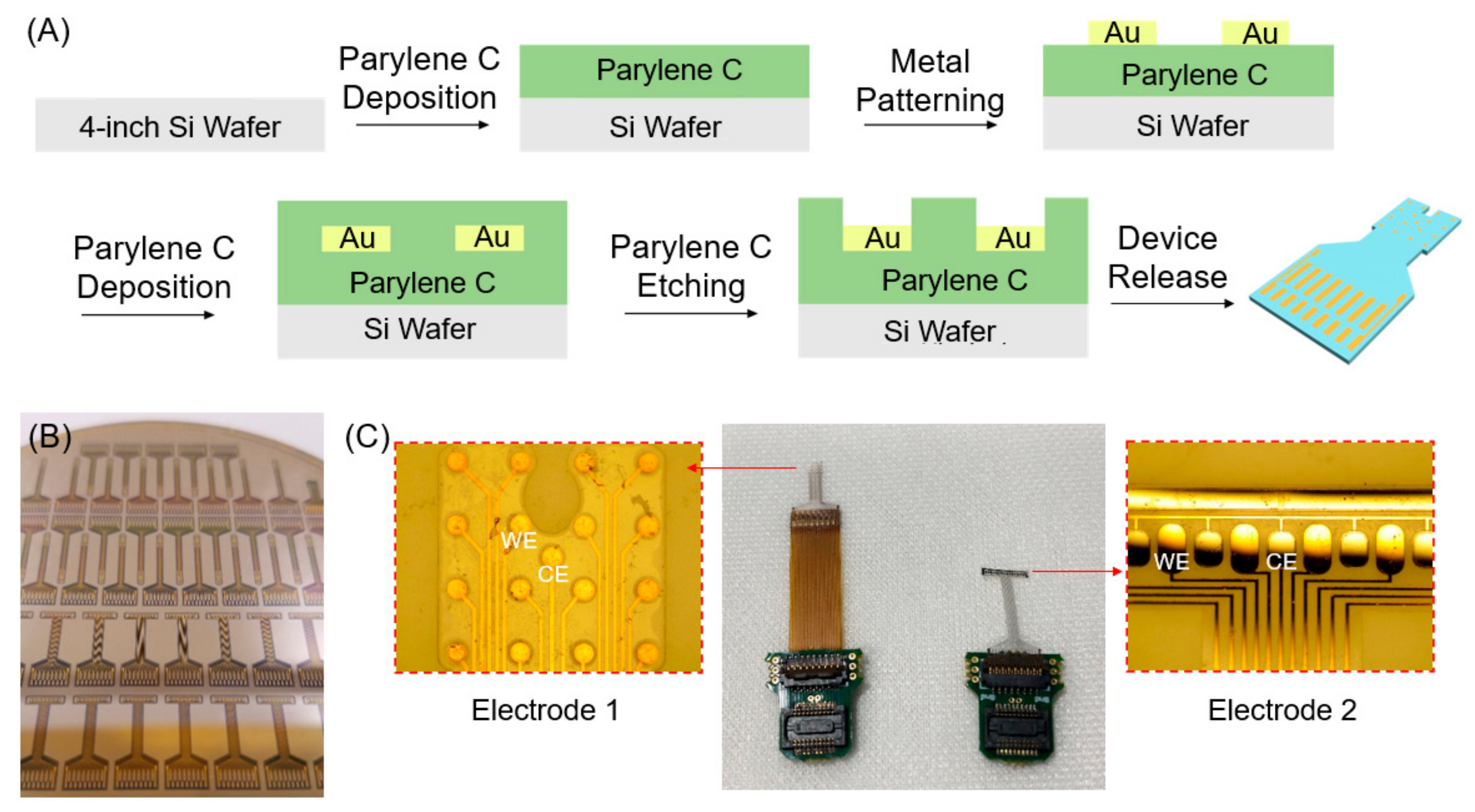
2.1. Fabrication of Flexible Microelectrode Arrays

We fabricated a flexible microelectrode array for sEMG recording on a 4-inch silicon (Si) wafer using the semiconductor fabrication process shown in Figure 1A. First, a 10 μm thick Parylene C layer was deposited on a 4-inch Si wafer using a Parylene coater. Then, gold (Au) and titanium (Ti) metal electrodes (Ti/Au = 10/200 μm) were patterned using photolithography and a subsequent lift-off process. A second layer of Parylene C (10 μm) was then deposited to encapsulate the entire wafer. The first and second Parylene C layers were patterned using photolithography and oxygen (O2) plasma dry etching. During the etching process, the electrode sites and pads were exposed for electrical connections between the skin, electrode array, and sEMG measurement equipment. Finally, the fabricated devices were released from the silicon wafer. Figure 1B shows several microelectrode arrays fabricated on a 4-inch wafer. Additional information regarding the fabrication process can be found in our previous papers [13,14,15].
Figure 1C shows the two types of electrode arrays with different electrode sizes, which are inserted in the zero-insertion force (ZIF) connectors on printed circuit boards (PCB) for connection to the recording system.
Electrode 1 has circle-shaped electrode sites of diameter 150 μm (area: 17,671 μm2), and Electrode 2 has a combination of half-ellipse and rectangle-shaped sites with a height of 300 μm and a width of 230 μm (area: 59,325 μm2). Different shapes and sizes of the electrodes could thus be selected for different sEMG recordings, depending on the target muscle. It should be noted that the slightly different shape could influence the recording results, although it is known that the electrode size has an integrative effect on the sEMG signal. For measuring the sEMG signal in this study, we mainly used Electrode 2, which showed a higher SNR and signal amplitude. We also compared these two electrodes to evaluate the effects of the electrode sizes, which will be discussed later.

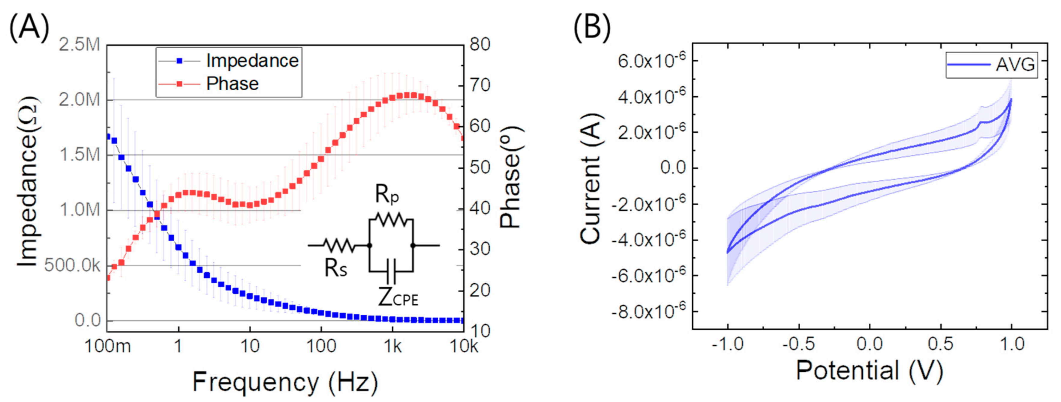
2.2. Electrochemical Impedance Spectroscopy (EIS) and Cyclic Voltammetry (CV)
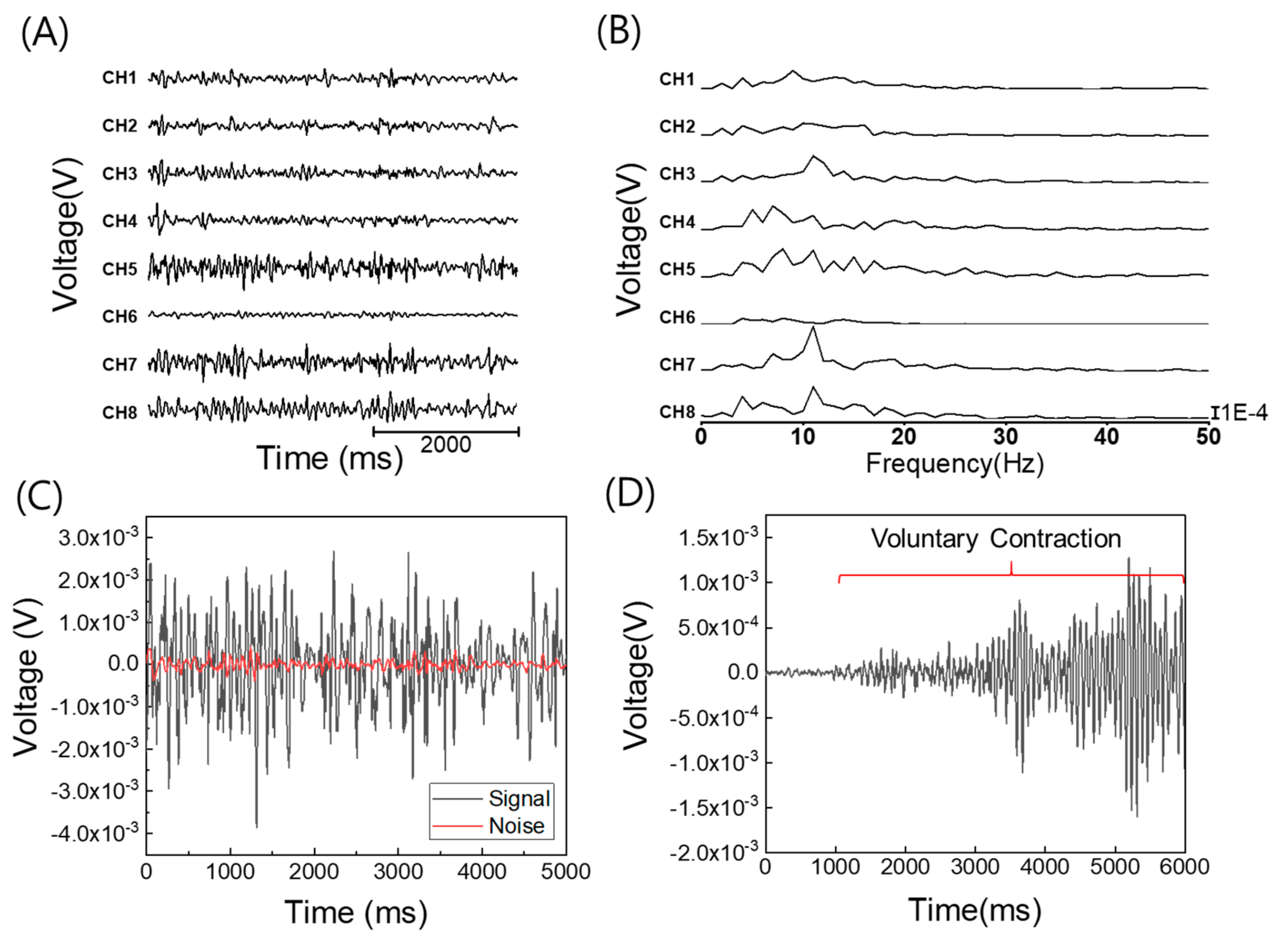
We characterized the fabricated microelectrodes using electrochemical impedance spectroscopy (EIS) and cyclic voltammetry (CV). EIS and CV were performed in phosphate buffered saline (PBS) with a potentiostat/galvanostat (PGSTAT128N, Metrohm Autolab). Platinum (Pt) was used as the counter electrode (CE), and silver/silver chloride (Ag/AgCl) was used as the reference electrode (RE). The frequency range of the EIS measurements was 0.1 Hz to 10 kHz. Each electrode in the 8-channel electrode array was measured, and the average impedance and standard deviation were calculated.

2.3. sEMG Recording

The sEMG recording was performed by connecting the microelectrode array to the electrophysiology system comprising a signal processor and an amplifier (TDT RZ5 and PZ2, Tucker Davis Technology Inc., Alachua, FL, USA). The recorded sEMG on the skin of the arm was extracted from the system and processed using MATLAB. To be specific, the isometric contraction of flexor carpi ulnaris muscle was recorded through a differential sEMG measurement. For the reproducible recording, the forearm was located at 120° with respect to the arm, and isometric contraction was achieved by manual resistance with 50% of the MVC level. To reduce the electrode-skin interface impedance, the skin had been cleaned with water before placing the microelectrode [6]. Additionally, we located a surface microelectrode on the muscle belly of flexor carpi ulnaris along the muscle fibers to avoid a reduction in signal [16,17]. For the data processing, we used a high-pass filter (HPF) and a low-pass filter (LPF). To reduce the impact of motion artifacts, we set the HPF cutoff frequency as 5 Hz [18]. The most meaningful sEMG signal is in the frequency range of 0–500 Hz; therefore, the LPF cutoff frequency was set to 500 Hz [19].

3. Results and Discussion

3.1. Electrochemical Impedance Spectroscopy (EIS) and Cyclic Voltammetry (CV)

Figure 2A shows the EIS measurement results, including the impedance and phase, depending on the frequency. The average impedance of a microelectrode ranged from 3.7 kΩ to 1.6 MΩ and the frequency ranged from 10 kHz to 0.1 Hz, with an average impedance of 13.9 kΩ at 1 kHz. The standard deviation of the impedance of each channel showed 1.43 kΩ, and its coefficient of variation was 10.3 percent. The average phase angle showed an increasing trend from 25° to 67°. Each channel of the array showed similar tendencies, indicating a good reliability within the microelectrode array. We also performed the equivalent circuit modeling of the electrode, and the extracted parameters from the equivalent circuit model (inset of Figure 2A) are as follows: solution resistance, Rs = 724.5 Ω; constant phase element, Z C P E = ( 1.985 7 ω n ) 1 × e π 2 n i (n = 0.67812); polarization resistance, Rp = 2.167 MΩ. These results indicate that the fabricated microelectrode array has a capacitive charge-carrying mechanism that is consistent with previous reports using gold electrodes [15]. Finally, CV was measured using the potentiostat/galvanostat at a scan rate of 1000 mV/s. As shown in Figure 2B, the peak current ranged from −6.81 × 10⁻⁶ to 5.22 × 10⁻⁶ A. This result shows a similar trend to that of other conventional large-sized gold-based electrodes [9,20], indicating that the fabricated 8-channel microelectrode array might be appropriate for sEMG recording.

3.2. sEMG Recording

Figure 3A shows the recorded sEMG signals from the eight different channels in the time domain. The time domain data were transformed to the frequency domain by applying fast Fourier transform (FFT) through MATLAB, as shown in Figure 3B. Each channel has a similar peak frequency that demonstrates the reliability of the microelectrode array. Using the fabricated microelectrode, we achieved an SNR of ~21.4 dB with a 4.28 dB standard deviation, as calculated from Equation (1). The root mean square (RMS) voltage of the sEMG signal was 0.853 mV, whereas the baseline noise was 78.4 μV, meaning that we can clearly distinguish the signal from noise, as shown in Figure 3C.
SNR = 10 × log s i g n a l   p o w e r b a s e l i n e   n o i s e   p o w e r .
We also performed the maximum voluntary contraction (MVC) experiment to verify the recording capability of the microelectrode array. The MVC experiment was conducted with the same electrode location as described in the method section, starting from the 0% MVC level and going to the 100% MVC level, each lasting 6–7 s [21]. According to Henneman’s size principle [22,23], more MUs start to fire as more load is added to the muscles. We observed the tendency of increasing signal with increasing voluntary contractions, which is consistent with the theory, as shown in Figure 3D.

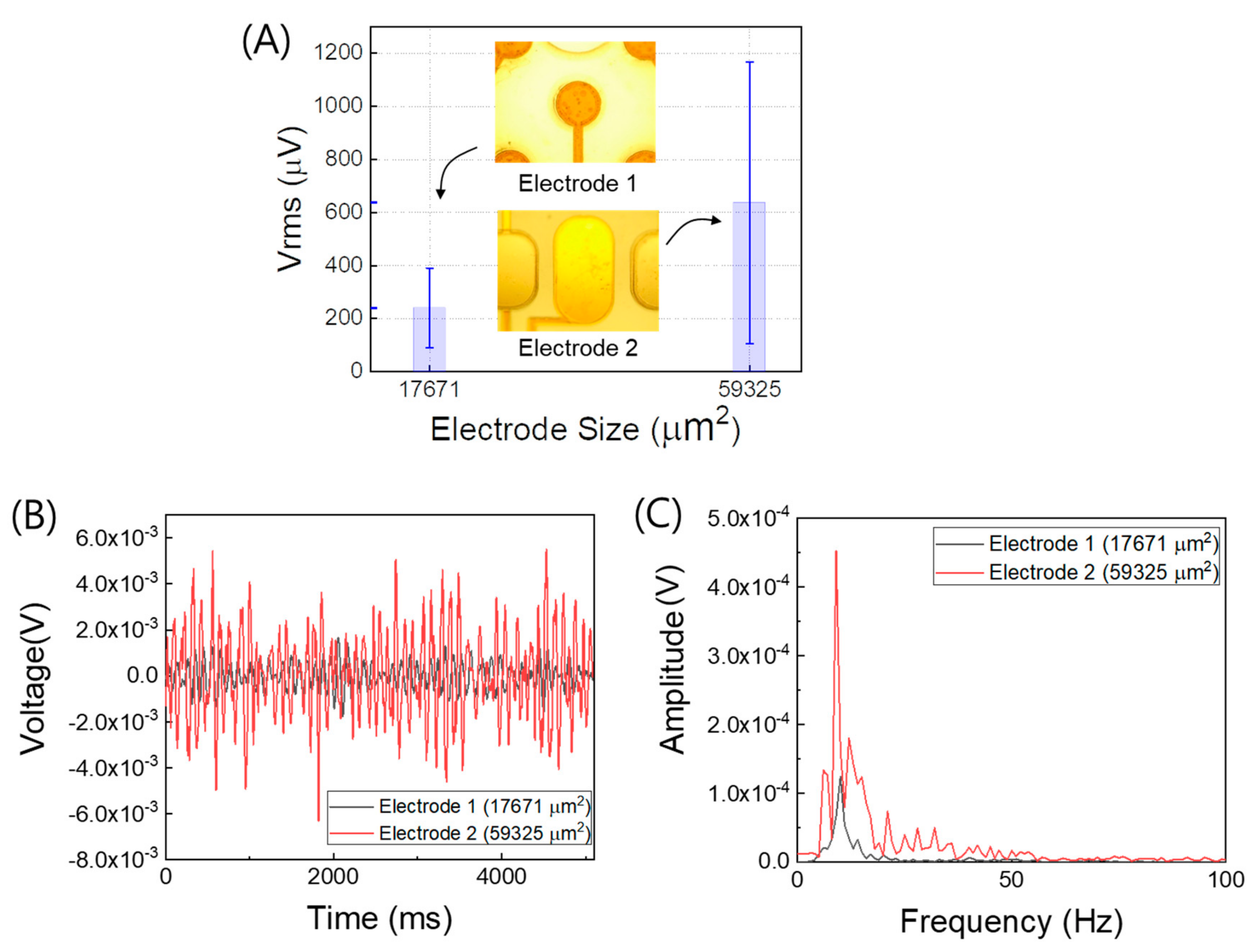
3.3. Electrode Size Effect on sEMG

The amplitude of the recorded EMG signal is an important parameter to achieve a high SNR electrode. Since the microelectrode has a relatively small electrode size compared to a conventional electrode, the recorded sEMG signal is sensitive to the electrode size. To observe the effect of the electrode size on the measurements, we compared our two microelectrode types: Electrode 1 of size 17,671 μm2 and Electrode 2 of size 59,325 μm2. The size of Electrode 2 is 3.357 times greater than that of Electrode 1 (Figure 4A). Assuming that the electrode size affects the amplitude of the sEMG signal, the sEMG was recorded in the same environment to minimize any errors attributable to the body condition. Figure 4A shows the root-mean-square amplitudes (Vrms) from the two microelectrode arrays (eight channels for each array). The Vrms of the larger electrode (Electrode 2) was 2.64 times greater than that of the smaller electrode (Electrode 1). The Vrms difference corresponds to the size difference between Electrodes 1 and 2 to some extent. Furthermore, a comparison between the data from the best channel of each microelectrode array shows the 2.85 times gap of the Vrms, as shown in Figure 4B, indicating consistent results with the earlier comparison. Further, the SNRs of Electrodes 1 and 2 were 16.9 and 21.4 dB, respectively, showing a higher SNR from the larger electrode (Electrode 2). This is attributed to the increased recording ability due to the enlarged electrode size. From the FFT signal processing shown in Figure 4C, we observe that both the electrodes have similar peaks in the frequency domain, indicating that both microelectrode arrays could reliably record sEMGs with different signal amplitudes. Although more studies are required to further investigate the effects of electrode size, the present results are expected to be of help for recording sEMGs using microscale flexible electrode arrays.

4. Conclusions

In this study, we demonstrated two Parylene C-based flexible multichannel microelectrode arrays for small-area sEMG recording. The proposed microelectrode arrays with electrode diameters of 150–300 μm were demonstrated to reliably record sEMG signals with a maximum SNR of 21.4 dB. Additionally, we experimentally showed that the recorded signal amplitude was affected by the electrode size by comparing electrodes of two different sizes (areas of 17,671 and 59,325 μm2). Although the signal amplitude could be improved using a large-sized electrode, the size of the electrode should be optimized depending on the purpose of use and the location of the small-area sEMG recording. The number of electrode sizes was limited to two in this work; therefore, more studies would be beneficial for microelectrode sEMG in the future. Nonetheless, we expect that the proposed microelectrode arrays could be used for surface EMG or in vivo EMG to measure the MUs in a specific area. In addition, the flexible and miniaturized microelectrode arrays can be readily reconfigured for wearable platforms with further research.

Author Contributions

Conceptualization, B.-J.C. and D.-W.P.; Data curation, B.-J.C., J.-H.K., W.-J.Y., D.-J.H. and J.P.; Investigation, B.-J.C., J.-H.K., W.-J.Y., D.-J.H. and J.P.; Project administration, D.-W.P.; Supervision, D.-W.P.; Writing—original draft, B.-J.C. and D.-W.P.; Writing—review and editing, D.-W.P. All authors have read and agreed to the published version of the manuscript.

Funding

The work was supported by the Basic Science Research Program through the National Research Foundation of Korea (NRF) funded by the Ministry of Science and ICT (grant no. 2018R1C1B6001529). The EDA tool was supported by the IC Design Education Center (IDEC), Korea.

Conflicts of Interest

The authors declare no conflict of interest.

References

  1. Drost, G.; Stegeman, D.F.; van Engelen, B.G.; Zwarts, M.J. Clinical applications of high-density surface EMG: A systematic review. J. Electromyogr. Kinesiol. 2006, 16, 586–602. [Google Scholar] [CrossRef] [PubMed]
  2. Mishra, V.K.; Bajaj, V.; Kumar, A. Classification of normal, ALS, and myopathy EMG signals using ELM classifier. In Proceedings of the 2016 2nd International Conference on Advances in Electrical, Electronics, Information, Communication and Bio-Informatics (AEEICB), Chennai, India, 27–28 February 2016; pp. 455–459. [Google Scholar]
  3. Hilton-Brown, P.; Stålberg, E. The motor unit in muscular dystrophy, a single fibre EMG and scanning EMG study. J. Neurol. Neurosurg. Psychiatr. 1983, 46, 981–995. [Google Scholar] [CrossRef] [PubMed] [Green Version]
  4. Komi, P.V.; Tesch, P. EMG frequency spectrum, muscle structure, and fatigue during dynamic contractions in man. Eur. J. Appl. Physiol. Occup. Physiol. 1979, 42, 41–50. [Google Scholar] [CrossRef] [PubMed]
  5. Jacob, J.M. Lumbar motor neuron size and number is affected by age in male F344 rats. Mech. Ageing Dev. 1998, 106, 205–216. [Google Scholar] [CrossRef]
  6. Hermens, H.J.; Freriks, B.; Disselhorst-Klug, C.; Rau, G. Development of recommendations for SEMG sensors and sensor placement procedures. J. Electromyogr. Kinesiol. 2000, 10, 361–374. [Google Scholar] [CrossRef]
  7. Kim, M.; Kim, T.; Kim, D.S.; Chung, W.K. Curved microneedle array-based sEMG electrode for robust long-term measurements and high selectivity. Sensors 2015, 15, 16265–16280. [Google Scholar] [CrossRef] [PubMed]
  8. Roberts, T.; De Graaf, J.B.; Nicol, C.; Hervé, T.; Fiocchi, M.; Sanaur, S. Flexible inkjet-printed multielectrode arrays for neuromuscular cartography. Adv. Healthc. Mater. 2016, 5, 1462–1470. [Google Scholar] [CrossRef] [PubMed]
  9. Kang, X.; Liu, J.; Tian, H.; Yang, B.; NuLi, Y.; Yang, C. Sputtered iridium oxide modified flexible parylene microelectrodes array for electrical recording and stimulation of muscles. Sens. Actuators B Chem. 2016, 225, 267–278. [Google Scholar] [CrossRef]
  10. Yamagiwa, S.; Sawahata, H.; Ishida, M.; Kawano, T. Micro-electrode arrays for multi-channel motor unit EMG recording. In Proceedings of the 2014 IEEE 27th International Conference on Micro Electro Mechanical Systems (MEMS), San Francisco, CA, USA, 26–30 January 2014; pp. 857–860. [Google Scholar]
  11. Ullah, K.; Khan, K.; Amin, M.; Attique, M.; Chung, T.-S.; Riaz, R. Multi-Channel Surface EMG Spatio-Temporal Image Enhancement Using Multi-Scale Hessian-Based Filters. Appl. Sci. 2020, 10, 5099. [Google Scholar] [CrossRef]
  12. Metallo, C.; White, R.D.; Trimmer, B.A. Flexible parylene-based microelectrode arrays for high resolution EMG recordings in freely moving small animals. J. Neurosci. Methods 2011, 195, 176–184. [Google Scholar] [CrossRef] [PubMed]
  13. Park, D.-W.; Schendel, A.A.; Mikael, S.; Brodnick, S.K.; Richner, T.J.; Ness, J.P.; Hayat, M.R.; Atry, F.; Frye, S.T.; Pashaie, R. Graphene-based carbon-layered electrode array technology for neural imaging and optogenetic applications. Nat. Commun. 2014, 5, 5258. [Google Scholar] [CrossRef] [PubMed]
  14. Park, D.-W.; Brodnick, S.K.; Ness, J.P.; Atry, F.; Krugner-Higby, L.; Sandberg, A.; Mikael, S.; Richner, T.J.; Novello, J.; Kim, H. Fabrication and utility of a transparent graphene neural electrode array for electrophysiology, in vivo imaging, and optogenetics. Nat. Protoc. 2016, 11, 2201–2222. [Google Scholar] [CrossRef] [PubMed]
  15. Park, D.-W.; Ness, J.P.; Brodnick, S.K.; Esquibel, C.; Novello, J.; Atry, F.; Baek, D.-H.; Kim, H.; Bong, J.; Swanson, K.I. Electrical neural stimulation and simultaneous in vivo monitoring with transparent graphene electrode arrays implanted in GCaMP6f mice. ACS Nano 2018, 12, 148–157. [Google Scholar] [CrossRef] [PubMed]
  16. Kendall, F.P.; McCreary, E.K.; Provance, G.P.; Rodgers, M.M.; Romani, W.A. Muscles, Testing and Function: With Posture and Pain, 5th ed.; Williams & Wilkins: Baltimore, MD, USA, 2005; pp. 247–283. [Google Scholar]
  17. Staudenmann, D.; Roeleveld, K.; Stegeman, D.F.; van Dieën, J.H. Methodological aspects of SEMG recordings for force estimation—A tutorial and review. J. Electromyogr. Kinesiol. 2010, 20, 375–387. [Google Scholar] [CrossRef] [PubMed]
  18. Fratini, A.; Cesarelli, M.; Bifulco, P.; Romano, M. Relevance of motion artifact in electromyography recordings during vibration treatment. J. Electromyogr. Kinesiol. 2009, 19, 710–718. [Google Scholar] [CrossRef] [PubMed]
  19. Van Boxtel, A.; Boelhouwer, A.; Bos, A. Optimal recording of electric, acoustic, and visual blink reflexes: Effects of EMG signal bandwidth and inter-electrode distance. Abstract. Psychophysiology 1996, 33 (Suppl. S1), S85. [Google Scholar]
  20. Diao, P.; Jiang, D.; Cui, X.; Gu, D.; Tong, R.; Zhong, B. Studies of structural disorder of self-assembled thiol monolayers on gold by cyclic voltammetry and ac impedance. J. Electroanal. Chem. 1999, 464, 61–67. [Google Scholar] [CrossRef]
  21. Vera-Garcia, F.J.; Moreside, J.M.; McGill, S.M. MVC techniques to normalize trunk muscle EMG in healthy women. J. Electromyogr. Kinesiol. 2010, 20, 10–16. [Google Scholar] [CrossRef] [PubMed]
  22. Henneman, E.; Somjen, G.; Carpenter, D.O. Functional significance of cell size in spinal motoneurons. J. Neurophysiol. 1965, 28, 560–580. [Google Scholar] [CrossRef] [PubMed]
  23. McPhedran, A.M.; Wuerker, R.B.; Henneman, E. Properties of motor units in a homogeneous red muscle (soleus) of the cat. J. Neurophysiol. 1965, 28, 71–84. [Google Scholar] [CrossRef] [PubMed]
Figure 1. Fabrication process and images of the microelectrode arrays. (A) Cross-sectional view of the microelectrode fabrication process. (B) Image of the fabricated electrode arrays in a 4-inch wafer. (C) Two types of electrode arrays with different electrode sizes: Electrode 1 with an area of 17,671 μm2; Electrode 2 with an area of 59,325 μm2 (WE: working electrode; CE: counter electrode).
Applsci 10 07364 g001
Figure 2. Electrochemical impedance spectroscopy (EIS) and cyclic voltammetry (CV) measurements. (A) Bode magnitude and phase plots for the fabricated Parylene C-based microelectrodes; (inset) equivalent circuit model used for electrode characterization. (B) CV results of the microelectrode array.
Applsci 10 07364 g002
Figure 3. Recorded sEMG (surface electromyography) data from the 8-channel microelectrode array. (A) A set of sEMG signals in the time domain. (B) A set of FFT (fast Fourier transform) data in the frequency domain. (C) Signal to baseline noise ratio (SNR). (D) sEMG signal change depending on the increasing load for measuring the maximum voluntary contraction (MVC).
Applsci 10 07364 g003
Figure 4. Comparison of the recorded sEMGs for different electrode sizes. (A) Averaged Vrms (root-mean-square voltage) of the recorded sEMG with Electrode 1 and Electrode 2. (B) Comparison between the best channel of each microelectrode array. (C) Averaged fast Fourier transform (FFT) data of Electrodes 1 and 2.
Applsci 10 07364 g004
Publisher’s Note: MDPI stays neutral with regard to jurisdictional claims in published maps and institutional affiliations.

