Article

Effect of Prooxidative Natural Products: Comparison of the OSI1 (YKL071w) Promoter Luciferase Construct from Yeast with an Nrf2/Keap Reporter System

by
Ivan Schlembach
1,2,†,
Andreas Uebachs
1,†,
Tim Caspers
3,†,
Athanassios Fragoulis
3,
Alan J. Slusarenko
1 and
Martin C. H. Gruhlke
1,*
1
Department of Plant Physiology (Bio III), RWTH Aachen University, Worringer Weg 1, 52056 Aachen, Germany
2
Leibniz Institute for Natural Product Research and Infection Biology, Beutenbergstraße 11a, 07745 Jena, Germany
3
Department of Anatomy and Cell Biology, Uniklinik RWTH Aachen University, 52056 Aachen, Germany
*
Author to whom correspondence should be addressed.
These authors contribute equally to this work.
Appl. Sci. 2020, 10(10), 3520; https://doi.org/10.3390/app10103520
Submission received: 21 April 2020 / Revised: 12 May 2020 / Accepted: 12 May 2020 / Published: 20 May 2020
(This article belongs to the Section Applied Biosciences and Bioengineering)

Abstract

:
The oxidative stress response (OSR) in yeast is under the control of oxidation-sensitive cysteines in the Yap1p transcription factor, and fusion of the Yap1p-dependent OS-induced promoter of the YKL071w gene (OSI1) to a luciferase coding sequence makes a sensitive reporter for OS induced by electrophiles. In mammalian cells, the OSR induced by electrophiles is coordinated in a mechanistically similar way via oxidation-sensitive cysteines in the kelch-like ECH-associated protein 1 (Keap1)– nuclear factor erythroid 2-related factor 2 / antioxidant response element ( Nrf2/ARE) system. Many electrophilic oxidants have already been independently shown to trigger both the Yap1 and Keap1 systems. Here, we investigated the responses of Yap1 and Keap1 reporters to sulforaphane (SFN), allyl isothiocyanate (AITC), phenylethyl isothiocyanate (PEITC), previously known to stimulate Keap1–Nrf2/ARE but not known to activate Yap1, and as a positive control, allicin, previously reported to stimulate both Yap1 and Nrf2. We have compared the reciprocal responsiveness of the respective reporter systems and show that the yeast reporter system can have predictive value for electrophiles that stimulate the mammalian Keap1–Nrf2/ARE system.

1. Introduction

Cells defend themselves against oxidative stress by having oxidation-sensitive proteins, which, when triggered, lead to the expression of antioxidant genes. The oxidative stress response in yeast is coordinated by the oxidation-sensitive Yap1p transcription factor, which upon oxidation of critical cysteine thiols, accumulates in the nucleus and transcribes antioxidant phase II genes [1]. Electrophilic alkylating agents such as the phytochemical allicin or the xenobiotic N-ethylmaleimide oxidize C-term cysteines in Yap1p. In contrast, H2O2, working in conjunction with Gpx3p and Ybp1p, causes intramolecular folding by internal disulfide bridge formation between N- and C-term cysteines in Yap1p. In both cases, the nuclear export protein binding site becomes masked, causing Yap1p to accumulate in the nucleus, thus enabling it to transcribe target genes [2,3]. In the course of our work with the sulfur-containing electrophile allicin (diallylthiosulfinate) that is produced as a defense substance by garlic (Allium sativum) upon tissue injury [4], we developed a reporter system for Yap1p activation by fusing the Yap1p-dependent promoter of the YKL071w open reading frame to a luciferase coding sequence [3]. A broad spectrum of oxidative and thiol-reactive substances induce the YKL071w gene, for example, allicin (27-fold) [5], the mycotoxin patulin (253-fold) [6], the phytochemical celastrol (31-fold) [7] and the pentose degradation product furfural (7-fold) [8]. We gave YKL071w the working name oxidative stress-induced1 (OSI1) [3].
Allicin has been shown to activate the Keap1–Nrf2/ARE system that, in mammalian cells, perceives electrophilic oxidants, and coordinates the OSR [9,10,11]. Keap1 (Kelch-like ECH-associated protein) in its reduced state has free cysteine thiols and associates with the transcription factor Nrf2, targeting it for ubiquitination and degradation in the cytosol. However, when the cysteine thiols in Keap1 are alkylated by electrophiles, Keap1 dissociates from Nrf2, which is thus stabilized and transported to the nucleus, where it associates with the co-transcription factor Maf and facilitates transcription of antioxidant phase II genes that have an antioxidant response element (ARE) in their promoter [12]. Reporter systems based on the Keap1–Nrf2/ARE response have been used to detect potentially useful electrophilic or pro-electrophilic drugs that activate phase II protective enzymes, and thus work physiologically as antioxidants [13]. For example, using ARE reporter systems, certain cancer chemotherapeutic agents were demonstrated to activate Nrf2 [14], and in the context of the role of oxidative stress in neurodegenerative disorders, various botanicals were shown to exert a protective effect via activation of ARE-regulated genes in astrocytes [15]. Another example is the LuSens assay system employing a human keratinocyte cell line transfected with an ARE::luciferase construct, which was developed to test for undesirable skin-sensitizing substances in response to EU legislation banning animal testing for cosmetic products [16,17]. In assays using the Keap1–Nrf2/ARE system, the phytochemical sulforaphane is usually employed as a positive control because it alkylates Keap1 efficiently and results in a large ARE-dependent response. Sulforaphane, alone or in combination with other substances, has been used to combat oxidative stress and reduce inflammation [18,19].
Because KEAP1 and Yap1 are both activated by the same chemical mechanisms, logically, if an electrophilic oxidant reacts with the sensitive cysteines in Yap1, it will most likely react chemically with the sensitive cysteines in Keap1, because in both systems these reactive cysteines have evolved to detect oxidative stress and coordinate the oxidative stress responses in their respective hosts. Indeed, very many such substances have already been independently shown to trigger both the Keap1 and Yap1 systems (Table 1).
In the work reported here, we test the sensitivity of the OSI1::luciferase reporter in yeast and the Keap1–Nrf2/ARE reporter in murine hepatocytes, in direct comparison, for four test substances. Allicin is known to activate both Yap1 and Keap1–Nrf2/ARE, but conversely, phenylethyl isothiocyanate (PEITC), allyl isothiocyanate (AITC), sulforaphane (SFN) are known to activate Keap1–Nrf2/ARE but it was unknown whether they activated Yap1. A congruence for response by the two reporter systems was shown in all cases. Furthermore, we tested the OSI1::luciferase reporter with monomethyl fumarate (MMF) and dimethyl fumarate (DMF), non-isothiocyanates but α-β-unsaturated electrophilic oxidants known to activate Keap1–Nrf2/ARE and 3 kavalactones (methysticin, yangonin, kavain) and a diterpene lactone (andrographolide), weak antioxidants which activate Keap1–Nrf2/ARE by a non-oxidative mechanism. We discuss the advantages offered by the yeast-based method compared to the animal cell method for large scale screening procedures.

2. Materials and Methods

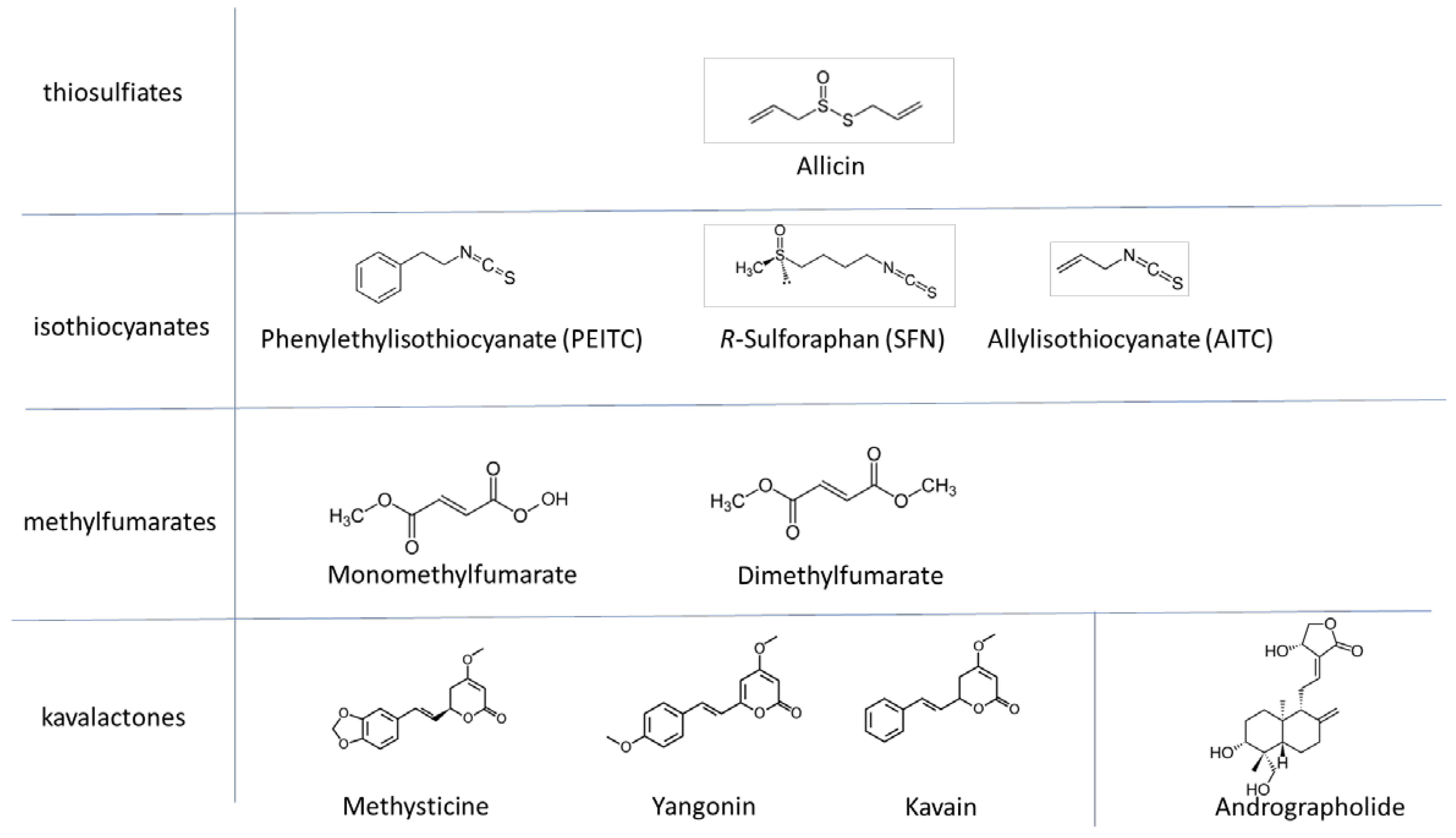
2.1. Test Substances

Allicin was synthesized according to our published protocol [35] and used as an aqueous solution. PEITC was purchased from Alpha Aesar Ltd. (Karlsruhe, Germany), AITC, and SFN from Sigma-Aldrich (Darmstadt, Germany). All these substances are water-soluble at the concentrations used and were used as aqueous solutions in the OSI1 tests. However, because it is sometimes necessary to dissolve sparingly soluble substances in DMSO, tests with the ARE reporter system were carried out with solutions of the test substances in 0.03% DMSO. No effect of DMSO on cell viability or induction of luminescence was observed up to 0.1%.

2.2. Hepatocyte Cell Culture

Primary murine hepatocytes were isolated, as described previously [36]. The cells were cultured in DMEM High Glucose medium (Sigma-Aldrich, Steinheim, Germany) supplemented with 10% heat-inactivated fetal bovine serum (Gibco, Madison, WI, USA) and 1% penicillin/streptomycin under normoxia at 37 °C. The cells were transfected with the SIN-lenti-ARE reporter gene plasmid that allows the measurement of Nrf2 activity by bioluminescence detection.
For CellTiter-Blue® (Promega, Walldorf, Germany), as well as ARE reporter gene assays, 3 × 10⁴ cells/well were seeded in a 96-well microtiter plate. Cells were treated with either an aqueous solution of allicin, or PEITC, AITC, and SFN, in up to 0.1% DMSO for 6 h. The DMSO control corresponds to the highest concentration of DMSO used in the treatment.
The protocol was approved by the regional authorities of North Rhine-Westphalia (Landesamt für Natur, Umwelt und Verbraucherschutz LANUV North Rhine-Westfaphalia, Germany, ID: 8.87-51.05.20.10.063).

2.3. Cell Titer Blue® (CTB®) Assay

The CTB® assay (Promega) was conducted as recommended by the manufacturer without any changes to the protocol.

2.4. ARE Reporter Gene Assay

The SIN-lenti-ARE reporter gene construct [37] was based on the addgene plasmid #24308, in which the luciferase gene is under the control of an upstream minimal promoter driven by multiple TCF (T-cell factor) response elements. The 7 × TCF sequences of the 7TFP plasmid (a kind gift from Roel Nusse, Department of Developmental Biology, Howard Hughes Medical Institute, Stanford Institute for Stem Cell Biology and Regenerative Medicine, Stanford University School of Medicine, Stanford, CA, USA [38]) were removed by enzymatic restriction using PstI and NheI and replaced with the antioxidant response element (ARE) consensus sequence of the NADPH:quinone oxidoreductase 1 promoter (ggtaccagtctagagtcacagtgacttggcaaaatcgctagc) [19,39]. Test restriction, gel electrophoresis, and DNA sequencing verified the resulting plasmid DNA. The production of lentiviral particles was conducted as described before [40]. Briefly, HEK293 cells were cotransfected with a second-generation lentiviral plasmid system containing SIN-lenti-ARE as transfer vector, psPAX2 as a packaging vector, and pMD2.G as the vector encoding for the envelope protein. Supernatants containing lentiviral particles were used for gene delivery.
Primary murine hepatocytes were transfected with the SIN-lenti-ARE reporter gene construct. Cells were lysed in passive lysis buffer (PLB) as recommended by the manufacturer (Promega). For the luciferase assay, 50 µL of the lysate was transferred to a white 96-well plate. Luciferase assay reagent I (LAR I) was injected automatically by the luminometer (Glomax 96, Promega). Sulforaphane (SFN: 7.5 µM) treatment was included as a positive control for Nrf2 activation. Measurement was conducted with default setting parameters (reagent volume 50 µL, delay 0.4 ms, integration time 10 s). Because sparingly soluble test substances often need to be dissolved in aqueous DMSO and then added to the test mix, a control with the highest DMSO end concentration but without test substance was performed.

2.5. Yeast Cell Cultivation

The BY4742 wildtype strain (Mat α; his3Δ1; leu2Δ0; lys2Δ0; ura3Δ0) was used. Yeast cells were cultivated on CSM-medium (7 g L−1 yeast nitrogen base containing (NH4)2SO4 without amino acids (ForMedium Ltd., Norwich, UK); 40 g L−1 glucose (Carl Roth, Karlsruhe, Germany); 0.8 g L−1 CSM complete dropout (ForMedium Ltd., Norwich, UK) at 28 °C and liquid culture was shaken at 220 rpm.

2.6. OSI1-Promoter::Luciferase Reporter Construct

Details of the cloning procedure have been published elsewhere [3]. The finished construct was amplified in E.coli DH5α and verified by colony PCR, restriction digestion, and sequencing the plasmid. It was transformed into BY4742 yeast cells by electroporation and plated out on uracil-lacking CSM medium.
Luciferin (Diagonal, Münster, Germany) was dissolved in 0.1 M sodium citrate, pH = 5 (Carl Roth, Karlsruhe, Germany) to a final concentration of 1 mM luciferin. Then, 100 µL of an overnight-culture in CSM-medium was adjusted to an OD600 = 0.5 and was treated with 25 µL of 5 × concentrated aqueous solution of test substance and 25 µL of luciferin-solution in black-sided, transparent-flat-bottomed 96-well microtiter plates. Luminescence was measured in a Tristar2S plate reader (Berthold, Bad Wildungen, Germany).

2.7. Statistics

Error bars show standard deviation. A normal distribution was assumed and a one-way ANOVA was performed with Dunett’s multiple comparisons test for significance (** p < 0.01).

3. Results

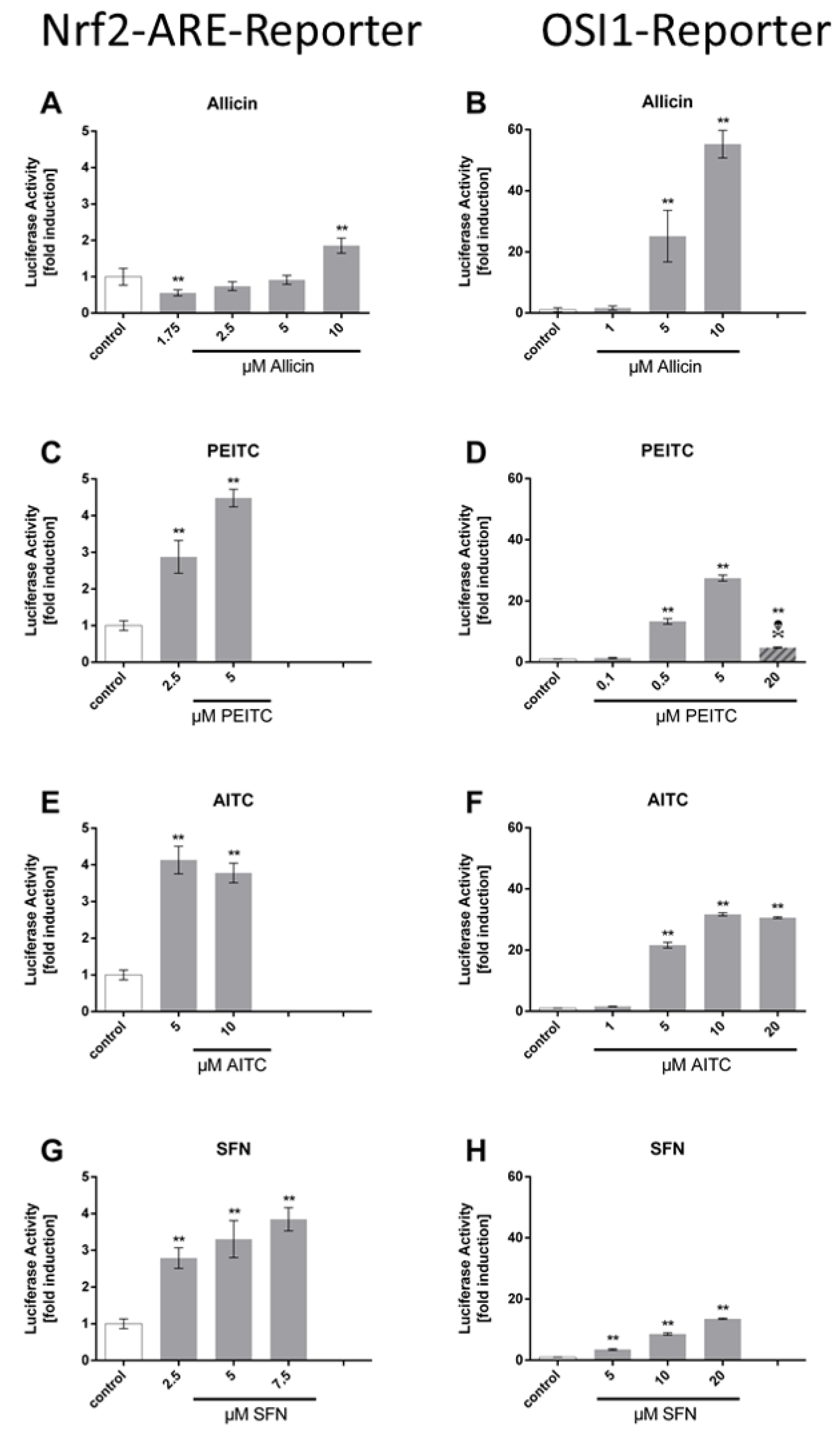
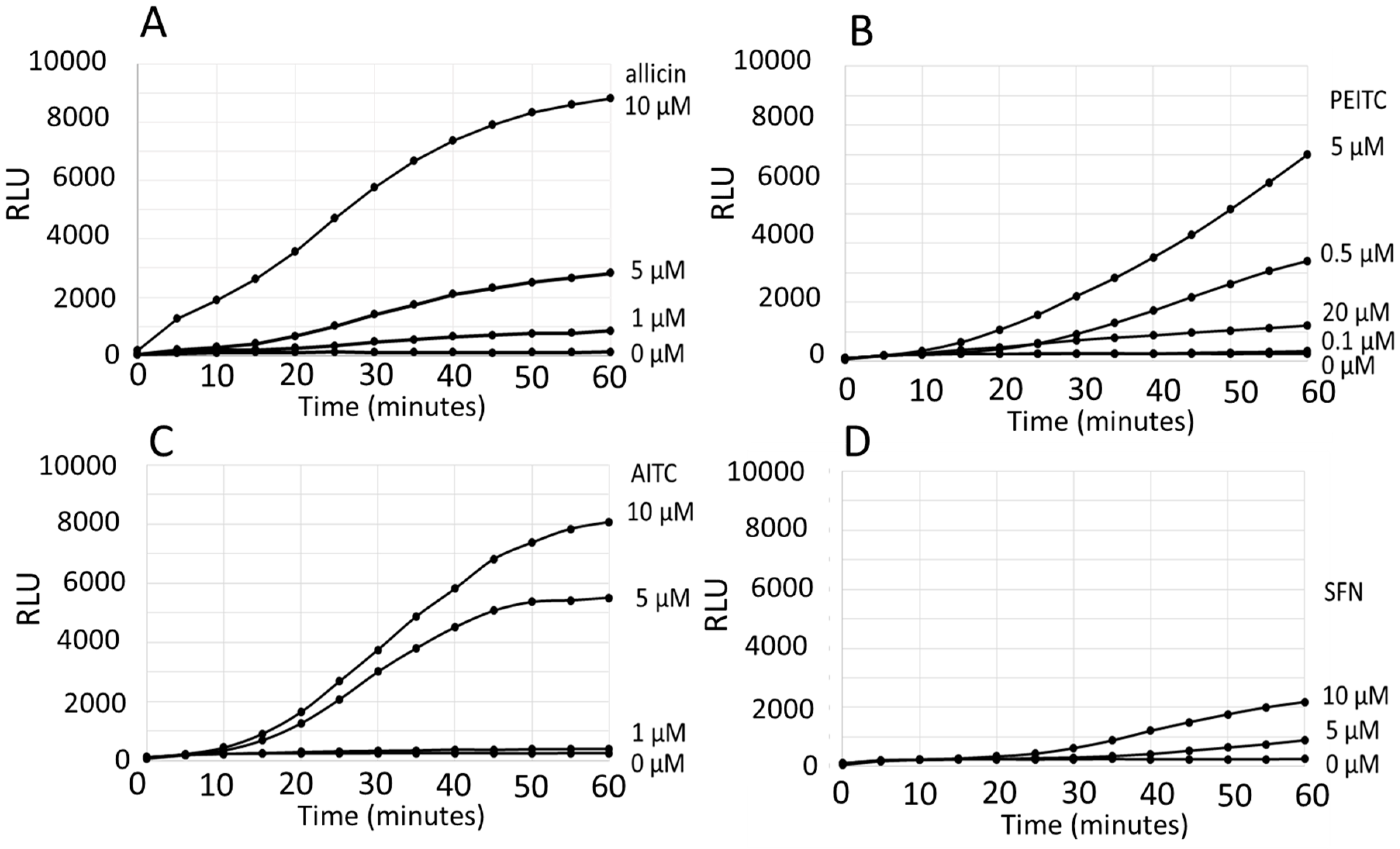
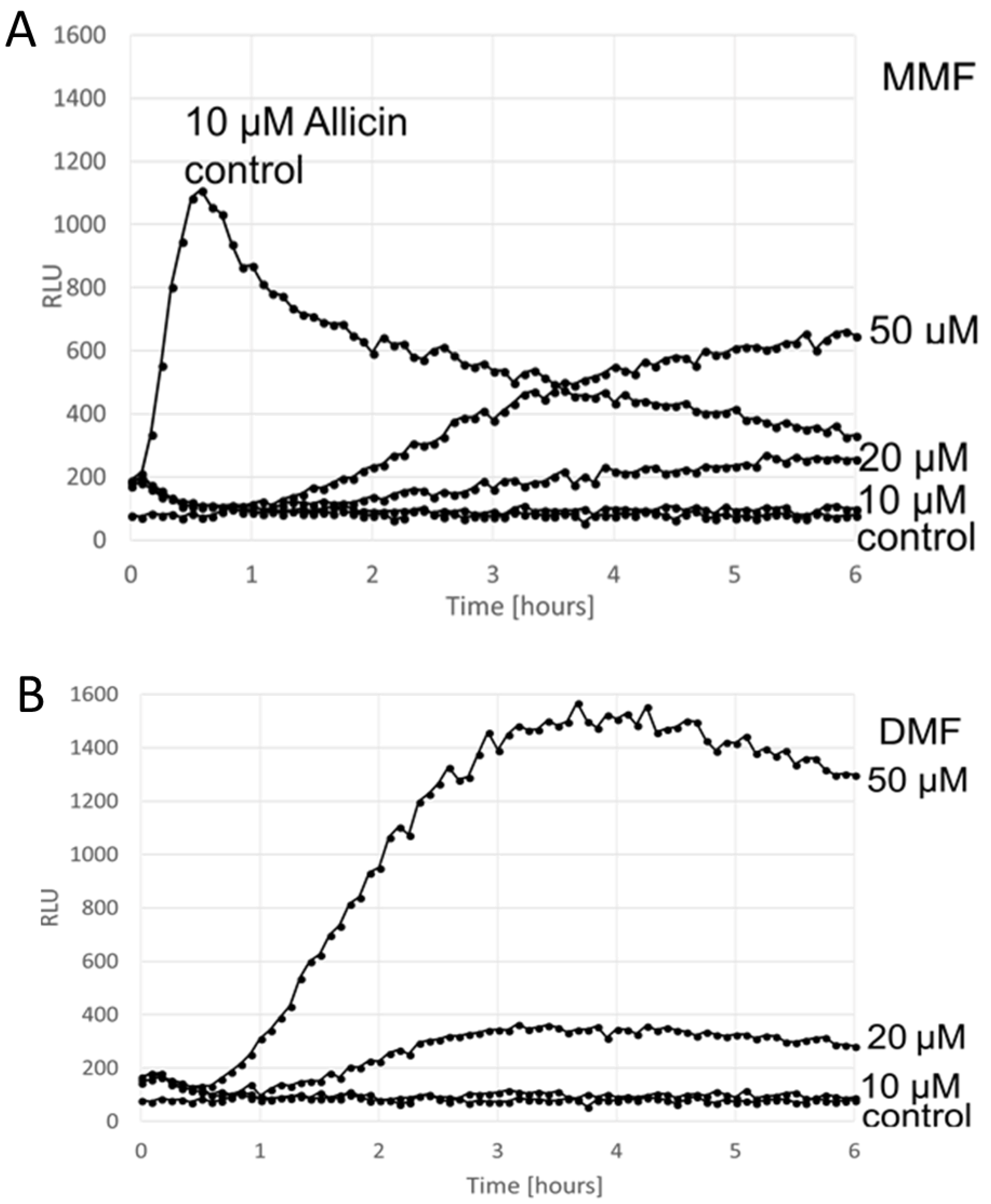
Comparison of the ARE Reporter System in Murine Hepatocytes and OSI1 in BY4742 Yeast Cells

The four test substances, allicin, phenylethyl isothiocyanate (PEITC), allyl isothiocyanate (AITC) and sulforaphane (SFN), were able to induce the luciferase reporter in both systems at low µM concentrations (Figure 1). In the ARE assay, which was measured after 6 h of co-cultivation, the concentration-dependent increase of the luminescence signal compared to non-treated controls ranged from ~2-fold (allicin) to ~4-fold (PEITC, AITC, SFN). Allicin showed significant ARE activation at 10 µM, PEITC at 2.5–5 µM, AITC at 5–10 µM, and SFN at 2.5–7.5 µM, respectively (Figure 1A,C,E,G). In comparison, after 1 h of co-cultivation with the four test substances, the OSI1 reporter in yeast cells showed a 15-fold (sulforaphane) to >50-fold induction (allicin) (Figure 1 B,D,F,H). The ARE reporter was more sensitive to sulforaphane (2.5 vs. 10 µM; Figure 1G,H) but the OSI1 reporter system was more sensitive to allicin (5 vs. 10 µM; Figure 1A,B) and PEITC (0.5 vs. 2.5 µM; Figure 1C,D).
Routinely, in the ARE assay with murine hepatocytes, an “end point” measurement is made, in this case, after 6 h of co-cultivation of the cells with the test substance. However, for the OSI1 reporter, no pre-cultivation of the yeast cells with the test substance is necessary, as the measurements in the 96-well plates are dynamic from time zero and show the increase in luminescence over time (Figure 2), i.e., a kinetic is established rather than an end-point measurement. Thus, it can be seen that within the first 30 min of exposure, all the test substances activated the OSI1 reporter in the low µM range. The OSI1 reporter was particularly responsive to PEITC (0.5 µM) and allicin (1 µM), while AITC and SFN showed clear responses in the 5–10 µM range (Figure 2A–D). The data from the measurements at the 1 h time point were incorporated into Figure 1B,D,F,G to facilitate a more direct comparison with the ARE reporter system.
With the exception of the thiosulfinate allicin, all of the substances tested in the previous section were isothiocyanates. We, therefore, screened the non-isothiocyanate α-β-unsaturated electrophilic substances monomethyl and dimethyl fumarate for activity with the OSI1::luciferase reporter. The response was measured over 6 h, and the dose-dependent kinetics of the OSI1::luciferase response is clearly visible (Figure 3). Interestingly, the OSI1 test system responded more rapidly to allicin than to mono- and dimethyl fumarate, and this emphasizes the advantage of the OSI1::luciferase reporter giving a kinetic response, compared to the ARE reporter which only gives a single time point reading.
Kavalactones are weak antioxidants present in the rhizome and roots of the kava plant (Piper methysticum G. Forst), which were demonstrated to activate Keap1–Nrf2/ARE by a non-oxidative route [41]. Thus, we would not expect them to stimulate the oxidation-sensitive trigger-cysteines of Yap1p and activate the OSI1::luciferase reporter. This was indeed the case and analytically pure samples of methysticin, yangonin, kavain and the diterpene lactone andrographolide [42] did not elicit luminescence up to a concentration of 400 µM (DMSO 5% (v/v); data not shown). This emphasizes that the Yap1p-dependent OSI1::luciferase reporter detects physiologically active electrophilic oxidants and not substances that activate Keap1–Nrf2/ARE by a non-oxidative route, for example, by affecting the binding pocket of the Keap1–Nrf2 complex.

4. Discussion

In this study, different substances were tested that are either known to be thiol-reactive (as is the case for thiosulfinates and isothiocyanates) or known to induce the Nrf2 system without oxidizing or modifying the relevant cysteine residues. An overview of the chemical structures is given in Figure 4.
Keap1–Nrf2/ARE-based assays represent an elegant and relatively easy-to-use approach to examine the oxidative potential of electrophilic substances on mammalian cells [16]. These assays are usually conducted with immortalized human or murine cell lines, thereby allowing high-throughput screening. However, these are cancer cell lines originating mostly from tumor biopsies. In the last few years, research has shown that, aside from its well-known antioxidant functions, Nrf2 has roles in the development and the progression of cancer. Furthermore, it has been shown that Nrf2 is also involved in many oxidative stress-independent intracellular processes, such as the cell cycle [40], metabolism [43,44], and differentiation [45]. Tumor cells have been shown to highjack the Nrf2/ARE system to adjust cellular processes to their specific requirements [46]. Moreover, many human solid cancers, as well as commonly used cell lines, show gain-of-function mutations of Nrf2 or loss-of-function mutations of Keap1 [47,48]. All these aspects make the use of cancer cell lines for Keap1–Nrf2/ARE-based assays at least questionable. For these reasons, we used freshly isolated primary cells in our study to better represent physiological conditions. In this case, the ARE-based assay requires the keeping and sacrifice of animals to obtain test cells, which are then transfected with ARE reporter construct before an assay. In the standard ARE protocol, from the onset of an assay with test substances, a six-hour incubation period is used and gives a point reading as output. Furthermore, the ARE procedure requires a prior assessment of the test substances for their effects on cell viability (e.g., CTB®) to decide on a suitable working concentration. In contrast, an overnight culture of yeast cells, already stably transformed with the OSI1 reporter construct, provides the starting inoculum for the log-phase cells used for the OSI1 reporter assay. The OSI1 reporter assay gives a continual output after the addition of test substances over any chosen concentration- and time-range, allowing for kinetic observations. Furthermore, the plate reader allows parallel measurements of luminescence and cell growth kinetics. Concentrations of test substance which reduce yeast cell growth show a reduced luminescence response compared to lower concentrations of the test substance. Thus, dose-dependency can be seen up to concentrations that begin to inhibit growth.
Furthermore, because cell culture itself is stressful, there is some induction of the ARE protection system leading to relatively high background luminescence in the controls. Thus, the OSI1-reporter has a further advantage in that it shows a several-fold higher signal-to-noise ratio in comparison to the ARE reporter and has a higher dynamic resolution.
In contrast to Keap1–Nrf2/ARE, the OSI1 reporter does not rely on slow translation events to accumulate the reporting transcription factor in the nucleus; therefore, its response is generally much faster, and the reaction to the stimulating substance can be followed easily in real-time. This enables the investigation of important kinetic parameters such as the uptake rate of the investigated substances or the bioavailability. The sensitivities of the ARE reporter and OSI1-reporter systems are similar for the tested substances and lie in the low µM range in both cases.
It should be emphasized, however, that the yeast system with the OSI1 reporter will mirror the Nrf2–Keap1/ARE system only with electrophilic oxidants that can attack trigger-cysteines in the respective sensor proteins. Substances that activate Nrf2 by non-oxidative mechanisms, such as the kavalactones and andrographolide, would not be expected to activate the OSI1 reporter, and indeed we show this to be so (Figure 3). Furthermore, because of differences in metabolism, yeast cells may, of course, be unable to modify some electrophilic compounds to convert them into active thiol oxidants as might happen in mammalian cells.
Nevertheless, the OSI1 reporter system offers a quick, sensitive, economical alternative to the ARE reporter system for detecting physiologically active electrophilic oxidants and has the added bonus that it does not require animal sacrifice. Indeed, a recent report also drew attention to similarities between the sensing of electrophiles in fungi via Yap1p activation and the Keap1–Nrf2/ARE in mammals [49].
An added advantage of working with yeast is that having identified an active substance, knock-out libraries of non-essential yeast genes and libraries maintaining knock-outs of essential genes as heterozygotes in diploid yeast lines are available for screens (www.euroscarf.de/index.php?name=News) to identify genes of interest related to the effects of the test substance [50].

Author Contributions

I.S. invented and established the OSI1 reporter system and performed initial experiments with allicin and other oxidants. A.U. performed the experiments with PEITC, AITC, SFM, MMF, DMF and kavalactones using the OSI1 reporter systems. T.C. performed all the experiments with the NRF2/ARE reporter system. A.F., M.C.H.G. and A.J.S. planned and supervised the experiments and wrote the manuscript. All authors have read and agreed to the published version of the manuscript.

Funding

This research was funded by RWTH Aachen University (MCHG, A.J.S.) and Undergraduate Research Opportunity Program (UROP) of RWTH Aachen University (Grants to A.U. and I.S.).

Acknowledgments

We thank Ulrike Noll for excellent technical assistance.

Conflicts of Interest

The authors declare no conflict of interest.

References

  1. Morano, K.A.; Grant, C.M.; Moye-Rowley, W.S. The Response to Heat Shock and Oxidative Stress in Saccharomyces cerevisiae. Genetics 2012, 190, 1157–1195. [Google Scholar] [CrossRef] [Green Version]
  2. Azevedo, D.; Tacnet, F.; Delaunay, A.; Rodrigues-Pousada, C.; Toledano, M.B. Two redox centers within Yap1 for H2O2 and thiol-reactive chemicals signaling. Free Radic. Biol. Med. 2003, 35, 889–900. [Google Scholar] [CrossRef]
  3. Gruhlke, M.C.H.; Schlembach, I.; Leontiev, R.; Uebachs, A.; Gollwitzer, P.U.G.; Weiss, A.; Delaunay, A.; Toledano, M.; Slusarenko, A.J. Yap1p, the central regulator of the S. cerevisiae oxidative stress response, is activated by allicin, a natural oxidant and defence substance of garlic. Free Radic. Biol. Med. 2017, 108, 793–802. [Google Scholar] [CrossRef] [PubMed]
  4. Borlinghaus, J.; Albrecht, F.; Gruhlke, M.C.H.; Nwachukwu, I.D.; Slusarenko, A.J. Allicin: Chemistry and Biological Properties. Molecules 2014, 19, 12591–12618. [Google Scholar] [CrossRef] [PubMed] [Green Version]
  5. Yu, L.; Guo, N.; Meng, R.; Liu, B.; Tang, X.; Jin, J.; Cui, Y.; Deng, X. Allicin-induced global gene expression profile of Saccharomyces cerevisiae. Appl. Microbiol. Biotechnol. 2010, 88, 219–229. [Google Scholar] [CrossRef] [PubMed]
  6. Iwahashi, Y.; Hosoda, H.; Park, J.-H.; Lee, J.-H.; Suzuki, Y.; Kitagawa, E.; Murata, S.M.; Jwa, N.-S.; Gu, M.B.; Iwahashi, H. Mechanisms of Patulin Toxicity under Conditions That Inhibit Yeast Growth. J. Agric. Food Chem. 2010, 54, 1936–1942. [Google Scholar] [CrossRef]
  7. Trott, A.; West, J.D.; Klaić, L.; Westerheide, S.D.; Silverman, R.B.; Morimoto, R.I.; Morano, K.A. Activation of Heat Shock and Antioxidant Responses by the Natural Product Celastrol: Transcriptional Signatures of a Thiol-targeted Molecule. Mol. Biol. Cell 2008, 19, 1104–1112. [Google Scholar] [CrossRef] [Green Version]
  8. Heer, D.; Heine, D.; Sauer, U. Resistance of Saccharomyces cerevisiae to High Concentrations of Furfural Is Based on NADPH-Dependent Reduction by at Least Two Oxireductases. Appl. Environ. Microbiol. 2009, 75, 7631–7638. [Google Scholar] [CrossRef] [Green Version]
  9. Bat-Chen, W.; Tal, G.; Irena, P.; Zvi, L.; Betty, S. Allicin purified from fresh garlic cloves induces apoptosis in colon cancer cells via Nrf2. Nutr. Cancer 2009, 62, 947–957. [Google Scholar] [CrossRef]
  10. Kansanen, E.; Kuosmanen, S.M.; Leinonen, H.; Levonen, A.-L. The Keap1-Nrf2 pathway: Mechanisms of activation and dysregulation in cancer. Redox Biol. 2013, 1, 45–49. [Google Scholar] [CrossRef] [Green Version]
  11. Niture, S.K.; Khatri, R.; Jaiswal, A.K. Regulation ofNrf2—An update. Free Radic. Biol. Med. 2014, 66, 36–44. [Google Scholar] [CrossRef] [PubMed] [Green Version]
  12. Liebler, D.C.; Guengerich, F.P. Elucidating mechanisms of drug-induced toxicity. Nat. Rev. Drug Discov. 2005, 4, 410–420. [Google Scholar] [CrossRef] [PubMed]
  13. Satoh, T.; McKercher, S.R.; Lipton, S.A. Nrf2/ARE-mediated antioxidant actions of pro-electrophilic drugs. Free Radic. Biol. Med. 2013, 65, 645–657. [Google Scholar] [CrossRef] [PubMed] [Green Version]
  14. Wang, X.J.; Hayes, J.D.; Wolf, C.R. Generation of a Stable Antioxidant Response Element–Driven Reporter Gene Cell Line and Its Use to Show Redox-Dependent Activation of Nrf2 by Cancer Chemotherapeutic Agents. Cancer Res. 2006, 66, 10983–10994. [Google Scholar] [CrossRef] [PubMed] [Green Version]
  15. Ajit, D.; Simonyi, A.; Li, R.; Chen, Z.; Hannik, M.; Fritsche, K.L.; Mossine, V.V.; Smith, R.E.; Dobbs, T.K.; Luo, R.; et al. Phytochemicals and botanical extracts regulate NF-κB and Nrf2/ARE reporter activities in DI TNC1 astrocytes. Neurochem. Int. 2016, 97, 49–56. [Google Scholar] [CrossRef] [PubMed] [Green Version]
  16. Ramirez, T.; Mehling, A.; Kolle, S.N.; Wruck, C.J.; Teubner, W.; Eltze, T.; Aumann, A.; Urbisch, D.; van Ravenzwaay, B.; Landsiedel, R. LuSens: A keratinocyte based ARE reporter gene assay for use in integrated testing strategies for skin sensitization hazard identification. Toxicol. In Vitro 2014, 28, 1482–1497. [Google Scholar] [CrossRef] [Green Version]
  17. Ramirez, T.; Stein, N.; Aumann, A.; Remus, T.; Edwards, A.; Norman, K.G.; Ryan, C.; Bader, J.E.; Fehr, M.; Burleson, F.; et al. Intra- and inter-laboratory reproducibility and accuracy of the LuSens assay: A reporter gene-cell line to detect keratinocyte activation by skin sensitizers. Toxicol. In Vitro 2016, 32, 278–286. [Google Scholar] [CrossRef] [Green Version]
  18. Cheung, K.L.; Khor, T.O.; Kong, A.N. Synergistic Effect of Combination of Phenethyl Isothiocyanate and Sulforaphane or Curcumin and Sulforaphane in the Inhibition of Inflammation. Pharmaceut. Res. 2009, 26, 224–231. [Google Scholar] [CrossRef]
  19. Fragoulis, A.; Laufs, J.; Müller, S.; Soppa, U.; Siegl, S.; Reiss, L.K.; Tohidnezhad, M.; Rosen, C.; Tenbrock, K.; Varoga, D.; et al. Sulforaphane has opposing effects on TNF-alpha stimulated and unstimulated synoviocytes. Arthritis Res. Ther. 2012, 14, R220. [Google Scholar] [CrossRef] [Green Version]
  20. Tirumalai, R.; Rajesh Kumar, T.; Mai, K.H.; Biswal, S. Acrolein causes transcriptional induction of phase II genes by activation of Nrf2 in human lung type II epithelial (A549) cells. Toxicol. Lett. 2002, 132, 27–36. [Google Scholar] [CrossRef]
  21. Ouyang, X.; Tran, Q.T.; Goodwin, S.; Wible, R.S.; Sutter, C.H.; Sutter, T.R. Yap1 activation by H2O2 or thiol-reactive chemicals elicits distinct adaptive gene responses. Free Radic. Biol. Med. 2011, 50, 1–13. [Google Scholar] [CrossRef] [PubMed]
  22. Chen, J.; Shaikh, Z.A. Activation of Nrf2 by cadmium and its role in protection against cadmium-induced apoptosis in rat kidney cells. Toxicol. Appl. Pharmacol. 2009, 241, 81–89. [Google Scholar] [CrossRef] [PubMed]
  23. Wemmie, J.A.; Wu, A.L.; Harshman, K.D.; Parker, C.S.; Moye-Rowley, W.S. Transcriptional activation mediated by the yeast AP-1 protein is required for normal cadmium tolerance. J. Biol. Chem. 1994, 269, 14690–14697. [Google Scholar]
  24. Piccirillo, S.; Filomeni, G.; Brune, B.; Rotilio, G.; Ciriolo, M.R. Redox mechanisms involved in the selective activation of Nrf2-mediated resistance versus p53-dependent apoptosis in adenocarcinoma cells. J. Biol. Chem. 2009, 284, 27721–27733. [Google Scholar] [CrossRef] [PubMed] [Green Version]
  25. Delaunay, A.; Isnard, A.-D.; Toledano, M.B. H2O2 sensing through oxidation of the Yap1 transcription factor. EMBO J. 2000, 19, 5157–5166. [Google Scholar] [CrossRef]
  26. Kanlaya, R.; Khamchun, S.; Kapincharanon, C.; Thongboonkerd, V. Protective effect of epigallocatechin-3-gallate (EGCG) via Nrf2 pathway against oxalate-induced epithelial mesenchymal transition (EMT) of renal tubular cells. Sci. Rep. 2016, 6, 30233. [Google Scholar] [CrossRef] [PubMed]
  27. Maeta, K.; Nomura, W.; Takatsume, Y.; Izawa, S.; Inoue, Y. Green Tea Polyphenols Function as Prooxidants To Activate Oxidative-Stress-Responsive Transcription Factors in Yeasts. Appl. Environ. Microbiol. 2007, 73, 572–580. [Google Scholar] [CrossRef] [Green Version]
  28. Covas, G.; Marinho, H.S.; Cyrne, L.; Antunes, F. Activation of Nrf2 by H2O2: De novo synthesis versus nuclear translocation. Meth. Enzymol. 2013, 528, 157–171. [Google Scholar]
  29. Ya, B.; Li, H.; Wang, H.; Wu, F.; Xin, Q.; Cheng, H.; Li, W.; Lin, N.; Ba, Z.; Zhang, R.; et al. 5-HMF attenuates striatum oxidative damage via Nrf2/ARE signaling pathway following transient global cerebral ischemia. Cell Stress Chaperone 2017, 22, 55–65. [Google Scholar] [CrossRef] [Green Version]
  30. Kim, D.; Hahn, J.-S. Roles of the Yap1 Transcription Factor and Antioxidants in Saccharomyces cerevisiae’s Tolerance to Furfural and 5-Hydroxymethylfurfural, Which Function as Thiol-Reactive Electrophiles Generating Oxidative Stress. Appl. Environ. Microbiol. 2013, 79, 5069–5077. [Google Scholar] [CrossRef] [Green Version]
  31. Nishimoto, S.; Koike, S.; Inoue, N.; Suzuki, T.; Ogasawara, Y. Activation of Nrf2 attenuates carbonyl stress induced by methylglyoxal in human neuroblastoma cells: Increase in GSH levels is a critical event for the detoxification mechanism. Biochem. Biophys. Res. Commun. 2017, 483, 874–879. [Google Scholar] [CrossRef]
  32. Maeta, K.; Izawa, S.; Okazaki, S.; Kuge, S.; Inoue, Y. Activity of the Yap1 Transcription Factor in Saccharomyces cerevisiae Is Modulated by Methylglyoxal, a Metabolite Derived from Glycolysis. Mol. Cell. Biol. 2004, 24, 8753–8764. [Google Scholar] [CrossRef] [PubMed] [Green Version]
  33. Ungvari, Z.; Bagi, Z.; Feher, A.; Recchia, F.A.; Sonntag, W.E.; Pearson, K.; de Cabo, R.; Csiszar, A. Resveratrol confers endothelial protection via activation of the antioxidant transcription factor Nrf2. Am. J. Physiol. Heart C 2010, 299, H18–H24. [Google Scholar] [CrossRef] [PubMed] [Green Version]
  34. Escoté, X.; Miranda, M.; Menoyo, S.; Rodríguez-Porrata, B.; Carmona-Gutiérrez, D.; Jungwirth, H.; Madeo, F.; Cordero, R.R.; Mas, A.; Tinahones, F.; et al. Resveratrol induces antioxidant defence via transcription factor Yap1p. Yeast 2012, 29, 251–263. [Google Scholar] [CrossRef] [PubMed] [Green Version]
  35. Albrecht, F.; Leontiev, R.; Jacob, C.; Slusarenko, A.J. An Optimized Facile Procedure to synthesize and purify Allicin. Molecules 2017, 22, 770. [Google Scholar] [CrossRef] [PubMed] [Green Version]
  36. Kaldenbach, M.; Giebeler, A.; Tschaharganeh, D.F.; Erschfeld, S.; Wasmuth, H.E.; Dolle, L.; Floege, J.; Trautwein, C.; Streetz, K.L. Hepatocyte growth factor/c-Met signalling is important for the selection of transplanted hepatocytes. Gut 2012, 61, 1209–1218. [Google Scholar] [CrossRef] [PubMed]
  37. Jansen, S.; Kress, E.; Fragoulis, A.; Wruck, C.J.; Wolf, R.; Grötzinger, J.; Michalek, M.; Pufe, T.; Tauberg, S.C.; Brandenburg, L.-O. Psoriasin has divergent effects on the innate immune responses of murine glial cells. J. Neurochem. 2017, 141, 86–99. [Google Scholar] [CrossRef] [Green Version]
  38. Fuerer, C.; Nusse, R. Lentiviral vectors to probe and manipulate the wnt signaling pathway. PLoS ONE 2010, 5, e9370. [Google Scholar] [CrossRef]
  39. Wruck, C.J.; Claussen, M.; Führmann, G.; Römer, L.; Schulz, A.; Pufe, T.; Waetzig, V.; Peipp, M.; Herdegen, T.; Götz, M.E. Luteolin protects rat PC 12 and C6 cells against MPP+ induced toxicity via an ERK dependent Keapl-Nrf2-ARE pathway. In Neuropsychiatric Disorders An Integrative Approach; Gerlach, M., Deckert, J., Double, K., Koutsilieri, E., Eds.; Springer: Vienna, Austria, 2007; Volume 72, pp. 57–67. [Google Scholar]
  40. Reddy, N.M.; Kleeberger, S.R.; Bream, J.H.; Fallon, P.G.; Kensler, T.W.; Yamamoto, M.; Reddy, S.P. Genetic disruption of the Nrf2 compromises cell-cycle progression by impairing GSH-induced redox signalling. Oncogene 2008, 27, 5821–5832. [Google Scholar] [CrossRef] [Green Version]
  41. Wruck, C.J.; Götz, M.E.; Herdegen, T.; Varoga, D.; Brandenburg, L.-O.; Pufe, T. Kavalactones Protect Neural Cells against Amyloid β Peptide-Induced Neurotoxicity via Extracellular Signal-Regulated Kinase 1/2-Dependent Nuclear Factor Erythroid 2-Related Factor 2 Activation. Mol. Pharmacol. 2008, 73, 1785–1795. [Google Scholar] [CrossRef] [Green Version]
  42. Wong, S.Y.; Tan, M.G.; Wong, P.T.; Herr, D.R.; Lai, M.K. Andrographolide induces Nrf2 and heme oxygenase 1 in astrocytes by activating p38 MAPK and ERK. J. Neuroinflamm. 2016, 3, 251. [Google Scholar] [CrossRef] [PubMed] [Green Version]
  43. Saito, T.; Ichimura, Y.; Taguchi, K.; Suzuki, T.; Mizushima, T.; Takagi, K.; Hirose, Y.; Nagahashi, M.; Iso, T.; Fukutomi, T.; et al. p62/Sqstm1 promotes malignancy of HCV-positive hepatocellular carcinoma through Nrf2-dependent metabolic reprogramming. Nat. Commun. 2016, 7, 12030. [Google Scholar] [CrossRef] [PubMed]
  44. Wu, Y.S.; Looi, C.Y.; Subramaniam, K.S.; Masamune, A.; Chung, I. Soluble factors from stellate cells induce pancreatic cancer cell proliferation via Nrf2-activated metabolic reprogramming and ROS detoxification. Oncotarget 2016, 7, 36719–36732. [Google Scholar] [CrossRef] [PubMed] [Green Version]
  45. Murakami, S.; Motohashi, H. Roles of Nrf2 in cell proliferation and differentiation. Free Radic. Biol. Med. 2015, 88, 168–178. [Google Scholar] [CrossRef] [Green Version]
  46. Taguchi, K.; Yamamoto, M. The KeAP1–NRF2 System in Cancer. Front. Oncol. 2017, 7. [Google Scholar] [CrossRef]
  47. Menegon, S.; Columbano, A.; Giordano, S. The Dual Roles of NRF2 in Cancer. Trends Mol. Med. 2016, 22, 578–593. [Google Scholar] [CrossRef]
  48. Shibata, T.; Ohta, T.; Tong, K.I.; Kokubu, A.; Odogawa, R.; Tsuta, K.; Asamura, H.; Yamamoto, M.; Hirohashi, S. Cancer related mutations in NRF2 impair its recognition by Keap1-Cul3 E3 ligase and promote malignancy. Proc. Natl. Acad. Sci. USA 2008, 105, 13568–13573. [Google Scholar] [CrossRef] [Green Version]
  49. Marinho, H.S.; Real, C.; Cyrne, L.; Soares, H.; Antunes, F. Hydrogen peroxide sensing, signaling and regulation of transcription factors. Redox Biol. 2014, 2, 535–562. [Google Scholar] [CrossRef] [Green Version]
  50. Leontiev, R.; Slusarenko, A.J. Finding the Starting Point for Mode-of-Action Studies of Novel Selenium Compounds: Yeast as a Genetic Toolkit. Curr. Org. Synth. 2017, 14, 1102–1108. [Google Scholar] [CrossRef]
Figure 1. Responses of the antioxidant response element (ARE) and the OS-induced promoter of the YKL071w gene (OSI1) reporters to allicin, phenylethyl isothiocyanate (PEITC), allyl isothiocyanate (AITC), sulforaphane (SFN). (A,C,E,G) show the ARE assay with murine hepatocytes transfected with the SIN-lenti-ARE gene construct as reporter. Measurements were taken 6 h after treatment (n = 8). (B,D,F,H) show the OSI1 assay with yeast cells transformed with the OSI1 promoter–luciferase gene construct as reporter (n = 3). Measurements are shown after 1 h of co-cultivation with the respective test substance. Attention is drawn to the different y-axis scales of the ARE and OSI1 tests. Error bars show standard deviation. A normal distribution was assumed, and a one-way ANOVA was performed with Dunett’s multiple comparisons test for significance (** p < 0.01). RLU is the abbreviation for Relative Luminescence Units.
Applsci 10 03520 g001
Figure 2. Dynamic response of yeast cells containing the OSI1::luciferase reporter to (A) allicin, (B) phenylethyl isothiocyanate (PEITC), (C) allyl isothiocyanate (AITC), (D) sulforaphane (SFN). Dose-dependency can be seen, and higher concentrations, which can already potentially damage the cells, show a lower luminescence signal, as can be seen in the example of 20 µM PEITC (B). RLU is the abbreviation for Relative Luminescence Units.
Applsci 10 03520 g002
Figure 3. Dynamic response of yeast cells containing the OSI1::luciferase reporter to (A) monomethyl fumarate (MMF) (B) dimethyl fumarate (DMF). MMF and DMF were dissolved in DMSO. In the assay was 5% (v/v). n = 3. RLU is the abbreviation for Relative Luminescence Units.
Applsci 10 03520 g003
Figure 4. Overview of the chemical structure of the tested compounds. The compounds can be roughly divided into five chemical groups. Allicin belongs to the thiosulfinates, which are typical for representatives of the genus Allium. The isothiocyanates AITC, PEITC, and sulforaphane belong to the isothiocyanates and are mainly found in the Brassicaceae family. Methyl fumarates are esters of fumaric acid and are synthetic in nature. All these compounds have a thiol reactivity in common. The kavalactones (methysticin, yangonin, kavain) and the diterpene lactone andrographolide are derived from the plant Piper methysticum G. Forst and activate the Keap/Nrf2 system in a non-oxidative way.
Applsci 10 03520 g004
Table 1. A selection of substances shown to activate both the Keap1–Nrf2/ARE and Yap1 oxidative stress response systems in mammals and in yeast, respectively.
SubstanceActivation of Keap1–Nrf2/AREActivation of Yap1
AcroleinTirumalai et al. (2002) [20]Ouyang et al. (2011) [21]
CadmiumChen and Shaikh (2009) [22]Wemmie et al. (1994) [23]
CelastrolTrott et al. (2008) [7]Trott et al. (2008) [7]
DiamidePiccirillo et al. (2009) [24]Delauney et al. (2000) [25]
epigallocatechin gallateKanlaya et al. (2016) [26]Maeta et al. (2007) [27]
hydrogen peroxideCovas et al. (2013) [28]Delauney et al. (2000) [25]
5-hydroxymethylfurfuralYa et al. (2017) [29]Kim and Hahn (2013) [30]
MethylglyoxalNishimoto et al. (2017) [31]Maeta et al. (2004) [32]
ResveratrolUngvari et al. (2010) [33]Escote et al. (2012) [34]

