Article

Field-Realistic Pendimethalin Exposure Induces Sublethal Alterations in the Gut and Malpighian Tubules of a Beneficial Ground Beetle


by
Maria Luigia Vommaro
1,*,
Piero Giulio Giulianini
2 and
Anita Giglio
1,*
1
Department of Biology, Ecology and Earth Science, University of Calabria, 87036 Rende, Italy
2
Department of Life Sciences, University of Trieste, 34128 Trieste, Italy
*
Authors to whom correspondence should be addressed.
Environments 2026, 13(7), 394; https://doi.org/10.3390/environments13070394
Submission received: 10 June 2026 / Revised: 2 July 2026 / Accepted: 9 July 2026 / Published: 10 July 2026
(This article belongs to the Section Environmental Pollution, Toxicology and Restoration)

Abstract

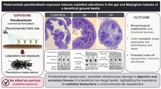
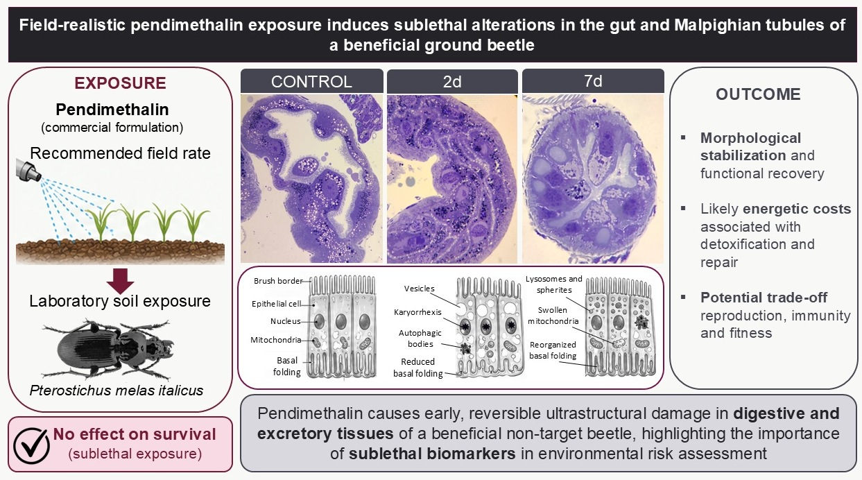
Herbicides are widely used in modern agriculture to control weeds and maintain crop productivity, but their persistence in soil raises concerns about unintended effects on non-target organisms. Pendimethalin, a dinitroaniline herbicide extensively applied to cereal and vegetable crops, is designed to target plant microtubules and is generally considered unlikely to pose genotoxic risks to animals. However, information on its sublethal effects on beneficial soil arthropods remains limited. In this study, we investigated the cytotoxic and histopathological effects of a commercial pendimethalin-based formulation on the ground beetle Pterostichus melas italicus, an ecologically relevant predatory species in agroecosystems. Adult males collected from an organic farm were exposed under laboratory conditions to soil treated at the recommended field dose and maintained for up to 7 days, corresponding to subchronic exposure. Individuals were sampled after 2 and 7 days, and the midgut and Malpighian tubules were analysed using histological and transmission electron microscopy. Exposure induced marked but non-lethal ultrastructural alterations, particularly in the Malpighian tubules, including reduction in the basal labyrinth, cytoplasmic vacuolisation, mitochondrial swelling, increased phagolysosome abundance, and nuclear karyorrhexis. These effects were transient under laboratory conditions and occurred without detectable impacts on survival, highlighting the Malpighian tubules as sensitive targets for the early detection of herbicide-induced physiological disturbances. However, the observed recovery may reflect compensatory physiological processes that could entail energetic costs and, under field conditions characterized by multiple concurrent stressors, potentially compromise physiological performance and predatory efficiency. Consequently, this study underscores the necessity of integrating sublethal ultrastructural biomarkers into environmental risk assessment frameworks for non-target beneficial insects.

Graphical Abstract

1. Introduction

The intensive utilisation of agrochemicals in conventional agriculture has given rise to persistent concerns regarding their unintended effects on non-target organisms, particularly soil-dwelling arthropods [1,2]. It is evident that, despite their primary function of suppressing plant growth, herbicides have the capacity to exert an indirect or direct influence on insects inhabiting treated fields [3,4,5]. The persistence of these substances in soil, where residues can remain active for extended periods ranging from weeks to months, has been demonstrated to amplify the potential for chronic exposure and subtle physiological disturbances in non-target organisms [6,7].
Pendimethalin, a dinitroaniline herbicide extensively employed in cereal and vegetable cultivation, exerts its function by interfering with microtubule formation and mitosis in plant cells [8]. Whilst the substance’s intensive application is intended to suppress plant life, its secondary impact on non-target fauna frequently manifests in the form of subtle physiological disturbances, as opposed to immediate mortality [9]. Dinitroanilines are widely regarded as posing a minimal risk of direct lethality or significant genotoxic damage to animals, primarily due to their low affinity for animal tubulin [10]. However, pendimethalin residues have been shown to exert cytotoxic and immunological effects in exposed non-target insects [9]. At field-realistic rates, it produces histopathological damage in the excretory–digestive system of Apis mellifera (Hymenoptera) [11], and it induces haemocyte genotoxicity (micronuclei, nuclear pleomorphism) in Harpalus rufipes (Coleopera) [12]. Comparable impacts occur across taxa, including increased male mortality in Tiphia vernalis (Hymenoptera) [13], reduced reproduction and growth in Folsomia candida (Collembola) and Eisenia fetida (Clitellata) at 1 mg Kg−1 [14], detoxification enzyme induction in A. mellifera ligustica and A. cerana cerana larvae [15], and impaired development in springtails (Xenylla welchi, Cyphoderus javanus; Collembola) [16,17]. At the systemic level, the herbicide produces immune alteration, such as reduced phagocytic activity in H. rufipes [12] and altered antimicrobial peptide (AMPs) production in tenebrionid beetles [18,19]. However, the mechanism of action in invertebrate systems remains poorly understood. Given its persistence in soil and high use frequency [7,20], further investigation of these sublethal effects on terrestrial arthropods is warranted.
Ground beetles (Coleoptera, Carabidae) represent a dominant group of beneficial arthropods in temperate agroecosystems, functioning as predators within the soil trophic web, where they contribute to biological pest control and serve as bioindicators of soil health [21,22]. The carabid Pterostichus melas italicus Dejean, 1828 is a widespread generalist, eurytopic, and thermophilous species predominantly associated with the clay soils of pastures, open forests, and cultivated terrains in Southern Europe [23]. Due to their soil-dwelling behaviour and trophic habits, carabids are especially susceptible to agrochemical exposure through both dermal contact and contaminated prey consumption [24].
The digestive and excretory systems, specifically the alimentary canal and Malpighian tubules, are particularly vulnerable to toxic insults as they play central roles in nutrient absorption, osmoregulation, and the detoxification of xenobiotics [25,26]. Specific cellular components, such as the mitochondria and the basal labyrinth, are established sensitive targets for environmental stressors [27,28]. For instance, Malpighian tubules have been identified as key structures for evaluating detoxification capacity through excretion [29]. In addition to their established contributions to waste filtration and osmotic regulation, Malpighian tubules function as decentralized “cell-autonomous immune entities” [30]. This functional overlap underscores why Malpighian tubules are sensitive indicators of systemic physiological disturbances [31]. Concurrently, the midgut (ventricle) is also particularly sensitive to toxic insults, playing a central role in nutrient absorption and featuring elongated gastric caeca that increase absorptive surface area [32]. Ultrastructural and histological alterations in these tissues can thus serve as early and sensitive biomarkers of physiological stress following exposure to environmental contaminants [33].
This study aims to test the hypothesis that a pendimethalin-based commercial herbicide, applied at the recommended field rate, induces early cytotoxic and ultrastructural alterations in the gut epithelium and Malpighian tubules of P. melas italicus exposed to contaminated soil under controlled laboratory conditions. This study is part of a broader project investigating the physiological responses to pendimethalin exposure in this species, including alterations in the gut microbiota [34]. By combining histological and ultrastructural analyses, we seek to characterize sublethal tissue alterations that could compromise insect fitness and contribute to the decline of beneficial insect populations in intensively managed agroecosystems.

2. Materials and Methods

2.1. Species Collection and Laboratory Maintenance

In October, adult specimens of Pterostichus melas were sampled (n = 187; 123 males and 64 females) at an organic olive grove located in San Marco Argentano, Calabria, Southern Italy (39°59′27.56″ N, 16°15′32.64″ E; elevation ca. 1202 m). The insects were captured using live pitfall traps, 9 cm diameter, plastic jars baited with fruit. Once collected, beetles were transported to the laboratory, identified via a dichotomous key [35], sexed, and housed in 5 L plastic boxes containing approximately 6 cm of soil collected from the same site in groups of 10 individuals for acclimation period. Environmental conditions were maintained at ~60% relative humidity under room temperature and natural daylight. Beetles were fed ad libitum with mealworms (stock population in the same laboratory) and organic apples. Female specimens were excluded from the ultrastructural and histopathological analyses to eliminate sex-specific physiological confounding factors, such as vitellogenesis and differential metabolic demands during egg production. For the soil-contact bioassays addressed in this study, the male cohort (n = 123; 63 untreated controls and 60 pendimethalin-treated individuals) was utilized. To simulate agrochemical exposure, males were exposed to a commercial pendimethalin-based formulation (Activus EC, product no. HRB00858-39, ADAMA Italia S.r.l., Grassobbio, Italy; 330 g L−1 active ingredient), herein referred to as PND, sprayed at the field-recommended rate (equivalent to 4 L/ha in typical cereal and vegetable systems). Considering a soil half-life of 24–34 days in sandy acidic conditions [6,36], a sub-chronic 7-day exposure protocol was designed. To accurately assess the impact of pendimethalin on beneficial soil fauna, it is essential to consider the temporal dynamics of its toxicity. In this study, we selected two time points, 2 and 7 days, to simulate different phases of field exposure. The 2-day interval represents the acute toxicity phase, occurring immediately after application when residues are most active. The 7-day interval allows for the evaluation of subchronic exposure, a period during which initial cytotoxic insults may either persist or trigger physiological recovery.
To prevent overcrowding and ensure true replication, insects were distributed across multiple independent boxes (maximum 10 individuals per box); each box had a surface area of 180.5 cm2. Each of the treatment replicates was filled with uncontaminated sandy soil (pH ~5) that had been sourced from the collection site. For the treatment of boxes, a solution of 7.2 µL of Activus diluted in 14 mL of distilled water was applied to the soil surface using a pipette, with the aim of simulating realistic field contact. The experimental setup focused on simulating dermal exposure via contact with the treated soil matrix, but there is also a potential for oral intake, as carabids drink and absorb water and soil particles from the substrate. Control boxes received an equivalent volume of distilled water. Following a 15 min period, a total of ten male beetles were introduced into each container. Survival was checked and recorded daily in both control and treated groups throughout the experimental period to ensure that the designated sampling intervals (days 2 and 7) captured sublethal physiological states rather than acute mortality events. To provide the required tissue samples, 5 individuals were randomly taken from one control box, and 5 individuals were randomly taken from two PND-treated replicate boxes. Each male was dissected for structural and ultrastructural evaluations for each experimental condition. Prior to dissection, individuals were cold-anesthetized for ~3 min at 0 °C, and dissected under a Zeiss stereomicroscope. Morphometric measurements of epithelial cells were performed on digitized images using ImageJ software (v. 1.54g; National Institutes of Health, Bethesda, MD, USA), and data are expressed as means ± standard deviation.

2.2. Electron Microscopy

Immediately following the dissection process, the alimentary canal and Malpighian tubules were removed and immersed in a freshly prepared fixative solution, comprising 2.5% glutaraldehyde, 1% paraformaldehyde, and 7.5% picric acid in 0.1 M phosphate buffer (PBS, pH 7.4) with 1.5% sucrose. Samples were fixed at 4 °C for 12 h, after which they were rinsed in PBS. They were then incubated in 1% osmium tetroxide in 0.1 M phosphate buffer at 4 °C for 2 h. Following rinses, the tissues were dehydrated through a graded acetone series and embedded in epoxy resin (Sigma Aldrich, Milan, Italy). Semithin resin sections, 1 µm thick, were obtained using an ultramicrotome Pabish Top Ultra 150 (W. Pabish S.p.A., Milan, Italy) and mounted onto round glass coverslips and were stained with 1% Toluidine Blue for light microscopy screening. After drying and adhesion of the sections, the coverslips were fixed onto aluminium SEM stubs with conductive carbon double-sided adhesive tape. To reduce charging and improve image stability, samples were coated with a thin carbon layer before observation, using the pulsed rod method with the sputter coater Quorum Q150T ES Plus (Quorum Technologies Ltd., Laughton, UK). Ultrastructural-like imaging was performed using a field-emission SEM Zeiss Gemini 300 (Carl Zeiss AG, Oberkochen, Germany) operated in TEM-like mode. Samples were analysed at a working distance of 5 mm and with an acceleration voltage of 5 kV; images were acquired using the 6-sector annular BSD detector, suitable for imaging backscattered electron contrast from thin sections. The signal acquired by the detector was inverted to obtain TEM-like images. For TEM, ultrathin sections (120 nm) were cut with a Leica Ultracut UCT ultramicrotome (Leica Microsystems GmbH, Vienna, Austria), stained with uranyl acetate and lead citrate, and examined with a Philips EM 208 microscope (Philips Electron Optics, Eindhoven, The Netherlands) operated at 100 kV. Images were captured using a Panasonic Lumix camera (Panasonic Corporation, Osaka, Japan). Images were acquired from selected representative regions of the sections for comparative morphological evaluation. Image acquisition was performed at the Interdepartmental Center for Advanced Microscopy (CIMA), University of Trieste, Italy.

2.3. Data Analysis

Statistical processing and data management were performed in R version 4.5.0 [37]. Cumulative survival curves were calculated using the Kaplan–Meier method and compared via the log-rank (Mantel–Cox) test. Morphometric measurements derived via ImageJ (version 1.54t, National Institutes of Health, Bethesda, MD, USA) were expressed as mean ± standard deviation.

3. Results

3.1. Survival Analysis

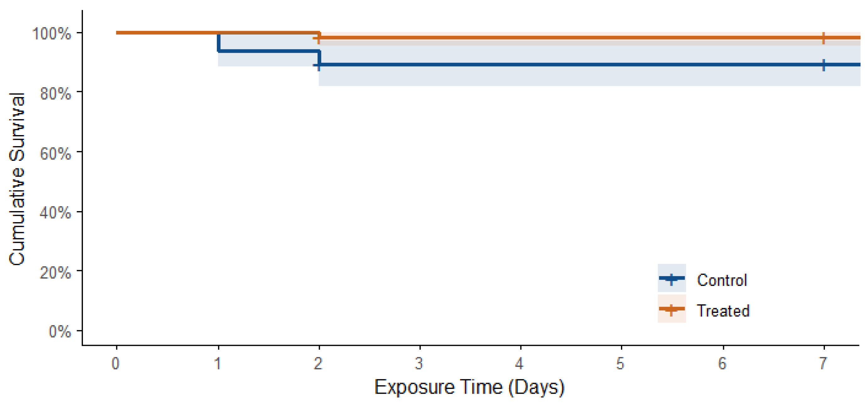
The cumulative survival of male P. melas did not differ significantly between the herbicide-treated cohort (final survival probability = 98.3%) and the untreated control group (final survival probability = 88.9%) over 7-day observation window (Figure 1). No statistically significant difference in survival was observed between cohorts (log-rank test, p = 0.36).

3.2. Light Microscopy and Histological Analysis

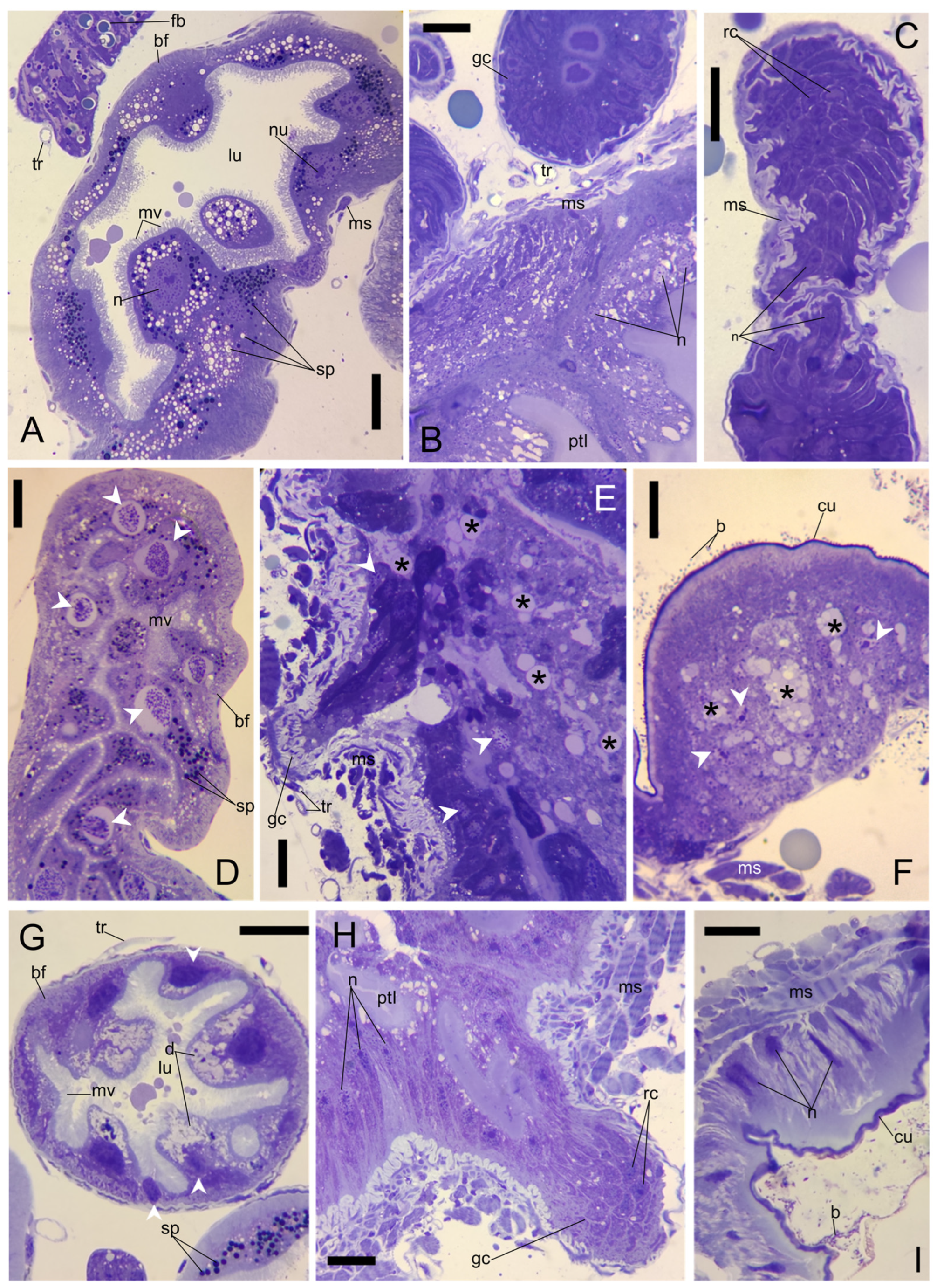
The histological analysis of the alimentary canal and Malpighian tubules in control specimens of P. melas italicus revealed a well-defined and intact tissue architecture, consistent with previous structural accounts of predatory ground beetles (Figure 2A–C). The Malpighian tubules appeared as long, blind-ended tubes with a regular diameter (diameter cross-Section 50–140 µm) and a narrow, clear lumen. The tubule wall was composed of a single layer of cuboidal epithelial cells that were located on a thin basal lamina (0.66 ± 0.17 µm; n = 12). The nuclei of these cells were euchromatic, and the apical brush border was distinctly visible (Figure 2A); the cytoplasm contained numerous refractive spherites and mineralized concretions. The midgut presented a continuous epithelium with prominent gastric caeca (Figure 2B). The digestive cells appeared columnar, with homogeneous cytoplasm and euchromatic nuclei, lying on a well-defined circular muscle layer. The apex of the gastric caeca showed distinct clusters of regenerative cells (Figure 2C). No evidence of cellular swelling, vacuolization, or nuclear fragmentation was observed in the control specimens.
Following a two-day exposure to PND (Figure 2D–F), the Malpighian tubules began to manifest early histopathological alterations, as evidenced by the presence of karyorrhectic nuclei (Figure 2D) and a slight irregularity in the basal folding thickness, which appeared visibly swollen (Figure 2D). In the midgut, small epithelial vesicles were observed at the insertion of the gastric caeca (Figure 2E), and karyorrhectic nuclei were also evident (Figure 2E). The rectum evinced sparse bacterial flora surrounding the cuticle layer at the lumen interface, while hindgut cells featured vesicles within the cytoplasm (Figure 2F).
After a seven-day exposure period, (Figure 2G–I) the Malpighian tubules displayed significant cyto-plasmic deposits in proximity to the lumen and nuclei exhibiting distinctive heterochromatin distribution patterns (Figure 2G). The gastric caeca maintained their structural integrity, exhibiting euchromatic nuclei (Figure 2H), while the colon retained a discernible cuticular layer, surrounded by a substantial layer of luminal bacteria (Figure 2I).

3.3. Ultrastructure of the Malpighian Tubules

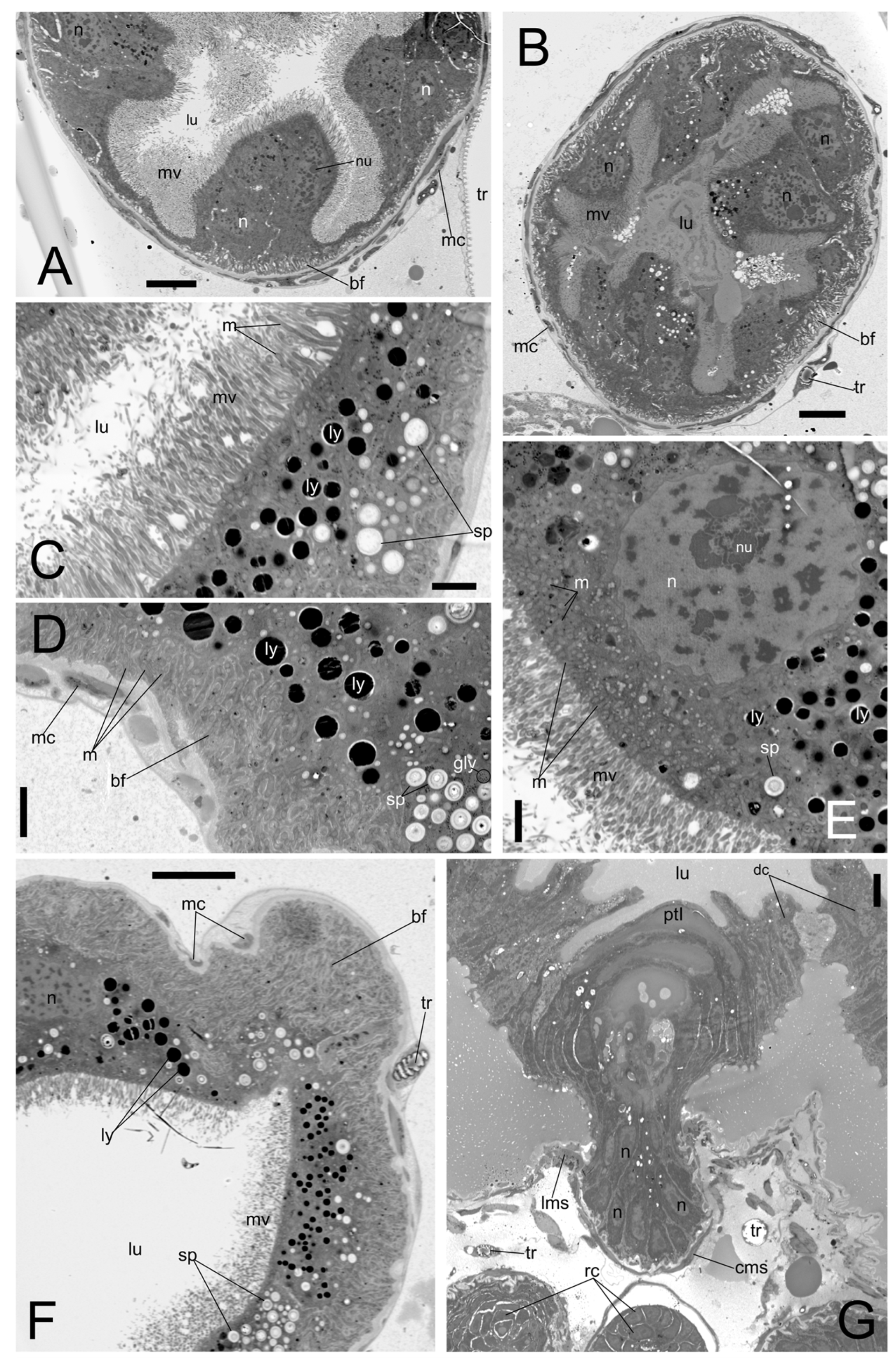
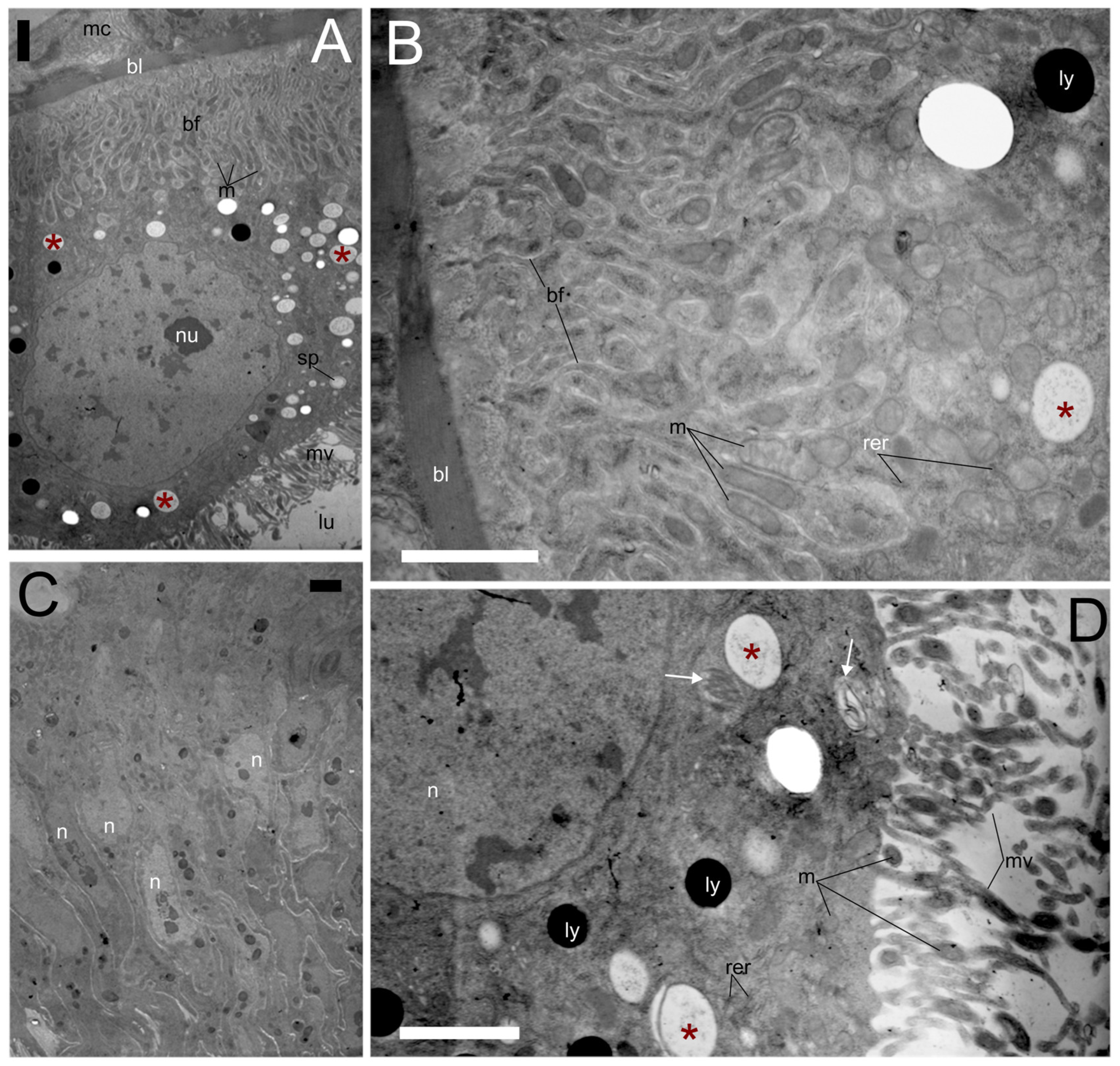
The ultrastructural analysis of Malpighian tubules from control beetles confirmed the high integrity of the excretory epithelium and digestive system (Figure 3 and Figure 4). In the Malpighian tubules, the cells were characterized by an extensive basal labyrinth (depth: 6.61 ± 2.76 µm; n = 14) consisting of deep and regular plasma membrane infoldings (Figure 3A–D,F). These infoldings compartmentalised numerous elongated mitochondria (length: 0.67 ± 0.22 µm; n = 20) with clearly visible cristae (Figure 4A,B). The apical surface, on the opposite side, displayed a dense, well-organized brush border (length: 4.13 ± 0.60 µm; n = 27) composed of tightly packed microvilli projecting into the lumen (Figure 3A–C,E). Numerous elongated mitochondria (length: 1.20 ± 0.32 µm; n = 10) were observed, some of which extended into the microvillar protrusions (Figure 4A,D). The cytoplasm contained a moderate number of electron dense lysosomes (diameter: 1.01 ± 0.36 µm; n = 34), and small spherites, which appear as round bodies (diameter: 1.23 ± 0.37 µm; n = 36) with concentric rings of electron-dense materials (Figure 3C–F and Figure 4A,D). The nuclei were characterised by their substantial size (major axes diameter: 11.40 ± 3.69 µm; minor axes diameter: 6.57 ± 1.91 n = 10) and euchromatin status, frequently featuring a prominent and centrally positioned nucleolus, suggesting a state of steady-state protein synthesis (Figure 3E and Figure 4A,D). The midgut is characterised by protruded gastric caeca, which display organised muscular layers, including circular and longitudinal muscle sheets (Figure 3G). In control adults, the midgut showed a well-developed peritrophic matrix. The ventricular lumen contained a type I peritrophic matrix, secreted directly by the epithelium (Figure 3G). Regenerative nidi, visible as clusters of small cells with euchromatic nuclei, were observed in the distal region of the gastric caeca (Figure 3G and Figure 4C).
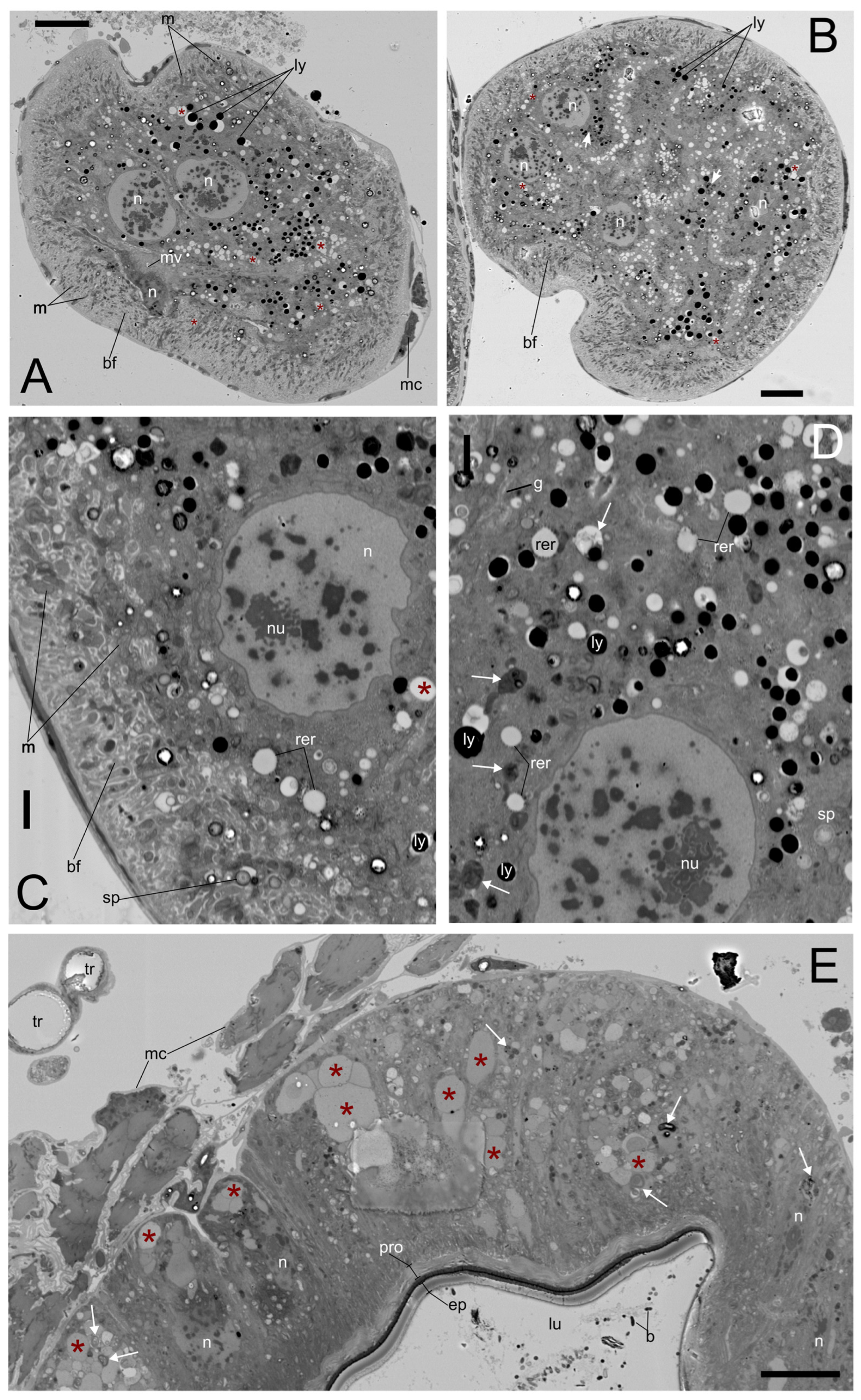
After 2 days of PND exposure, early ultrastructural changes become evident in both Malpighian tubules and hindgut epithelium (Figure 5). In Malpighian tubules, epithelial cells showed marked cytoplasmic vacuolization accompanied by initial chromatin clumping and irregularly shaped nuclei (Figure 5A–D; major axes diameter: 11.30 ± 1.97 µm; minor axes diameter: 9.36 ± 1.59 n = 6). The basal region displayed early modifications of the basal labyrinth, characterized by a reduction in the depth of membrane infoldings (depth: 6.32 ± 2.65 µm; n = 23) and enlargement of basal invaginations (Figure 5C). Numerous electron-dense lysosomes (diameter: 0.95 ± 0.28 µm; n = 35) were observed throughout the cytoplasm, particularly in the perinuclear area (Figure 5A–D), together with autophagic-like bodies and myelinic figures (Figure 5C,D). In the hindgut, the epithelium appeared extensively vesiculated, with widespread cytoplasmic vesicles and autophagic vacuoles (Figure 5E). The lumen was lined by cuticular layers (epicuticle and procuticle), and a sparse bacterial presence was detectable (Figure 5E). Occasional detachment of the muscle coat was also noted.
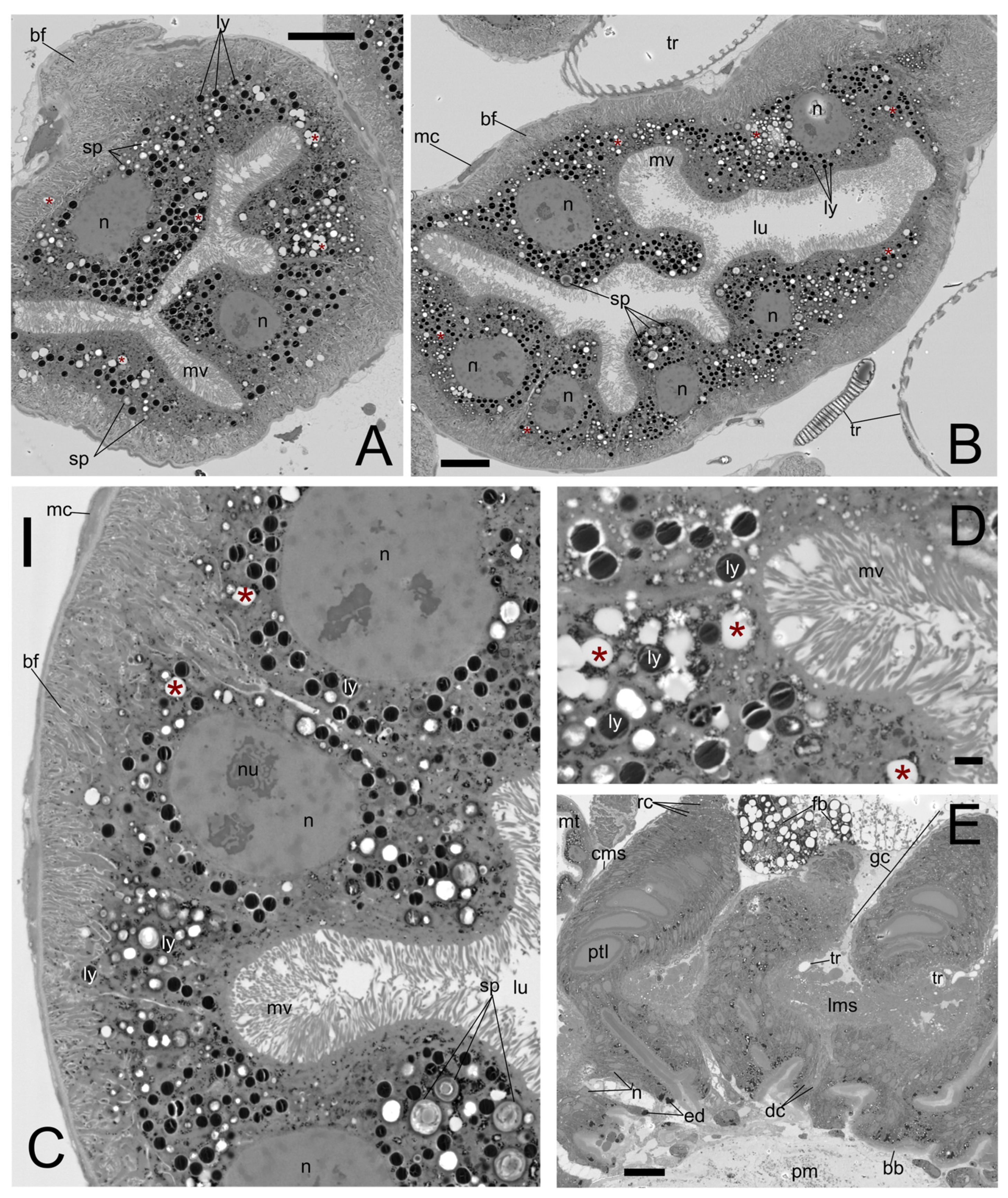
Conversely, after 7 days of PND exposure, Malpighian tubules showed a markedly metabolically active epithelium (Figure 6), with a cytoplasm densely packed with spherites (laminated concretions; diameter: 1.16 ± 0.47 µm; n = 15) and numerous electron-dense lysosomes (Figure 6A–D). The basal labyrinth appeared strongly expanded (depth: 7.25 ± 2.18 µm; n = 18), characterized by deep and abundant plasma membrane infoldings (Figure 5A–C). Nuclei (major axes diameter: 11.96 ± 2.73 µm; minor axes diameter: 8.30 ± 1.44 n = 8) appeared predominantly euchromatic and contained prominent nucleoli (Figure 6A–C), indicating sustained cellular activity. In addition, the apical cytoplasm displayed intense vesiculation with multiple electron-lucent vesicles (diameter: 1.12 ± 0.25 µm; n = 15) (Figure 6D). The gastric caeca exhibited an overall structural recovery, with a well-preserved organization of the epithelium and surrounding circular and longitudinal muscle layers (Figure 6E). Regenerative cells were evident within the distal portion of the caeca (Figure 6E), supporting epithelial renewal. The luminal compartment retained a continuous peritrophic matrix, while the brush border appeared well defined and regular, suggesting restoration of digestive surface integrity (Figure 6E).
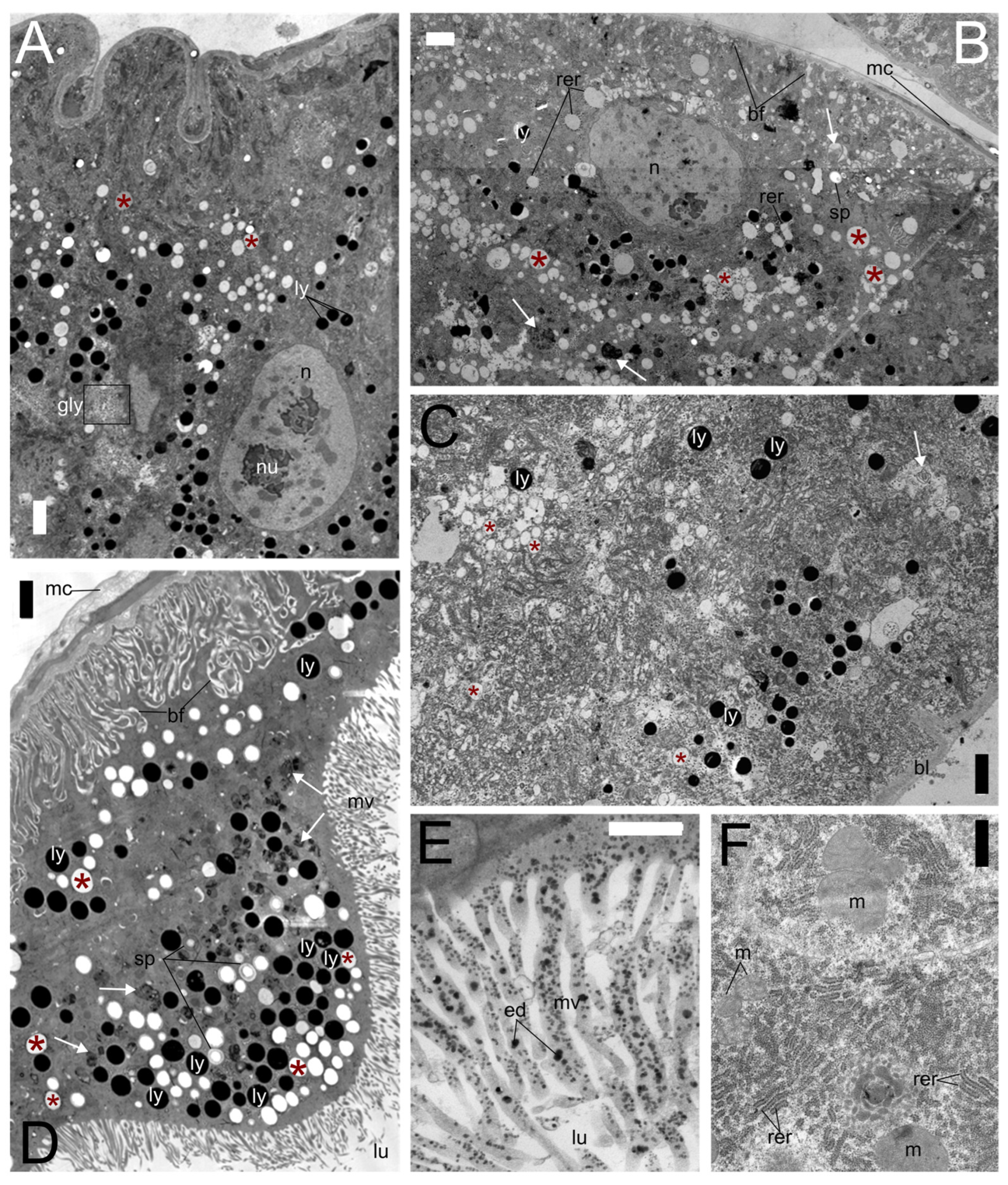
Compared with controls, the main differences in PND-treated specimens (Table 1) consisted of an increased abundance of electron-lucent vesicles and lysosomes. This was already evident after 2 days of exposure (Figure 7A–D) and was massively present after 7 days of exposure (diameter: 1.03 ± 0.23 µm; n = 46), together with a marked proliferation of the endoplasmic reticulum. In particular, both smooth endoplasmic reticulum (Figure 7C) and rough endoplasmic reticulum appeared strongly expanded, showing enlarged and swollen cisternae after 2 days (Figure 7B,C), while a further increase in rough endoplasmic reticulum development was observed after 7 days (Figure 7F). At 2 days, the cytoplasm also contained glycogen granules (Figure 7A). Autophagic activity became more evident with prolonged exposure, with autophagic-like bodies clearly detectable in the cytoplasm, particularly after 7 days (Figure 7C,D). In addition, electron-dense deposits were observed at the apical region of the cells, appearing dispersed along the microvilli (length: 2.48 ± 0.52 µm; n = 18) of the brush border (Figure 7E).

4. Discussion

In this study, the observed tissue alterations were explicitly non-lethal and occurred without any detectable effects on overall beetle survival, underscoring the importance of looking beyond mortality to assess environmental risk. The Malpighian tubules of P. melas emerged as highly sensitive indicators of pendimethalin-induced stress. The initial alterations observed after 2 days of exposure, including karyorrhexis and cytoplasmic vacuolization, are recognized biomarkers of sublethal toxicity, indicating a rapid response to chemical insult [28]. The appearance of karyorrhectic and pyknotic nuclei marks a critical threshold of cellular degeneration. Karyorrhexis, defined as the destructive fragmentation of the nucleus, and pyknosis, characterized by chromatin condensation, are recognized precursors of irreversible cell death [38]. Comparative studies in other carabids, such as Calathus fuscipes, suggest that chromatin clumping and nuclear degradation reflect a progressive inactivation of nuclear components and a significant reduction in transcriptional activity [33]. Similar nuclear alterations identified in the haemocytes of ground beetle Harpalus rufipes have been regarded as hallmarks of both necrotic and apoptotic processes triggered by pendimethalin exposure [12]. Comparable stress-related alterations have been reported in other insects, such as Apis mellifera, where salivary gland cells showed chromatin compaction, nuclear blebbing, DNA/nucleolar fragmentation, and autophagic features [39]. While PND is generally considered safe for animals, its potential for genotoxicity is highlighted even by in vitro studies on calf thymus DNA and by observations of DNA strand breaks in human lymphocytes and rat bone-marrow cells [40,41,42] and to its potential ability to bind DNA [43].
The documented cytoplasmic vacuolization may be a functional hallmark of both a failure in osmoregulation and an active sequestration strategy. The electron-lucent vesicles, potentially corresponding to macropinosomes, may arise from a dynamic homeostatic attempt to isolate toxic PND molecules from sensitive cytoplasmic organelles, as observed in H. rufipes haemocytes following PND exposure [12]. Comparable alterations have been reported in the Malpighian tubules of the neuropteran Ceraeochrysa claveri upon Pyriproxyfen exposure [44] and in the midgut of the darkling beetle Blaps polychresta exposed to nickel oxide nanoparticles [45], where these morphological abnormalities signal significant cellular injury. Analogous biomarkers of early stress have also been documented in other models of environmental intoxication, including the cockroach Blattella germanica. In this species, exposure to heavy metals (Hg, Pb, and Cr) results in marked pathological anomalies in the midgut, characterized by increased cellular volume and extensive cytoplasmic vacuolization [46]. Furthermore, acute intoxication with polychlorinated biphenyls in B. germanica induced severe Malpighian tubule degeneration, including karyorrhexis, cytoplasmic vacuolization, and loss of cellular architecture [47]. These rapid systemic responses are consistent with findings in pollinators such as A. mellifera, orally exposed to PND, which exhibit similar nuclear and cytoplasmic damage within 24 to 72 h of exposure [11]. However, the transition to a “stabilized” morphology by day 7, characterized by euchromatic nuclei with prominent nucleoli and a significantly expanded basal labyrinth, could suggest a phase of intense metabolic compensation rather than permanent failure. This structural adaptation might suggest an upregulation of active transport processes to meet the high energetic demands of xenobiotic excretion. The transient nature of these effects in P. melas, with tissue morphology stabilizing by day 7, aligns with broader entomological observations indicating that structural recovery is facilitated over time by epithelial turnover and herbicide dissipation. For instance, in A. mellifera workers, the midgut and ileum epithelia were found to be structurally intact just one week after the initial PND exposure, with recovery facilitated by the regular renewal of intestinal cells and the reduction in herbicide levels over time [11]. Similarly, in the ground beetle H. rufipes, initial cytotoxic damage to haemocytes was found to be transient, with the organism restoring normal cell density and functionality following herbicide dissipation [12]. This capacity for restoration could be driven by the activation of regenerative cell nidi located in the distal portion of the midgut caeca, which serve as a vital survival strategy in insects to replace damaged or apoptotic cells and maintain gut homeostasis [48,49]. Comparable strategies have been described in other soil-dwelling arthropods, such as the springtail Orchesella cincta, which periodically renews its midgut epithelium during moulting and eliminates accumulated contaminants together with the shed cells [49]. Although these regenerative mechanisms appear sufficient to restore tissue morphology under controlled laboratory conditions, their energetic costs may become substantial under field conditions, where simultaneous exposure to multiple environmental stressors could reduce physiological performance and compromise ecological fitness. Consequently, the transient ultrastructural alterations documented here should not be interpreted as evidence of negligible toxicity but rather as indicators of a robust, yet potentially costly, adaptive response [50].
The basal infoldings are associated with numerous mitochondria, whose expansion may reflect an upregulation of ion and metabolite transport from the haemolymph to sustain the energetic demands of detoxification [25]. Although PND primarily acts by disrupting plant microtubules and arresting mitosis [10], the mitochondrial swelling and membrane blebbing observed in P. melas suggest the activation of secondary stress-related cytotoxic pathways. Mitochondria are considered among the primary targets of PND toxicity in animals, as demonstrated in human lymphocytes exposed to pendimethalin and rat bone-marrow cells, where the herbicide induced oxidative stress, mitochondrial membrane potential dysfunction, DNA damage, and apoptotic responses [42]. The observed mitochondrial pathologies in P. melas, specifically the swelling of the matrix and degradation of the cristae, could be interpreted as markers of a collapse in the mitochondrial membrane potential [42] following pendimethalin exposure. This interpretation aligns with discriminative cytotoxicity models indicating that oxidative shifts in the mitochondrial environment often occur as an early indicator of stress before total cell membrane failure [51].
In contrast to the Malpighian tubules, which reported severe alterations in the first phase of the exposure, the gastric caeca remained structurally intact, and the colon maintained its cuticular lining throughout the treatment. This structural resilience could be likely attributed to the protective role of the peritrophic matrix and the intestinal cuticle, which act as primary physical and chemical barriers against ingested toxicants [52]. The presence of a cuticular intima in the hindgut of beetles provides an additional layer of protection not found in the midgut [32]. The recovery of the digestive brush border and the presence of active regenerative nidi by day 7 suggest a high rate of epithelial cell turnover, a common strategy in insects to maintain gut homeostasis following pesticide ingestion [11]. However, the swelling and expansion of the basal labyrinth observed by day 7 may indicate a shift from acute stress to metabolic compensation. A similar functional remodelling has been reported in the Malpighian tubules of the cicada Meimuna mongolica during developmental niche shifts, involving denser microvilli and dissolution of secretory vacuoles to support rapid waste excretion and metabolic activity [53]. This “metabolically active” state might suggest that P. melas possesses a robust physiological plasticity, allowing for functional recovery after the initial acute impact of PND.
The cellular response in P. melas following herbicide exposure is marked by a proliferation of spherites. These concentrically lamellar organelles, abundant in the Malpighian tubules of invertebrates, are not merely static waste deposits but function as dynamic compartments involved in detoxification, biomineralization, and metabolic storage [54,55]. In addition, they may serve as reservoirs of essential minerals and organic precursors, including amino acids, supporting the energetic demands associated with physiological recovery [55,56,57,58]. The increase in laminated concretions within tubule cells may therefore reflect an active sequestration mechanism for xenobiotics and toxic metabolites. Similar mobilization of spherite-associated reserves has been described during periods of intense metabolic reorganization, such as winter diapause and increased rough endoplasmic reticulum development prior to the epigean phase [55,59]. Comparable ultrastructural responses, including spherite accumulation, mitochondrial swelling, rough endoplasmic reticulum vesiculation, and loss of microvillar organization, have also been reported in the Malpighian tubules of the dipteran Boettcherisca peregrina after heavy metal exposure and in the neuropteran C. claveri following pyriproxyfen treatment [60,61]. Moreover, the high abundance of autophagic-like bodies observed in the tubules after PND exposure supports the hypothesis of intense cellular turnover. Through the sequestration and degradation of damaged cellular components, autophagy contributes to cellular homeostasis, biomolecule recycling, and tissue repair [33,62,63]. In the Malpighian tubules of P. melas, this enhanced cytoplasmic turnover likely facilitates the removal of herbicide-damaged material, supporting recovery from acute physiological stress and the restoration of metabolic activity observed by day 7. In P. melas, the concurrent proliferation of spherites, lysosomes, and autophagic structures following pendimethalin exposure therefore suggests a coordinated physiological response aimed at xenobiotic isolation and maintenance of tissue functionality.
Pendimethalin exposure has been shown to induce dysbiosis in a previous study on the same species, altering the richness and diversity of the gut microbial community [34]. Interestingly, certain gut bacteria (e.g., Enterobacter sp., Pseudomonas sp.) are known to assist in the degradation of dinitroaniline herbicides, suggesting that the microbiota assists the host in metabolic detoxification, which supports the transition to a metabolically active state occurring in a subchronic phase. These interspecific interactions are critical, as herbicide-induced shifts in the microbiome can delay development and alter host susceptibility to natural pathogens [64,65]. Moreover, Malpighian tubules express all essential molecular components of both the Toll and IMD signalling pathways, allowing them to synthesize and secrete antimicrobial peptides in response to either pathogen invasion or chemical stress [31]. Consequently, herbicide-induced ultrastructural damage to these tissues does not merely impair the filtration of the haemolymph or the maintenance of ionic and osmotic balance. Such damage could directly compromise the insect’s innate immune defence system, reduce its overall immune competence and significantly increase its vulnerability to pathogens [29,66].
The stabilization of tissue morphology suggests that P. melas activates effective physiological defences to maintain homeostasis. However, this morphologic stabilization likely represents an energetically costly physiological compensation rather than a complete restoration of the baseline pre-exposure state [67]. The redirection of resources toward detoxification (lysosome/spherite production) and tissue repair (regenerative cell activity) often results in physiological trade-offs with other vital life-history traits, such as reproduction and development [67]. As documented in other insect models, anthropogenic stressors can redefine an organism’s energy budget, forcing a prioritization of immediate survival and maintenance over long-term traits such as reproduction and immune competence [68]. Ultimately, while these defences allow for morphological stabilization, underlying cellular stress mechanisms may persist, and the long-term metabolic burden could potentially reduce the predatory efficiency and ecological fitness of carabid beetles in intensively managed agroecosystems.

5. Conclusions

This study demonstrates that exposure to field-realistic concentrations of pendimethalin induces pronounced yet transient ultrastructural alterations in the Malpighian tubules and midgut of a non-target organism. The Malpighian tubules emerged as particularly sensitive targets, revealing early biomarkers of herbicide-induced cellular stress, including nuclear degeneration (karyorrhexis), cytoplasmic vacuolization, mitochondrial swelling, and enhanced autophagic activity. Although many of these alterations appeared transient and partially stabilized after prolonged exposure, the observed recovery likely reflects an energetically costly physiological compensation rather than a complete absence of toxic impact. The coordinated activation of detoxification pathways, regenerative processes, and xenobiotic sequestration mechanisms highlights the remarkable plasticity of this carabid species in coping with herbicide-induced stress. However, the observed morphological stabilisation occurred under optimal, stress-free laboratory conditions, characterized by stable environmental parameters and unrestricted food availability. In natural agricultural landscapes, beneficial insects rarely experience such conditions. Under natural agricultural conditions, where insects are simultaneously challenged by fluctuating temperatures, limited prey resources, repeated agrochemical applications, and other environmental stressors, the energetic costs associated with continuous detoxification and tissue remodelling may become substantially greater. Such increased metabolic investment could ultimately reduce the energy available for growth, reproduction, dispersal, and predatory activity, with potential consequences for individual fitness and the ecosystem services provided by this species. Future studies integrating ultrastructural, physiological, behavioural, and energetic endpoints under environmentally realistic exposure scenarios will be essential to determine whether the compensatory responses documented here translate into measurable ecological costs. Overall, our findings demonstrate that apparently reversible cellular alterations should not be interpreted as evidence of negligible toxicity and support the inclusion of sensitive sublethal biomarkers, particularly those based on Malpighian tubule ultrastructure, in environmental risk assessment frameworks for herbicides and other soil contaminants affecting beneficial arthropods.

Author Contributions

Conceptualization, M.L.V. and A.G.; methodology, M.L.V. and A.G.; investigation, M.L.V. and A.G. conduced field sampling, exposure assays, dissection and sample preparation; P.G.G. performed the ultrastructural analyses; resources, A.G. and P.G.G.; visualization, M.L.V.; writing—original draft preparation, M.L.V.; writing—review and editing, M.L.V., A.G. and P.G.G.; supervision, A.G.; funding acquisition, A.G. All authors have read and agreed to the published version of the manuscript.

Funding

This research was funded by the Italian Ministry of University and Research (MUR) through the REINFORCe Project, Agritech Spoke 2, under the National Recovery and Resilience Plan (PNRR), funded by the European Union—NextGenerationEU (Grant No. CN00000022; CUP E63C22000920005). The APC was waived as part of an invited submission from the journal.

Data Availability Statement

The original contributions presented in this study are included in the article. Further inquiries can be directed to the corresponding author(s).

Acknowledgments

Authors thank Giampiero Ventura for allowing access to the sampled fields.

Conflicts of Interest

The authors declare no conflicts of interest.

References

  1. Prosser, R.S.; Anderson, J.C.; Hanson, M.L.; Solomon, K.R.; Sibley, P.K. Indirect Effects of Herbicides on Biota in Terrestrial Edge-of-Field Habitats: A Critical Review of the Literature. Agric. Ecosyst. Environ. 2016, 232, 59–72. [Google Scholar] [CrossRef]
  2. Köhler, H.R.; Triebskorn, R. Wildlife Ecotoxicology of Pesticides: Can We Track Effects to the Population Level and Beyond? Science 2013, 341, 759–765. [Google Scholar] [CrossRef] [PubMed]
  3. Sharma, A.; Jha, P.; Reddy, G.V.P. Multidimensional Relationships of Herbicides with Insect-Crop Food Webs. Sci. Total Environ. 2018, 643, 1522–1532. [Google Scholar] [CrossRef] [PubMed]
  4. Pereira, J.L.; Da Silva, A.A.; Picanço, M.C.; De Barros, E.C.; Jakelaitis, A. Effects of Herbicide and Insecticide Interaction on Soil Entomofauna under Maize Crop. J. Environ. Sci. Health B 2005, 40, 45–54. [Google Scholar] [CrossRef] [PubMed]
  5. Capinera, J.L. Direct and Indirect Effects of Herbicides on Insects. In Weed Control: Sustainability, Hazards, and Risks in Cropping Systems Worldwide; CRC Press: Boca Raton, FL, USA, 2018; pp. 76–91. [Google Scholar]
  6. Strandberg, M.; Scott-Fordsmand, J.J. Effects of Pendimethalin at Lower Trophic Levels—A Review. Ecotoxicol. Environ. Saf. 2004, 57, 190–201. [Google Scholar] [CrossRef] [PubMed]
  7. Vighi, M.; Matthies, M.; Solomon, K.R. Critical Assessment of Pendimethalin in Terms of Persistence, Bioaccumulation, Toxicity, and Potential for Long-Range Transport. J. Toxicol. Environ. Health B Crit. Rev. 2017, 20, 1–21. [Google Scholar] [PubMed]
  8. Rose, M.T.; Cavagnaro, T.R.; Scanlan, C.A.; Rose, T.J.; Vancov, T.; Kimber, S.; Kennedy, I.R.; Kookana, R.S.; Van Zwieten, L. Impact of Herbicides on Soil Biology and Function; Elsevier Inc.: Amsterdam, The Netherlands, 2016; Volume 136, ISBN 9780128046814. [Google Scholar]
  9. Giglio, A.; Vommaro, M.L. Dinitroaniline Herbicides: A Comprehensive Review of Toxicity and Side Effects on Animal Non-Target Organisms. Environ. Sci. Pollut. Res. 2022, 29, 76687–76711. [Google Scholar] [CrossRef] [PubMed]
  10. Nyporko, A.Y.; Yemets, A.I.; Brytsun, V.N.; Lozinsky, M.O.; Blume, Y.B. Structural and Biological Characterization of the Tubulin Interaction with Dinitroanilines. Cytol. Genet. 2009, 43, 267–282. [Google Scholar] [CrossRef]
  11. Vommaro, M.L.; Giglio, A. Cytotoxic and Genotoxic Effects of a Pendimethalin-Based Herbicide in Apis mellifera. Ecotoxicol. Environ. Saf. 2024, 280, 116565. [Google Scholar] [CrossRef] [PubMed]
  12. Vommaro, M.L.; Giulianini, P.G.; Giglio, A. Pendimethalin-Based Herbicide Impairs Cellular Immune Response and Haemocyte Morphology in a Beneficial Ground Beetle. J. Insect Physiol. 2021, 131, 104236. [Google Scholar] [CrossRef] [PubMed]
  13. Oliver, J.B.; Reding, M.E.; Moyseenko, J.J.; Klein, M.G.; Mannion, C.M.; Bishop, B. Survival of Adult Tiphia vernalis (Hymenoptera: Tiphiidae) After Insecticide, Fungicide, and Herbicide Exposure in Laboratory Bioassays. J. Econ. Entomol. 2009, 99, 288–294. [Google Scholar] [CrossRef] [PubMed]
  14. Belden, J.B.; Phillips, T.A.; Clark, B.W.; Coats, J.R. Toxicity of Pendimethalin to Nontarget Soil Organisms. Bull. Environ. Contam. Toxicol. 2005, 74, 769–776. [Google Scholar] [CrossRef] [PubMed]
  15. He, Q.; Yang, Q.; Liu, Q.; Hu, Z.; Gao, Q.; Dong, Y.; Xiao, J.; Yu, L.; Cao, H. The Effects of Beta-Cypermethrin, Chlorbenzuron, Chlorothalonil, and Pendimethalin on Apis mellifera ligustica and Apis cerana cerana Larvae Reared In Vitro. Pest Manag. Sci. 2022, 78, 1407–1416. [Google Scholar] [PubMed]
  16. Haque, A.; Das Gupta, R.; Chakravorty, P.P. Effect of Two Herbicides on Xenylla welchi (Hexapoda:Collembola) under Laboratory Conditions. Bull. Environ. Contam. Toxicol. 2011, 86, 583–586. [Google Scholar] [CrossRef] [PubMed]
  17. Chakravorty, P.P.; Haque, A.; Sanyal, S.; Dasgupta, R. Effect of Herbicides on Cyphoderus javanus (Hexapoda: Collembola) under Laboratory Conditions. J. Entomol. Zool. Stud. 2015, 3, 220–223. [Google Scholar]
  18. Vommaro, M.L.; Zanchi, C.; Angelone, T.; Giglio, A.; Kurtz, J. Herbicide Exposure Alters the Effect of the Entomopathogen Beauveria bassiana on Immune Gene Expression in Mealworm Beetles. Environ. Pollut. 2023, 338, 122662. [Google Scholar] [CrossRef] [PubMed]
  19. Vommaro, M.L.; Korša, A.; Lindeza, A.S.; Giglio, A.; Kurtz, J. The Combined Effect of Herbicide and Bacillus thuringiensis Exposure Delays Development in the Red Flour Beetle. J. Invertebr. Pathol. 2024, 207, 108227. [Google Scholar] [CrossRef] [PubMed]
  20. Belden, J.B.; Phillips, T.A.; Henderson, K.L.; Clark, B.W.; Lydy, M.J.; Coats, J.R. Persistence, Mobility, and Bioavailability of Pendimethalin and Trifluralin in Soil. In Proceedings of the ACS Symposium Series; American Chemical Society: Washington, DC, USA, 2003; Volume 853, pp. 167–177. [Google Scholar]
  21. Koivula, M.J. Useful Model Organisms, Indicators, or Both? Ground Beetles (Coleoptera, Carabidae) Reflecting Environmental Conditions. ZooKeys 2011, 100, 287–317. [Google Scholar] [CrossRef] [PubMed]
  22. Rainio, J.; Niemelä, J. Ground Beetles (Coleoptera: Carabidae) as Bioindicators. Biodivers. Conserv. 2003, 12, 487–506. [Google Scholar] [CrossRef]
  23. Sunderland, K.D. Invertebrate Pest Control by Carabids. In The Agroecology of Carabid Beetles; Holland, J.M., Ed.; Intercept Limited: Andover, UK, 2002; pp. 165–214. [Google Scholar]
  24. Benítez, H.A.; Lemic, D.; Püschel, T.A.; Gašparić, H.V.; Kos, T.; Barić, B.; Bažok, R.; Živković, I.P. Fluctuating Asymmetry Indicates Levels of Disturbance between Agricultural Productions: An Example in Croatian Population of Pterostichus melas melas (Coleptera: Carabidae). Zool. Anz. 2018, 276, 42–49. [Google Scholar] [CrossRef]
  25. Beyenbach, K.W.; Skaer, H.; Dow, J.A.T. The Developmental, Molecular, and Transport Biology of Malpighian Tubules. Annu. Rev. Entomol. 2010, 55, 351–374. [Google Scholar] [CrossRef] [PubMed]
  26. Gillott, C. Entomology; Springer Science & Business Media: Berlin/Heidelberg, Germany, 2005; ISBN 1402031831. [Google Scholar]
  27. Catae, A.F.; Roat, T.C.; De Oliveira, R.A.; Ferreira Nocelli, R.C.; Malaspina, O. Cytotoxic Effects of Thiamethoxam in the Midgut and Malpighian Tubules of Africanized Apis mellifera (Hymenoptera: Apidae). Microsc. Res. Tech. 2014, 77, 274–281. [Google Scholar] [CrossRef] [PubMed]
  28. Malaspina, O.; da Silva-Zacarin, E.C.M. Cell Markers for Ecotoxicological Studies in Target Organs of Bees. J. Morphol. Sci. 2017, 23, 303–309. [Google Scholar] [CrossRef]
  29. Giglio, A.; Brandmayr, P. Structural and Functional Alterations in Malpighian Tubules as Biomarkers of Environmental Pollution: Synopsis and Prospective. J. Appl. Toxicol. 2017, 37, 889–894. [Google Scholar] [CrossRef] [PubMed]
  30. Farina, P.; Bedini, S.; Conti, B. Multiple Functions of Malpighian Tubules in Insects: A Review. Insects 2022, 13, 1001. [Google Scholar] [CrossRef] [PubMed]
  31. Khan, S.A.; Han, Y.S. Current Status of Innate Immune Responses in Malpighian Tubules of Insects: A Review Focusing on the Toll Signaling Pathway. Entomol. Res. 2024, 54, e12716. [Google Scholar] [CrossRef]
  32. Donato, S.; Vommaro, M.L.; Tromba, G.; Giglio, A. Synchrotron X-Ray Phase Contrast Micro Tomography to Explore the Morphology of Abdominal Organs in Pterostichus melas italicus Dejean, 1828 (Coleoptera, Carabidae). Arthropod Struct. Dev. 2021, 62, 101044. [Google Scholar] [CrossRef] [PubMed]
  33. Giglio, A.; Cavaliere, F.; Giulianini, P.G.; Mazzei, A.; Talarico, F.; Vommaro, M.L.; Brandmayr, P. Impact of Agrochemicals on Non-Target Species: Calathus fuscipes Goeze 1777 (Coleoptera: Carabidae) as Model. Ecotoxicol. Environ. Saf. 2017, 142, 522–529. [Google Scholar] [CrossRef] [PubMed]
  34. Giglio, A.; Vommaro, M.L.; Gionechetti, F.; Pallavicini, A. Gut Microbial Community Response to Herbicide Exposure in a Ground Beetle. J. Appl. Entomol. 2021, 145, 986–1000. [Google Scholar] [CrossRef]
  35. Allegro, G. Tabella Illustrata Di Determinazione per Le Specie Del Genere Pterostichus Note in Italia (Coleoptera, Carabidae, Pterostichinae). Mem. Della Soc. Entomol. Ital. 2021, 98, 65–91. [Google Scholar] [CrossRef]
  36. Kocárek, M.; Artikov, H.; Voríšek, K.; Boruvka, L. Pendimethalin Degradation in Soil and Its Interaction with Soil Microorganisms. Soil Water Res. 2016, 11, 213–219. [Google Scholar] [CrossRef]
  37. R Core Team. R: A Language and Environment for Statistical Computing (Version 4.4.1) [Computer Software]; R Foundation for Statistical Computing: Vienna, Austria, 2025. [Google Scholar]
  38. Ferrari, A.; Polidori, C.; Trisoglio, C.F.; Bonasoro, F. Increasing Road Cover in Urban Areas Is Associated with Greater Midgut Histological Damage in a Primitively Eusocial Bee. Insectes Sociaux 2024, 71, 331–341. [Google Scholar] [CrossRef]
  39. Silva-Zacarin, E.C.M.; Taboga, S.R.; Silva de Moraes, R.L.M. Nuclear Alterations Associated to Programmed Cell Death in Larval Salivary Glands of Apis mellifera (Hymenoptera: Apidae). Micron 2008, 39, 117–127. [Google Scholar] [CrossRef] [PubMed]
  40. Ahmad, M.I.; Zafeer, M.F.; Javed, M.; Ahmad, M. Pendimethalin-Induced Oxidative Stress, DNA Damage and Activation of Anti-Inflammatory and Apoptotic Markers in Male Rats. Sci. Rep. 2018, 8, 17139. [Google Scholar] [CrossRef] [PubMed]
  41. Patel, S.; Bajpayee, M.; Pandey, A.K.; Parmar, D.; Dhawan, A. In Vitro Induction of Cytotoxicity and DNA Strand Breaks in CHO Cells Exposed to Cypermethrin, Pendimethalin and Dichlorvos. Toxicol. Vitr. 2007, 21, 1409–1418. [Google Scholar] [CrossRef] [PubMed]
  42. Ansari, S.M.; Saquib, Q.; Attia, S.M.; Abdel-Salam, E.M.; Alwathnani, H.A.; Faisal, M.; Alatar, A.A.; Al-Khedhairy, A.A.; Musarrat, J. Pendimethalin Induces Oxidative Stress, DNA Damage, and Mitochondrial Dysfunction to Trigger Apoptosis in Human Lymphocytes and Rat Bone-Marrow Cells. Histochem. Cell Biol. 2018, 149, 127–141. [Google Scholar] [CrossRef] [PubMed]
  43. Ahmad, I.; Ahmad, A.; Ahmad, M. Binding Properties of Pendimethalin Herbicide to DNA: Multispectroscopic and Molecular Docking Approaches. Phys. Chem. Chem. Phys. 2016, 18, 6476–6485. [Google Scholar] [CrossRef] [PubMed]
  44. Scudeler, E.L.; Barroso, G.; Daquila, B.V.; de Carvalho, S.F.; Conte, H.; dos Santos, D.C. Pyriproxyfen Exposure Compromises Cocoon Spinning and Damages the Malpighian Tubules of the Nontarget Predator Ceraeochrysa claveri (Neuroptera: Chrysopidae). Environ. Pollut. 2024, 363, 125255. [Google Scholar] [CrossRef] [PubMed]
  45. El-Samad, L.M.; Bakr, N.R.; El-Ashram, S.; Radwan, E.H.; Abdul Aziz, K.K.; Hussein, H.K.; El Wakil, A.; Hassan, M.A. Silver Nanoparticles Instigate Physiological, Genotoxicity, and Ultrastructural Anomalies in Midgut Tissues of Beetles. Chem. Biol. Interact. 2022, 367, 110166. [Google Scholar] [CrossRef] [PubMed]
  46. Zhang, Y.; Lambiase, S.; Fasola, M.; Gandini, C.; Grigolo, A.; Laudani, U. Mortality and Tissue Damage by Heavy Metal Contamination in the German Cockroach, Blattella germanica (Blattaria, Blattellidae). Ital. J. Zool. 2001, 68, 137–145. [Google Scholar] [CrossRef]
  47. Lambiase, S.; Lambiase, S.; Zhang, Y.; Morbini, P.; Fasola, M.; Bernocchi, G.; Roda, E.; Grigolo, A. Tissue Damage after Acute Intoxication by Polychlorinated Biphenyls (PCBs) in Cockroaches Blattella germanica. Eur. J. Histochem. 2004, 49, 189–198. [Google Scholar]
  48. Dias, W.C.S.; Teixeira, É.W.; Serrão, J.E. Regenerative Cells in the Midgut of the Honey Bee Apis mellifera (Apidae: Apini) Queens with Different Ages. Braz. Arch. Biol. Technol. 2024, 67, e24230473. [Google Scholar] [CrossRef]
  49. Rost, M.M. Ultrastructural Changes in the Midgut Epithelium in Podura aquatica L. (Insecta, Collembola, Arthropleona) during Regeneration. Arthropod Struct. Dev. 2006, 35, 69–76. [Google Scholar] [CrossRef] [PubMed]
  50. Posthuma, L.; Hogervorst, R.F.; Van Straalen, N.M. Adaptation to soil pollution by cadmium excretion in natural populations of Orchesella cincta (L.) (Collembola). Arch. Environ. Contam. Toxicol. 1992, 22, 146–156. [Google Scholar] [CrossRef] [PubMed]
  51. Kim, H.; Yoon, S.C.; Lee, T.Y.; Jeong, D. Discriminative Cytotoxicity Assessment Based on Various Cellular Damages. Toxicol. Lett. 2009, 184, 13–17. [Google Scholar] [CrossRef] [PubMed]
  52. Terra, W.R. The Origin and Functions of the Insect Peritrophic Membrane and Peritrophic Gel. Arch. Insect Biochem. Physiol. 2001, 47, 47–61. [Google Scholar] [CrossRef] [PubMed]
  53. Yuan, F.; Lian, P.; Liu, L.; Xie, Z.; Li, Z.; Li, T.; Wei, C. Age-Associated Morphology and Transcriptome Analyses Reveal Functional Transformation of Malpighian Tubules of Cicadas in Response to Development and Niche Shift. Insect Mol. Biol. 2025, 34, 863–877. [Google Scholar] [CrossRef] [PubMed]
  54. Cintra-Socolowski, P.; Nocelli, R.; Roat, T.; Silva-Zacarin, E.; Malaspina, O. Comparative Physiology of Malpighian Tubules: Form and Function. Open Access Insect Physiol. 2016, 6, 13–23. [Google Scholar] [CrossRef]
  55. Lipovšek, S.; Novak, T.; Dariš, B.; Hofer, F.; Leitinger, G.; Letofsky-Papst, I. Ultrastructure of Spherites in the Midgut Diverticula and Malpighian Tubules of the Harvestman Amilenus aurantiacus during the Winter Diapause. Histochem. Cell Biol. 2022, 157, 107–118. [Google Scholar] [CrossRef] [PubMed]
  56. Ballan-Dufrançais, C. Localization of Metals in Cells of Pterygote Insects. Microsc. Res. Tech. 2002, 56, 403–420. [Google Scholar] [CrossRef] [PubMed]
  57. Lipovšek, S.; Letofsky-Papst, I.; Hofer, F.; Pabst, M.A.; Devetak, D. Application of Analytical Electron Microscopic Methods to Investigate the Function of Spherites in the Midgut of the Larval Antlion Euroleon nostras (Neuroptera: Myrmeleontidae). Microsc. Res. Tech. 2012, 75, 397–407. [Google Scholar] [CrossRef] [PubMed]
  58. Pigino, G.; Migliorini, M.; Paccagnini, E.; Bernini, F.; Leonzio, C. Fine Structure of the Midgut and Malpighian Papillae in Campodea (Monocampa) Quilisi Silvestri, 1932 (Hexapoda, Diplura) with Special Reference to the Metal Composition and Physiological Significance of Midgut Intracellular Electron-Dense Granules. Tissue Cell 2005, 37, 223–232. [Google Scholar] [CrossRef] [PubMed]
  59. Lipovšek, S.; Novak, T.; Janžekovič, F.; Weiland, N.; Leitinger, G. Malpighian Tubule Cells in Overwintering Cave Crickets Troglophilus cavicola (Kollar, 1833) and T. Neglectus Krauss, 1879 (Rhaphidophoridae, Ensifera). PLoS ONE 2016, 11, e0158598. [Google Scholar] [CrossRef] [PubMed]
  60. Wu, G.X.; Gao, X.; Ye, G.Y.; Li, K.; Hu, C.; Cheng, J.A. Ultrastructural Alterations in Midgut and Malpighian Tubules of Boettcherisca peregrina Exposure to Cadmium and Copper. Ecotoxicol. Environ. Saf. 2009, 72, 1137–1147. [Google Scholar] [CrossRef] [PubMed]
  61. Scudeler, E.L.; de Carvalho, S.F.; Garcia, A.S.G.; Santorum, M.; Padovani, C.R.; dos Santos, D.C. Midgut and Fat Body: Multisystemic Action of Pyriproxyfen on Non-Target Organism Ceraeochrysa claveri (Neuroptera: Chrysopidae). Environ. Pollut. 2022, 293, 118580. [Google Scholar] [CrossRef] [PubMed]
  62. Carneiro, L.S.; Martinez, L.C.; de Oliveira, A.H.; Cossolin, J.F.S.; de Resende, M.T.C.S.; Gonçalves, W.G.; Medeiros-Santana, L.; Serrão, J.E. Acute Oral Exposure to Imidacloprid Induces Apoptosis and Autophagy in the Midgut of Honey Bee Apis mellifera Workers. Sci. Total Environ. 2022, 815, 152847. [Google Scholar] [CrossRef] [PubMed]
  63. Lipovšek, S.; Vajs, T.; Dariš, B.; Novak, T.; Kozel, P. Autophagic Activity in the Midgut Cells of Three Arachnids Responds Selectively to Different Modes of Overwintering in Caves. Protoplasma 2025, 262, 531–544. [Google Scholar] [CrossRef] [PubMed]
  64. Lee, J.-H.; Lee, K.-A.; Lee, W.-J. Microbiota, Gut Physiology, and Insect Immunity. In Advances in Insect Physiology; Academic Press: Cambridge, MA, USA, 2017; Volume 52, pp. 111–138. [Google Scholar]
  65. Yuan, X.; Pan, Z.; Jin, C.; Ni, Y.; Fu, Z.; Jin, Y. Gut Microbiota: An Underestimated and Unintended Recipient for Pesticide-Induced Toxicity. Chemosphere 2019, 227, 425–434. [Google Scholar] [CrossRef] [PubMed]
  66. Chahine, S.; O’Donnell, M.J. Interactions between Detoxification Mechanisms and Excretion in Malpighian Tubules of Drosophila melanogaster. J. Exp. Biol. 2011, 214, 462–468. [Google Scholar] [CrossRef] [PubMed]
  67. Khan, M.K.; Rolff, J. Insect Immunity in the Anthropocene. Biol. Rev. 2024, 100, 698–723. [Google Scholar] [CrossRef] [PubMed]
  68. Rebar, D.; Xiao, T.; Murdock, E. Anthropogenic Light Impacts Life-History Traits and Induces a Trade-Off in Female Field Crickets. Behav. Ecol. 2024, 35, arae064. [Google Scholar] [CrossRef]
Figure 1. Kaplan–Meier survival curves of male Pterostichus melas italicus exposed to a field-recommended rate of pendimethalin (Treated; n = 60) and untreated controls (Control; n = 63) over 7 days. Vertical ticks indicate right-censored individuals removed alive during scheduled destructive samplings for microscopy.
Environments 13 00394 g001
Figure 2. Semithin sections (toluidine blue–stained) of Pterostichus melas italicus alimentary canal and Malpighian tubules. Control group (AC): (A) Malpighian tubules with regular epithelium, euchromatic nuclei (n), and spherites (sp); (B) Midgut showing gastric caeca (gc) supported by a circular muscle layer (ms); (C) High magnification of gastric caecum apex with regenerative cell (rc) clusters. Two days after PND exposure (DF): (D) Malpighian tubule longitudinal section with karyorrhectic nuclei (white arrowheads); (E) Midgut epithelium at caecum insertion exhibiting vesicles (asterisks) and karyorrhexis (white arrowheads); (F) Rectum with cuticular lining (cu), epithelial vesicles (asterisks), and sparse bacterial flora (b). Seven days after PND exposure (GI): (G) Malpighian tubules containing cytoplasmic deposits (d) and nuclei with condensed heterochromatin (arrowheads); (H) Structurally restored gastric caecum with euchromatic nuclei; (I) Colon with distinct cuticular layer and luminal bacteria (b). Abbreviations: bf, basal foldings; fb, fat body; lu, lumen; mv, microvilli; ptl, type I peritrophic matrix; tr, tracheoles. Scale bar: 20 µm.
Environments 13 00394 g002
Figure 3. TEM-like micrographs of the Malpighian tubules and midgut of Pterostichus melas italicus from the control group. (A,B) General view of the Malpighian tubule epithelium, cross section, showing a single layer of cells with euchromatic nuclei (n) and a regular apical brush border (mv). (C) Detail of the apical cytoplasm highlighting closely packed microvilli and numerous mitochondria (m). (D) Basal region of a tubule cell showing a basal labyrinth (bf) with associated mitochondria. (E) High magnification of apical region, nucleus with a prominent nucleolus (nu) and cytoplasmic lysosomes (ly). (F) Cross section of Malpighian tubule resting on the muscle coat (mc) with visible tracheoles (tr) associated. (G) In the gastric caecum of the (G) section, the regenerative nidi (rc) can be observed in the distal portion and organised muscular layers (cms, lms). The presence of digestive cells (dc) in contact with the type I peritrophic matrix (ptI) is evident in the luminal region. Abbreviations: lu, lumen; sp, spherites. Scale bars: (A,B,F,G) 10 µm; (D) 3 µm; (E) 2 µm; (C) 200 nm.
Environments 13 00394 g003
Figure 4. Transmission electron micrographs of the Malpighian tubules and midgut of Pterostichus melas italicus from the control group. (A,B) General view of the Malpighian tubule epithelium showing a single layer of cells with euchromatic nuclei (n) and a regular brush border of microvilli (mv) surrounding the lumen (lu); the basal region is characterized by a basal labyrinth with regular plasma membrane infoldings (bf) resting on a distinct basal lamina (bl). (B) High magnification of the cytoplasm showing numerous mitochondria (m) associated with the basal foldings and scattered lysosomes (ly). (C) Detail of the gastric caecum characterized by organized regenerative nidi resting on the muscle layers. (D) Detail of the apical portion of a digestive cell showing mitochondria (m) within the microvilli, lysosomes (ly) and vesicle (asterisk) dispersed in the cytoplasm. Myelinic figures (white arrows) are also visible. Abbreviations: bl, basal lamina; rer, rough endoplasmic reticulum; asterisks, vesicles. Scale bars: (AD) 2 µm.
Environments 13 00394 g004
Figure 5. TEM-like micrographs of the alimentary canal of Pterostichus melas italicus following exposure to PND for a period of two days. (A,B) Cross-sections of Malpighian tubules exhibiting early signs of cellular stress, including cytoplasmic vacuolization (asterisks) and initial chromatin clumping in the nuclei (n). (C) Detail of the basal labyrinth showing a reduction in the depth of the membrane infoldings and an enlargement of the invaginations (bf). (D) Perinuclear region showing a proliferation of lysosomes (ly) and autophagic-like bodies (white arrows). (E) Hindgut epithelium displaying multiple vesicles within the cytoplasm, with a luminal portion that is lined by cuticle layers. Abbreviations: b, bacteria; ep, epicuticle; lu, lumen; m, mitochondria; mc, muscle cells; mv, microvilli; nu, nucleolus; pro, procuticle; sp, spherites; tr, tracheoles; asterisks, vesicles. Scale bars: (A,B,E) 10 µm; (C,D) 2 µm.
Environments 13 00394 g005
Figure 6. TEM-like micrographs of the alimentary canal of Pterostichus melas italicus after exposure to PND for 7 days. Cross (A) and oblique (B) sections of Malpighian tubules showing a metabolically active epithelium, characterized by a high density of spherites (sp) and lysosomes (ly). (C) View of the basal region of Malpighian tubule reveals a notably expanded basal labyrinth with deep infoldings (bf) and a substantial number of mitochondria (m). (D) Apical cytoplasm showing intense vesiculation and numerous lysosomes (ly). (E) Gastric caeca showing structural recovery with organized muscle sheets (cms, lms) and functional regenerative cells (rc) at the apex of the extrusion. Abbreviations: bb, brush border; cms, circular muscles; dc, digestive cells; ed, electron-dense deposits; fb, fat body; gc, gastric caecum; lms, longitudinal muscles; lu, lumen; mc, muscle cells; mt, Malpighian tubule; mv, microvilli; n, nucleus; nu, nucleolus; pm, peritrophic matrix; ptl, type I peritrophic matrix; tr, tracheoles; asterisks, vesicles. Scale bars: (E) 30 µm, (A,B) 10 µm; (C) 3 µm, (D) 1 µm.
Environments 13 00394 g006
Figure 7. Transmission electron micrographs of the Malpighian tubules of Pterostichus melas italicus after exposure to PND. (AC) 2 days of exposure: (A,B) Basal portion lined by muscle cells (mc), showing reduced basal foldings (bf) and extensive cytoplasmic vacuolization, with enlarged rough endoplasmic reticulum (rer) and autophagic-like bodies (white arrows). (C) Detail of cytoplasm with lysosomes (ly), numerous vesicles (asterisks) and smooth endoplasmic reticulum (ser). (DF) 7 days of exposure: (D) Apical portion of a cell showing the lumen surrounded by microvilli (mv) and increased density of spherites (sp), ly and autophagic bodies (white arrows); (E) Luminal portion showing microvilli (mv) with dispersed electron-dense deposits (ed) and enlargements; (F) Cytoplasmic region with a significantly expanded rough endoplasmic reticulum (rer) and enlarged, swollen mitochondria (m). Abbreviations: bl, basal lamina; gly, glycogen granules; lu, lumen; n, nucleus; nu, nucleolus; sp, spherites; asterisks, vesicles. Scale bars: (A,B,D) 2 µm; (C) 2 µm; (F) 500 nm; (E) 200 nm.
Environments 13 00394 g007
Table 1. Summary of primary histopathological and ultrastructural alterations observed in the Malpighian tubules and midgut of Pterostichus melas italicus following subchronic pendimethalin exposure.
Major Histopathological and Ultrastructural LesionsMalpighian TubulesMidgutDiagnostic Features and Pathological Signification
Nuclear degenerationday 2day 2Early acute cytotoxic insult. Chromatin condensation along the inner nuclear membrane, nucleolar segregation, and focal karyorrhexis
Marked cytoplasmic vacuolizationday 2
(severe)
day 2Dilation of endoplasmic reticulum cisternae forming macro electron-lucent vesicles
day 7
(persistent)
day 7
(localized)
Pronounced mitochondrial swellingday 2
(severe)
Mitochondrial rounding, loss or disruption of cristae
day 7
(residual)
Disruption and dilation of the basal labyrinthday 2 Severe enlargement and architectural disruption of the basal plasma membrane infoldings
Apical blebbing and cellular fragment shedding day 2Disruption of apical boundaries with protrusion of cytoplasmic blebs and shedding of debris into the lumen
Widening of extracellular basal spaces day 2Intercellular fluid accumulation flanking the basal lamina of the digestive epithelium
Prominent macroautophagic pathwayday 7day 7Marked proliferation of autophagosomes and autophagic vacuoles containing sequestered organelles, as well as numerous myelin-like figures and lysosomes
Deposits at the apical lumen sideday 7 Cytoplasmic deposits of non-recognised material in proximity to the lumen and in microvilli
Active mineralized spherite accumulationday 2 Increased formation of concentric, laminated intracellular concretions (spherites) within the cytoplasm
day 7
Disclaimer/Publisher’s Note: The statements, opinions and data contained in all publications are solely those of the individual author(s) and contributor(s) and not of MDPI and/or the editor(s). MDPI and/or the editor(s) disclaim responsibility for any injury to people or property resulting from any ideas, methods, instructions or products referred to in the content.