Article

Protective Effect of Resveratrol Improves Systemic Inflammation Responses in LPS-Injected Lambs

1
College of Animal Science and Technology, Shihezi University, Shihezi 832003, China
2
Northwest Institute of Eco-Environment and Resources, Chinese Academy of Sciences, Lanzhou 730000, China
3
Desert Animal Adaptations and Husbandry, Wyler Department of Dryland Agriculture, Blaustein Institutes for Desert Research, Ben-Gurion University of the Negev, Beer Sheva 8410500 Israel
*
Authors to whom correspondence should be addressed.
Animals 2019, 9(11), 872; https://doi.org/10.3390/ani9110872
Submission received: 15 October 2019 / Revised: 23 October 2019 / Accepted: 24 October 2019 / Published: 28 October 2019
(This article belongs to the Collection Nutraceuticals and Animal Physiology: Performance and Welfare)

Abstract

:

Simple Summary

China’s livestock industry has been transforming from traditional extensive systems to highly intensive systems. Highly intensive livestock production often causes immune stress to animals, which makes them more susceptible to infections. The aim of this study was to examine whether resveratrol alleviates inflammation in lambs. Results showed that resveratrol attenuated the LPS-evoked inflammatory responses in lambs by suppressing expression levels of inflammatory cytokines and blocking NF-κB and MAPK signaling pathways. Based on these studies, resveratrol has the potential to be a promising therapeutic reagent for multiple inflammatory illnesses caused by immune stress.

Abstract

Highly intensive livestock production often causes immune stress to animals, which makes them more susceptible to infections. The aim of this study was to examine whether resveratrol (Res) alleviates inflammation in lambs. In Experiment 1, 16 male lambs were injected with lipopolysaccharides (LPS) at an initial dose of 0.25, 1.25, and 2.5 μg/kg body weight (BW) for 9 days. Average daily gain and blood parameters were measured and clinical symptoms were recorded. In Experiment 2, 20 male lambs were injected intravenously with LPS (0 mg/kg) + Res (0 mg), LPS (2.5 μg /kg) + Res (0 mg, 82.5 mg, 165 mg, 330 mg), 4 h after LPS injection. Jugular blood was collected from each lamb to determine white blood cell (WBC) counts and the expression of inflammatory genes. In Experiment 1, all LPS-treated lambs showed clinical signs of sickness including rhinorrhea, lethargy, and shivering, and systemic inflammatory responses of increased inflammatory genes levels and cortisol concentration. The lambs had increased respiratory and heart rates and rectal temperature and decreased average daily gain and feed intake. In Experiment 2, resveratrol significantly reduced WBCs and the expression levels of several genes associated with inflammation response (TLR4, NF-κB, c-jun) and inhibited the signaling cascades of NF-κB and MAPKs by down-regulating the expression levels of inflammatory cytokines (IL-1β, IL-4, IL-6, TNF-α, IFN-γ) induced by LPS. Resveratrol attenuated the LPS-evoked inflammatory responses in lambs by suppressing expression levels of inflammatory cytokines, and blocking NF-κB and MAPK signaling pathways.

1. Introduction

China’s livestock industry has been transforming from traditional extensive systems to highly intensive systems. Livestock are vulnerable to immune stress under intensive conditions, which can lead to infections and outbreaks of animal epidemics [1].
Intravenous injection of bacterial lipopolysaccharides (LPS) simulates infection in livestock and can be used for the study of immune inflammatory responses without the risk of using a live pathogen [2,3,4]. LPS is a major component of the outer membrane of gram-negative bacteria such as Escherichia coli and a chief member of the pathogen-associated molecular patterns [5]. It binds to the CD14/TLR4/MD2 receptor complex initiating the activation of intracellular signaling pathways, including mitogen-activated protein kinases (MAPK) and nuclear factor κB (NF-κB). It also stimulates the expression of inflammatory mediators, including interleukin-1β (IL-1β), tumor necrosis factor alpha (TNF-α), interleukin-6 (IL-6), and interferon gamma (IFN-γ), causing pathological reactions such as inflammation, fever, and shock to the body [3,6]. The pathogenesis of many diseases, including inflammatory bowel disease, sepsis and cancer, involves inflammation [7]. Consequently, inhibition of the expression of inflammatory factors or inflammatory genes is an important target for preventing or treating various diseases.
Resveratrol (trans-3, 4’, 5-trihydroxystilbene), a natural plant phytoalexin, is widely distributed in many plants. It occurs in high concentrations in grape skins, peanuts and wines and possesses anti-inflammatory, anti-cancerogen and antioxidant properties [8,9,10,11]. Studies involving resveratrol treatment in animals with inflammatory diseases have demonstrated downregulation of inflammation-induced biomarkers, including pro-inflammatory mediators (IL-1β, IL-6, TNF-α, MCP-1, IFN-γ, and NF-κB) [12,13,14,15] and upregulation of inflammation-reduced biomarkers, including anti-oxidant protein (SOD) and total WBC counts [16,17]. Calabrese et al. [18] classified resveratrol as a hormetic modulator in diseases, which included inflammation and infections.
Anti-inflammatory effects of resveratrol have been largely confirmed in rodents, but there have been few studies on livestock. To fill this gap, the aim of this study was to measure responses to the administration of resveratrol in LPS-challenged lambs. Lambs were chosen because intensive sheep production is an important and expanding enterprise in China and lambs are very susceptible to diseases. These data are necessary prior to consideration of resveratrol as an immunomodulatory intervention agent in clinical practice and as a potential feed additive in livestock production systems.

2. Materials and Methods

All experimental procedures were in accordance with the animal welfare legislation and approved by the Academic Committee of Northwestern Institute of Eco-Environment Resources, Chinese Academy of Sciences (protocol number: CAS201810082). The animals were supplied by the Gaolan Ecological and Agricultural Integrated Experimental Station in Gansu, China.

2.1. Experiment 1

Sixteen healthy male Hu lambs (4 to 5 months of age and 22.2 ± 0.52 kg BW) were penned individually with free access to food and water. Composition and energy yield of the diet are presented in Table 1. Air temperature was maintained at 13 ± 1.4 °C and relative humidity at 81% ± 9.5%. After 5 days of adaptation to the conditions, each lamb received a jugular injection of LPS isolated from Escherichia coli O111:B4 (Sigma, St. Louis, MO, USA) at 08:00 on days 1, 3, 5, 7 and 9 to induce a chronic inflammatory response [2,4]. Initial doses of 0 (control group, n = 4), 0.25 (LPSL group, n = 4), 1.25 (LPSM group, n = 4) and 2.5 (LPSH group, n = 4) µg LPS/kg BW were injected, which were increased by 20% at each subsequent injection. Lambs are sensitive to LPS [19], and, consequently, the increase was only 20% to avoid the risk of sensitization and mortalities.
The lambs were weighed at the beginning and at the end of the experiment (days 0 and 10). Dry matter intakes were recorded daily by weighing the feed offered and the feed remains. Rectal temperature of each lamb was measured 3 h after LPS injection.
Ten mL jugular vein blood samples were collected in tubes with EDTA before morning feeding on day 0 and 3 h after the LPS challenge on days 1, 3, 5, 7, and 9. White blood cells were counted in 2 mL of blood from each lamb on days 0, 1, 3, 5, 7, and 9 (Prang XFA6000 Automatic Blood Cell Analyzer, Nanjing, China). Five mL blood were centrifuged at 3500 rmp for 15 min at 4 °C, and plasma was stored at −80 °C until analysis. Another 5 mL of blood were used for RNA extraction.

2.2. Experiment 2

Twenty healthy male Hu lambs (5 months of age and 25.2 ± 0.54 kg BW) were penned individually and were offered free access to food and water. Air temperature was maintained at 5 ± 1.0 °C and relative humidity at 84 ± 4.4%. They were divided randomly into five groups (n = 4 per group) and after 5 days of adaptation to the conditions, each lamb received a jugular injection of resveratrol (Res; purity ≥ 98%, Shaanxi Xinzhikang Biological Technology Co., Ltd. Xi’an, China) diluted in 50% medical alcohol (Control: 0 mg, LPS: LPS + Res 0 mg; ResL: LPS + Res 82.5 mg; ResM: LPS + Res 165 mg; or ResG: LPS + Res 330 mg) daily at 08:00. LPS was isolated from Escherichia coli O111:B4 (Sigma, St. Louis, MO, USA), diluted in saline and delivered via jugular injection at 08:30 on days 15 and 17. An initial dose of 2.5 µg LPS/kg BW was increased by 20% at each subsequent injection, whereas the lambs in the control group received an equal volume of saline and medical alcohol. Resveratrol is difficult to dissolve in normal saline but is soluble in medical alcohol and 50% medical alcohol has been used in studies [20]. For the purpose of this study, it was important to deliver very accurate amounts of resveratrol into the lambs; intrajugular injection was the most accurate method and, therefore, was employed.
Jugular vein blood samples (12 mL) were collected in vacuum tubes with EDTA at 4 h after resveratrol injection from each lamb on day 17 and white blood cells were counted in 2 mL whole blood (Prang XFA6000 Automatic Blood Cell Analyzer, Nanjing, China). Five mL of blood were centrifuged at 3500 rmp for 15 min at 4 °C, and the plasma was stored at −80 °C until analysis and another 5 mL of blood were used for RNA extraction.

2.3. Enzyme Linked Immunosorbent Assays

Plasma cortisol concentration was measured using an enzyme-linked immunosorbent assay (ELISA) kit (Shanghai Elisa Biotech Co., Ltd., Shanghai, China) according to the manufacturer’s protocol, with some modifications. Briefly, 50 μL of standard/sample and 100 μL of horseradish peroxidase (HRP)-conjugate reagent were added to the antibody-coated 96-well plates, covered with an adhesive strip and then incubated for 60 min at 37 °C. The plates were then washed manually 4 times and chromogen solution A (50 μL) and chromogen solution B (50 μL) were added to each well, mixed gently and incubated for 15 min at 37 °C. Stop solution (50 μL) was then added and, within 15 min, the optical density was read at 450 nm using a microtiter plate reader.

2.4. Quantitative Real-Time PCR

The relative gene expression was quantified by quantitative real-time PCR (qPCR) using a qPCR kit (TB Green™ Premix Ex Taq™ II, TaKaRa, Tokyo, Japan), done on a Mx3000P Real-Time PCR System (Stratagene, La Jolla, CA, USA).
Total RNA was extracted from blood samples using TRIzol® reagent (Invitrogen; ThermoFisher Scientific, Inc., Waltham, MA, USA) according to the manufacturer’s protocol. Reverse transcription was done on RNA with the FastKing RT Kit (with gDNase; TIABGEN Biotech Co. Ltd, Beijing, China). The mRNA expression levels of IL-1β, IL-4, IL-6, IFN-γ, TNF-α, C3, TLR4, NF-κB, and c-jun in the blood samples were detected by quantitative real-time PCR using a TB Green™ Premix Ex Taq™ II Kit (Tli RNaseH Plus) (TaKaRa, Tokyo, Japan) as specified by the manufacturer. The reference gene for normalization was β-actin. Primers (Table 2) were synthesized by AuGCT DNA-Syn Biotechnology Co., Ltd. (Beijing, China). The reaction was done in a 20 μL total reaction volume, which included 10 μL of the 2× TB Green™ Premix Ex Taq™ II, 0.4 μL of the ROX reference dye (50×), 0.8 μL each of the forward and reverse primers (10 μM), 2 μL of the cDNA template, and 6 μL of sterilized water. Relative fold changes in expression of candidate genes were obtained using the 2−ΔΔCt method. The Ct values were used to calculate ΔCt values for genes of interest [Ct (test)−Ct(reference)]. All measurements were done in triplicate.

2.5. Statistical Analysis

Data are presented as means ± SD. One-way ANOVA was used to test for differences among groups using SPSS software version 17.0 (SPSS, Inc., Chicago, IL, USA). The Levene test was used to test for homogeneity of variances and the null hypothesis was that all variances were equal. A resulting p-value under 0.05 meant that variances were not equal. Dunnett’s test correction was used when the equality of variances assumption held, and Tamhane’s T2 post hoc test was used otherwise. Difference between means was accepted to be statistically significant at p < 0.05 and as a trend at 0.05 < p < 0.10.

3. Results

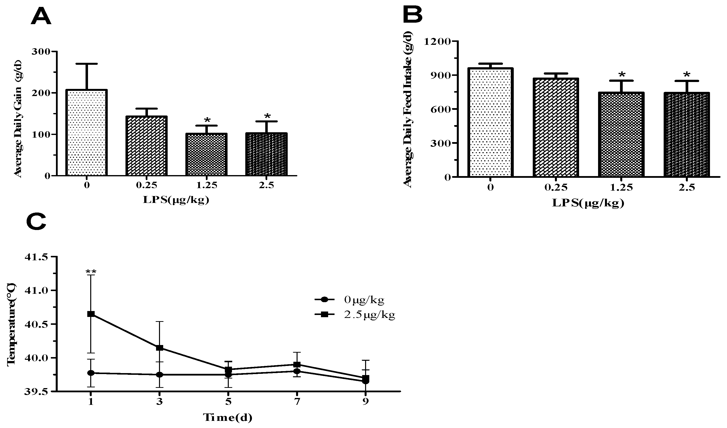
3.1. Average Daily Gain, Feed Intake, and Clinical Symptoms after LPS-Challenge

Within the first 3 hours after LPS administration, six lambs in the LPSM and LPSH groups had diarrhoea and, 60 minutes after administration on days 1 and 3, all lambs increased their respiratory rate, and seven lambs had rhinorrhea, became lethargic, and shivered. Heart rate in the LPSH group was significantly higher than in the control group (Supplementary Materials Table S1). Subsequent LPS injections resulted in lethargy, shivering, and hyperventilation in the lambs, but no diarrhoea was observed. The LPSM (p < 0.05) and LPSH (p < 0.05) groups had lower average daily gain (ADG; Figure 1A) and average daily feed intake (ADFI; Figure 1B) compared to the control lambs. LPSH lambs had a higher rectal temperature (Figure 1C) on day 1 (p < 0.01) than controls, however, there was no difference among groups in the following days. Lambs administered with LPS had a significant decrease in the number of white blood cells (Figure 2A, p < 0.05) in a dose-independent manner on days 1 and 3 after administration, compared with the control group.

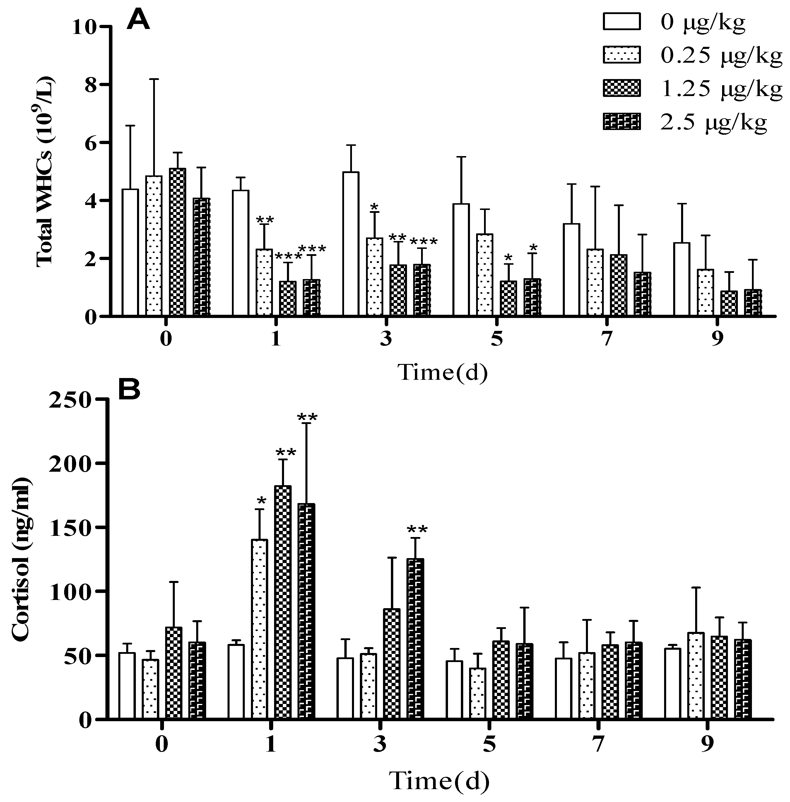
3.2. LPS Challenge Induced Systemic Inflammation in Lambs

Plasma cortisol concentration remained constant on days 0, 1, 3, and 5 in control lambs, but with LPS injection, cortisol concentration increased in a dose-independent manner at 3 h after LPS-challenge on day 1 and returned to pretreatment level on day 5. Following subsequent administration of LPS, plasma cortisol concentration did not differ from the control group (Figure 2B, p < 0.05). The expressions of IFN-γ and TLR4 in blood were higher than in the control group after the LPS challenge on days 1 and 3 (Figure 3A–B, p < 0.05), in particular in the LPSH group. On days 5, 7, and 9, there was no significant difference between the LPS injected groups and the control group.

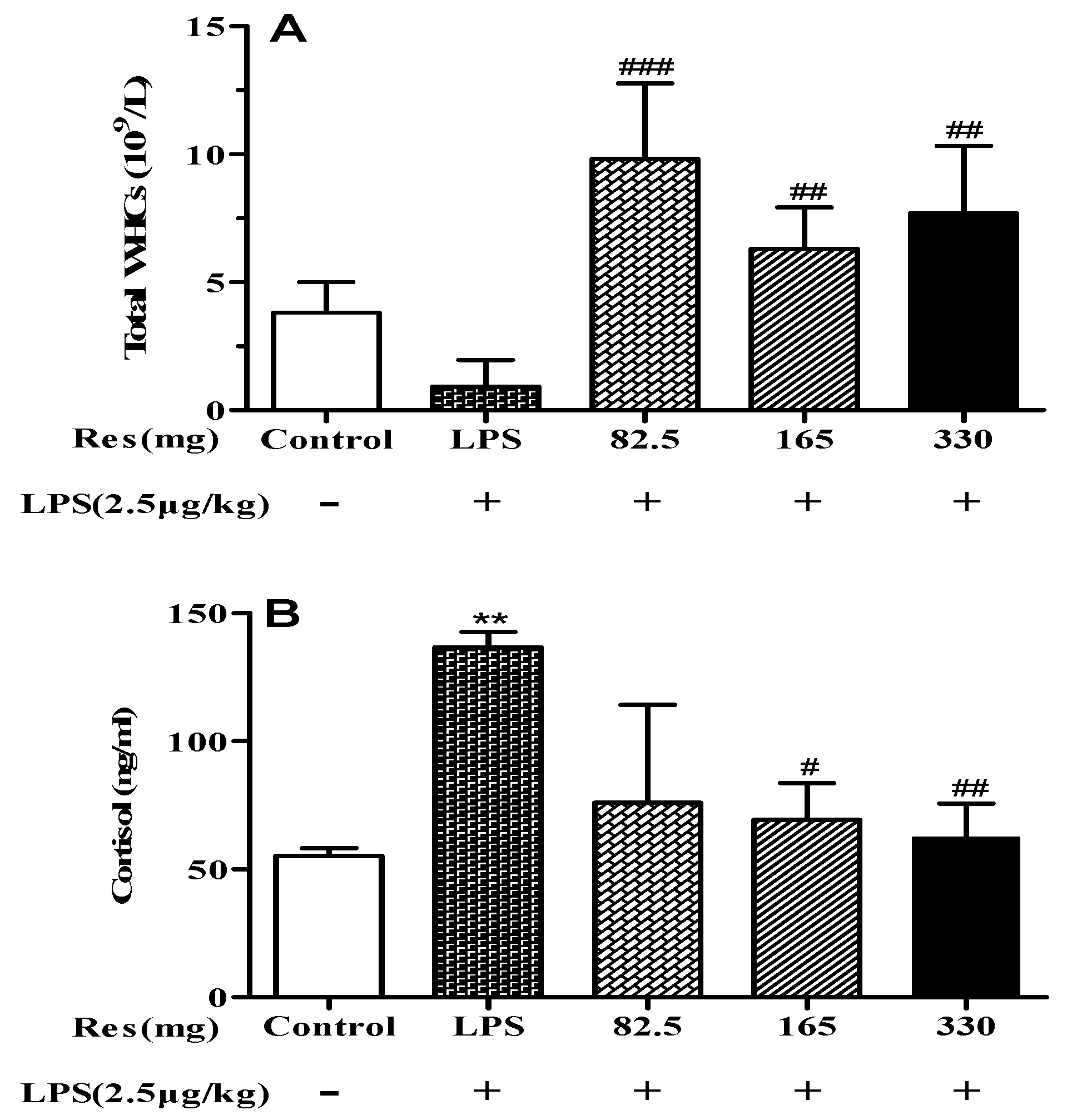
3.3. Effect of Resveratrol on Blood Parameters in LPS-Challenged Lambs

LPS injected lambs had lower WBCs (Figure 4A) and higher concentrations of plasma cortisol (Figure 4B) than the control group. The resveratrol treatment enhanced WBCs and decreased the concentration of plasma cortisol in lambs subjected to an LPS challenge (p < 0.05, Figure 4).

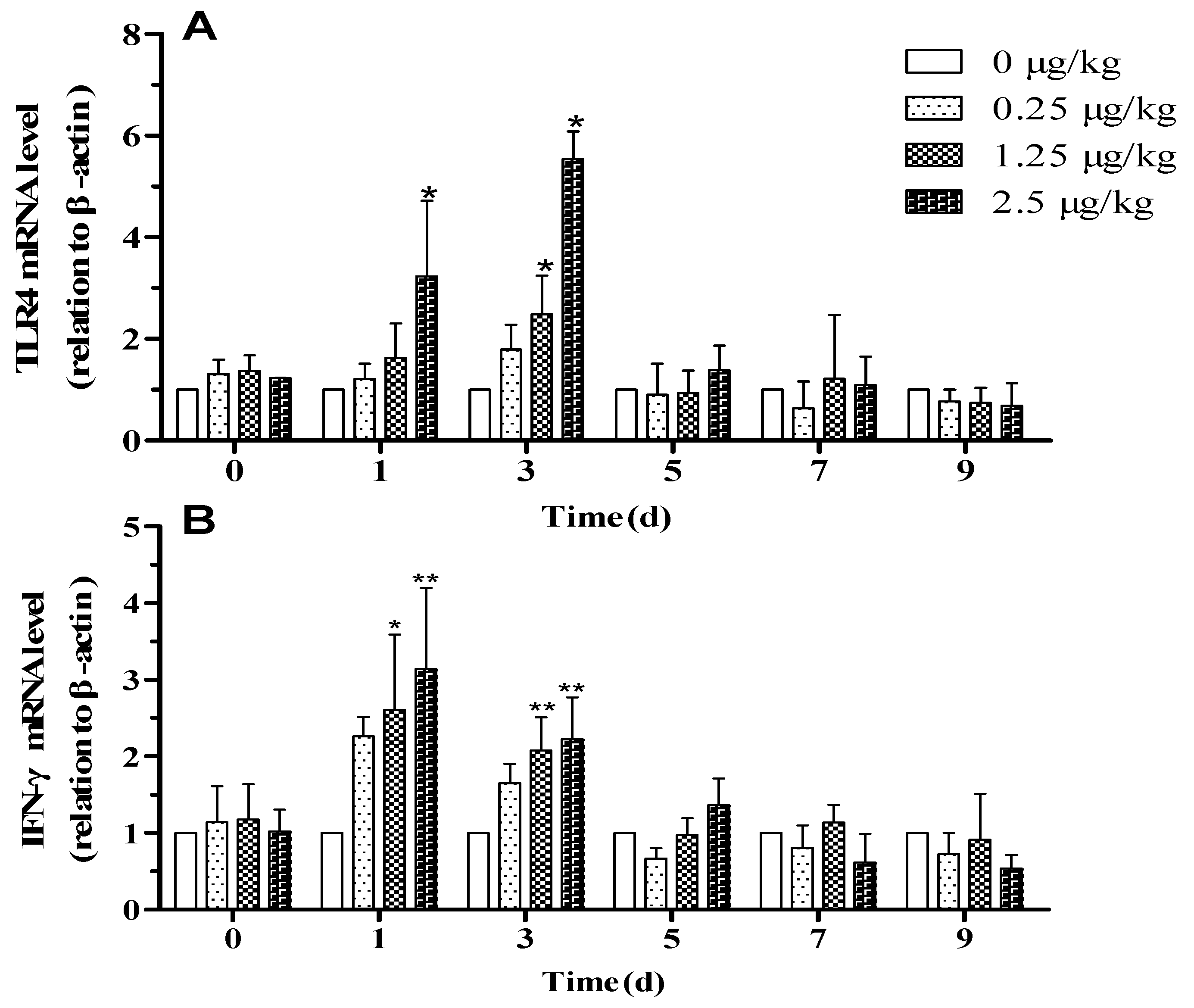
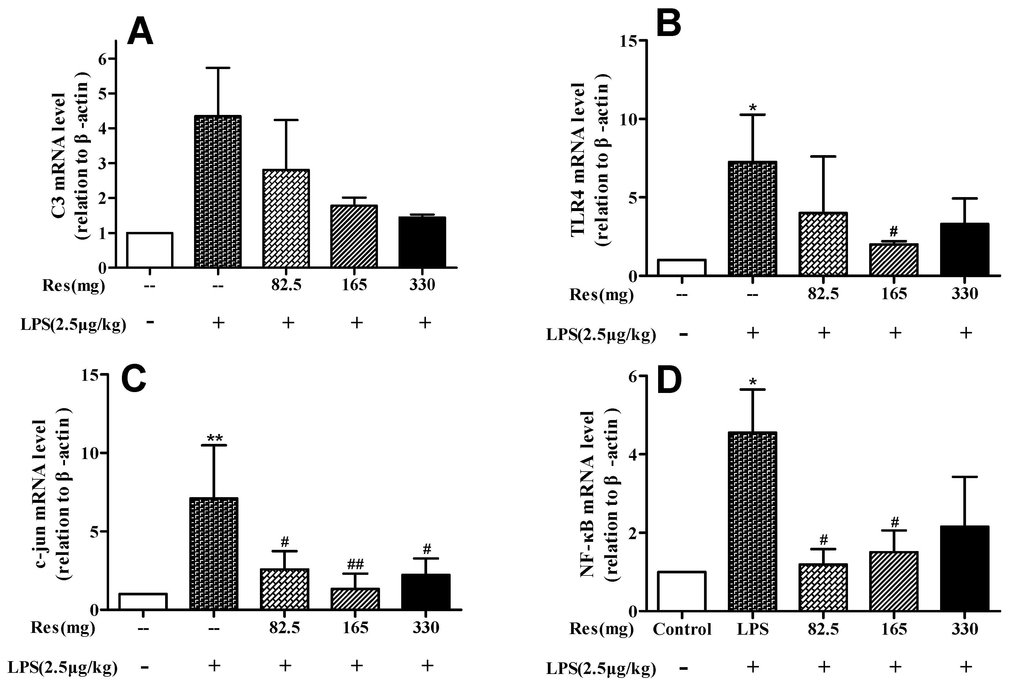
3.4. Effect of Resveratrol on Expression of Genes Following the LPS Challenge

When compared to the control group, LPS injected lambs had a tendency to increase the expression levels of C3, while lambs with resveratrol treatment had a tendency to reduce the levels (p < 0.10; Figure 5A). The mRNA levels of TLR4 (Figure 5B), c-jun (Figure 5C), and NF-κB (Figure 5D) in the LPS injected lambs were significantly higher than in the control group (p < 0.05 or p < 0.01); however, resveratrol at 165 mg suppressed the expression of these genes compared with the LPS group (p < 0.05 or p < 0.01).

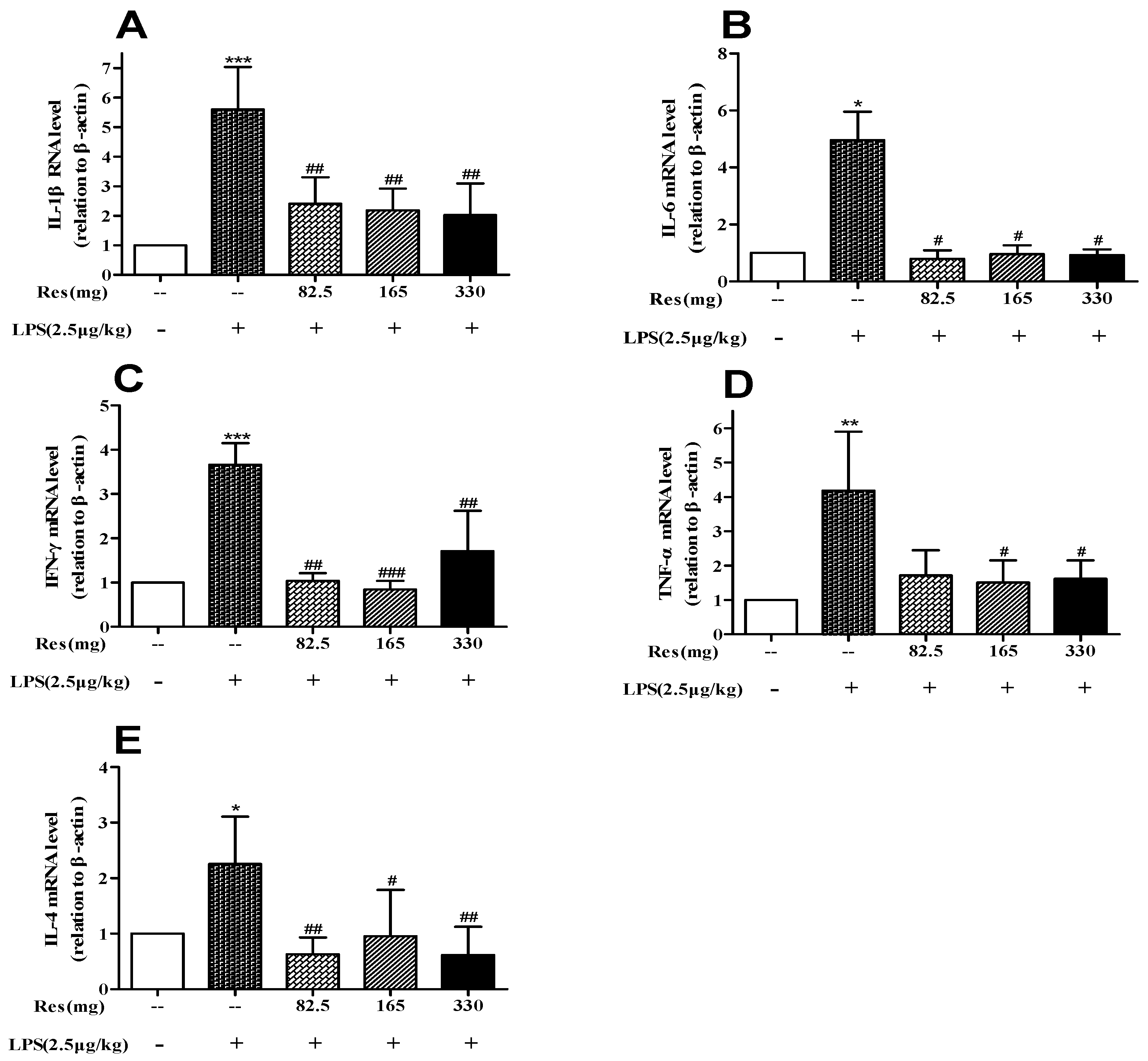
3.5. Effect of Resveratrol on Expression of Inflammatory Cytokines Following LPS Challenge

LPS administration upregulated the levels of IL-1β (p < 0.001; Figure 6A), IL-6 (p < 0.05; Figure 6B), IFN-γ (p < 0.001; Figure 6C), TNF-α (Figure 6D; p < 0.01), and IL-4 (p < 0.05; Figure 6E). After the administration of 165 mg and 330 resveratrol, the expressions of these genes were lowered significantly.

4. Discussion

In recent years, overuse of antibiotics has led to a rise in bacterial resistance to antibiotics, and has become a worldwide concern. Faced with this situation, natural plants are being examined as possible alternatives to antibiotics. Resveratrol has the potential to become an effective natural anti-bacterial drug as it possesses properties of anti-oxidation [13], anti-inflammation [21], anti-platelet aggregation [22,23], microcirculation improvement [24], vascular endothelium protection [25], nervous system protection [26], immunity enhancement, and aging delay. The main finding of this study was that resveratrol provided vital protection against inflammation induced by LPS in lambs.

4.1. LPS Challenge Induced Systemic Inflammation in Lambs

The pathogenesis of many diseases, including inflammatory bowel disease, Crohn’s disease, gout, and cancer, involves inflammation [19]. To reveal the mechanism of inflammatory disease and to find effective counter-measures for the chronic diseases, LPS, as a non-pathogenic immune activator, is being used to establish animal models. An intrajugular injection of 2.5 μg/kg LPS resulted in different expressions of inflammation-related genes in ewes [2]. Yates et al. [27] reported that an intrajugular injection of 0.75 and 1.5 μg/kg LPS caused a dose-dependent inflammatory response, manifested as an increase in rectal temperature, serum cortisol, and serum insulin. A single injection of LPS in animals only induced a short-term acute immune stress, which caused a sharp increase in plasma concentrations of inflammatory cytokines that returned to the pre-injection levels after 4 to 8 h [2,27,28]. Therefore, to reduce the tolerance and avoid the risk of mortality due to LPS in lambs in the present study, an initial dose of 0.25, 1.25, or 2.5 μg/kg LPS was injected every other day with a dose increase of 20% each time to examine the putative effect of resveratrol on inflammatory responses.
LPS induced an inflammatory state in lambs, especially at the high dose of 2.5 µg/kg LPS. The hypothalamic–pituitary–adrenal (HPA) axis is the major system for stress regulation, and cortisol, a hormone from the adrenal cortex, is secreted for its anti-inflammatory actions. In this study, plasma cortisol concentration increased on the first day after administration of LPS then returned to the pre-injection level on the fifth day, which indicated an increased secretion caused by a response of the hypothalamic–pituitary–adrenal axis [29]. The WBCs in the LPSM and LPSH groups were lower than in the control group after administration on days 1, 3, and 5, as was also reported for pigs [4]. According to Wang et al. [17], the reason for blood leukopenia was that the WBCs adhered to the endothelial cells after LPS injection, and the WBCs infiltrated into the tissue through the vascular endothelial barrier. The lower LPS dose (LPSL) did not affect the expressions of IFN-γ and TLR4, whereas the higher doses (LPSM and LPSH) increased them to an even greater extent than the control group after the LPS challenge on days 1 and 3. However, the magnitude of cortisol response and the expressions of IFN-γ and TLR4 to LPS administration were attenuated with successive injections. This might be due to a lower amplitude of response, relative to the first challenge, and could be associated with the instability and short half-life of inflammatory factors in blood [4]. These findings demonstrated an adaptive endocrine response and an activation of intracellular signaling pathways to repeated inflammatory stimuli of LPS.

4.2. Resveratrol Improves Systemic Inflammation Response in LPS-Injected Lambs

The complement system begins with the formation of antigen–antibody immune complexes, thereby creating a chemo-attractant for immune cells, leading to a release of proteolytic enzymes from intracellular granules, and modulation of cytokine production [30]. The complement component C3 is the central factor in all three activation pathways (classical, alternative, and lectin) [31]. Hadfield et al. [2] reported that the mRNA level C3 in blood of ewes increased for 6 h after LPS administration, but there was no change in C3 at 0–4 h. In this study, LPS had a tendency to increase the expression level of C3 when compared to the control group, whereas lambs treated with resveratrol had a tendency to reduce the level of C3. It was reported that the enhanced transcription of the C3 gene induced by LPS is targeted by the c-jun transcription factor [32]. C-jun is a component of the AP-1 transcription complex, which is considered to be the major target and key factor in inflammatory diseases that regulate inflammatory processes. The activity of AP-1 is regulated by mitogen-activated protein kinase (MAPK) [33]. In the present study, the mRNA level of c-jun was enhanced substantially after LPS treatment in the lambs; however, this up-regulation was suppressed markedly by resveratrol. The dose of 165 mg resveratrol showed the best results, indicating that the proper dose of resveratrol is important in alleviating inflammation by the MAPK signal pathway. Studies in rodents also demonstrated resveratrol-mediated protection against LPS-induced inflammation [34,35], which suggests that resveratrol is not species specific in its actions and can be used, most likely, on all animals.
Previous research demonstrated that the inflammatory response to LPS was associated with activation of toll-like receptors (TLRs) whose family members are receptors of the innate immune system that recognize pathogen-associated molecular patterns. Among them, TLR4 is considered to be a major LPS signaling receptor [36]. Stimulation of TLR4 triggers activation of transcription factors, which, in turn, enhance the synthesis of inflammatory genes, including cytokines. The formation of an LPS-TLR4/MD-2 complex and the subsequent recruitment of MyD88 adaptor protein induces activation of MAPKs, as well as the transcriptional NF-κB, leading to the secretion of various inflammatory mediators [36]. NF-κB is present in the cytoplasm as a hetero-trimeric complex composed of three subunits: p50, p65, and IκB. After activation of the complex, phosphorylation and degradation of IκB exposes nuclear localization signals on the p50/p65 complex, leading to their nuclear translocation and binding to the specific regulated sequences in the DNA, thus controlling gene transcription [37]. NF-κB signaling is an inducer of LPS, as well as MAPK signaling, while the attenuation of this signaling by resveratrol contributes to its anti-inflammatory effect [10,38]. NF-κB and AP-1 were key downstream factors of TLR4 and resveratrol was shown to block the expression of TLR4 and suppress the phosphorylation of NF-κB and expression of AP-1 in high glucose-induced mesangial cells [39]. In this study, resveratrol prevented NF-κB and TLR4 gene upregulation caused by LPS.
The production of inflammatory cytokines such as IL-1β, IL-4, IL-6, TNF-α, and IFN-γ have strong influence on inflammatory responses and serve as markers in LPS-induced inflammation [40,41,42,43]. TNF-α acts as a first signal that enhances the production of other pro-inflammatory cytokines such as IL-1β. IL-1β, a subtype of IL-1, is thought to mediate inflammatory actions by inducing the expression of pro-inflammatory genes and the production of secondary cytokines [44]. In contrast, IL-4 is often associated with suppression or reduction of inflammatory responses, mainly by inhibiting the secretion of IL-1β, IL-6, and TNF-α [45,46]. Studies have shown that resveratrol regulates the expression of cytokines induced by LPS through the NF-κB signaling pathway. It can activate transcription and promote production of TNF-α, IL-1β, IL-4, IL-6, and IFN-γ genes. Then NF-κB is activated again, which leads to further amplification of the initial inflammatory signal, aggravating the body damage and microcirculatory disorders [37]. In the present study, resveratrol at 165 and 330 mg suppressed the secretion of cytokines, and showed a positive trend to narrow the gap caused by LPS, which indicated that resveratrol regulated immune abnormalities caused by LPS.

5. Conclusions

Resveratrol attenuated the LPS-evoked inflammatory activation in lambs by suppressing expression levels of pro-inflammatory cytokines such as IL-1β, IL-6, IFN-γ, and TNF-α and anti-inflammatory cytokines IL-4, as well as the release of cortisol and the reduction of leucocytes. Resveratrol induced anti-inflammation actions by decreasing complement activity and blocking NF-κB and MAPK signaling pathways. Based on these studies, resveratrol has the potential to be a promising therapeutic reagent for multiple inflammatory illnesses caused by immune stress. Perhaps it can be added to feed in intensively-raised livestock systems, but this still warrants further investigation.

Supplementary Materials

The following are available online at https://www.mdpi.com/2076-2615/9/11/872/s1, Table S1: Effect of lipopolysaccharides (LPS) on Heart rate in lambs

Author Contributions

Conceptualization, Y.L., G.Y. and Z.Z.; methodology, Y.L. and D.J.; software, Y.L.; validation, J.Z., G.Y. and Z.Z.; formal analysis, H.L.; investigation, K.J.; resources, J.G.; data curation, Y.L. and M.Z.; writing—original draft preparation, Y.L.; writing—review and editing, J.Z., M.Z. and A.D.; visualization, H.L.; supervision, K.J.; project administration, G.Y.; funding acquisition, G.Y. All authors participated in writing the final draft of the manuscript and agreed on the final format.

Funding

This work was supported by the Hundred Talents Program of the Chinese Academy of Sciences (NO: Y629721002), Special Funds for Scientific and Technological Innovation and Development in Gansu Province (NO: Y939BD1003), Open subject of Provincial and ministerial co-construction of national key laboratories.

Acknowledgments

We are very grateful to three anonymous reviewers for their helpful comments and to the staff of the Gaolan Ecological and Agricultural Integrated Experimental Station (Gansu, China) for their help with the research.

Conflicts of Interest

The authors declare that they have no conflict of interest.

References

  1. Kumar, B. Stress and its impact on farm animals. Front. Biosci. 2012, 4, 1759–1767. [Google Scholar] [CrossRef]
  2. Hadfield, J.M.; Bowdridge, E.C.; Holásková, I.; Elsasser, T.H.; Dailey, R.A. Breed-specific differences in the immune response to lipopolysaccharide in ewes. J. Anim. Sci. 2018, 96, 4220–4228. [Google Scholar] [CrossRef] [PubMed]
  3. Benjamin, A.; Korkmaz, F.; Elsasser, T.; Kerr, D. Neonatal lipopolysaccharide exposure does not diminish the innate immune response to a subsequent lipopolysaccharide challenge in Holstein bull calves. J. Dairy Sci. 2016, 99, 5750–5763. [Google Scholar] [CrossRef] [PubMed]
  4. Campos, P.H.; Merlot, E.; Damon, M.; Noblet, J.; Le Floc’h, N. High ambient temperature alleviates the inflammatory response and growth depression in pigs challenged with Escherichia coli lipopolysaccharide. Vet. J. 2014, 200, 404–409. [Google Scholar] [CrossRef] [PubMed]
  5. Kallapura, G.; Pumford, N.; Hernandez-Velasco, X.; Hargisand, B.M.; Tellez, G. Mechanisms involved in lipopolysaccharide derived ROS and RNS oxidative stress and septic shock. J. Microbiol. Res. Rev. 2014, 2, 6–11. [Google Scholar]
  6. Plessers, E.; Watteyn, A.; Wyns, H.; Pardon, B.; De Backer, P.; Croubels, S. Study of the immunomodulatory properties of gamithromycin and dexamethasone in a lipopolysaccharide inflammation model in calves. Res. Vet. Sci. 2015, 103, 218–223. [Google Scholar] [CrossRef]
  7. Harris, R.E. Inflammation in the Pathogenesis of Chronic Diseases: The Cox-2 Controversy; Springer Science & Business Media: Berlin/Heidelberg, Germany, 2007. [Google Scholar]
  8. Singh, A.P.; Singh, R.; Verma, S.S.; Rai, V.; Kaschula, C.H.; Maiti, P.; Gupta, S.C. Health benefits of resveratrol: Evidence from clinical studies. Med. Res. Rev. 2019, 39, 1851–1891. [Google Scholar] [CrossRef]
  9. Berman, A.Y.; Motechin, R.A.; Wiesenfeld, M.Y.; Holz, M.K. The therapeutic potential of resveratrol: A review of clinical trials. NPJ Precis. Oncol. 2017, 1, 35. [Google Scholar] [CrossRef]
  10. Bobermin, L.D.; Roppa, R.H.A.; Quincozes-Santos, A. Adenosine receptors as a new target for resveratrol-mediated glioprotection. Biochim. Biophys. Acta Mol. Basis Dis. 2019, 1865, 634–647. [Google Scholar] [CrossRef]
  11. Bigagli, E.; Cinci, L.; Paccosi, S.; Parenti, A.; D’Ambrosio, M.; Luceri, C. Nutritionally relevant concentrations of resveratrol and hydroxytyrosol mitigate oxidative burst of human granulocytes and monocytes and the production of pro-inflammatory mediators in LPS-stimulated RAW 264.7 macrophages. Int. Immunopharmacol. 2017, 43, 147–155. [Google Scholar] [CrossRef]
  12. Mathew, L.M.; Woode, R.A.; Axiak-Bechtel, S.M.; Amorim, J.R.; DeClue, A.E. Resveratrol administration increases phagocytosis, decreases oxidative burst, and promotes pro-inflammatory cytokine production in healthy dogs. Vet. Immunol. Immunopathol. 2018, 203, 21–29. [Google Scholar] [CrossRef] [PubMed]
  13. Jia, R.; Li, Y.; Cao, L.; Du, J.; Zheng, T.; Qian, H.; Gu, Z.; Jeney, G.; Xu, P.; Yin, G. Antioxidative, anti-inflammatory and hepatoprotective effects of resveratrol on oxidative stress-induced liver damage in tilapia (Oreochromis niloticus). Comp. Biochem. Physiol. C Toxicol. Pharmacol. 2019, 215, 56–66. [Google Scholar] [CrossRef] [PubMed]
  14. Jha, R.K.; Ma, Q.; Lei, Z.; Sha, H. Resveratrol ameliorates the deleterious effect of severe acute pancreatitis. Cell Biochem. Biophys. 2012, 62, 397–402. [Google Scholar] [CrossRef] [PubMed]
  15. Tian, Y.; Ma, J.; Wang, W.; Zhang, L.; Xu, J.; Wang, K.; Li, D. Resveratrol supplement inhibited the NF-κB inflammation pathway through activating AMPKα-SIRT1 pathway in mice with fatty liver. Mol. Cell. Biochem. 2016, 422, 75–84. [Google Scholar] [CrossRef] [PubMed]
  16. Bereswill, S.; Muñoz, M.; Fischer, A.; Plickert, R.; Haag, L.M.; Otto, B.; Kühl, A.A.; Loddenkemper, C.; Göbel, U.B.; Heimesaat, M.M. Anti-Inflammatory Effects of Resveratrol, Curcumin and Simvastatin in Acute Small Intestinal Inflammation. PLoS ONE 2010, 5, e15099. [Google Scholar] [CrossRef] [PubMed]
  17. Wang, Y.; Cui, H.; Niu, F.; Liu, S.L.; Li, Y.; Zhang, L.M.; Du, H.B.; Zhao, Z.G.; Niu, C.Y. Effect of Resveratrol on Blood Rheological Properties in LPS-Challenged Rats. Front. Physiol. 2018, 9, 1202. [Google Scholar] [CrossRef] [Green Version]
  18. Calabrese, E.J.; Mattson, M.P.; Calabrese, V. Resveratrol commonly displays hormesis: Occurrence and biomedical significance. Hum. Exp. Toxicol. 2010, 29, 980–1015. [Google Scholar] [CrossRef]
  19. Salvesen, Ø.; Reiten, M.R.; Espenes, A.; Bakkebø, M.K.; Tranulis, M.A.; Ersdal, C. LPS-induced systemic inflammation reveals an immunomodulatory role for the prion protein at the blood-brain interface. J. Neuroinflamm. 2017, 14, 106. [Google Scholar] [CrossRef] [Green Version]
  20. Gupta, Y.; Briyal, S.; Chaudhary, G. Protective effect of trans-resveratrol against kainic acid-induced seizures and oxidative stress in rats. Pharmacol. Biochem. Behav. 2002, 71, 245–249. [Google Scholar] [CrossRef]
  21. Narayanan, B.A.; Narayanan, N.K.; Nargi, D. Natural anti-inflammatory agent resveratrol inhibit mediators of inflammation and cancer. Proc. Am. Assoc. Cancer Res. 2006, 47, 920–921. [Google Scholar]
  22. Shen, M.Y.; Hsiao, G.; Liu, C.L.; Fong, T.H.; Lin, K.H.; Chou, D.S.; Sheu, J.R. Inhibitory mechanisms of resveratrol in platelet activation: Pivotal roles of p38 MAPK and NO/cyclic GMP. Br. J. Haematol. 2007, 139, 475–485. [Google Scholar] [CrossRef] [PubMed]
  23. Olas, B.; Wachowicz, B. Resveratrol, a phenolic antioxidant with effects on blood platelet functions. Platelets 2005, 16, 251–260. [Google Scholar] [CrossRef] [PubMed]
  24. Sha, H.; Ma, Q.; Jha, R.K.; Wu, Z.; Qingyuan, Z.; Wang, Z.; Ma, Z.; Luo, X.; Liu, C. Resveratrol suppresses microcirculatory disturbance in a rat model of severe acute pancreatitis. Cell Biophys. 2013, 67, 1059–1065. [Google Scholar] [CrossRef] [PubMed]
  25. Chen, F.; Qian, L.H.; Deng, B.; Liu, Z.M.; Zhao, Y.; Le, Y.Y. Resveratrol protects vascular endothelial cells from high glucose–Induced apoptosis through Inhibition of NADPH oxidase activation–driven oxidative stress. CNS Neurosci. Ther. 2013, 19, 675–681. [Google Scholar] [CrossRef] [PubMed]
  26. Dasgupta, S.; Bandyopadhyay, M. Neuroprotective mode of action of resveratrol in central nervous system. Pharma. Nutrition 2013, 1, 90–97. [Google Scholar] [CrossRef]
  27. Shah, Z.; Ali, T. Inflammation the kingpin of chronic diseases naturopathy as an alternative source of treatment: An updated overview. J. Drug Deliv. Ther. 2019, 9, 478–483. [Google Scholar] [CrossRef]
  28. Yates, D.T.; Loest, C.A.; Ross, T.T.; Hallford, D.M.; Carter, B.H.; Limesand, S.W. Effects of bacterial lipopolysaccharide injection on white blood cell counts, hematological variables, and serum glucose, insulin, and cortisol concentrations in ewes fed low- or high-protein diets. J. Anim. Sci. 2011, 89, 4286–4293. [Google Scholar] [CrossRef]
  29. Beishuizen, A.; Thijs, L.G. Endotoxin and the hypothalamo-pituitary-adrenal (HPA) axis. J. Endotoxin Res. 2003, 9, 3–24. [Google Scholar]
  30. Wu, C.C.; Huang, Y.S.; Chen, J.S.; Huang, C.F.; Su, S.L.; Lu, K.C.; Lin, Y.F.; Chu, P.; Lin, S.H.; Sytwu, H.K. Resveratrol Ameliorates renal damage, increases expression of heme oxygenase-1, and has anti-complement, anti-oxidative, and anti-apoptotic effects in a murine model of membranous nephropathy. PLoS ONE 2015, 10, e0125726. [Google Scholar] [CrossRef]
  31. Elvington, M.; Liszewski, M.K.; Bertram, P.; Kulkarni, H.S.; Atkinson, J.P. A C3(H2O) recycling pathway is a component of the intracellular complement system. J. Clin. Investig. 2017, 127, 970–981. [Google Scholar] [CrossRef]
  32. Chen, M.; Wang, C.; Li, Z.; Chen, J. Involvement of JNK signaling pathway in lipopolysaccharide-induced complement C3 transcriptional activation from amphioxus Branchiostoma belcheri. Fish Shellfish. Immunol. 2019, 86, 196–203. [Google Scholar] [CrossRef] [PubMed]
  33. Karin, M. The Regulation of AP-1 Activity by mitogen-activated protein kinases. J. Biol. Chem. 1995, 270, 16483–16486. [Google Scholar] [CrossRef] [PubMed]
  34. Zhang, X.; Wang, Y.; Xiao, C.; Wei, Z.; Wang, J.; Yang, Z.; Fu, Y. Resveratrol inhibits LPS-induced mice mastitis through attenuating the MAPK and NF-κB signaling pathway. Microb. Pathog. 2017, 107, 462–467. [Google Scholar] [CrossRef] [PubMed]
  35. Wang, G.; Hu, Z.; Fu, Q.; Song, X.; Cui, Q.; Jia, R.; Zou, Y.; He, C.; Li, L.; Yin, Z. Resveratrol mitigates lipopolysaccharide-mediated acute inflammation in rats by inhibiting the TLR4/NF-κBp65/MAPKs signaling cascade. Sci. Rep. 2017, 7, 45006. [Google Scholar] [CrossRef] [PubMed]
  36. Moynagh, P.N. The NF-kappaB pathway. J. Cell Sci. 2005, 118, 4589–4592. [Google Scholar] [CrossRef] [PubMed]
  37. Liu, T.; Zhang, L.; Joo, D.; Sun, S.C. NF-kappaB signaling in inflammation. Signal Transduct. Target. Ther. 2017, 2. [Google Scholar] [CrossRef]
  38. Gorina, R.; Font-Nieves, M.; Márquez-Kisinousky, L.; Santalucia, T.; Planas, A.M. Astrocyte TLR4 activation induces a proinflammatory environment through the interplay between MyD88-dependent NFκB signaling, MAPK, and Jak1/Stat1 pathways. Glia 2011, 59, 242–255. [Google Scholar] [CrossRef]
  39. Deng, Y.; Gong, W.; Li, Q.; Wu, X.; Wu, L.; Zheng, X.; Chen, W.; Huang, H. Resveratrol inhibits high glucose-induced activation of AP-1 and NF-κB via SphK1/S1P2 pathway to attenuate mesangial cells proliferation and inflammation. J. Funct. Foods 2019, 55, 86–94. [Google Scholar] [CrossRef]
  40. Nielsen, O.H.; Køppen, T.; Rüdiger, N.; Horn, T.; Eriksen, J.; Kirman, I.; Nielsen, D.O.H. Involvement of interleukin-4 and-10 in inflammatory bowel disease. Dig. Dis. Sci. 1996, 41, 1786–1793. [Google Scholar] [CrossRef]
  41. Laflamme, N.; Lacroix, S.; Rivest, S. An essential role of interleukin-1beta in mediating NF-kappaB activity and COX-2 transcription in cells of the blood-brain barrier in response to a systemic and localized inflammation but not during endotoxemia. J. Neurosci. 1999, 19, 10923–10930. [Google Scholar] [CrossRef]
  42. Barnes, T.C.; Anderson, M.E.; Moots, R.J. The Many Faces of Interleukin-6: The Role of IL-6 in Inflammation, Vasculopathy, and Fibrosis in Systemic Sclerosis. Int. J. Rheumatol. 2011, 2011, 721608. [Google Scholar] [CrossRef]
  43. Zhang, J. Yin and yang interplay of IFN-gamma in inflammation and autoimmune disease. J. Clin. Investig. 2007, 117, 871–873. [Google Scholar] [CrossRef]
  44. Kim, K.S.; Jung, H.; Shin, I.K.; Choi, B.R.; Kim, D.H. Induction of interleukin-1 beta (IL-1β) is a critical component of lung inflammation during influenza A (H1N1) virus infection. J. Med. Virol. 2015, 87, 1104–1112. [Google Scholar] [CrossRef]
  45. Rubinstein, A.I.; Rubinstein, D.B. Interleukin-4 (IL-4) inhibits secretion of IL-1 beta, tumor necrosis factor aipha, and IL-6 by human monocytes. Blood 1990, 76, 1392–1397. [Google Scholar] [CrossRef]
  46. Paul, W.E. Interleukin-4 a prototypic immunoregulatory lymphokine. Blood 1991, 77, 1859–1870. [Google Scholar] [CrossRef]
Figure 1. Effect of 2.5 μg /kg body weight lipopolysaccharides (LPS) on (A) average daily gain, (B) average daily feed intake and (C) rectal temperature in lambs. Values are expressed as means ± SD. * p < 0.05, ** p < 0.01, and *** p < 0.001, compared with the control group.
Animals 09 00872 g001
Figure 2. Effect of 0.25, 1.25, and 2.5 μg/kg LPS on (A) white blood cells (WBCs) and (B) plasma cortisol concentration in lambs. Values are expressed as means ± SD. * p < 0.05, ** p < 0.01, and *** p < 0.001, compared with the control group.
Animals 09 00872 g002
Figure 3. Effect of 0.25, 1.25, and 2.5 μg/kg LPS on expression of inflammatory markers in blood of lambs. (A) Toll-like receptor-4 (TLR4) and (B) Interferon-γ (IFN-γ). The values are normalized to control values and expressed as means ± SD. * p < 0.05, ** p < 0.01, and *** p < 0.001, compared with the control group.
Animals 09 00872 g003
Figure 4. Effect of resveratrol on: (A) the number of total white blood cells (WBCs) and (B) concentration of plasma cortisol in LPS-challenged lambs. Values are normalized to control values and expressed as means ± SD. * p < 0.05, ** p < 0.01, and *** p < 0.001, compared with the control group; # p < 0.05, ## p < 0.01, and ### p < 0.001, compared with the LPS treatment.
Animals 09 00872 g004
Figure 5. Effect of resveratrol on expression of immune gene C3 (A) and inflammatory markers in peripheral WBCs in LPS-challenged lambs, (B) TLR4, (C) c-jun, and (D) NF-κB. Values are normalized to control values and expressed as means ± SD. * p < 0.05, ** p < 0.01, and *** p < 0.001, compared with the control group; # p < 0.05, ## p < 0.01, and ### p < 0.001, compared with the LPS treatment.
Animals 09 00872 g005
Figure 6. Effect of resveratrol on expression of pro-inflammatory cytokines (A) IL-1β, (B) IL-6, (C) IFN-γ, (D) TNF-α, and (E) anti-inflammatory cytokines IL-4 in LPS-challenged lambs. The values are normalized to control values and expressed as means ± SD. * p < 0.05, ** p < 0.01, and *** p < 0.001, compared with the control group; # p < 0.05, ## p < 0.01, and ### p < 0.001, compared with the LPS treatment.
Animals 09 00872 g006
Table 1. Ingredient and nutrient levels of the diet offered the lambs in Experiments 1 and 2.
Ingredient Nutrient 2
Ingredient, % of DM
Oat hay25.00ME (MJ/kg) 9.50
Corn stalk15.00Crude protein (%)10.89
Corn31.50Neutral detergent fiber (%)43.83
Soybean meal4.00Acid detergent fiber (%)16.71
Wheat bran4.50Ca (%)0.63
DDGS3.00P (%)0.45
Molasses3.00
Barley grain8.50
NaCl0.50
CaHPO40.80
Limestone0.70
Soybean oil1.50
Sodium bicarbonate1.00
Premix 11.00
DM: Dry Matter; ME: Metabolic Energy; DDGS: Distillers Dried Grains with Solubles. 1 The premix provided the following per kg of diets: Vit A, 12 000 IU; Vit D, 2 000 IU; Vit E, 30 IU; Cu, 12 mg; Fe, 64 mg; Mn, 56 mg; Zn, 60 mg; I, 1.2 mg; Se, 0.4 mg; Co, 0.4 mg. 2 ME was a calculated value, while the other nutrient levels were measured values.
Table 2. List of primer sequences for quantitative real-time PCR.
GeneForward Primer 5’-3’Reverse Primer 5’-3’Product LengthAnnealing Temperature (℃)
C3GCACTGTCCACCAACCTCAATCAGGCTTCTGCTTCTCCA8758
TLR4GGCATCATCTTCATCGTCCTCCACTCCAGGTAGGTGTTCC9958
NF-κBCATCAGCCAGCGCATCCAGACGCACGGCATTCAGGTCGTAGTC8661
c-junAGCGGATCAAGGCGGAGAGGCCTGAGCATATTGGCGGTGGAC15555
IL-1βGCAGGCAGTGTCGGTCATCGCCTCAGGTCATCATCACGGAAGAC8258
IL-4GCGGACTTGACAGGAATCTCAGCCAGCGTACTTGTACTCGTCTTGGC8063
IL-6ACACTGACATGCTGGAGAAGATGCGCCGCAGCTACTTCATCCGAATAG13261
IFN-RATGTTTCATTTGCCACCATCCGGTTACGCTTGCTTTGCCTTATGT8160
TNF-αCTGGCGGAGGAGGTGCTCTCGGAGGAAGGAGAAGAGGCTGAGG8559
β-actinAGCCTTCCTTCCTGGGCATGGAGGACAGCACCGTGTTGGCGTAGA11360

