Communication

Clinical, Histological and Genetic Characterisation of a Disorder of Sexual Development in a Pygmy Goat

The Royal (Dick) School of Veterinary Studies and the Roslin Institute, University of Edinburgh, Easter Bush Campus, Roslin, Midlothian EH25 9RG, Scotland, UK
*
Author to whom correspondence should be addressed.
Animals 2025, 15(7), 976; https://doi.org/10.3390/ani15070976
Submission received: 12 February 2025 / Revised: 20 March 2025 / Accepted: 26 March 2025 / Published: 28 March 2025
(This article belongs to the Section Small Ruminants)

Simple Summary

This study presents a comprehensive investigation of a disorder of sexual development (DSD) in a pygmy goat, expanding current understanding beyond polled intersex syndrome. The individual goat presented with both male and female characteristics. Histological and genetic investigations revealed the presence of testicular tissue and implied an XY karyotype, resembling Swyer syndrome in humans. These findings contribute to the understanding of DSDs in goats, albeit further genetic investigations would be needed to determine the underlying cause.

Abstract

A 10-month-old pygmy goat was examined at the Royal (Dick) School of Veterinary Studies at the University of Edinburgh in January 2022, having been referred with a suspected diagnosis of a disorder of sexual development (DSD). The animal displayed a predominantly female phenotype (vulva with enlarged clitoris and vagina) but some male external characteristics such as a beard, larger body size, and larger horns. It also typically exhibits male behaviours such as mounting, aggression, and flehmen response. Computed tomography (CT) detected a bicornuated tubular fluid-filled structure connected to bulbous soft tissue masses. Subsequent exploratory laparotomy and reproductive tract removal identified underdeveloped uterine horns connected to ductus deferens and gonads, which were confirmed as testicles by histological examination. No spermatogenesis was detected, and no ovarian tissue was apparent. Blood levels of testosterone showed a marked decrease post-surgery (2.10 nmol/L to <0.03 nmol/L). Whole genome sequencing suggested that the patient may have an XY male karyotype, although cytological analysis was not possible to confirm the XY karyotype.

1. Introduction

Historically, individuals with characteristics of both male and female phenotypes were described as intersex, hermaphrodites, pseudohermaphrodites, or sex reversals. Following a change in nomenclature regarding humans with such characteristics [1], these and a variety of other congenital presentations that arise from anomalous sexual differentiation are now classified together as disorders of sexual development (DSD) [2]. The entire spectrum of DSD can be categorised according to chromosomal, gonadal, and phenotypic presentation. Accordingly, chromosomal or genetic DSD results from anomalies in sex chromosomes and combination, whereas gonadal DSD describes normal XX or XY karyotype but discordance with the gonadal and/or external genital phenotype [3].
DSD is well documented in goats as they have a higher frequency of DSD than other domestic species (up to 10–15% in some breeds) [4]. These cases occur almost exclusively in polled (hornless) 60, XX individuals, a condition known as polled intersex syndrome (PIS) [5,6]. PIS is a heritable trait associated with an autosomal recessive expression, which exhibits a link to inheritance with the hornless locus, which has a dominant mode of inheritance. Individuals that are XX homozygous for the polled gene demonstrate a range of masculinisation, with testicular embryonic development in the absence of an SRY gene [5,6,7]. Rare examples of chimeric individuals with a XX/XY karyotype, such as freemartins, have been described [8]; nevertheless, only about 1% of heterosexual twin pregnancies in goats result in freemartins [9]. Swyer syndrome, a human condition describing a suite of presentations where an individual has a male XY karyotype but has female genitalia and a female or mixed phenotype [10], has been reported in a variety of domestic bovids, including cattle [11,12] buffalo [13], and sheep [14,15], but no unifying genetic basis has been found. This syndrome is also associated with an underdeveloped reproductive tract, elongated gonads (streak gonads), and, in some cases, clitoromegaly [10,16], as observed in the specimen of this study.
Although a variety of genetic, hormonal, and/or environmental possibilities exist to explain the development of a DSD, cases in livestock species rarely benefit from the comprehensive investigations made available for companion animals. Thus, full classification of individuals with ambiguous sexual features may be lacking. This work describes the clinical, histopathological, and genomic investigation of a phenotypically mixed female horned pygmy goat in order to characterise a DSD.

2. Materials and Methods

2.1. Animal Material

The affected individual was a 10-month-old assumed female pygmy goat (Capra aegagrus hircus). It was referred to the Farm Animal Hospital at the Royal (Dick) School of Veterinary Studies, University of Edinburgh, in January 2022 with a suspected diagnosis of a disorder of sexual development (DSD).

2.2. Diagnostic Imaging

Following clinical examination, transabdominal ultrasound (Mindray DP-30; IMV imaging, Strathclyde, Scotland) was undertaken using a 5.0–8.5 MHz micro-convex sector transducer, followed by computed tomography (CT) under general anaesthesia to evaluate the internal reproductive structures. CT was performed with the patient in sternal recumbency, using a 64-slice SOMATOM Definition AS (Siemens, Erlangen, Germany).

2.3. Surgical and Histological Examination

Exploratory laparotomy was performed under general anaesthesia, and the uterine and gonadal tissues were removed en bloc by standard surgical methods. The excised reproductive organs were fixed in 10% neutral buffered formalin, from which selected samples were paraffin-embedded and processed as 5-μm sections and stained with haematoxylin and eosin. Paired venous blood samples (heparin and EDTA tubes) were obtained prior to and two weeks after surgery for measurement of testosterone concentration and genetic analyses (stored frozen until analysis). A blood sample for cytological analysis to confirm the karyotype was not collected at the time, and as such, we used an approach based on whole-genome sequencing analysis as an alternative.

2.4. Samples, DNA Extraction and Sequencing

The suspected DSD goat sample was referred to as sample “DSD Goat”. DNA was extracted from peripheral blood by the commercial sequencing provider Neogen. Illumina short-read, paired-end (PE) libraries were prepared by Neogen and sequenced on the Illumina NovaSeq platform to a depth of 15 million reads per sample. Sequencing data from a control male and control female goat was used to facilitate comparative analysis. These two animals, “Male control” and “Female Control” (Table S1), were mixed-breed dairy goats with whole genome sequencing available from a previous study [17]. The two control animals were sequenced to a higher depth of coverage than the patient (30X) DSD goat (15X), so these data were down-sampled to create alignments of comparable coverage. Our hypothesis was that the read coverage would act as a proxy for the karyotype and that an XY or intermediate karyotype could be identified by the proportion of reads mapping to each sex chromosome. This method has been used successfully to investigate ploidy in cell lines in livestock [18].

2.5. Illumina Read Mapping

Raw reads from all samples were quality trimmed and had remnant adapter sequences removed with Trim Galore, a Perl wrapper for Cutadapt v.1.18 [19] and FastQC v.0.12.1 [20]. Reads were mapped to two reference genomes: Capra hircus Saanen_v1 genome (GCA_015443085.1), derived from a European Swiss dairy goat breed, and the official goat reference genome ARS1.2 (GCF_001704415.2), derived from a San Clemente Island goat. The Saanen genome has an assembled Y chromosome, whereas the Y chromosome on the ARS1.2 reference assembly is currently on unplaced scaffolds, so the Saanen reference was used to examine this region. Reads were mapped using the Burrow-wheeler aligner v. 2.1.0 (BWA) [21] with the mem algorithm before converting to bam format and sorting with samtools v.1.13 [22]. PCR duplicates were removed with Picard MarkDuplicates [23], and mapping statistics were calculated with samtools flagstat and idxstats, bedtools genomecov [24], and mosdepth [25].

2.6. Determination of Karyotype from Sequence Data

To assess the sex chromosome complement, a number of measurements based on sequencing coverage ratios were calculated: the X:Y coverage ratio, X:autosome ratio, and Y:autosome ratio. As genetic females have two X chromosomes (XX) and males have one X and one Y (XY), a female karyotype would show roughly similar coverage of the X chromosomes compared to autosomes and low coverage of the Y chromosome. In a male (XY) karyotype, the single X chromosome would be expected to have approximately half the coverage of the autosomes, and also roughly half coverage for Y chromosome reads.
Chromosome 10 was selected as the comparative autosome.

2.7. SRY Promotor Mutation

Heidari et al. [15] identified a T to G mutation on the SRY promotor region of a sheep with Swyer syndrome, which they speculate may be implicated in the disease. To investigate whether the patient had this mutation, the Y chromosome region was extracted from bam alignment files and imported into Geneious v. 8.1.9 [26]. Consensus sequences were obtained specifying the fewest ambiguities option and coding bases with <3 read coverage coded as missing (N). The SRY promotor region corresponding to that used in the multi-species alignment of Heidari et al. [15] was extracted from each sample. Ten sequences from Heidari et al. [15] were downloaded from Genbank, representing their query sequence individual (SRY+, Swyer syndrome sheep), four male Ovis spp., two male Capra hircus samples, one male Rupicapra pyrenaica (chamois) sample, one male Ammotragus lervia (barbary) sample, and one male Bos taurus sequence. Alignments were performed with MUSCLE [27] in the Geneious platform [26], with default settings and the maximum number of iterations set to five. The final alignment was inspected for the T to G mutation at position 303 [15].

3. Results

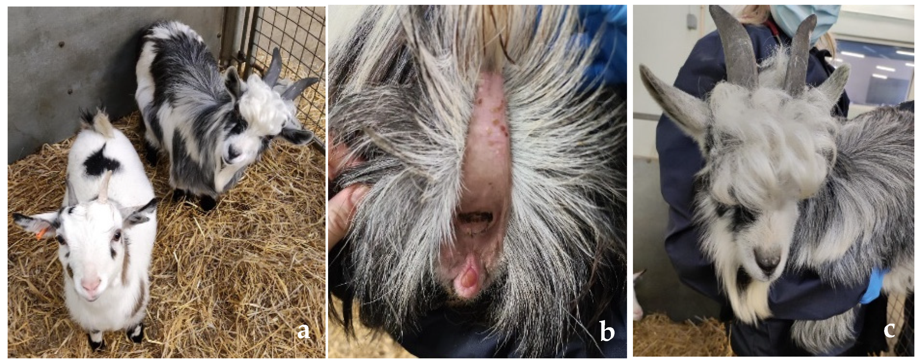
3.1. Clinical Phenotype

On clinical examination, abnormalities were restricted to the reproductive organs. The patient presented with a mixed phenotype with female external genitalia (vulva with a markedly enlarged and prominent clitoris and vagina) (Figure 1a,b) and some male external characteristics such as a beard, larger body size, and larger horns (Figure 1c). Moreover, the patient expressed male-like behavior (mounting, aggression, and flehmen response).

3.2. Diagnostic Imaging

CT images revealed a fluid-filled tubular organ dorsal to the urinary bladder (Figure S1a), which branched into a left and right horn to which bulbous soft tissue opacity gonads (Figure S1b) were attached by narrow tethers.

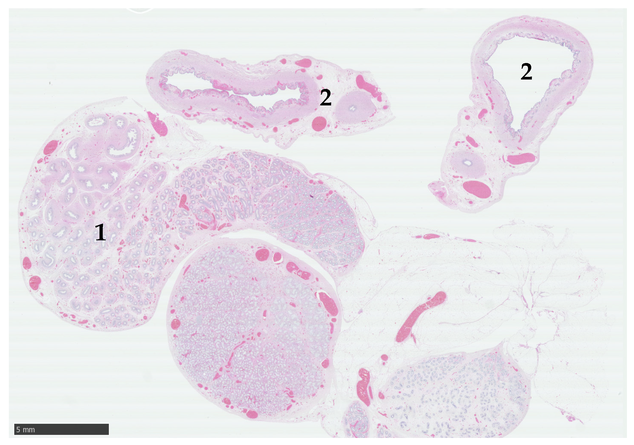
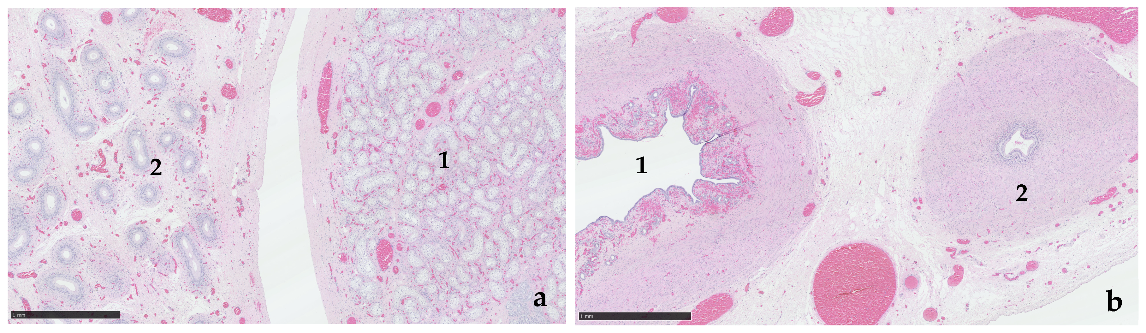
3.3. Exploratory Laparotomy and Histological Examination

Exploratory laparotomy revealed the fluid-filled, bifurcated, hypoplastic uterus dorsal to the urinary bladder. The uterine horns terminated in white, thick-walled tissue, leading to what appeared grossly to resemble hypoplastic testicular gonads (Figure S2).
Histological assessment of the gonads confirmed paired testes encased in abundant adipose tissue (Figure 2). Bilaterally, the majority of the tissue was represented by well-developed epididymis, devoid of speratozoa. Each gonad contained a single nodule of seminiferous tubules formed by multiple lobular clusters of amphophilic polygonal cells, with large clear cytoplasmic vacuoles (presumptive Sertoli cells), with no evidence of spermatogenesis. Leydig cells were not visible in the interstitial tissue (Figure 3a). On one side, the vas deferens were accompanied by a uterine horn (Figure 3b).

3.4. Blood Testosterone Concentrations

The testosterone concentration prior to surgery was 2.10 nmol/L, while the result two weeks post-operatively was <0.03 nmol/L. This reduction is indicative of the presence of functional testicular tissue. Nevertheless, the pre-surgery testosterone value is low compared to the normal testosterone value for male white goats in January (7.31 nmol/L) [28].

3.5. Illumina Sequencing Read Mapping

DSD Goat had a similar X-fold sequencing coverage when aligned to either the San Clemente or Saanen reference genomes (15X, Table S1). As the Saanen genome had a complete Y chromosome contig, this genome was used for further analysis. Downsampling of the comparative samples, Male Control and Female Control resulted in 16X and 15X coverage, respectively (Table S1).
Read mapping ratios of Male Control and Female Control were consistent with what would be expected for a male XY and a female XX karyotype (Table S2). The read mapping pattern of the Goat DSD suggested the presence of a Y chromosome (0.3M mapped reads), the coverage for the X chromosome was intermediate (12X) between the male (8X) and the female (14X), and median X coverage for the Y chromosome was much lower than in the male dairy goat—3X compared to 8X. DSD Goat also had a higher X:chr10 ratio (0.8) than the male (0.5) and a lower Y:chr10 ratio (0.2 compared to 0.5, Table S2).

3.6. SRY Promotor Mutation

Read mapping of DSD Goat data to the SRY region showed lower read coverage than in the Male control. DSD Goat did not have this mutation, nor did it show any other mutation in the promotor region that may be associated with Swyer syndrome.

4. Discussion

This study describes a case of a horned pygmy goat presenting with a DSD. The majority of caprine DSD relates to the polled intersex syndrome; however, the presence of horns and the potential of being XY suggests that PIS is unlikely to be the underlying cause of this individual’s condition. Testicular feminisation syndrome or insensitivity to androgen hormones [29] is also unlikely as it prevents the formation of a uterus and secondary male characteristics. Rare instances of XY individuals with testicular tissue and female ductal systems derived from the embryonic Müllerian duct are classified as exhibiting persistent Müllerian duct syndrome (PMDS). This situation can arise due to failure to express functional AMH or its receptor, AMHR2. Various mutations in either AMH or AMHR2 have been identified in humans [30], and it is well recognised in Miniature Schnauzer dogs, in which it is associated with an AMHR2 mutation with an autosomal recessive mode of inheritance [31]. A single report is described in a goat, but no genetic basis was investigated [32].
The SRY gene is located on the Y chromosome and encodes the SRY protein, which is one of the SOX9 transcription factors responsible for the development of the male reproductive tissues [33]. Deletions or mutations of SRY on any coding or regulatory region can disrupt male development and cause XY female sex reversal. In sheep, a mutation on the SRY promotor has been shown to lead to hermaphrodism [15], and, in cattle, deletion of SRY was associated with Swyer Syndrome-like condition in 7 out of 8 animals [11]. Humans with the condition lack the SRY gene (SRY-) in around 10–15% of cases, and mutations in the SRY gene (SRY+ individuals) are found in a further 10–15% of individuals [16,34]. Raudsepp et al. [35] suggest a distinction between SRY- and SRY+ Swyer syndrome individuals in horses, where the former lacks testes, and the latter may exhibit underdeveloped testes.
Freemartinism, which describes XX female-to-male, intersex presentations when gestation is shared with a male twin, would be unlikely as this individual was reportedly a single kid. However, being single-born does not completely exclude this condition, as multiple single-born freemartin heifers have been reported [36]. This situation may occur as a result of in utero death of a male co-twin, which influenced the sexual development of its female twin (with varying degrees of masculinisation) prior to its loss. Another rare cause for an XX/XY karyotype is mosaicism, in which two or more populations of cells arise from a single zygote due to recombination or mutation in mitosis or meiosis [3].
Read mapping coverage to the Y chromosome and SRY regions is lower than is observed for the control male despite down-sampling data for even coverage across the genome. The chromosome coverage ratio for Y:chr10 was also lower than expected for a male. These findings align well with what would be expected from a mosaic or chimeric individual, where the presence of Y-chromosome-containing cells is reduced compared to a typical male, and X chromosome coverage is elevated relative to an XY male but lower than an XX female. However, pygmy goats are a composite breed with a complex history, including the merging of breeds with dwarfism and from disparate geographic origins. It would not be surprising if pygmy goats exhibited unique Y chromosome structure and genetic divergence from other goat breeds. However, they are missing from previous studies characterising the genome-wide [37] and Y-chromosome diversity [38] of goat breeds globally. Due to a dearth of available comparative data, it has not been possible to explore their Y chromosome phylogeny in detail here, although determining which major Y chromosome lineage pygmy goats belong to and whether Y chromosome introgression and admixture is observable would be informative.
Identification of chimera/mosaic specimens requires further cytogenetic testing, such as karyotyping. This involves cell culture, preparation, and staining [39] from a freshly collected blood sample. In this case, karyotyping was not possible due to the lack of funding at the time of the clinical case and the impossibility of collecting fresh blood samples after the discharge of the patient. If the opportunity arises to collect suitable blood and or tissue samples in the future, this analysis will be performed and reported as a follow-up study. New methods to perform cytological analysis on frozen blood that will help to facilitate this are becoming available, although the volumes of blood required can be prohibitive due to sample collection limitations from clinical cases [40]. Possible further work could also include PCR and Sanger sequencing of SRY and other male-specific Y-chromosomal genes (DMRT1, USP9Y, UTY, and DDX3Y, for example) and other key genes in sexual development, such as AMHR2. In addition, variant calling of this sample with the VarGoats dataset [38] and the generation of a pygmy goat breed-specific reference genome to avoid reference mapping bias would add valuable genomic resources for pygmy goats.

5. Conclusions

The pygmy goat, in this case, appears to be an XY DSD and is potentially SRY+. To confirm the karyotype, both conventional and molecular cytogenetic analysis will be required.

Supplementary Materials

The following supporting information can be downloaded at: https://www.mdpi.com/article/10.3390/ani15070976/s1, Figure S1. Computed tomography images. (a) Dorsal to the urinary bladder a fluid filled tubular structure is noted, which splits into a left and right branch at the level of the urinary bladder. This is consistent with a fluid filled uterus; (b) Dextrally, at the end of the uterine horn, there is a thin soft tissue band which travels laterodorsally. A similar structure can be seen sinistrally, however both end in bulbous soft tissue structures, which are located dextrally to the midline. Figure S2. Gross morphology of the reproductive tract. Two small, tubular, bicornuated, filled-fluid structures resembling uterine horns (blue lines), thick-walled white tubular structure attached to the end of both horns resembling the vas deferens (purple lines), small rounded soft tissue masses resembling testis or ovotestis (green lines). Table S1. Number of Illumina reads mapped to the Saanen_v.1 and ARS1.2 genomes for the patient (DSD Goat), Male control and Female Control. Table S2. Read mapping statistics for the regions chr10, chrX and chrY.

Author Contributions

Conceptualization, N.S. and E.L.C.; methodology, N.S. and E.L.C.; formal analysis, M.M.M.; Investigation, A.L.C., M.M.M., L.L., J.P. and C.D.; resources, A.L.C. and F.M.; data curation, A.L.C., M.M.M., E.L.C. and J.P.; writing—original draft preparation, A.L.C., M.M.M., J.P. and C.D.; writing—review and editing, A.M. (Alexandra Malbon), A.M. (Alastair Macrae), E.L.C., R.K., F.M. and N.S.; supervision, A.M. (Alexandra Malbon), A.M. (Alastair Macrae), E.L.C., R.K., F.M. and N.S.; project administration, A.L.C., A.M. (Alastair Macrae) and N.S.; funding acquisition, A.M. (Alastair Macrae), N.S. and E.L.C. All authors have read and agreed to the published version of the manuscript.

Funding

Clinical and histopathological examinations were funded by internal RDSVS sources. Whole genome sequencing and analysis was funded by Institute Strategic Programme grants awarded to the Roslin Institute BBS/E/RL/230001A and BBS/E/D/10002070. For the purpose of open access, the author has applied a CC by public copyright licence to any Author Accepted Manuscript version arising from this submission.

Institutional Review Board Statement

An Institutional Review Board Statement was not sought at the time as this was referred to the RDSVS Farm Animal Hospital for clinical investigation. After initial investigations and under veterinary guidance, the gonado hysterectomy was considered the best treatment option for this animal.

Informed Consent Statement

Informed consent was obtained from the patient’s owner.

Data Availability Statement

The raw whole genome sequencing data for the 10-month-old pygmy goat (DSD Goat) presented in the study are openly available in the Short Read Archive under BioProject PRJNA1232017.

Acknowledgments

We would like to thank the owners of the patient and the referring vet, the clinicians involved in the anaesthesia and surgery of the patient, Miguel Gozalo Marcilla and Anhad Anhad Bir Singh Sidhu, and Mazdak Salavati for his collaboration on the genetic investigation.

Conflicts of Interest

The authors declare no conflicts of interest.

References

  1. Hughes, I.A. Consensus statement on management of intersex disorders. Arch. Dis. Child. 2005, 91, 554–563. [Google Scholar] [CrossRef]
  2. Poth, T.; Breuer, W.; Walter, B.; Hecht, W.; Hermanns, W. Disorders of sex development in the dog—Adoption of a new nomenclature and reclassification of reported cases. Anim. Reprod. Sci. 2010, 121, 197–207. [Google Scholar] [CrossRef] [PubMed]
  3. Rothkopf, A.C.; John, R.M. Understanding Disorders of Sexual Development. J. Pediatr. Nurs. 2014, 29, e23–e34. [Google Scholar] [CrossRef]
  4. Yang, S.; Han, H.; Li, J.; Zhang, Y.; Zhao, J.; Wei, H.; Hasi, T.; Lv, H.; Zhao, X.; Quan, K. Transcriptomic analysis of gene expression in normal goat ovary and intersex goat gonad. Reprod. Domest. Anim. 2021, 56, 12–25. [Google Scholar] [CrossRef] [PubMed]
  5. Simon, R.; Lischer, H.E.L.; Pieńkowska-Schelling, A.; Keller, I.; Häfliger, I.M.; Letko, A.; Schelling, C.; Lühken, G.; Drögemüller, C. New genomic features of the polled intersex syndrome variant in goats unraveled by long-read whole-genome sequencing. Anim. Genet. 2020, 51, 439–448. [Google Scholar] [CrossRef] [PubMed]
  6. Furet, J.-P.; Vaiman, D.; Vigier, B.; Servel, N.; Cribiu, E.P.; Cotinot, C.; Grosclaude, F.; Pailhoux, E.; Taourit, S.; Chaffaux, S.; et al. A 11.7-kb deletion triggers intersexuality and polledness in goats. Nat. Genet. 2001, 29, 453–458. [Google Scholar] [CrossRef]
  7. Boulanger, L.; Pannetier, M.; Gall, L.; Allais-Bonnet, A.; Elzaiat, M.; Le Bourhis, D.; Daniel, N.; Richard, C.; Cotinot, C.; Ghyselinck, N.B.; et al. FOXL2 Is a Female Sex-Determining Gene in the Goat. Curr. Biol. 2014, 24, 404–408. [Google Scholar] [CrossRef]
  8. Vallenzasca, C.; Galli, A. Cytogenetical and histopathologic study of two cases of polled goats. Andrologia 1990, 22, 289–290. [Google Scholar] [CrossRef]
  9. Rychlik, T.; Kozubska-Sobocinska, A.; Rejduch, B.; Sikora, J. The phenomenon of cell chimerism in goats. Vet. Med. 2005, 50, 311–314. [Google Scholar] [CrossRef]
  10. Swyer, G.I.M. Male Pseudohermaphroditism: A Hitherto Undescribed Form. Br. Med. J. 1955, 2, 709–712. [Google Scholar] [CrossRef]
  11. Berry, D.P.; Herman, E.K.; Carthy, T.R.; Jennings, R.; Bandi-Kenari, N.; O’Connor, R.E.; Mee, J.F.; O’Donovan, J.; Mathews, D.; Stothard, P. Characterisation of eight cattle with Swyer syndrome by whole-genome sequencing. Anim. Genet. 2023, 54, 93–103. [Google Scholar] [CrossRef]
  12. Chapman, H.M.; Bruere, A.N.; Jaine, P.M. XY gonadal dysgenesis in a Charolais heifer. Anim. Reprod. Sci. 1978, 1, 9–18. [Google Scholar] [CrossRef]
  13. Iannuzzi, L.; Di Meo, G.P.; Perucatti, A.; Incarnato, D.; Palo, R.D.; Zicarelli, L. Reproductive disturbances and sex chromosome abnormalities in two female river buffaloes. Vet. Rec. 2004, 154, 823–824. [Google Scholar] [CrossRef]
  14. Ferrer, L.M.; Monteagudo, L.V.; García de Jalón, J.A.; Tejedor, M.T.; Ramos, J.J.; Lacasta, D. A Case of Ovine Female XY Sex Reversal Syndrome Not Related to Anomalies in the Sex-Determining Region Y (SRY). Cytogenet. Genome Res. 2009, 126, 329–332. [Google Scholar] [CrossRef] [PubMed]
  15. Heidari, F.; Rahbaran, M.; Mirzaei, A.; Tabatabaii, M.; Shokrpoor, S.; Mahjoubi, F.; Ara, M.S.; Akbarinejhad, V.; Gharagozloo, F. The study of a hermaphroditic sheep caused by a mutation in the promoter of SRY gene. Vet. Anim. Sci. 2023, 21, 100308. [Google Scholar] [CrossRef] [PubMed]
  16. King, T.F.J.; Conway, G.S. Swyer syndrome. Curr. Opin. Endocrinol. Diabetes Obes. 2014, 21, 504–510. [Google Scholar] [CrossRef]
  17. Muriuki, C.; Bush, S.J.; Salavati, M.; McCulloch, M.E.B.; Lisowski, Z.M.; Agaba, M.; Djikeng, A.; Hume, D.A.; Clark, E.L. A Mini-Atlas of Gene Expression for the Domestic Goat (Capra hircus). Front. Genet. 2019, 10, 1080. [Google Scholar] [CrossRef]
  18. Johnsson, M.; Hickey, J.M.; Jungnickel, M.K. Building in vitro tools for livestock genomics: Chromosomal variation within the PK15 cell line. BMC Genom. 2024, 25, 49. [Google Scholar] [CrossRef]
  19. Martin, M. Cutadapt removes adapter sequences from high-throughput sequencing reads. EMBnet. J. 2011, 17, 10. [Google Scholar] [CrossRef]
  20. Andrews, S. FastQC: A Quality Control Tool for High Throughput Sequence Data. Available online: http://www.bioinformatics.babraham.ac.uk/projects/fastqc/ (accessed on 3 July 2024).
  21. Li, H.; Durbin, R. Fast and accurate short read alignment with Burrows–Wheeler transform. Bioinformatics 2009, 25, 1754–1760. [Google Scholar] [CrossRef]
  22. Danecek, P.; Bonfield, J.K.; Liddle, J.; Marshall, J.; Ohan, V.; Pollard, M.O.; Whitwham, A.; Keane, T.; McCarthy, S.A.; Davies, R.M. Twelve years of SAMtools and BCFtools. Gigascience 2021, 10, giab008. [Google Scholar] [CrossRef] [PubMed]
  23. Toolkit, P. Picard Toolkit. Available online: https://broadinstitute.github.io/picard/ (accessed on 3 July 2024).
  24. Quinlan, A.R.; Hall, I.M. BEDTools: A flexible suite of utilities for comparing genomic features. Bioinformatics 2010, 26, 841–842. [Google Scholar] [CrossRef]
  25. Pedersen, B.S.; Quinlan, A.R.; Hancock, J. Mosdepth: Quick coverage calculation for genomes and exomes. Bioinformatics 2018, 34, 867–868. [Google Scholar] [CrossRef]
  26. Kearse, M.; Moir, R.; Wilson, A.; Stones-Havas, S.; Cheung, M.; Sturrock, S.; Buxton, S.; Cooper, A.; Markowitz, S.; Duran, C.; et al. Geneious Basic: An integrated and extendable desktop software platform for the organization and analysis of sequence data. Bioinformatics 2012, 28, 1647–1649. [Google Scholar] [CrossRef] [PubMed]
  27. Edgar, R.C. MUSCLE: Multiple sequence alignment with high accuracy and high throughput. Nucleic Acids Res. 2004, 32, 1792–1797. [Google Scholar] [CrossRef]
  28. Polat, H.; Dellal, G.; Baritci, I.; Pehlivan, E. Annual Change of the Testosterone Hormone in Male White Goats. Agric. Sci. China 2011, 10, 312–316. [Google Scholar] [CrossRef]
  29. Singh, P. Testicular Feminizing Syndrome to Androgen Insensitivity Syndrome—A Walk Through Half a Century. Indian Pediatr. Case Rep. 2021, 1, 77–79. [Google Scholar] [CrossRef]
  30. Alanazi, A.B.; Aldhowayan, A.; Almuhanna, M.M.; Alghamdi, A.M. Persistent Mullerian duct syndrome (PMDS): Case report and review of literature. Urol. Case Rep. 2022, 42, 102031. [Google Scholar] [CrossRef]
  31. Smit, M.M.; Ekenstedt, K.; Minor, K.; Lim, C.; Leegwater, P.A.J.; Furrow, E. Prevalence of the AMHR2 mutation in Miniature Schnauzers and genetic investigation of a Belgian Malinois with persistent Müllerian duct syndrome. Reprod. Domest. Anim. 2017, 53, 371–376. [Google Scholar] [CrossRef]
  32. Haibel, G.K.; Rojko, J.L. Persistent müllerian duct syndrome in a goat. Vet. Pathol. 1990, 27, 135–137. [Google Scholar] [CrossRef]
  33. She, Z.-Y.; Yang, W.-X. Sry and SoxE genes: How they participate in mammalian sex determination and gonadal development? Semin. Cell Dev. Biol. 2017, 63, 13–22. [Google Scholar] [CrossRef] [PubMed]
  34. Da Silva Rios, S.; Mazzaro Monteiro, I.C.; Gonçalves Braz dos Santos, L.; Garcia Caldas, N.; Rios Chen, A.C.; Rios Chen, J.; Spindola Camargo Silva, H. A Case of Swyer Syndrome Associated with Advanced Gonadal Dysgerminoma Involving Long Survival. Case Rep. Oncol. 2015, 8, 179–184. [Google Scholar] [CrossRef] [PubMed]
  35. Raudsepp, T.; Durkin, K.; Lear, T.L.; Das, P.J.; Avila, F.; Kachroo, P.; Chowdhary, B.P. Molecular heterogeneity of XY sex reversal in horses. Anim. Genet. 2010, 41, 41–52. [Google Scholar] [CrossRef] [PubMed]
  36. Wijeratne, W.V.; Munro, I.B.; Wilkes, P.R. Heifer sterility associated with single-birth freemartinism. Vet. Rec. 1977, 100, 333–336. [Google Scholar] [CrossRef]
  37. Denoyelle, L.; Talouarn, E.; Bardou, P.; Colli, L.; Alberti, A.; Danchin, C.; Del Corvo, M.; Engelen, S.; Orvain, C.; Palhière, I.; et al. VarGoats project: A dataset of 1159 whole-genome sequences to dissect Capra hircus global diversity. Genet. Sel. Evol. 2021, 53, 86. [Google Scholar] [CrossRef]
  38. Consortium, V.; Nijman, I.J.; Rosen, B.D.; Bardou, P.; Faraut, T.; Cumer, T.; Daly, K.G.; Zheng, Z.; Cai, Y.; Asadollahpour, H.; et al. Geographical contrasts of Y-chromosomal haplogroups from wild and domestic goats reveal ancient migrations and recent introgressions. Mol. Ecol. 2022, 31, 4364–4380. [Google Scholar] [CrossRef]
  39. Binz, R.L.; Burns, K.; Pathak, R. Protocol for preparation and staining of chromosomes isolated from mouse and human tissues for conventional and molecular cytogenetic analysis. STAR Protoc. 2024, 5, 102897. [Google Scholar] [CrossRef]
  40. Braudeau, C.; Salabert-Le Guen, N.; Chevreuil, J.; Rimbert, M.; Martin, J.C.; Josien, R. An easy and reliable whole blood freezing method for flow cytometry immuno-phenotyping and functional analyses. Cytom. Part B Clin. Cytom. 2021, 100, 652–665. [Google Scholar] [CrossRef]
Figure 1. Phenotypical appearance of DSD goat. (a) Phenotypical differences between female pygmy goat (left) and DSD goat (right). Larger horns, beard, and hairier head are characteristics of the DSD goat; (b) Vulva with prominent clitoris. (c) Male-like phenotypic characteristics of the DSD goat.
Animals 15 00976 g001
Figure 2. Histological section of gonad and tubular genitalia from a pygmy goat with a disorder of sexual development. The subgross section reveals a small testis with prominent epididymis (1), and two transverse sections of the associated tubular system show paired structures (2). Haematoxylin and eosin (H&E). Scale bar = 5 mm.
Animals 15 00976 g002
Figure 3. (a) Testicular tissue (1) displays numerous seminiferous tubules lined by vacuolated Sertoli cells. Efferent ductules within the epididymis (2) are devoid of spermatozoa. H&E. Scale bar = 1 mm. (b). Paired (unilateral) tubular genitalia. A well-vascularised glandular endometrium is present surrounding the larger-calibre tubular genital structure (1). A muscular ductus deferens is present (2). H&E. Scale bar = 1 mm.
Animals 15 00976 g003
Disclaimer/Publisher’s Note: The statements, opinions and data contained in all publications are solely those of the individual author(s) and contributor(s) and not of MDPI and/or the editor(s). MDPI and/or the editor(s) disclaim responsibility for any injury to people or property resulting from any ideas, methods, instructions or products referred to in the content.

