Article

Preliminary Evidence of Chlamydiosis in Koalas of the Greater Geelong Region, Victoria: A Potential Emerging Threat?

1
Sydney School of Veterinary Science, The University of Sydney, Sydney, NSW 2006, Australia
2
Koala Clancy Foundation, P.O. Box 20, Bacchus Marsh, VIC 3340, Australia
*
Author to whom correspondence should be addressed.
Animals 2025, 15(14), 2048; https://doi.org/10.3390/ani15142048
Submission received: 26 May 2025 / Revised: 9 July 2025 / Accepted: 10 July 2025 / Published: 11 July 2025
(This article belongs to the Special Issue Koalas Management: Ecology and Conservation)

Simple Summary

Chlamydial disease is a major contributor to the decline in koala populations in Australia. In the states where koalas are listed as being endangered, chlamydiosis is closely monitored in many populations. In Victoria, however, where the species is considered overabundant, management practices differ, and less research has been conducted to monitor the presence of chlamydiosis. Populations of koalas in the You Yangs Regional Park and Brisbane Ranges National Park in Victoria have been declining, but the reasons behind this remain unexplored. The objective of our study was to assess the presence of disease within these two koala populations. We found that 66.7% of koalas tested positive for Chlamydia pecorum, providing a possible explanation for the observed population decline. This finding highlights the importance of disease surveillance in Victorian koala populations to prevent potential dramatic declines similar to those observed in other states and to inform the development of effective management strategies.

Abstract

Koalas (Phascolarctos cinereus) face many threats, with one of them being the disease chlamydiosis caused by the bacterium Chlamydia pecorum, a cause for infertility and one of the main causes of population decline. In New South Wales, Queensland, and the Australian Capital Territory, the koala is an endangered species, but in Victoria, koalas are widespread and overabundant in some areas. The koala populations in the You Yangs Regional Park (YYRP) and Brisbane Ranges National Park (BRNP) in Victoria are reported to be in decline, although there have been no studies looking into population density or disease. The aim of this study was to investigate the presence of C. pecorum in the koala populations of the YYRP and BRNP to understand whether disease may be a cause of concern. DNA was extracted from scats, and qPCR was used to detect the presence and load of C. pecorum. C. pecorum was found in 12/18 (66.7%) scat samples. Clinical signs of disease, age, and sex did not affect infection shedding load. These results confirm the presence of chlamydiosis within the YYRP and BRNP koala populations. More detailed studies are recommended to quantify prevalence of the disease in these populations and to understand the consequences of chlamydiosis in these areas.

1. Introduction

Koalas (Phascolarctos cinereus) have a complex conservation status across Australia [1]. The species is declining in New South Wales (NSW), Queensland, and in the Australian Capital Territory, primarily due to deforestation and disease, and is listed as endangered in these jurisdictions under federal legislation [2]. In contrast, koalas in Victoria are considered secure [3] and occur at high densities (≥2 koalas per hectare) in some regions, often to the point of overabundance, particularly in island populations or areas with poor habitat connectivity and limited dispersal opportunities [4,5]. These localised high-density populations have historically contributed to the perception that koalas are abundant throughout the state. However, their distribution across Victoria is uneven and shaped by extensive historical clearing and habitat fragmentation, and robust population data are lacking for much of their range [6,7]. Management strategies for the species in Victoria in areas with high-density populations include translocation and fertility control [3,8,9] to mitigate the impacts of localised over-browsing [10], nutritional stress, and in extreme cases, koala mortality [5,11].
The most significant disease for koalas is chlamydiosis, caused by the Gram-negative bacterium Chlamydia pecorum which replicates within cells [12]. Infection can be established in many different body systems, including the eye, where it can cause conjunctivitis, leading to scarring and blindness; the urinary tract, causing cystitis; and the reproductive tract, causing salpingitis and endometritis in females and epididymitis and orchitis in males [13,14], which can result in scarring and infertility in both sexes [13]. Chlamydiosis impacts koalas differently across regions, with Victoria seemingly experiencing lower infection rates [15], while other states face more severe outbreaks, contributing to population declines and increased conservation concerns [16]. Some populations of koalas in NSW and Queensland have up to a 94% infection rate, causing irreversible infertility [17,18] and high mortality in free-ranging individuals [19]. Compared to these populations, the incidence and severity of ocular and urogenital clinical signs are often decreased or absent in Victorian populations [20]. Nevertheless, chlamydiosis has contributed to local population declines in Victoria, including Walkerville, Phillip Island, and the Ballarat region [9,10,21]. The combined prevalence of C. pecorum in populations from South Gippsland, Raymond Island, Cape Otway, and Mallacoota was 49% in 2018 [22].
The koalas of the You Yangs Regional Park (YYRP) and Brisbane Ranges National Park (BRNP) in Victoria are regularly monitored by citizen scientists of the Koala Clancy Foundation (https://www.koalaclancyfoundation.org.au/). A decline in numbers has recently been observed in the populations (unpublished data), reflecting general declines in the region [23], and chlamydial clinical signs have been observed, but disease presence has not been confirmed. Previous studies of koalas in these areas focused on individual recognition by nose markings [24], natural drinking behaviour [25], and investigations into how koalas stay hydrated [26]. However, there is no information on the presence of disease in these locations. Monitoring disease in wildlife is vital to be able to manage its spread and impact [27]. This is especially important in Victoria, where management strategies are developed with secure populations in mind, but the true status of some is unknown due to limited monitoring and field research [28]. This may result in population declines going unnoticed and the increasing conservation significance of koalas in Victoria. Understanding the regional variations in chlamydiosis in koalas is crucial for developing effective conservation strategies given that the disease’s impact differs significantly across populations in different areas.
The use of scats to detect C. pecorum within a koala population is non-invasive and cost effective and can be carried out on a large scale [29]. The tests have 100% specificity, while sensitivity varies depending on the clinical presentation of disease, with scats from individuals with clear clinical signs of urogenital disease having 78% sensitivity, and those from koalas without obvious clinical signs having a sensitivity of 58% [29]. Despite its low sensitivity, this method is practical and allows for an initial investigation of disease in areas where chlamydiosis has never been investigated. The aim of this study was to evaluate the presence of C. pecorum in koalas from the YYRP and the BRNP through the isolation of chlamydial DNA from scats. Monitoring and maintaining healthy populations may prevent Victorian koalas from facing the same 2050 extinction predictions of NSW koalas [30].

2. Materials and Methods

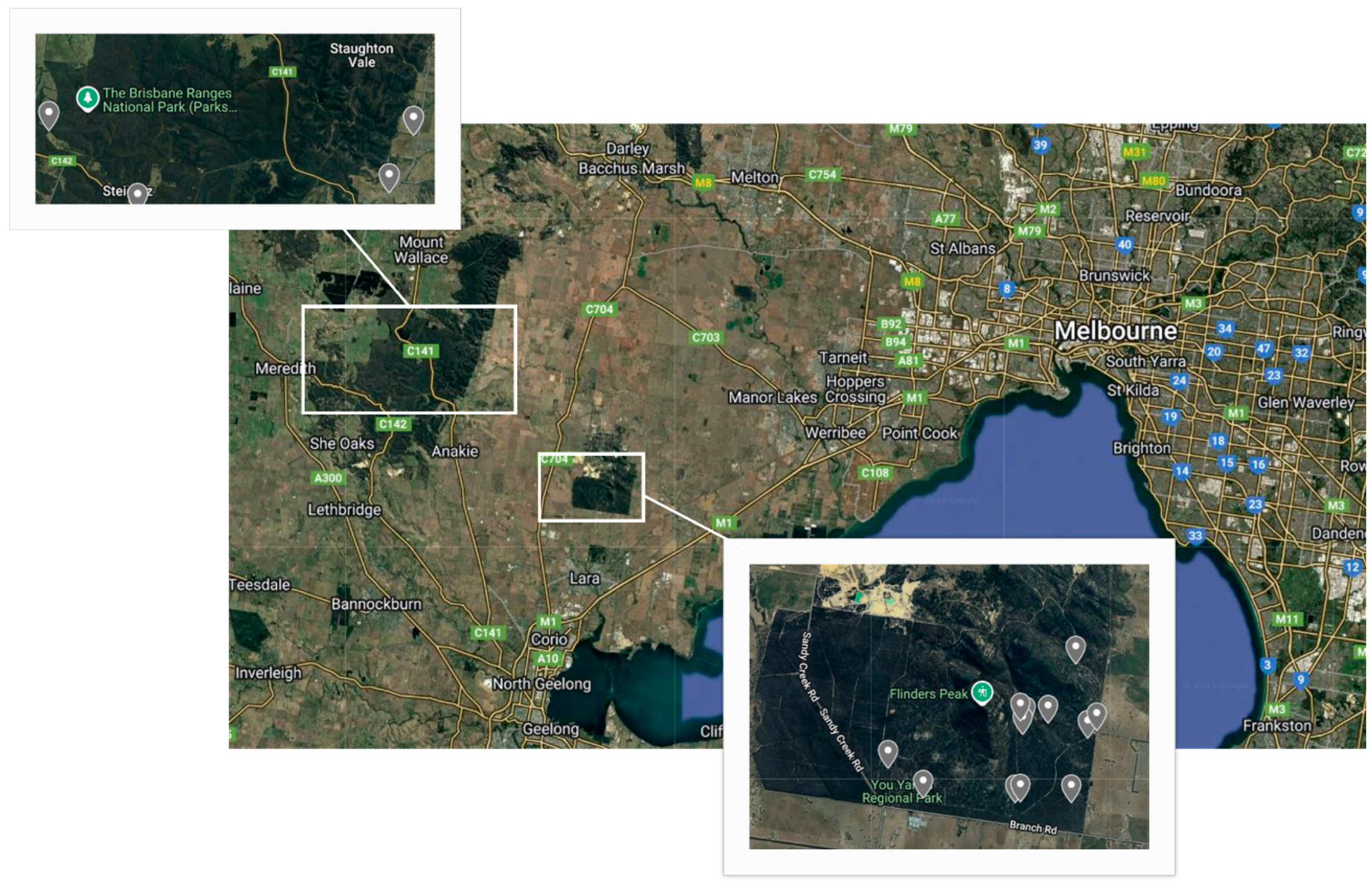
2.1. Sample Collection

Koala scats were collected opportunistically between October and November 2023 from the YYRP and the BRNP in the Greater Geelong Region of Victoria (Figure 1) during regular individual koala monitoring by the Koala Clancy Foundation (https://www.koalaclancyfoundation.org.au/). At the time of collection, the koalas were identified from the ground using the nose pattern identification technique [24], and any information from previous regular monitoring of the individuals was recorded, including sex, estimated age, and breeding history if female. Presence or absence of clinical signs of disease, such as conjunctivitis or wet bottom, were also recorded if visible. Scats were collected using scat collection kits (Integrated Sciences) only if fresh (i.e., covered in mucus and shiny) and if deposited while koalas were observed. Samples were then kept frozen at −18 °C until shipped overnight to the Koala Health Hub (KHH) at the Sydney School of Veterinary Science at the University of Sydney to be analysed.

2.2. DNA Extraction and Quantitative Polymerase Chain Reaction (qPCR) Analysis

DNA was extracted from scat samples using the ISOLATE Faecal DNA Kit (Bioline, Memphis, TN, USA). Thawed samples were vortexed, and a mixture of collection solution and scat pellet was homogenised using FastPrep™ (MP Biomedicals, Santa Ana, CA, USA) and then centrifuged. The supernatant underwent further centrifugation and was filtered through a Spin II A Filter (MP Biomedicals, Santa Ana, CA, USA). After adding Faecal DNA Binding Buffer, the mixture was processed through a spin column with sequential wash steps. DNA was then eluted, passed through a Spin II B Filter pre-treated (MP Biomedicals, Santa Ana, CA, USA) with Faecal Prep Solution, and centrifuged to obtain the final extracted DNA. The extracted DNA was then processed using the CFX Opus 96 Real-Time PCR System (Bio-Rad, Hercules, CA, USA). Quality controls were included alongside the test samples and underwent the same extraction and analysis procedures. These included blank extractions made during each batch extraction of DNA to ensure no cross contamination occurred during handling. A PCR positive control and a PCR negative control were included to ensure the PCR reaction was working and to check for the absence of contamination. A multiplex, TaqMan® (Thermo Fisher Scientific, Waltham, MA, USA) probe assay was used to test for the presence of koala DNA via beta-actin mRNA, C. pecorum (ompB), and Chlamydia genus (23S rRNA). These specific DNA sequences were used as they are highly conserved targets over all genotypes.
qPCR results were interpreted by first looking at the beta actin levels to ensure adequate levels of koala DNA were present in the samples (Table 1). Samples lacking adequate beta-actin levels were considered as failing quality control and excluded from analysis. This was followed by the interpretation of Chlamydia genus and C. pecorum levels to differentiate samples into four categories, namely negative (no C. pecorum detected), positive (C. pecorum DNA was clearly detected), weak positive (C. pecorum DNA was detected but at a low level), and suspect (C. pecorum could not be definitively excluded), depending on their cycle threshold (CT) values (Table 1). For the purposes of analysis, we took a conservative approach and considered all weak positives and suspect results as positive. To quantify the amount of chlamydial shedding, ΔCT values were then calculated by subtracting the CT of C. pecorum from the CT of the beta-actin housekeeping gene (to account for quality of swab yield), with higher chlamydial shedding being represented by a lower ΔCT value (as in [31]).

2.3. Statistical Analysis

All analyses of data were performed using R, version 4.2.2 [32]. A Q-Q plot was generated using the ‘qqnorm’ function to ensure data were normally distributed. We then used a linear model using the package ‘lme4’ [33] to test the effects of sex, age, and urogenital and ocular signs on C. pecorum infection shedding severity (using ΔCT values). Location could not be included in the models as all samples collected from Brisbane Ranges National Park were positive for C. pecorum.

3. Results

A total of 19 samples (15 from the YYRP and 4 from the BRNP) were collected from individual koalas (8 females, 10 males, and 1 of unknown sex). All koalas were adults (age range of 3–12 years) except for one dependent joey of around 10 months of age. Three koalas had clinical signs of C. pecorum infection in both the ocular area (i.e., conjunctivitis) and urogenital tract (i.e., perineum staining) upon observation at the time of scat collection. Two koalas had ocular signs only, and the rest of the koalas had no clinical signs or were unable to be evaluated from the ground. All samples except 1 had successful amplification: 6/18 (33.3%) samples tested negative for C. pecorum, 6/18 (33.3%) tested positive, 5/18 (27.8%) were weak positives, and 1/18 (5.6%) was suspect positive.
There was no effect of sex (F1,3 = 0.014, p = 0.914) and age (F1,3 = 0.039, p = 0.857) on C. pecorum shedding severity. The presence of ocular (F1,3 = 0.149, p = 0.725) and urogenital (F1,3 = 0.676, p = 0.471) signs of chlamydiosis also had no effect on C. pecorum shedding status.

4. Discussion

This study investigated the presence of C. pecorum infection in a limited number of koalas from the BRNP and the YYRP, Victoria, where testing had not been conducted previously, using non-invasive DNA scat analysis. Five koalas presented clinical signs of C. pecorum upon observation at the time of scat collection, including five with ocular signs and three showing urogenital signs. We found that 66.7% of the samples tested positive for C. pecorum, with all individuals from the BRNP and 8/15 of koalas from the YYRP being infected. The presence of C. pecorum within these populations may have been historical, as koalas were translocated to the BRNP in the 1940s from Philip Island, Victoria, where C. pecorum infection was already present [4]. However, koalas located near Warrnambool, in the South West Coast region, Victoria, from the same island source population, had only a 7% prevalence of C. pecorum and no observable loss of fertility [8], indicating that the disease may be recent in some areas. Hence, it is possible that the animals became infected at the location where they were translocated [9]. The high rate of disease we found was unexpected, as previous studies between 2015 and 2018 found that infection in Victorian koala populations was lower (1–49%) than the rate found in our study [15,20,22]. This increased disease rate may have been facilitated by different factors, including stress [34] and koala retrovirus (KoRV), with higher viral loads being positively correlated with the severity of chlamydiosis in southern koalas [35,36]. Habitat quality is decreased in the YYRP with invasive weeds such as boneseed (Chrysanthemoides monilifera) and bridal creeper (Myriophyllum asparagoides) covering extensive areas of the park and impacting the local flora and fauna by preventing the growth of native plants [37,38], including eucalypt species, potentially reducing the regrowth of trees used by koalas. The YYRP and BRNP are also surrounded by extensive agricultural land and are neighbouring several sand mines, limiting the movement of koalas and potentially resulting in increased stressors [16,34]. As the YYRP is situated within a rain shadow of the Otway Ranges (www.parks.vic.gov.au, accessed on 16 April 2025), climatic conditions, including droughts and heatwaves experienced in the area, may also compound the prevalence of disease in koalas. A recent study found that weather can affect koala immune function and increase the expression of stress receptor pathways, possibly increasing susceptibility to infection [39].
The sensitivity of the DNA detection of C. pecorum in scats can be dependent on the presence of clinical signs, with tests from individuals less affected by clinical disease, showing a lower sensitivity [29]. We did not observe severe clinical signs in any of the koalas in this study. However, Victorian koalas carry C. pecorum ompA genotype B, which differs from the genotype found in northern koalas [15,40], where the use of scats for C. pecorum detection was developed [29]. This appears to result in milder clinical signs [15,20], which would increase the likelihood of false negatives when using scats to test for C. pecorum within a population [41], potentially leading to an underestimation of the true disease burden in our study. This does not lower the value of using scats to test for C. pecorum as a preliminary exploratory tool for the disease as this method does not require invasive capture and handling, which would be required to gather direct ocular and urogenital swabs.
Females and males appeared equally susceptible to chlamydiosis in our study areas as we found no effect of sex on the shedding rate of C. pecorum. Our sample size was limited, but other studies conducted in NSW [31], Queensland [42], South Australia [35], and Victoria [20] also showed no effect of sex on shedding and/or the infection rate of individuals. This result can probably be ascribed to the sexually transmitted nature of C. pecorum disease, with sexually mature males and females both spreading the infection during mating season [16]. However, other studies found that females were more likely to show a higher incidence of visible signs of urogenital infection than males [31,43]. More samples from individuals with observable clinical signs may be needed from the YYRP and BRNP to be able to detect a relationship between sex and urogenital tract disease within these populations.
Estimated age also showed no effect on C. pecorum shedding, consistent with previous studies [20,42]. In koalas, mature age can sometimes be associated with an increased likelihood of C. pecorum infection due to the sexually transmitted nature of this disease [16]. However, in this study, we sampled only adult koalas that were sexually mature (except a joey which was sampled opportunistically while its mother was sampled), and these koalas did not differ much in shedding load and clinical signs, showing that the disease can affect sexually active individuals of different ages in similar ways [15].
All of the females with high historical fecundity tested positive for chlamydiosis, and the female who had the highest shedding load (i.e., the lowest ΔCT value) was also the one with the highest number of joeys recorded in the past, indicating that chlamydial infections in the YYRP may be recent. It is also possible that recent infections involve a different and more virulent Chlamydia genotype [44], particularly if they are associated with decreased fecundity. This may have contributed to the initial observations of population declines. Chlamydiosis is generally linked to reduced reproductive rates in females [35], and the decrease in fertility observed in female koalas is often due to reproductive tract changes, including unilateral or bilateral ovarian cysts, changes in uterus appearance, and associated structures [16]. These changes are often not apparent or identifiable without the use of ultrasound or abdominal palpation [16]. Even in areas where reproductive rates are very low, reproductive disease signs in females are sometimes undetectable [42]. Further investigations of reproductive tract pathology in female koalas of these populations would provide valuable insights into the structural impacts of C. pecorum infection in these areas.

5. Conclusions

This study has identified chlamydial disease for the first time in these populations and found a higher-than-expected infection rate, potentially responsible for the decline in koala numbers observed in these areas. In this initial investigation, we had a small sample size (n = 18), which prevented us from investigating disease variations between the BRNP and YYRP populations. Future studies should aim to include a greater number of samples to improve the robustness of the findings. Our study confirms that the scat analysis method is a valuable primary assessment tool to detect C. pecorum in populations where chlamydial disease status is unknown. The next step is to carry out a large-scale population disease assessment, including the capture and close examination of koalas.
Our findings are consistent with the observed decline in koala numbers around Australia and the increased prevalence of C. pecorum infection within many koala populations. Further investigations into the disease in these important koala populations would be beneficial. Longitudinal studies would allow for the progression and impact of C. pecorum within these populations to be recorded and for tailored management strategies to be developed.

Author Contributions

Conceptualisation, V.S.A.M. and J.D.; methodology, V.S.A.M., J.D. and G.K.; software, V.S.A.M. and G.K.; validation, G.K. and V.S.A.M.; formal analysis, V.S.A.M. and G.K.; investigation, G.K. and J.D.; resources, V.S.A.M. and J.D.; data curation, G.K. and V.S.A.M.; writing—original draft preparation, G.K.; writing—review and editing, V.S.A.M., G.K. and J.D.; visualisation, V.S.A.M.; supervision, V.S.A.M.; project administration, V.S.A.M.; funding acquisition, V.S.A.M. All authors have read and agreed to the published version of the manuscript.

Funding

This research was funded by the Research and Enquiry Program of the Sydney School of Veterinary Science at the University of Sydney.

Institutional Review Board Statement

Samples were collected opportunistically by the Koala Clancy Foundation as part of their ongoing koala monitoring activities.

Informed Consent Statement

Not applicable.

Data Availability Statement

The data that support the findings of this study are available from the corresponding author upon request. There are no restrictions on data availability.

Acknowledgments

Thanks to the staff of the Koala Clancy Foundation for the collection of the koala scat samples. We would also like to acknowledge Andrea Casteriano from the Koala Health Hub for providing assistance with the scat DNA extraction and qPCR analysis.

Conflicts of Interest

The authors declare no conflicts of interest.

Abbreviations

The following abbreviations are used in this manuscript:
YYRPYou Yangs Regional Park
BRNPBrisbane Ranges National Park
DNADeoxyribonucleic acid
qPCRQuantitative polymerase chain reaction
NSWNew South Wales
KHHKoala Health Hub
PCRPolymerase chain reaction
mRNAMessenger ribonucleic acid
ompBOuter membrane protein B
CTCycle threshold
ΔCTDelta cycle threshold
KoRVKoala retro virus
QC-failQuality control fail

References

  1. Melzer, A.; Carrick, F.; Menkhorst, P.; Lunney, D.; John, B.S. Overview, Critical Assessment, and Conservation Implications of Koala Distribution and Abundance. Conserv. Biol. 2000, 14, 619–628. [Google Scholar] [CrossRef]
  2. Department of Climate Change Energy the Environment and Water. Koala Listing Under National Environmental Law; Department of Climate Change Energy the Environment and Water: Canberra, ACT, Australia, 2022. Available online: https://www.dcceew.gov.au/environment/biodiversity/threatened/species/koalas/listing-under-national-environmental-law (accessed on 16 April 2025).
  3. Department of Energy Environment and Climate Action. Victorian Koala Management Strategy; Victorian Government: Melbourne, VIC, Australia, 2023.
  4. Menkhorst, P. Hunted marooned re-introduced contracepted: Ahistory of Koala management in Victoria. In Too Close for Comfort: Contentious Issues in Human-Wildlife Encounters; Lunney, D., Munn, A., Meikle, W., Eds.; Royal Zoological Society of New South Wales: Sydney, NSW, Australia, 2008. [Google Scholar]
  5. Whisson, D.A.; Dixon, V.; Taylor, M.L.; Melzer, A. Failure to respond to food resource decline has catastrophic consequences for koalas in a high-density population in southern Australia. PloS ONE 2016, 11, e0144348. [Google Scholar] [CrossRef] [PubMed]
  6. Wedrowicz, F.; Wright, W.; Schlagloth, R.; Santamaria, F.; Cahir, F. Landscape, koalas and people: A historical account of koala populations and their environment in South Gippsland. Aust. Zool. 2017, 38, 518–536. [Google Scholar] [CrossRef]
  7. Menkhorst, P.W. Mammals of Victoria: Distribution, Ecology and Conservation; Oxford University Press: Oxford, UK, 1995. [Google Scholar]
  8. Whisson, D.A.; Ashman, K.R. When an iconic native animal is overabundant: The koala in southern Australia. Conserv. Sci. Pract. 2020, 2, e188. [Google Scholar] [CrossRef]
  9. Santamaria, F.; Schlagloth, R. The effect of Chlamydia on translocated Chlamydia-naïve koalas: A case study. Aust. Zool. 2016, 38, 192–202. [Google Scholar] [CrossRef]
  10. Martin, R.W. Overbrowsing decline of a population of the koala Phascolarctos cinereus in Victoria, III. Population dynamics. Wildl. Res. 1985, 12, 377–385. [Google Scholar] [CrossRef]
  11. Ramsey, D.S.; Brown, G.W.; Tolsma, A.D. Towards a Habitat Condition Assessment Method for Guiding the Management of Overabundant Koala Populations; Arthur Rylah Institute for Environmental Research Technical Report Series No. 272; Department of Environment, Land, Water and Planning: Heidelberg, VIC, Australia, 2016.
  12. Quigley, B.L.; Timms, P. Helping koalas battle disease—Recent advances in Chlamydia and koala retrovirus (KoRV) disease understanding and treatment in koalas. FEMS Microbiol. Rev. 2020, 44, 583–605. [Google Scholar] [CrossRef]
  13. Higgins, D.P.; Hemsley, S.; Canfield, P.J. Immuno-histochemical demonstration of the role of Chlamydiaceae in renal, uterine and salpingeal disease of the koala, and demonstration of Chlamydiaceae in novel sites. J. Comp. Pathol. 2005, 133, 164–174. [Google Scholar] [CrossRef]
  14. Johnston, S.D.; Deif, H.H.; McKinnon, A.; Theilemann, P.; Griffith, J.E.; Higgins, D.P. Orchitis and epididymitis in koalas (Phascolarctos cinereus) infected with Chlamydia pecorum. Vet. Pathol. 2015, 52, 1254–1257. [Google Scholar] [CrossRef]
  15. Legione, A.R.; Patterson, J.L.S.; Whiteley, P.L.; Amery-Gale, J.; Lynch, M.; Haynes, L.; Gilkerson, J.R.; Polkinghorne, A.; Devlin, J.M.; Sansom, F.M. Sansom, Identification of unusual Chlamydia pecorum genotypes in Victorian koalas (Phascolarctos cinereus) and clinical variables associated with infection. J. Med. Microbiol. 2016, 65, 420–428. [Google Scholar] [CrossRef]
  16. Polkinghorne, A.; Hanger, J.; Timms, P. Recent advances in understanding the biology, epidemiology and control of chlamydial infections in koalas. Vet. Microbiol. 2013, 165, 214–223. [Google Scholar] [CrossRef] [PubMed]
  17. Hulse, L.; Beagley, K.; Larkin, R.; Nicolson, V.; Gosálvez, J.; Johnston, S. The effect of Chlamydia infection on koala (Phascolarctos cinereus) semen quality. Theriogenology 2021, 167, 99–110. [Google Scholar] [CrossRef] [PubMed]
  18. Robbins, A.; Hanger, J.; Jelocnik, M.; Quigley, B.L.; Timms, P. Longitudinal study of wild koalas (Phascolarctos cinereus) reveals chlamydial disease progression in two thirds of infected animals. Sci. Rep. 2019, 9, 13194. [Google Scholar] [CrossRef] [PubMed]
  19. Fernandez, C.M.; Schmertmann, L.J.; Higgins, D.P.; Casteriano, A.; Irinyi, L.; Mella, V.S.A.; Crowther, M.S.; Meyer, W.; Krockenberger, M.B. Genetic differences in Chlamydia pecorum between neighbouring sub-populations of koalas (Phascolarctos cinereus). Vet. Microbiol. 2019, 231, 264–270. [Google Scholar] [CrossRef]
  20. Patterson, J.L.S.; Lynch, M.; Anderson, G.A.; Noormohammadi, A.H.; Legione, A.; Gilkerson, J.R.; Devlin, J.M. Prevalence and clinical significance of chlamydia infection in island and mainland populations of Victorian koalas (Phascolarctos cinereus). J. Wildl. Dis. 2015, 51, 309–317. [Google Scholar] [CrossRef]
  21. Every, K. Evaluation of a decline in population of the koala, Phascolarctos cinereus (Goldfuss) in Ventnor Reserve, Phillip I., Vic., by means of a triple-count technique. Wildl. Res. 1986, 13, 517–525. [Google Scholar] [CrossRef]
  22. Wedrowicz, F.; Mosse, J.; Wright, W.; Hogan, F.E. Using non-invasive sampling methods to determine the prevalence and distribution of Chlamydia pecorum and koala retrovirus in a remnant koala population with conservation importance. Wildl. Res. 2018, 45, 366–380. [Google Scholar] [CrossRef]
  23. Adams-Hosking, C.; McBride, M.F.; Baxter, G.; Burgman, M.; de Villiers, D.; Kavanagh, R.; Lawler, I.; Lunney, D.; Melzer, A.; Menkhorst, P.; et al. Use of expert knowledge to elicit population trends for the koala (Phascolarctos cinereus). Divers. Distrib. 2016, 22, 249–262. [Google Scholar] [CrossRef]
  24. Duffy, J.; Stragliotto, T.; Mella, V.S.A. On the nose: Validating a novel, non-invasive method to identify individual koalas using unique nose patterns. Wildl. Res. 2024, 51, 1–8. [Google Scholar] [CrossRef]
  25. Mella, V.S.A.; Orr, C.; Hall, L.; Velasco, S.; Madani, G. An insight into natural koala drinking behaviour. Ethology 2020, 126, 858–863. [Google Scholar] [CrossRef]
  26. Flanagan, C.; Krockenberger, M.B.; Van Stan, J.T.; Duffy, J.; Mella, V.S.A. Koalas (Phascolarctos cinereus) and stemflow: Drinking more than just water. Austral Ecol. 2025, 50, e70076. [Google Scholar] [CrossRef]
  27. Grogan, L.F.; Berger, L.; Rose, K.; Grillo, V.; Cashins, S.D.; Skerratt, L.F. Surveillance for emerging biodiversity diseases of wildlife. PLoS Pathog. 2014, 10, e1004015. [Google Scholar] [CrossRef] [PubMed]
  28. Ashman, K.R.; Watchorn, D.J.; Whisson, D.A. Prioritising research efforts for effective species conservation: A review of 145 years of koala research. Mammal Rev. 2019, 49, 189–200. [Google Scholar] [CrossRef]
  29. Cristescu, R.H.; Miller, R.L.; Schultz, A.J.; Hulse, L.; Jaccoud, D.; Johnston, S.; Hanger, J.; Booth, R.; Frère, C.H. Developing noninvasive methodologies to assess koala population health through detecting Chlamydia from scats. Mol. Ecol. Resour. 2019, 19, 957–969. [Google Scholar] [CrossRef]
  30. NSW Parliament. Koala Populations and Habitat in New South Wales—Report 3; Parliament of New South Wales: Sydney, NSW, Australia, 2020.
  31. Simpson, S.J.; Higgins, D.P.; Timms, P.; Mella, V.S.A.; Crowther, M.S.; Fernandez, C.M.; McArthur, C.; Phillips, S.; Krockenberger, M.B. Efficacy of a synthetic peptide Chlamydia pecorum major outer membrane protein vaccine in a wild koala (Phascolarctos cinereus) population. Sci. Rep. 2023, 13, 15087. [Google Scholar] [CrossRef]
  32. R Core Team. R: A Language and Envronment for Statistical Computing, R Foundation for Statistical Computing: Vienna, Austria, 2024.
  33. Bates, D.; Mächler, M.; Bolker, B.; Walker, S. Fitting Linear Mixed-Effects Models Using lme4. J. Stat. Softw. 2015, 67, 1–48. [Google Scholar] [CrossRef]
  34. Narayan, E. Physiological stress levels in wild koala sub-populations facing anthropogenic induced environmental trauma and disease. Sci. Rep. 2019, 9, 6031. [Google Scholar] [CrossRef]
  35. Fabijan, J.; Caraguel, C.; Jelocnik, M.; Polkinghorne, A.; Boardman, W.S.J.; Nishimoto, E.; Johnsson, G.; Molsher, R.; Woolford, L.; Timms, P.; et al. Chlamydia pecorum prevalence in South Australian koala (Phascolarctos cinereus) populations: Identification and modelling of a population free from infection. Sci. Rep. 2019, 9, 6261. [Google Scholar] [CrossRef]
  36. Tarlinton, R.; Meers, J.; Hanger, J.; Young, P. Real-time reverse transcriptase PCR for the endogenous koala retrovirus reveals an association between plasma viral load and neoplastic disease in koalas. J. Gen. Virol. 2005, 86, 783–787. [Google Scholar] [CrossRef]
  37. Turner, P.J.; Scott, J.K.; Spafford, H. The ecological barriers to the recovery of bridal creeper (Asparagus asparagoides (L.) Druce) infested sites: Impacts on vegetation and the potential increase in other exotic species. Austral Ecol. 2008, 33, 713–722. [Google Scholar] [CrossRef]
  38. Roberts, D. A history of boneseed control in the You Yangs Regional Park, Victoria. Plant Prot. Q. 2008, 23, 51. [Google Scholar]
  39. Fernandez, C.M.; Krockenberger, M.B.; Ho, S.Y.; Crowther, M.; Mella, V.S.; Jelocnik, M.; Wilmott, L.; Higgins, D. Novel typing scheme reveals emergence and genetic diversity of Chlamydia pecorum at the local management scale across two koala populations. Vet. Microbiol. 2024, 293, 110085. [Google Scholar] [CrossRef] [PubMed]
  40. Jackson, M.; Giffard, P.; Timms, P. Outer membrane protein a gene sequencing demonstrates the polyphyletic nature of koala Chlamydia pecorum isolates. Syst. Appl. Microbiol. 1997, 20, 187–200. [Google Scholar] [CrossRef]
  41. Wan, C.; Loader, J.; Hanger, J.; Beagley, K.; Timms, P.; Polkinghorne, A. Using quantitative polymerase chain reaction to correlate Chlamydia pecorum infectious load with ocular, urinary and reproductive tract disease in the koala (Phascolarctos cinereus). Aust. Vet. J. 2011, 89, 409–412. [Google Scholar] [CrossRef]
  42. Jackson, M.; White, N.; Giffard, P.; Timms, P. Epizootiology of Chlamydia infections in two free-range koala populations. Vet. Microbiol. 1999, 65, 255–264. [Google Scholar] [CrossRef]
  43. Nyari, S.; Waugh, C.A.; Dong, J.; Quigley, B.L.; Hanger, J.; Loader, J.; Polkinghorne, A.; Timms, P. Epidemiology of chlamydial infection and disease in a free-ranging koala (Phascolarctos cinereus) population. PLoS ONE 2017, 12, e0190114. [Google Scholar] [CrossRef]
  44. Fernandez, C.M.; Krockenberger, M.B.; Crowther, M.; Mella, V.S.; Wilmott, L.; Higgins, D.P. Genetic markers of Chlamydia pecorum virulence in ruminants support short term host-pathogen evolutionary relationships in the koala, Phascolarctos cinereus. Infect. Genet. Evol. 2023, 116, 105527. [Google Scholar] [CrossRef]
Figure 1. Study sites and locations of koala (Phascolarctos cinereus) scat collections in the You Yangs Regional Park (bottom) and Brisbane Ranges National Park (top), Victoria, Australia.
Animals 15 02048 g001
Table 1. The result categories determined by the cycle threshold (CT) value limits of the Quantitative Polymerase Chain Reaction (qPCR) for Chlamydia pecorum. Beta-actin is used as a housekeeping gene to account for the quality of the swab yield. Quality control fail (QC-fail) means that a sample did not meet the required quality standards to be included in the analysis.
Beta-ActinChlamydia pecorumChlamydia Genus (23S)Result
CT ≤ 32no amplificationno amplificationNegative
CT ≤ 32CT ≤ 34CT ≤ 34Positive
CT ≤ 32CT 34–40CT 34–40Weak positive
CT > 32CT 34–40CT 34–40Weak positive
CT ≤ 32no amplificationCT 34+Suspect
CT > 32no amplificationno amplificationQC-fail
no amplificationno amplificationno amplificationQC-fail
Disclaimer/Publisher’s Note: The statements, opinions and data contained in all publications are solely those of the individual author(s) and contributor(s) and not of MDPI and/or the editor(s). MDPI and/or the editor(s) disclaim responsibility for any injury to people or property resulting from any ideas, methods, instructions or products referred to in the content.

