Surface Electromyography of the Longissimus and Gluteus Medius Muscles in Greyhounds Walking and Trotting on Ground Flat, Up, and Downhill


Simple Summary
Abstract
1. Introduction
2. Materials and Methods
2.1. Dogs
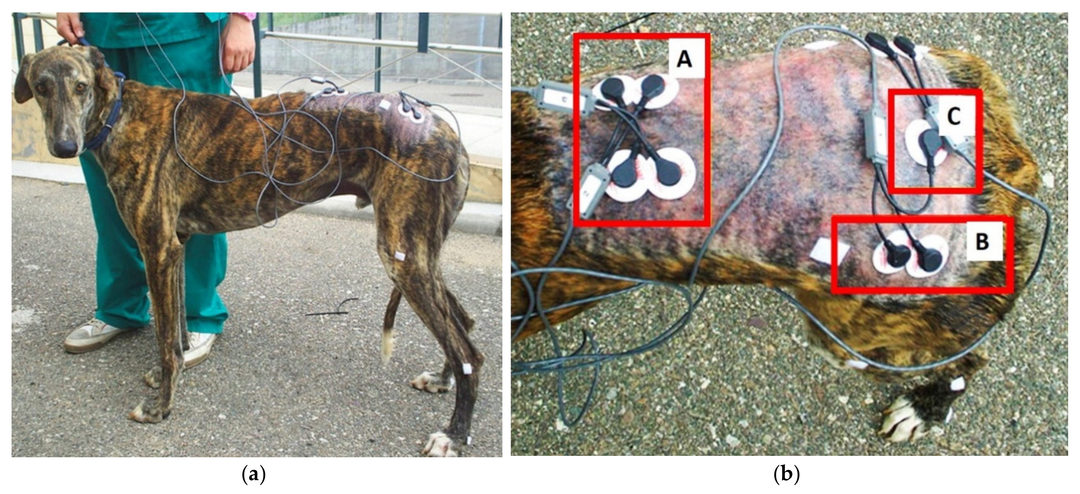
2.2. Data Collection
2.3. Data Processing and Analysis
3. Results
4. Discussion
4.1. Longissimus Dorsi Muscle
4.2. Gluteus Medius Muscle
5. Conclusions
Author Contributions
Funding
Conflicts of Interest
References
- Kirkby Shaw, K.; Alvarez, L.; Foster, S.A.; Tomlinson, J.E.; Shaw, A.J.; Pozzi, A. Fundamental principles of rehabilitation and musculoskeletal tissue healing. Vet. Surg. 2020, 49, 22–32. [Google Scholar] [CrossRef] [PubMed]
- Holler, P.J.; Brazda, V.; Dal-Bianco, B.; Lewy, E.; Mueller, M.C.; Peham, C.; Bockstahler, B.A. Kinematic motion analysis of the joints of the forelimbs and hind limbs of dogs during walking exercise regimens. Am. J. Vet. Res. 2010, 71, 734–740. [Google Scholar] [CrossRef] [PubMed]
- Evans, H.E. Miller’s Anatomy of the Dog, 3rd ed.; WB Saunders Co: Philadelphia, PA, USA, 1993. [Google Scholar]
- Pierotti, D.J.; Roy, R.R.; Gregor, R.J.; Edgerton, V.R. Electromyographic activity of cat hindlimb flexors and extensors during locomotion at varying speeds and inclines. Brain Res. 1989, 481, 57–66. [Google Scholar] [CrossRef]
- Carlson-Kuhta, P.; Trank, T.V.; Smith, J.L. Forms of Forward Quadrupedal Locomotion. II. A Comparison of Posture, Hindlimb Kinematics, and Motor Patterns for Upslope and Level Walking. J. Neurophysiol. 1998, 79, 1687–1701. [Google Scholar] [CrossRef] [PubMed]
- Smith, J.L.; Carlson-Kuhta, P.; Trank, T.V. Forms of forward quadrupedal locomotion. III. A comparison of posture, hindlimb kinematics, and motor patterns for downslope and level walking. J. Exp. Biol. 1998, 79, 1702–1716. [Google Scholar] [CrossRef] [PubMed]
- Wada, N.; Akatani, J.; Miyajima, N.; Shimojo, K.; Kanda, K. The role of vertebral column muscles in level versus upslope treadmill walking—An electromyographic and kinematic study. Brain Res. 2006, 1090, 99–109. [Google Scholar] [CrossRef] [PubMed]
- Wada, N.; Miyajima, N.; Akatani, J.; Shimojo, K.; Kanda, K. Electromyographic activity of m. longissimus and the kinematics of the vertebral column during level and downslope treadmill walking in cats. Brain Res. 2006, 1103, 140–144. [Google Scholar] [CrossRef]
- Crook, T.C.; Wilson, A.; Hodson-Tole, E. The effect of treadmill speed and gradient on equine hindlimb muscle activity. Equine Vet. J. 2010, 42, 412–416. [Google Scholar] [CrossRef]
- Robert, C.; Valette, J.P.; Denoix, J.M. The effects of treadmill inclination and speed on the activity of three trunk muscles in the trotting horse. Equine Vet. J. 2001, 33, 466–472. [Google Scholar] [CrossRef] [PubMed]
- Deban, S.M.; Schilling, N.; Carrier, D.R. Activity of extrinsic limb muscles in dogs at walk, trot and gallop. J. Exp. Biol. 2012, 215, 287–300. [Google Scholar] [CrossRef] [PubMed]
- Schilling, N.; Carrier, D.R. Function of the epaxial muscles in walking, trotting and galloping dogs: Implications for the evolution of epaxial muscle function in tetrapods. J. Exp. Biol. 2010, 213, 1490–1502. [Google Scholar] [CrossRef] [PubMed]
- Goslow, F.E.; Seeherman, H.F.; Taylor, C.R.; CcCutchln, M.N.; Heglund, N.C. Electrical Activity and Relative Length Changes of Dog Limb Muscles as a Function of Speed and Gait. J. Exp. Biol. 1981, 94, 15–42. [Google Scholar] [PubMed]
- Schilling, N.; Carrier, D.R. Function of the epaxial muscles during trotting. J. Exp. Biol. 2009, 212, 1053–1063. [Google Scholar] [CrossRef] [PubMed][Green Version]
- Schilling, N.; Fischbein, T.; Yang, E.P.; Carrier, D.R. Function of the extrinsic hindlimb muscles in trotting dogs. J. Exp. Biol. 2009, 212, 1036–1052. [Google Scholar] [CrossRef]
- Lauer, S.K.; Hillman, R.B.; Li, L.; Hosgood, G.L. Effects of treadmill inclination on electromyographic activity and hind limb kinematics in healthy hounds at a walk. Am. J. Vet. Res. 2009, 70, 658–664. [Google Scholar] [CrossRef]
- McLean, H.; Millis, D.; Levine, D. Surface Electromyography of the Vastus Lateralis, Biceps Femoris, and Gluteus Medius in Dogs During Stance, Walking, Trotting, and Selected Therapeutic Exercises. Front. Vet. Sci. 2019, 6, 221. [Google Scholar] [CrossRef]
- Breitfuss, K.; Franz, M.; Peham, C.; Bockstahler, B. Surface Electromyography of the Vastus Lateralis, Biceps Femoris, and Gluteus Medius Muscle in Sound Dogs During Walking and Specific Physiotherapeutic Exercises. Vet. Surg. 2015, 44, 588–595. [Google Scholar] [CrossRef]
- Bockstahler, B.; Kraeutler, C.; Holler, P.; Kotschwar, A.; Vobornik, A.; Peham, C. Pelvic Limb Kinematics and Surface Electromyography of the Vastus Lateralis, Biceps Femoris, and Gluteus Medius Muscle in Dogs with Hip Osteoarthritis. Vet. Surg. 2012, 41, 54–62. [Google Scholar] [CrossRef]
- Fischer, S.; Nolte, I.; Schilling, N. Adaptations in Muscle Activity to Induced, Short-Term Hindlimb Lameness in Trotting Dogs. PLoS ONE 2013, 8, e80987. [Google Scholar] [CrossRef]
- Valentin, S.; Zoldos, R.R. Surface electromyography in animal biomechanics: A systematic review. J. Electromyogr. Kines. 2016, 28, 167–183. [Google Scholar] [CrossRef]
- Drüen, S.; Böddeker, J.; Nolte, I.; Wefstaedt, P. Ground reaction forces of the canine hindlimb: Are there differences between gait on treadmill and force plate? Berl. Munch. Tierarztl. Wochenschr. 2010, 123, 339–345. [Google Scholar] [PubMed]
- Böddeker, J.; Drüen, S.; Nolte, I.; Wefstaedt, P. Comparative Motion Analysis of the Canine Hind Limb During Gait on Force Plate and Treadmill. Berl. Munch. Tierarztl. Wochenschr. 2010, 123, 431–439. [Google Scholar] [PubMed]
- Bockstahler, B.B.; Gesky, R.; Mueller, M.; Thalhammer, J.G.; Peham, C.; Podbregar, I. Correlation of Surface Electromyography of the Vastus Lateralis Muscle in Dogs at a Walk with Joint Kinematics and Ground Reaction Forces. Vet. Surg. 2009, 38, 754–761. [Google Scholar] [CrossRef] [PubMed]
- Robert, C.; Valette, J.P.; Degueurce, C.; Denoix, J.M. Correlation between surface electromyography and kinematics of the hindlimb of horses at trot on a treadmill. Cells Tissues Organs 1999, 165, 113–122. [Google Scholar] [CrossRef] [PubMed]
- Robert, C.; Valette, J.P.; Denoix, J.M. The effects of treadmill inclination and speed on the activity of two hindlimb muscles in the trotting horse. Equine Vet. J. 2000, 32, 312–317. [Google Scholar] [CrossRef]
- Araújo, J.F.; Rodrigues, F.B.; Abadia, F.G.; Gervásio, F.M.; Mendonça, G.B.N.; Damasceno, A.D.; Vieira, M.F. Electromyographic analysis of the gait cycle phases of boxer dogs. Arq. Bras. Med. Vet. Zootec. 2016, 68, 931–937. [Google Scholar] [CrossRef]
- Bockstahler, B.; Prickler, B.; Lewy, E.; Holler, P.J.; Vobornik, A.; Peham, C. Hind limb kinematics during therapeutic exercises in dogs with osteoarthritis of the hip joints. Am. J. Vet. Res. 2012, 73, 1371–1376. [Google Scholar] [CrossRef]
- Ritter, D.A.; Nassar, P.N.; Fife, M.; Carrier, D.R. Epaxial muscle function in trotting dogs. J. Exp. Biol. 2001, 204, 3053–3064. [Google Scholar]
- Hamilton, S.; Millis, D.L.; Taylor, R.A.; Levine, D. Therapeutics exercises. In Canine Rehabilitation and Physical Therapy, 1st ed.; Millis, D.L., Levine, D., Taylor, R.A., Eds.; Saunders: St. Louis, MI, USA, 2004; pp. 244–263. [Google Scholar]
- McCauley, L.; Van Dyke, J.B. Therapeutic exercise. In Canine Sports Medicine and Rehabilitation, 1st ed.; Zink, M.C., Van Dyke, J.B., Eds.; Wiley-Blacwell: Ames, IA, USA, 2013; pp. 132–157. [Google Scholar]
- Bockstahler, B. Methods of Physiotherapy. In Essential Facts of Physiotherapy in Dogs and Cats Rehabilitation and Pain Management, 1st ed.; Bockstahler, B., Levine, D., Millis, D., Eds.; PE VetVerLag: Babenhausen, Germany, 2004; pp. 64–65. [Google Scholar]
- Starr, L. Rehabilitation for Geriatric Patients. In Canine Sports Medicine and Rehabilitation, 1st ed.; Zink, M.C., Van Dyke, J.B., Eds.; Wiley-Blacwell: Ames, IA, USA, 2013; pp. 349–369. [Google Scholar]
- Dycus, D.L.; Levine, D.; Marcellin-Little, D.J. Physical Rehabilitation for the Management of Canine Hip Dysplasia. Vet. Clin. Small Anim. 2017, 47, 823–850. [Google Scholar] [CrossRef]


| Exercise | Speed (m/s) | Stride Duration (s) | Stance Phase Duration (%) |
|---|---|---|---|
| Walk downhill | 1.00 ± 0.13 | 0.80 ± 0.11 a | 63.07 ± 2.01 a |
| Walk flat | 1.40 ± 0.15 | 0.78 ± 0.06 a,b | 62.25 ± 1.45 a |
| Walk uphill | 0.94 ± 0.11 | 0.82 ± 0.10 b | 64.66 ± 2.54 |
| Trot downhill | 2.07 ± 0.32 | 0.51 ± 0 c | 44.23 ± 3.08 b,c |
| Trot flat | 2.93 ± 0.29 | 0.49 ± 0.02 c | 42.70 ± 3.70 b |
| Trot uphill | 1.84 ± 0.30 | 0.52 ± 0.03 c | 43.08 ± 4.39 c |
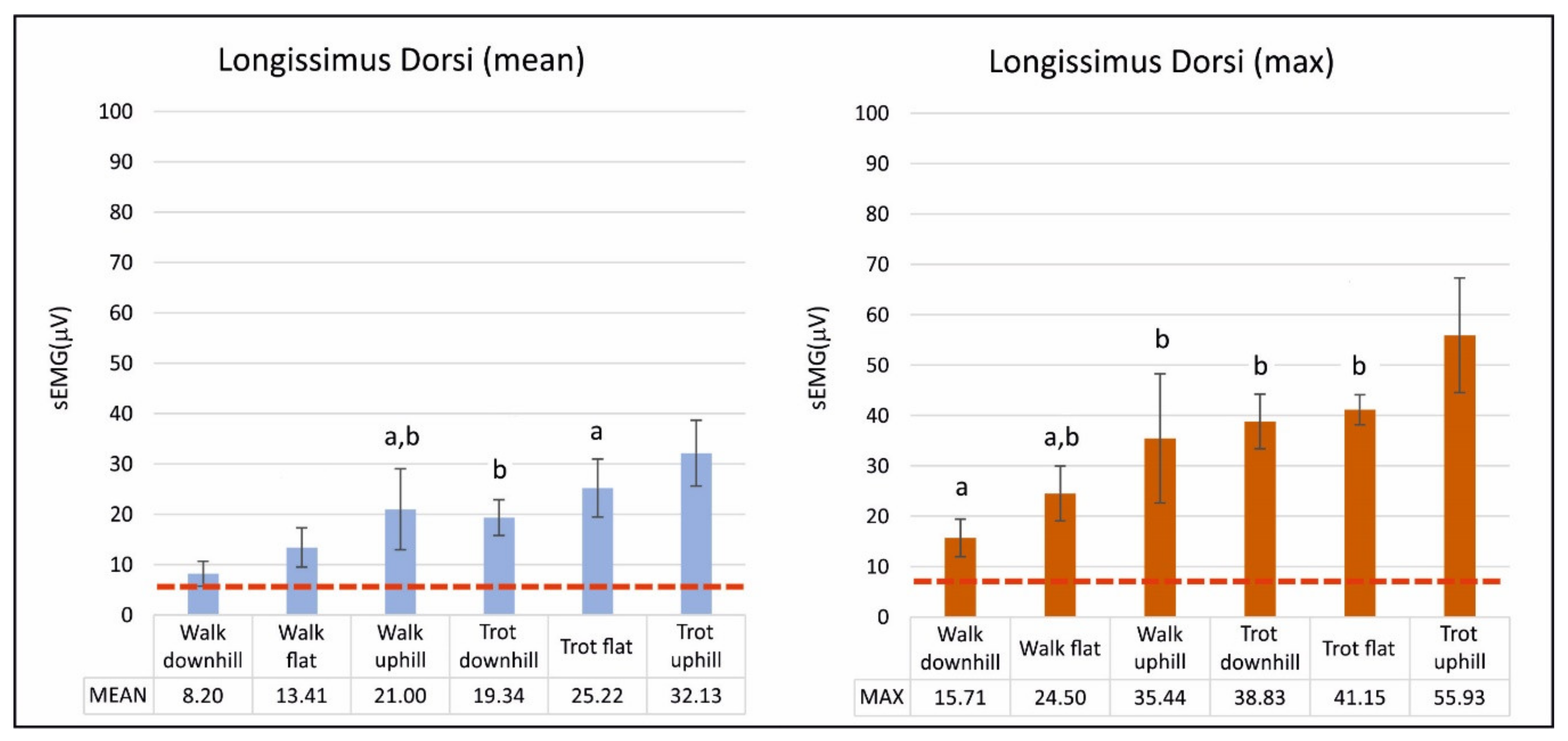
| Exercise | Mean | Maximum |
|---|---|---|
| Standing | 6.56 ± 1.1 | |
| Walk downhill | 8.20 ± 2.45 | 15.71 ± 3.73 a |
| Walk flat | 13.41 ± 3.88 | 24.50 ± 5.45 a,b |
| Walk uphill | 21.00 ± 8.02 a,b | 35.44 ± 12.82 b |
| Trot downhill | 19.34 ± 3.55 b | 38.83 ± 5.40 b |
| Trot flat | 25.22 ± 5.77 a | 41.15 ± 2.99 b |
| Trot uphill | 32.13 ± 6.54 | 55.93 ± 11.40 |
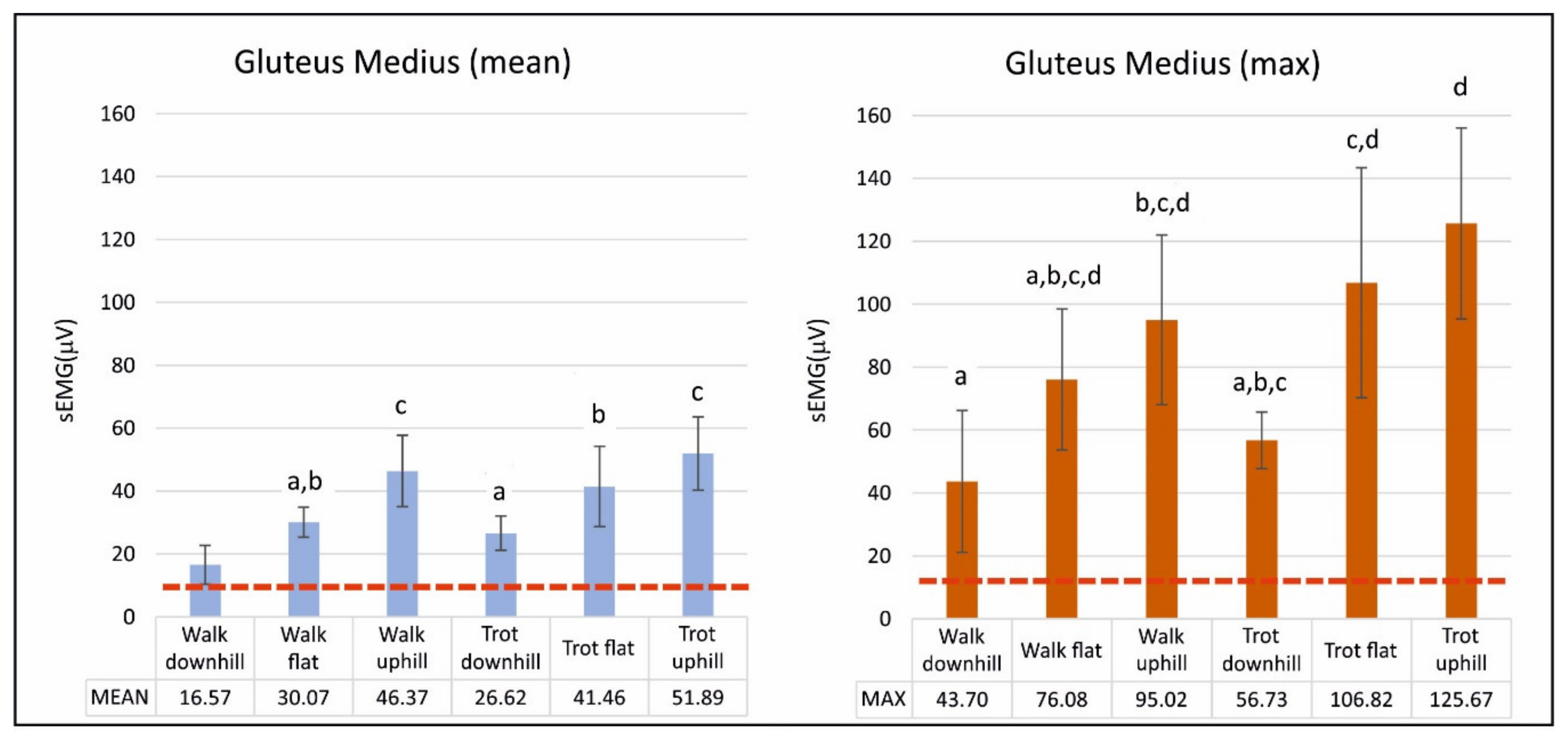
| Exercise | Mean | Maximum |
|---|---|---|
| Standing | 10.82 ± 4.76 | |
| Walk downhill | 8.20 ± 2.45 | 43.70 ± 22.54 a |
| Walk flat | 16.57 ± 6.17 a, b | 76.08 ± 22.40 a, b, c, d |
| Walk uphill | 46.37 ± 11.32 c | 95.02 ± 27.00 b, c, d |
| Trot downhill | 26.62 ± 5.46 a | 56.73 ± 9.02 a, b, c |
| Trot flat | 41.46 ± 12.80 b | 106.82 ± 36.60 c, d |
| Trot uphill | 51.89 ± 11.65 c | 125.67 ± 30.33 d |
© 2020 by the authors. Licensee MDPI, Basel, Switzerland. This article is an open access article distributed under the terms and conditions of the Creative Commons Attribution (CC BY) license (http://creativecommons.org/licenses/by/4.0/).
Share and Cite
Miró, F.; Galisteo, A.M.; Garrido-Castro, J.L.; Vivo, J. Surface Electromyography of the Longissimus and Gluteus Medius Muscles in Greyhounds Walking and Trotting on Ground Flat, Up, and Downhill. Animals 2020, 10, 968. https://doi.org/10.3390/ani10060968
Miró F, Galisteo AM, Garrido-Castro JL, Vivo J. Surface Electromyography of the Longissimus and Gluteus Medius Muscles in Greyhounds Walking and Trotting on Ground Flat, Up, and Downhill. Animals. 2020; 10(6):968. https://doi.org/10.3390/ani10060968
Chicago/Turabian StyleMiró, Francisco, Alfonso M. Galisteo, Juan L. Garrido-Castro, and Joaquín Vivo. 2020. "Surface Electromyography of the Longissimus and Gluteus Medius Muscles in Greyhounds Walking and Trotting on Ground Flat, Up, and Downhill" Animals 10, no. 6: 968. https://doi.org/10.3390/ani10060968
APA StyleMiró, F., Galisteo, A. M., Garrido-Castro, J. L., & Vivo, J. (2020). Surface Electromyography of the Longissimus and Gluteus Medius Muscles in Greyhounds Walking and Trotting on Ground Flat, Up, and Downhill. Animals, 10(6), 968. https://doi.org/10.3390/ani10060968