Article

Gut Microbiota Dynamics in Natural Populations of Pintomyia evansi under Experimental Infection with Leishmania infantum

by
Rafael José Vivero
1,*,
Victor Alfonso Castañeda-Monsalve
1,
Luis Roberto Romero
2,
Gregory D. Hurst
3,
Gloria Cadavid-Restrepo
1 and
Claudia Ximena Moreno-Herrera
1,*
1
Grupo de Microbiodiversidad y Bioprospección, Laboratorio de Biología Celular y Molecular, Universidad Nacional de Colombia sede Medellín, Street 59A #63–20, Medellín 050003, Colombia
2
Grupo Investigaciones Biomédicas, Universidad de Sucre, Street 16B #13B-80, Sincelejo, Sucre 700001, Colombia
3
Institute of Infection, Veterinary, and Ecological Sciences, Leahurst Campus, University of Liverpool, Neston, Wirral, Liverpool CH64 7TE, UK
*
Authors to whom correspondence should be addressed.
Microorganisms 2021, 9(6), 1214; https://doi.org/10.3390/microorganisms9061214
Submission received: 23 February 2021 / Revised: 6 May 2021 / Accepted: 19 May 2021 / Published: 4 June 2021
(This article belongs to the Special Issue Leishmania and Leishmaniasis)

Abstract

:
Pintomyia evansi is recognized by its vectorial competence in the transmission of parasites that cause fatal visceral leishmaniasis in rural and urban environments of the Caribbean coast of Colombia. The effect on and the variation of the gut microbiota in female P. evansi infected with Leishmania infantum were evaluated under experimental conditions using 16S rRNA Illumina MiSeq sequencing. In the coinfection assay with L. infantum, 96.8% of the midgut microbial population was composed mainly of Proteobacteria (71.0%), followed by Cyanobacteria (20.4%), Actinobacteria (2.7%), and Firmicutes (2.7%). In insect controls (uninfected with L. infantum) that were treated or not with antibiotics, Ralstonia was reported to have high relative abundance (55.1–64.8%), in contrast to guts with a high load of infection from L. infantum (23.4–35.9%). ASVs that moderately increased in guts infected with Leishmania were Bacillus and Aeromonas. Kruskal–Wallis nonparametric variance statistical inference showed statistically significant intergroup differences in the guts of P. evansi infected and uninfected with L. infantum (p < 0.05), suggesting that some individuals of the microbiota could induce or restrict Leishmania infection. This assay also showed a negative effect of the antibiotic treatment and L. infantum infection on the gut microbiota diversity. Endosymbionts, such as Microsporidia infections (<2%), were more often associated with guts without Leishmania infection, whereas Arsenophonus was only found in guts with a high load of Leishmania infection and treated with antibiotics. Finally, this is the first report that showed the potential role of intestinal microbiota in natural populations of P. evansi in susceptibility to L. infantum infection.

1. Introduction

The midgut bacterial genera of phlebotomine sand flies, such as Acinetobacter, Enterobacter, Pseudomonas, Bacillus, Serratia, Burkholderia, Erwinia, and Pantoea, among others, have been recorded using microbial and molecular approaches for wild-caught and laboratory-reared populations (Telleira et al., 2018; Omondi and Demir, 2020; Kelly et al., 2017). Environmental factors (temperature, precipitation, humidity, and inorganic toxicants), geographical distribution, development stages, organs, sex, feeding habits, and interactions with endosymbionts (e.g., Wolbachia) can also influence the diversity and composition of gut microbiota in sandfly populations [1,2,3].
Several studies with phlebotomines from America have also demonstrated the importance and potential role of intestinal microbiota in susceptibility and competence in developing Leishmania infection [2]. Recently, in vivo trials of Lutzomyia longipalpis coinfection with different Leishmania species and bacterial isolates (Lysinibacillus, Serratia, and Pseudocitrobacter) reported significant differences, suggesting a powerful effect on inhibiting Leishmania survival, and causing the death of promastigotes in a few hours or days [4]. Other studies showed that bacterial richness and diversity progressively decreased in Leishmania infantum-infected sand flies as the parasite numbers increased [3]. There exist differences in the microbiota composition based on the distinct physiological stages of the adult insects such as Lutzomyia intermedia [5]. The antibiotic-mediated perturbation of the midgut microbiota can also influence sand flies that cannot support parasite growth and metacyclogenesis [3]. These data suggested that the microbiota may determine the ability of disease transmission.
Sand flies can also mount general and specific humoral immune responses (via AMPs and reactive oxygen species) to the presence of both Gram-positive and Gram-negative bacteria [6]. The parasite must compete for space and nutrients with the resident gut bacteria and evade the presence of molecules produced by the bacteria and the digestive enzymes of the host [7,8]. The eco-epidemiological complexity of the vectors in America, protozoan pathogens, and microbiota relationship need to be studied further in depth to identify targets (symbionts or endemic bacteria) that can be used for developing paratransgenic strategies for controlling the sandfly populations and blocking their capacity to transmit leishmaniasis to humans.
To date, no comprehensive studies (tritrophic interactions) on gut microbiota, Leishmania, and endosymbionts in natural populations of phlebotomine sand flies from Colombia have been reported. Previous studies have reported Wolbachia in wild-caught populations of Pintomyia evansi from Colombia [9], which have dominant intestinal bacteria such as Acinetobacter, Enterobacter, Pseudomonas, Ochrobactrum and Methylobacterium. These were identified through culture-dependent techniques [10] and with the use of high-throughput sequencing (Illumina MiSeq, Illumina Inc., San Diego, CA, USA) [11]. Due to various conditions listed as easy access, and the recognition of the specific collection area, knowledge of patterns of abundance, and the medical importance of P. evansi in the Caribbean ecoregion (vector of visceral leishmaniasis), this species was selected to reinforce, deepen, and understand the dynamics of the gut microbiota in the presence of L. infantum infection, in treatment with antibiotics, and with the potential presence of some endosymbionts such as Wolbachia and Cardinium, among others.

2. Materials and Methods

2.1. Ethics Statement

Sandfly collection was performed following the parameters of Colombian decree no. 1376 under resolution no. 0207 of 090320 of the Ministry of Environment and Sustainable Development, and permission contract no. 121 of 2016, OTROSí No. 25, for access to genetic resources and its derivative products. Sand flies were collected on private property and permission was received from landowners before sampling.

2.2. Study Area and Survey of Sand Flies

Entomological sampling was performed in November 2019, during a period of high rainfall. P. evansi was collected in the Villa Paz locality, in the periurban areas of the Municipality of Ovejas, Department of Sucre, Colombia. For this, a Shannon-type light trap was used, located in a peridomiciliary environment, between 18:00 and 22:00. Insects were captured with an oral aspirator to complete 100 individuals per muslin cage (20 × 20 cm). A total of four cages were obtained and placed in expanded polystyrene boxes. The samples were transported alive to the Biomedical Research Laboratory of the University of Sucre, where it was kept at 27–29 °C and with 80–90% relative humidity. The sand flies in the four cages were fed with a sterilized sucrose solution (30%) during transport to the laboratory.

2.3. Infections under Experimental Conditions of P. evansi with L. infantum

To evaluate the effect of the gut microbiota of P. evansi on its susceptibility to infection with Leishmania, two treatments were considered. The first female group of P. evansi (n = 200) was fed with a 30% sucrose solution, whereas the second female group (n = 200) was fed with a sugar solution supplemented with a tetracycline and rifampicin antibiotic cocktail (50 μg/μL; Table 1).
The P. evansi group treated only with sugar (control) was not included in this study due to the limitations of the number of individuals collected, and the decision to have a better significant representation of females exposed to Leishmania parasites; however, it is also necessary to indicate that we previously reported the gut microbiota core community of P. evansi (same location) in this condition in previous research [11].
For the test, defibrillated rabbit blood was used [12]. A strain of L. infantum (Trypanosomatid Strain Bank of the Biomedical Research Laboratory, University of Sucre (Sincelejo, Sucre, Colombia) was grown and maintained in RPMI 1640 culture medium (Gibco™). Using a hemocytometer, L. infantum promastigotes were counted during the exponential growth phase and added to the decomplemented blood for a final concentration of 5 × 10⁶ parasites/mL.
The infection test was conducted 48 h after the capture of the phlebotomines. The sand flies were depleted of the sugar solution, with and without antibiotics, 24 h before the experiment. Using glass feeders covered with 1- to 3-day-old chicken skin membranes, each group of phlebotomines was supplemented with the decomplemented blood containing the parasites (parasite viability previously checked by microscope at 400× (88). Feeding of the females was performed for 2 h in darkness and under temperature and relative humidity conditions described previously. The feeder was subsequently removed, and a blood sample was again examined under a microscope to verify parasite viability. All procedures were performed following the biosecurity standards for managing potential vector insects. The fed females were kept in muslin cages and supplied with a sucrose solution as a feeding source. On the next postinfection day, the phlebotomine females were separated into non-fed and fed, the latter being kept alive in the laboratory.

2.4. Sandfly Washing Procedure and Midgut Dissection

To determine Leishmania infection, fed females were dissected 5 days after infection [13,14]. Phlebotomines were sacrificed in 2% Extran® (Thomas Scientific, Swedesboro, NJ, USA) detergent solution for 30 s and subsequently placed in a 1× phosphate-buffered saline (PBS) solution Gibco™ (Waltham, MA, USA) to keep the tissues hydrated. The dissection process of each female was performed in a drop of 1× PBS deposited on a slide plate, under a Carl Zeiss™ stereomicroscope (Oberkochen, Germany). Aided with micro stilettos, the head was removed from the thorax. Then, the digestive tract was removed, dragging the last three segments of the abdomen while holding the rest of the body. Later, the insects were examined under a Carl Zeiss™ (Oberkochen, Germany) microscope at 400× for intestinal parasite search. According to parasite detection and load, the phlebotomines were categorized and grouped as (1) uninfected, (2) insects with low parasite load (1–100 parasites), and (3) with high parasite load (>100 parasites) (Table 1), following the initial protocol of Tesh and Modi, 1984, implemented by Santamaria et al., 2005 [13].
The morphological structures, such as spermathecae, were removed aseptically for classical taxonomic identification. The rest of the body was grouped and used to detect Wolbachia and Cardinium, as described previously [15,16]. During dissection, parasite visualization, and taxonomic confirmation, some females of Lutzomyia gomezi individuals were also found incidentally and included in this study as a control to compare the intestinal microbiota variations between sandfly species. L. gomezi can also transmit cutaneous leishmaniasis [17], hence a group of guts was included in the study.

2.5. DNA Extraction of Guts from P. evansi

DNA was obtained from groups of P. evansi using the ZR Tissues & Insect DNA miniPrep (Zymo Research, Irvine, CA, USA) extraction kit, according to the manufacturer’s instruction, and eluted in a total volume of 100 µL. DNA quantification was performed on a Nanodrop 2000 (Thermo Fisher Scientific, Santa Clara, CA, USA; Table 1). The amplification potential of the 16S rDNA was tested via polymerase chain reaction (PCR) using the primers 27F and 1492R [11]. After confirmation, DNA was dried and sent to sequencing services, where the previous purification step was performed before library construction.

2.6. Bacterial 16S rRNA Gene Fragment PCR Amplification and Sequencing

PCR amplicon libraries of the 16S rDNA V4 region were prepared using total DNA as a template, according to the protocol described by the EMP 16S Illumina Amplicon Protocol (https://www.earthmicrobiome.org/protocols-and-standards/16s/ accessed on 15 December 2020, Earth Microbiome Project, 2017) [18], using the primers 515F and 806R with a PrimeStar HS DNA Polymerase (Takara, Kusatsu, Japan) cocktail mix, according to the manufacturer’s instructions [19]. The first PCR round consisted of an initial step of 5 min at 95 °C, followed by 20 cycles of denaturation at 98 °C for 10 s, annealing at 50 °C for 15 s, and extension at 72 °C for 45 s, and a final step at 4 °C until further processing. From these reactions, 1 µL was transferred to fresh PCR cocktail mixes, with each sample containing a corresponding and overlapping primer with two distinct indices and Illumina adapters [20], and subsequently run for 10 cycles using the same temperature and times of the first round. The presence and expected size of PCR products were assessed by agarose gel electrophoresis. The amplified products were purified, normalized, and pooled using the SequalPrep Normalization Plate (Thermo Fisher Scientific, Santa Clara, CA, USA) and subjected to 250 bp paired-end Illumina MiSeq (Illumina Inc., San Diego, CA, USA) sequencing.

2.7. Bioinformatics and Statistical Analysis of the Microbiota Data

For the demultiplexed 16S amplicon raw pair-end sequence datasets from each sample, the DADA2 software package (https://github.com/benjjneb/dada2 accessed on 15 December 2020) was used following a sequential pipeline for filtering, denoizing, chimeras, and merging [21]. This resulted in assembled datasets for each amplicon, with trimmed reads based on quality, cut out of the primer sequences, deletion of potentially chimeric sequences to detect the counts of each unique amplicon sequence variant (ASV) across all samples, and their classification using the RDP Naive Bayesian Classifier with taxonomic reference Silva database release 132 (https://www.arb-silva.de/documentation/release-132/ accessed on 15 December 2020) [22]. α- and β-Diversity metrics were estimated using the phyloseq software package (https://joey711.github.io/phyloseq/ accessed on 15 December 2020) [23]. Likewise, a β-diversity analysis of microbial communities associated with the established groups of guts of P. evansi infected with Leishmania was performed, using a Principal Coordinate Analysis (PCoA) of Bray–Curtis dissimilarities of 16S rRNA data, filtered as ASVs at the genus level. The plot was obtained in the PAST 4.0 package. Also were used a heat map to analyze the beta diversity in the Microbiome Analyst (https://www.microbiomeanalyst.ca (accessed on 15 December 2020)).

3. Results

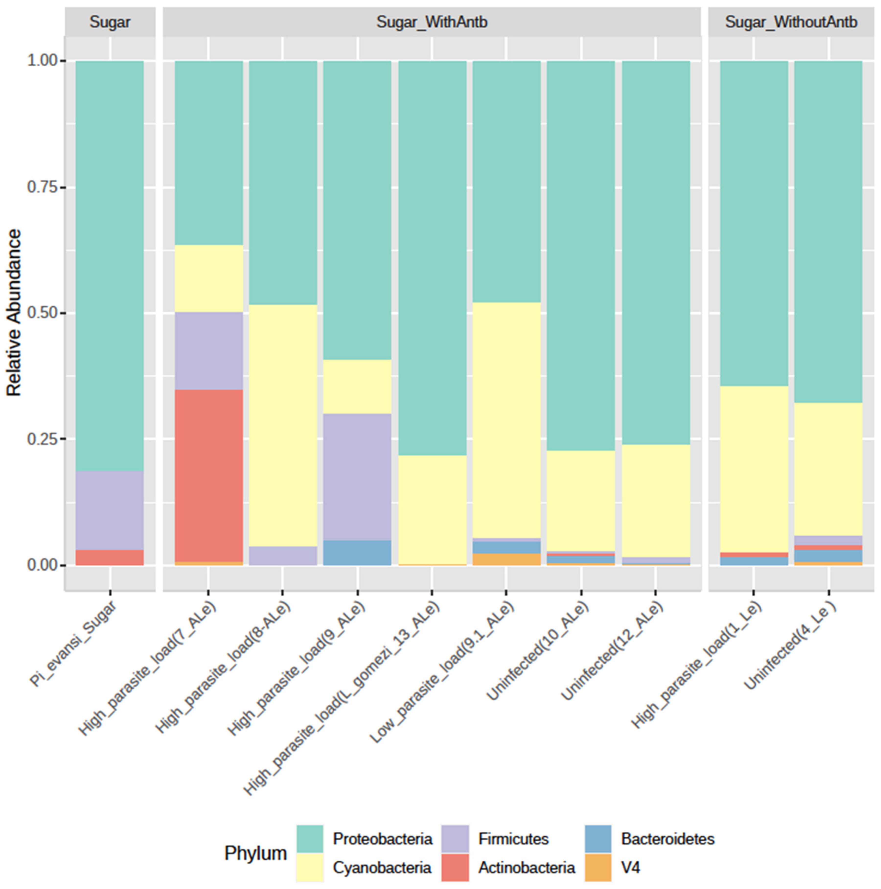
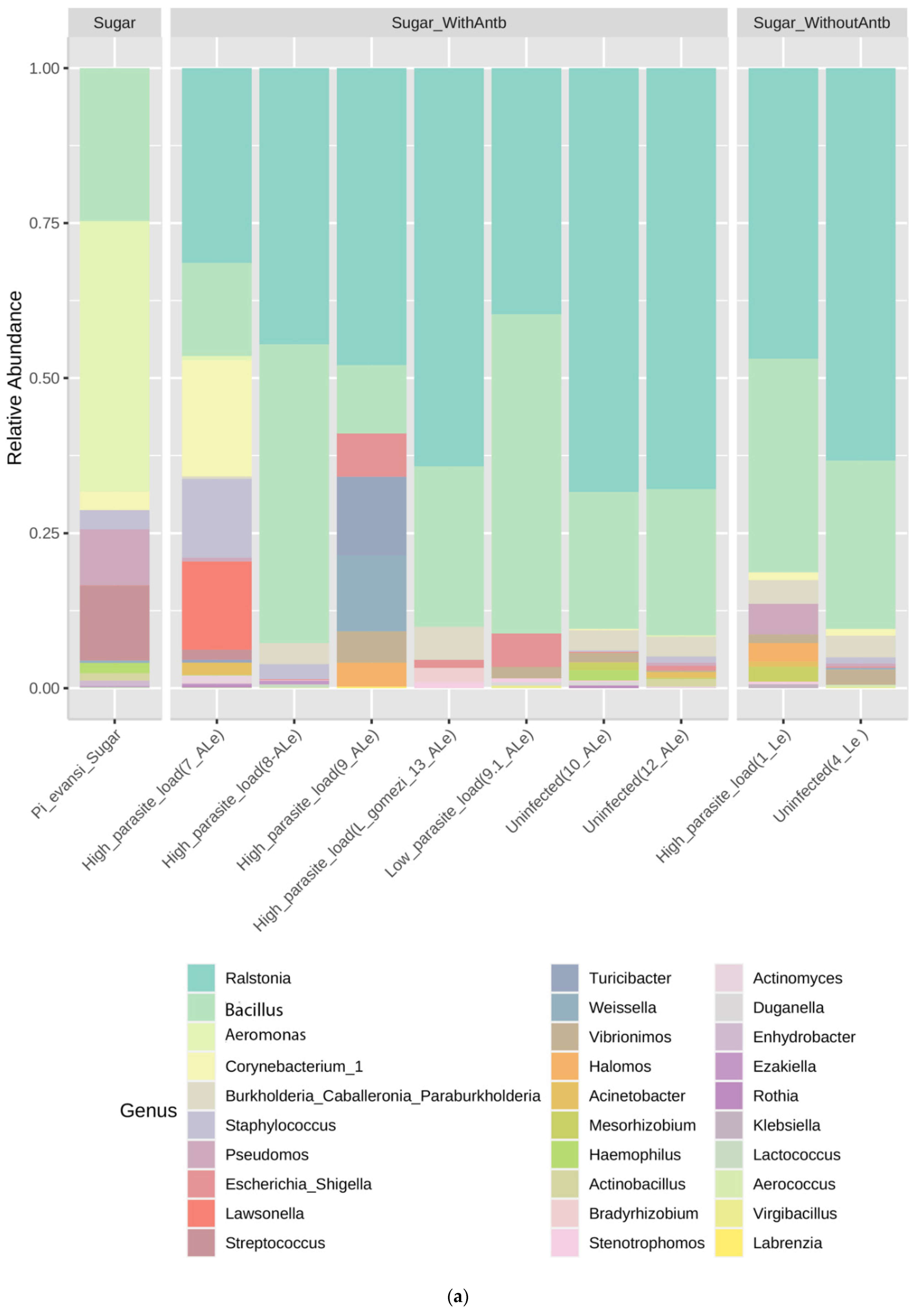
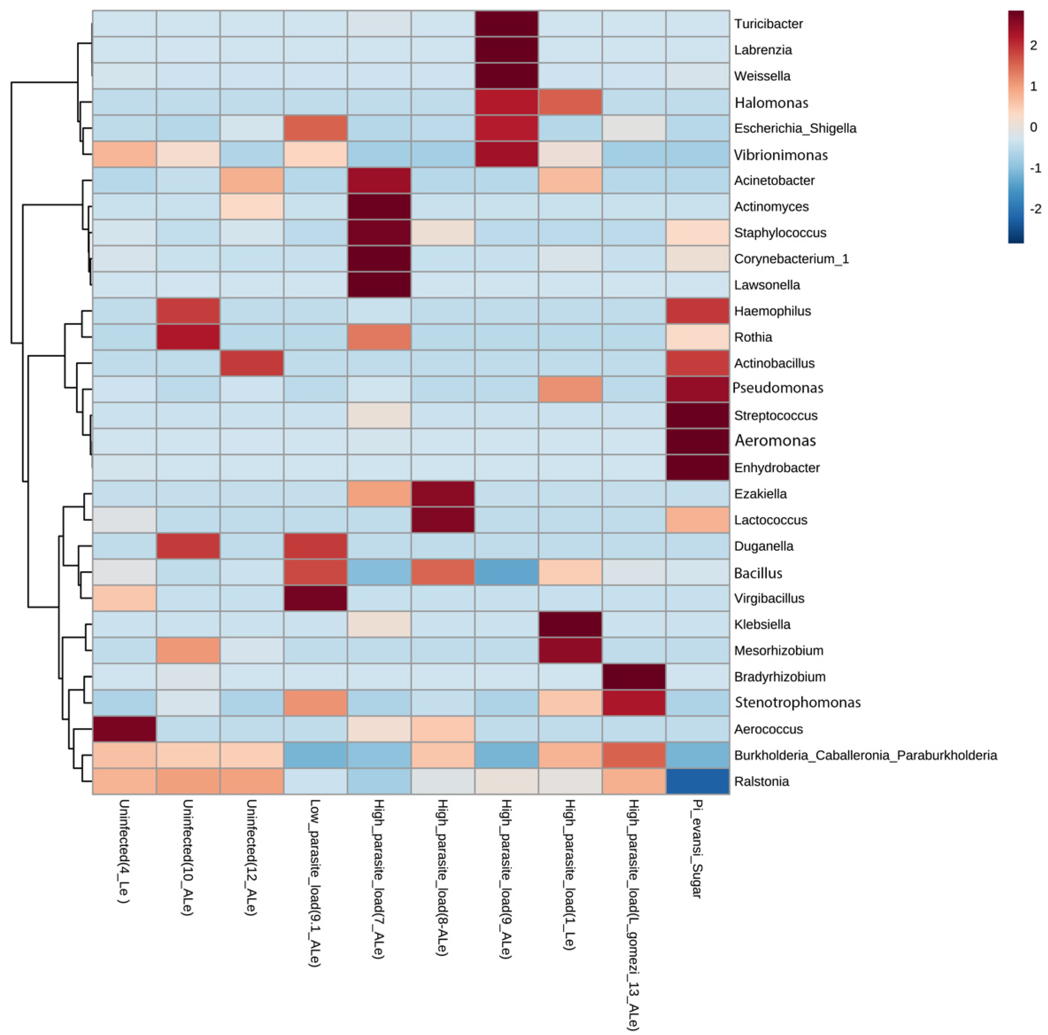
3.1. P. evansi Gut Microbiota Composition

The total reads, the number of ASVs, and the five most abundant phylum-family-genus from 16S rRNA gene amplicon sequencing were obtained from 11 samples of P. evansi guts, untreated and treated with an antibiotic cocktail (based on quality and taxonomy classification; Table 2). In the coinfection assay, 99.3% of the microbial population was composed of Proteobacteria (71.0%), Cyanobacteria (20.4%), Actinobacteria (2.7%), Firmicutes (2.7%), Bacteroidetes (1.8%), and Acidobacteria (1.0%; Figure 1).

3.2. Influence of Leishmania and Antibiotic Cocktail on Gut Microbiota

Gut pools uninfected with L. infantum (treated or untreated with antibiotics) reported the Ralstonia genera to have high relative abundance (55.1–64.8%; Figure 2a), as opposed to groups with a high load of L. infantum infection (23.4–35.9%). ASVs that moderately increased in guts infected with Leishmania were Bacillus (29.3–36%) (Figure 2a). Burkholderia, Corynebacterium, Aeromonas and Staphylococcus have an abundance similar to most samples of Pi. evansi (Figure 2a). L. gomezi with a high L. infantum infection rate presented a similar ASV profile to that of P. evansi guts uninfected with Leishmania (Figure 2a). The sample of Pi. evansi only fed with sugar has a microbiota more diverse before of the exposition to Leishmania or antibiotics (Figure 2a).

3.3. Endosymbiont Detection

No ASVs of Wolbachia, Cardinium, Rickettsia, and Flavobacterium were found in the groups of P. evansi. The rest of the body (head, thorax, legs, tegument, and reproductive structures) of P. evansi females treated and untreated with antibiotics was also identified as negative for Wolbachia by conventional PCR. However, other endosymbionts, such as Microsporidia (<2%), were detected in midgut groups without Leishmania infection; in groups treated (10-ALe 0.17%; ASVs = 1867 and 12-ALe 0.02%; ASVs = 20) and untreated with an antibiotic mixture (4Le 0.4%; ASVs = 258) containing a low load of Leishmania infection (9b-ALe+ 1.9%; ASVs = 542); and in a single group of intestines with a high load of Leishmania infection treated with the antibiotic cocktail (7-ALe+ 0.3%; ASVs = 281). Additionally, Arsenophonus was found in a gut group with a high load of Leishmania infection treated with antibiotics (8-ALe+ 4%; ASVs = 2239).

3.4. Diversity of Gut Microbiota in P. evansi

To analyze α-diversity, most of the intestinal groups treated (with and without antibiotics) and subjected to Leishmania infection showed indices of richness (observed Chao 1), diversity (Shannon and Simpson), and dominance of ASVs represented by values not exceeding 50, 2, and 0.7, respectively (Figure 2b), which are considered low compared to natural populations of P. evansi unexposed to these treatments or insectary conditions. This result suggested an antibiotic perturbation on the microbiota diversity and the potential specialization or dominance of some communities. However, a different expression of diversity profiles was presented in two gut groups with a high infection load (7-ALe+_High) and without Leishmania infection (10-ALe_Uninfected), both treated with antibiotics with values higher than those exposed above.
β-Diversity, measured by phyloseq software package version 3.8 (Bray–Curtis distance matrix) and Past package version 4.04 (Plot of Principal Coordinate Analysis-PCoA), suggested differences in the structure and composition of ASVs between the uninfected gut group and L. infantum guts infected with a high and low load (Figure 2c,d). However, at the intergroup level, a sample of intestines (7-ALe+_High) with a high rate of Leishmania infection resulted in a divergent ASV composition (Figure 2c). Kruskal–Wallis nonparametric variance statistical inference and heatmap showed statistically significant intergroup differences in guts of P. evansi infected and uninfected with L. infantum (<0.05), suggesting the impact of microbiota that may induce or restrict Leishmania infection in natural populations (Figure 3). Finally, the L. gomezi group with a high rate of Leishmania infection showed statistically significant differences in the Mann–Whitney paired test (p < 0.005), as well as in the sample 7-ALe+_High (p < 0.0005), as opposed to the rest of the groups of the bioassay. The intragroup analysis also showed statistically significant differences between the ASV communities (p < 0.0012), which is demonstrated by the distances in the PCoA (Figure 2d) and the heatmap (Figure 3).

4. Discussion

This study contributes information on the influence and potential role of the gut microbiota on the experimental establishment of L. infantum in a natural population of P. evansi, achieved through sequencing strategies and solid bioinformatics that analyzed richness and microbial diversity. Experiments were also performed to explore the behavior of some specific endosymbionts.
First, this study suggested that a fraction of the intestinal microbiota of P. evansi females may have a protective role and/or prevent the development or establishment of L. infantum. This hypothesis may be associated with the high parasite infection load in the group of guts treated with the antibiotic cocktail. The microbiota also has a fundamental role in the induction, maturation, and function of the host immune system, modulating host protection from pathogens and infectious diseases [3,24]. Bacteria may also directly inhibit pathogen development, either by hindering the necessary interactions between the pathogen and vector epithelium or through the production of antiparasitic molecules [25], such as antimicrobial peptides (e.g., defensins) [26] and pigment (prodigiosin), mainly through Gram-negative bacteria [27].
Treatment with antibiotics decreased the richness and diversity of gut microbiota on P. evansi, but the Leishmania infection increased. As in other studies, these findings strengthened the theory that any manipulation that reduces the size and/or diversity of the natural microbiota should enhance the ability of Leishmania to establish infections in sand flies or other pathogens in mosquitoes [28]. However, in Phlebotomus dubosqui, treatment with the antibiotics results in females highly refractory to the development of transmissible infections [29]. The capacity of the gut bacterial symbionts or resistant microbiota to generate appropriate nutrient stress and osmotic conditions is required for promastigote differentiation and survival [29,30], suggesting that significant differences occur if sandfly populations are in the laboratory or wild.
For sand flies, the few studies that have addressed this relationship also support the role of natural microbiota in inhibiting parasite development [31]. Ralstonia directly impacted the establishment of L. infantum in P. evansi. Its relative abundance was high (65%) in groups of guts uninfected or infected with a low load of Leishmania infection. However, the ASVs that moderately increased in guts infected with a high load of Leishmania, such as Bacillus and Aeromonas (Figure 2a), frequently isolated bacteria in sand flies [1,3,10,11]. Defined as an ASV similar to Ralstonia solanacearum at the species level, these can be acquired by adult or larval-stage sand flies from several sources. R. solanacearum is a Gram-negative and plant pathogenic bacterium [32]. The pathogenic lifestyles of this bacterium are attributed to ecological adaptation and genomic convergence during vertical evolution [32]. The frequency, density, and diversity of phylotypes of R. solanacearum in insects are poorly documented, but there are recent reports on Illeis [33], Oscinella, Aphis mellifera, Chelisoches, and Dolichiderus associated with crops, which postulated that these species could transport R. solanacearum [34]. Natural populations of L. longipalpis, Lutzomyia cruzi, and L. intermedia were found in sand flies from Brazil and Colombia [5,35,36]. Further studies are needed to understand the mechanism of interaction between Leishmania and Ralstonia.
Other ASVs were found in the gut microbiota of P. evansi, with a significant abundance of Staphylococcus, Corynebacterium, and Burkholderia. The first two were previously detected in Phlebotomus papatasi, Phlebotomus argentipes, Phlebotomus perniciosus, and L. longipalpis [37,38,39,40], whereas the others have not been registered for sand flies. To date, no information exists on the role of these bacteria in the interaction between Leishmania and sand flies.
Second, this study also showed that the endosymbionts (Wolbachia and Cardinium) were not found by either conventional PCR or next-generation sequencing. Instead, they were positively related to a natural variation in the frequency of infection, to abundance, or to the seasonality of these endosymbionts concerning the behavior of insects. This study did not attribute the absence of these two endosymbionts to the treatment with the mixture of antibiotics supplied to females of P. evansi. However, an important finding was the presence of Microsporidia and Arsenophonus in the intestinal microbiota of P. evansi. The first was more frequent in groups of intestines with no or a low load of the Leishmania infection.
This result was interesting because of the previously reported impact of Microsporidia on parasites, such as Plasmodium [41], suggesting the potential influence of this endosymbiont on Leishmania. Unlike Arsenophonus that was only detected in a group of intestines with a high load of Leishmania infection, this was the first record of Arsenophonus in sand flies from America. Arsenophonus has been described to significantly contribute to virus transmission in plants [42,43,44] and has been identified in parasitic wasps, triatomine bugs, psyllids, whiteflies, aphids, ticks, ant lions, hippoboscids, streblids, bees, lice, bat flies, louse flies, and two plant species [45]. The manipulation of host reproduction has been demonstrated by Arsenophonus [46], but some strains isolated from a divergent range of arthropods showed no evidence of sex ratio distortion activity [47]. Arsenophonus can be easily established in triatomines under laboratory conditions and influence the modification of intestinal microbiota over time and vector competition [48].
Finally, during taxonomic identification in the process of intestinal dissection, we found that a group of infected guts with Leishmania corresponded to a female of L. gomezi. Because of the importance of L. gomezi (endophilic/anthropophilic species) as a vector of the cutaneous leishmaniasis in Colombia [49], principally associated with highly intervened areas on the Caribbean coast, we consider its inclusion, highlighting the surprise experimental infection and first report with L. infantum. Despite the potential significance of L. gomezi as a vector of L. panamensis and L. braziliensis [50], few types of research have aimed to describe the associated microbiota under states of interaction with Leishmania. We found a microbiota profile consisting mainly of Ralstonia ASVs, followed by Burkholderia-Caballeronia-Paraburkholderia and ASVs without taxonomic assignment (NA) in a significant percentage. Complementary studies should be carried out to increase the information on the gut microbiota of this vector.

5. Conclusions

The inclusion of a larger number of groups of guts of P. evansi uninfected with Leishmania may improve the analysis. However, this study was done with natural and wild populations, so their abundance and susceptibility to infection are subject to variations. In summary, it is the first study that showed the potential role of the gut microbiota in natural populations of P. evansi on their susceptibility to L. infantum infection. This study also showed that treatment with antibiotics reduces the richness and diversity of microbiota, but Leishmania infection increases, indicating that the microbiota can be a barrier to the establishment and development of promastigotes in P. evansi. Finally, in vivo coinfection studies are needed to better understand Leishmania–microbiota–sand fly interactions and identify microbiome communities or effector molecules determinant in blocking or reducing the development and establishment of Leishmania.

Author Contributions

Conceptualization, R.J.V., G.C.-R., and C.X.M.-H.; data curation, R.J.V., V.A.C.-M., and G.D.H.; lab, R.J.V., V.A.C.-M., L.R.R.; methodology, R.J.V., L.R.R., and V.A.C.-M.; project administration, R.J.V., G.C.-R., G.D.H., and C.X.M.-H.; resources, G.D.H., R.J.V., and G.C.-R.; supervision, R.J.V., G.C.-R., and G.D.H.; validation, R.J.V., G.C.-R., and C.X.M.-H.; visualization, R.J.V. and V.A.C.-M.; writing—original draft, R.J.V.; writing—review and editing, R.J.V., G.C.-R., and C.X.M.-H. All authors have read and agreed to the published version of the manuscript.

Funding

This work was supported by the GCRF Networks in Vector-Borne Disease Research, which was co-founded by BBSRC, MRC, and NERC, and supported by the ANTI-VeC grant AV/PP0018/1 “Beyond Wolbachia: Determining heritable microbe incidence, prevalence, and impact on sandfly vector species” and the project Hermes 47050 of the Universidad Nacional de Colombia Sede Medellín.

Institutional Review Board Statement

Not applicable.

Informed Consent Statement

Not applicable.

Acknowledgments

We acknowledge the experimental support of the Biomédicas Research Group of the University of Sucre and its leader the Eduar E. Bejarano, for surveying insects at the locations in the Department of Sucre and supervision during the experimental infection with Leishmania. We thank the different communities we visited during our surveys in Antioquia, Caldas, and Cundinamarca for giving us access to their facilities and for their hospitality and collaboration with fieldwork. We acknowledge to the Howard Junca for their technical and scientific support.

Conflicts of Interest

The authors declare no conflict of interest. The funders had no role in the design of the study; in the collection, analysis, or interpretation of data; in the writing of the manuscript; or in the decision to publish the results.

References

  1. Telleria, E.L.; Martins-Da-Silva, A.; Tempone, A.J.; Traub-Csekö, Y.M. Leishmania, microbiota and sand fly immunity. Parasitology 2018, 145, 1336. [Google Scholar] [CrossRef] [PubMed] [Green Version]
  2. Omondi, Z.N.; Demir, S. Bacteria composition and diversity in the gut of sand fly: Impact on Leishmania and sand fly development. Int. J. Trop. Insect. Sci. 2021, 41, 25. [Google Scholar] [CrossRef]
  3. Kelly, P.H.; Bahr, S.M.; Serafim, T.D.; Ajami, N.J.; Petrosino, J.F.; Meneses, C.; Kirby, J.R.; Valenzuela, J.G.; Kamhawi, S.; Wilsone, M.E. The gut microbiome of the vector Lutzomyia longipalpis is essential for survival of Leishmania infantum. mBio 2017, 8, e01121-16. [Google Scholar] [CrossRef] [Green Version]
  4. Campolina, T.B.; Villegas, L.E.M.; Monteiro, C.C.; Pimenta, P.F.P.; Secundino, N.F.C. Tripartite interactions: Leishmania, microbiota and Lutzomyia longipalpis. PLoS Negl. Trop. Dis. 2020, 14, e0008666. [Google Scholar] [CrossRef]
  5. Monteiro, C.C.; Villegas, L.E.; Campolina, T.B.; Pires, A.C.; Miranda, J.C.; Pimenta, P.F.; Secundino, N.F. Bacterial diversity of the American sand fly Lutzomyia intermedia using high-throughput metagenomic sequencing. Parasites Vectors 2016, 9, 480. [Google Scholar] [CrossRef] [Green Version]
  6. Telleria, E.L.; Sant’Anna, M.R.; Alkurbi, M.O.; Pitaluga, A.N.; Dillon, R.J.; Traub-Csekö, Y.M. Bacterial feeding, Leishmania infection and distinct infection routes induce differential defensin expression in Lutzomyia longipalpis. Parasites Vectors 2013, 6, 12. [Google Scholar] [CrossRef] [Green Version]
  7. Telleria, E.L.; de Araújo, A.P.; Secundino, N.F.; d’Avila-Levy, C.M.; Traub-Csekö, Y.M. Trypsin-like serine proteases in Lutzomyia longipalpis—Expression, activity and possible modulation by Leishmania infantum chagasi. PLoS ONE 2010, 5, e10697. [Google Scholar] [CrossRef] [PubMed] [Green Version]
  8. Sant’Anna, M.R.; Diaz-Albiter, H.; Mubaraki, M.; Dillon, R.J.; Bates, P.A. Inhibition of trypsin expression in Lutzomyia longipalpis using RNAi enhances the survival of Leishmania. Parasites Vectors 2009, 2, 62. [Google Scholar] [CrossRef] [Green Version]
  9. Vivero, R.J.; Cadavid-Restrepo, G.; Herrera, C.X.; Soto, S.I. Molecular detection and identification of Wolbachia in three species of the genus Lutzomyia on the Colombian Caribbean coast. Parasites Vectors 2017, 10, 110. [Google Scholar] [CrossRef] [PubMed] [Green Version]
  10. Vivero, R.J.; Jaramillo, N.G.; Cadavid-Restrepo, G.; Soto, S.I.; Herrera, C.X. Structural differences in gut bacteria communities in developmental stages of natural populations of Lutzomyia evansi from Colombia’s Caribbean coast. Parasites Vectors 2016, 9, 496. [Google Scholar] [CrossRef] [Green Version]
  11. Vivero, R.J.; Villegas-Plazas, M.; Cadavid-Restrepo, G.E.; Herrera, C.X.M.; Uribe, S.I.; Junca, H. Wild specimens of sand fly phlebotomine Lutzomyia evansi, vector of leishmaniasis, show high abundance of Methylobacterium and natural carriage of Wolbachia and Cardinium types in the midgut microbiome. Sci. Rep. 2019, 9, 17746. [Google Scholar] [CrossRef] [Green Version]
  12. Tesh, R.B.; Modi, G.B. A simple method for experimental infection of phlebotomine sand flies with Leishmania. Am. J. Trop. Med. Hyg. 1984, 33, 41. [Google Scholar] [CrossRef]
  13. Santamaría, E.; Ponce, N.; Puerta, C.; Ferro, C. Validación de la PCR en la detección de parásitos de Leishmania (Viannia) spp. en Lutzomyia (Diptera: Psychodidae) como herramienta en la definición de especies vectores. Biomédica 2005, 25, 1349. [Google Scholar] [CrossRef] [Green Version]
  14. Santamaría, E.; Castillo, M.; Cárdenas, R.; Bello, F.; Ayala, M.; Ferro, C. Competencia vectorial de las especies de Lutzomyia del grupo verrucarum (Diptera, Psychodidae) en un foco endémico de Leishmania braziliensis en Reventones, Cundinamarca. Biomédica 1999, 19, 115. [Google Scholar] [CrossRef] [Green Version]
  15. Braig, H.R.; Zhou, W.; Dobson, S.L.; O’Neill, S.L. Cloning and characterization of a gene encoding the major surface protein of the bacterial endosymbiont Wolbachia pipientis. J. Bacteriol. 1998, 180, 2373. [Google Scholar] [CrossRef] [Green Version]
  16. Weeks, A.R.; Velten, R.; Stouthamer, R. Incidence of a new sex-ratio-distorting endosymbiotic bacterium among arthropods. Proc. Biol. Sci. 2003, 270, 1857. [Google Scholar] [CrossRef] [PubMed] [Green Version]
  17. Valderrama, A.; Tavares, M.G.; Dilermando Andrade Filho, J. Phylogeography of the Lutzomyia gomezi (Diptera: Phlebotominae) on the Panama Isthmus. Parasites Vectors 2014, 7, 9. [Google Scholar] [CrossRef] [Green Version]
  18. Illumina. 16S Illumina Amplicon Protocol: Earth Microbiome Project; Illumina: San Diego, CA, USA, 2017. [Google Scholar]
  19. Caporaso, J.G.; Lauber, C.L.; Walters, W.A.; Berg-Lyons, D.; Huntley, J.; Fierer, N.; Owens, S.M.; Betley, J.; Fraser, L.; Bauer, M.; et al. Ultra-high-throughput microbial community analysis on the Illumina HiSeq and MiSeq platforms. ISME J. 2012, 6, 1621. [Google Scholar] [CrossRef] [Green Version]
  20. Rath, S.; Heidrich, B.; Pieper, D.H.; Vital, M. Uncovering the trimethylamine-producing bacteria of the human gut microbiota. Microbiome 2017, 5, 54. [Google Scholar] [CrossRef] [Green Version]
  21. Callahan, B.J.; McMurdie, P.J.; Rosen, M.J.; Han, A.W.; Johnson, A.J.; Holmes, S.P. DADA2: High-resolution sample inference from Illumina amplicon data. Nat. Methods 2016, 13, 581. [Google Scholar] [CrossRef] [PubMed] [Green Version]
  22. Wang, Q.; Garrity, G.M.; Tiedje, J.M.; Cole, J.R. Naive Bayesian classifier for rapid assignment of rRNA sequences into the new bacterial taxonomy. Appl. Environ. Microbiol. 2007, 73, 5261. [Google Scholar] [CrossRef] [Green Version]
  23. McMurdie, P.J.; Holmes, S. Phyloseq: An R package for reproducible interactive analysis and graphics of microbiome census data. PLoS ONE 2013, 8, e61217. [Google Scholar] [CrossRef] [PubMed] [Green Version]
  24. Tabbabi, A.; Watanabe, S.; Mizushima, D.; Caceres, A.G.; Gomez, E.A.; Yamamoto, D.S.; Cui, L.; Hashiguchi, Y.; Kato, H. (Comparative Analysis of Bacterial Communities in Lutzomyia ayacuchensis Populations with Different Vector Competence to Leishmania Parasites in Ecuador and Peru. Microorganisms 2020, 9, 68. [Google Scholar] [CrossRef]
  25. Cirimotich, C.M.; Ramirez, J.L.; Dimopoulos, G. Native microbiota shape insect vector competence for human pathogens. Cell Host Microbe 2011, 10, 307. [Google Scholar] [CrossRef] [PubMed] [Green Version]
  26. Telleria, E.L.; Sant’Anna, M.R.; Ortigão-Farias, J.R.; Pitaluga, A.N.; Dillon, V.M.; Bates, P.A.; Traub-Cseko, Y.M.; Dillon, R.J. Caspar-like gene depletion reduces leishmania infection in sand fly host Lutzomyia longipalpis. J. Biol. Chem. 2012, 287, 12985. [Google Scholar] [CrossRef] [Green Version]
  27. Moraes, C.S.; Seabra, S.H.; Castro, D.P.; Brazil, R.P.; de Souza, W.; Garcia, E.S.; Azambuja, P. Leishmania (leishmania) chagasi interactions with Serratia marcescens: Ultrastructural studies, lysis and carbohydrate effects. Exp. Parasitol. 2008, 118, 561. [Google Scholar] [CrossRef] [PubMed]
  28. Dong, Y.; Manfredini, F.; Dimopoulos, G. Implication of the mosquito midgut microbiota in the defense against malaria parasites. PLoS Pathog. 2009, 5, e1000423. [Google Scholar] [CrossRef] [Green Version]
  29. Louradour, I.; Monteiro, C.C.; Inbar, E.; Ghosh, K.; Merkhofer, R.; Lawyer, P.; Paun, A.; Smelkinson, M.; Secundino, N.; Lewis, M.; et al. The midgut microbiota plays an essential role in sand fly vector competence for Leishmania major. Cell Microbiol. 2017, 19, 12755. [Google Scholar] [CrossRef] [Green Version]
  30. Dillon, R.J. A transdisciplinary consideration of sand flies & leishmaniasis. C. R. Biol. 2019, 342, 272. [Google Scholar]
  31. Sant’Anna, M.R.; Diaz-Albiter, H.; Aguiar-Martins, K.; Al Salem, W.S.; Cavalcante, R.R.; Dillon, V.M.; Bates, P.A.; Genta, F.A.; Dillon, R.J. Colonisation resistance in the sand fly gut: Leishmania protects Lutzomyia longipalpis from bacterial infection. Parasites Vectors 2014, 7, 329. [Google Scholar] [CrossRef] [Green Version]
  32. Remenant, B.; de Cambiaire, J.C.; Cellier, G.; Jacobs, J.M.; Mangenot, S.; Barbe, V.; Lajus, A.; Vallener, D.; Medigue, C.; Fegan, M.; et al. Ralstonia syzygii, the blood disease bacterium and some Asian R. solanacearum strains form a single genomic species despite divergent lifestyles. PLoS ONE 2011, 6, e24356. [Google Scholar] [CrossRef]
  33. Yun, J.H.; Roh, S.W.; Whon, T.W.; Jung, M.J.; Kim, M.S.; Park, D.S.; Yoon, C.; Nam, Y.-D.; Kim, Y.-J.; Choi, J.-H.; et al. Insect gut bacterial diversity determined by environmental habitat, diet, developmental stage, and phylogeny of host. Appl. Environ. Microbiol. 2014, 80, 5254. [Google Scholar] [CrossRef] [PubMed] [Green Version]
  34. Montong, V.; Salaki, S. Insects as carriers of Ralstonia solanacearum phylotype IV on kepOK banana flowers in South Minahasa and Minahasa districts. Int. J. ChemTech Res. 2020, 13, 100. [Google Scholar] [CrossRef]
  35. Sant’Anna, M.R.; Darby, A.C.; Brazil, R.P.; Montoya-Lerma, J.; Dillon, V.M.; Bates, P.A.; Dillon, R.J. Investigation of the bacterial communities associated with females of Lutzomyia sand fly species from South America. PLoS ONE 2012, 7, e42531. [Google Scholar] [CrossRef] [PubMed] [Green Version]
  36. McCarthy, C.B.; Diambra, L.A.; Rivera Pomar, R.V. Metagenomic analysis of taxa associated with Lutzomyia longipalpis, vector of visceral leishmaniasis, using an unbiased high-throughput approach. PLoS Negl. Trop. Dis. 2011, 5, e1304. [Google Scholar] [CrossRef]
  37. Mukhopadhyay, J.; Braig, H.R.; Rowton, E.D.; Ghosh, K. Naturally occurring culturable aerobic gut flora of adult Phlebotomus papatasi, vector of Leishmania major in the Old World. PLoS ONE 2012, 7, e35748. [Google Scholar] [CrossRef] [PubMed] [Green Version]
  38. Maleki-Ravasan, N.; Oshaghi, M.A.; Hajikhani, S.; Saeidi, Z.; Akhavan, A.A.; Gerami-Shoar, M.; Shirazi, M.H.; Yakhchali, B.; Rassi, Y.; Afshar, D. Aerobic microbial community of insectary population of Phlebotomus papatasi. J. Arthropod. Borne Dis. 2014, 8, 69. [Google Scholar] [PubMed]
  39. Fraihi, W.; Fares, W.; Perrin, P.; Dorkeld, F.; Sereno, D.; Barhoumi, W.; Sbissi, I.; Cherni, S.; Chelbi, I.; Durvasula, R.; et al. An integrated overview of the midgut bacterial flora composition of Phlebotomus perniciosus, a vector of zoonotic visceral leishmaniasis in the western Mediterranean basin. PLoS Negl. Trop. Dis. 2017, 11, e0005484. [Google Scholar] [CrossRef] [PubMed]
  40. Gouveia, C.; Asensi, M.D.; Zahner, V.; Rangel, E.F.; Oliveira, S.M. Study on the bacterial midgut microbiota associated to different Brazilian populations of Lutzomyia longipalpis (Lutz & Neiva) (Diptera: Psychodidae). Neotrop. Entomol. 2008, 37, 597. [Google Scholar] [CrossRef] [Green Version]
  41. Herren, J.K.; Mbaisi, L.; Mararo, E.; Makhulu, E.E.; Mobegi, V.A.; Butungi, H.; Mancini, M.; Oundo, J.W.; Teal, E.T.; Pinaud, S.; et al. A microsporidian impairs Plasmodium falciparum transmission in Anopheles arabiensis mosquitoes. Nat. Commun. 2020, 11, 2187. [Google Scholar] [CrossRef]
  42. Gottlieb, Y.; Zchori-Fein, E.; Mozes-Daube, N.; Kontsedalov, S.; Skaljac, M.; Brumin, M.; Sobol, I.; Czosnek, H.; Vavre, F.; Fleury, F.; et al. The transmission efficiency of tomato yellow leaf curl virus by the whitefly Bemisia tabaci is correlated with the presence of a specific symbiotic bacterium species. J. Virol. 2010, 84, 9310. [Google Scholar] [CrossRef] [PubMed] [Green Version]
  43. Rana, V.S.; Singh, S.T.; Priya, N.G.; Kumar, J.; Rajagopal, R. Arsenophonus GroEL interacts with CLCuV and is localized in midgut and salivary gland of whitefly B. tabaci. PLoS ONE 2012, 7, e42168. [Google Scholar] [CrossRef] [PubMed]
  44. Raina, H.S.; Rawal, V.; Singh, S.; Daimei, G.; Shakarad, M.; Rajagopal, R. Elimination of Arsenophonus and decrease in the bacterial symbionts diversity by antibiotic treatment leads to increase in fitness of whitefly, Bemisia tabaci. Infect. Genet. Evol. 2015, 32, 224. [Google Scholar] [CrossRef]
  45. Nováková, E.; Hypša, V.; Moran, N.A. Arsenophonus, an emerging clade of intracellular symbionts with a broad host distribution. BMC Microbiol. 2009, 9, 143. [Google Scholar] [CrossRef] [Green Version]
  46. Duron, O.; Bouchon, D.; Boutin, S.; Bellamy, L.; Zhou, L.; Engelstädter, J.; Hurst, G.D. The diversity of reproductive parasites among arthropods: Wolbachia do not walk alone. BMC Biol. 2008, 6, 143. [Google Scholar] [CrossRef] [PubMed] [Green Version]
  47. Dale, C.; Beeton, M.; Harbison, C.; Jones, T.; Pontes, M. Isolation, pure culture, and characterization of “Candidatus arsenophonus arthropodicus,” an intracellular secondary endosymbiont from the hippoboscid louse fly Pseudolynchia canariensis. Appl. Environ. Microbiol. 2006, 72, 2997. [Google Scholar] [CrossRef] [Green Version]
  48. Gumiel, M.; da Mota, F.F.; de Sousa Rizzo, V.; Sarquis, O.; de Castro, D.P.; Lima, M.M.; de Souza Garcia, E.; Carels, N.; Azambuja, P. Characterization of the microbiota in the guts of Triatoma brasiliensis and Triatoma pseudomaculata infected by Trypanosoma cruzi in natural conditions using culture independent methods. Parasites Vectors 2015, 8, 245. [Google Scholar] [CrossRef] [PubMed]
  49. Duque, P.; Vélez, I.; Morales, M.; Sierra, D. Sand flies fauna involved in the transmission of cutaneous leishmaniasis in Afro-Colombian and Amerindian communities of Choco, Pacific Coast of Colombia. Neotrop. Entomol. 2004, 33, 255–264. [Google Scholar] [CrossRef]
  50. Cortés, A.; Pérez-Doria, A.; Bejarano, E. Flebotomíneos (Diptera: Psychodidae) antropofílicos de importancia en salud pública en Los Montes de María, Colombia. Revista Cubana de Medicina Tropical 2009, 61, 220–225. [Google Scholar]
Figure 1. Gut microbiota composition at the phylum level in wild specimens of several natural populations of P. evansi infected with Leishmania and treated with antibiotics. The relative abundance of ASVs that were called to the taxonomic rank of the phylum.
Microorganisms 09 01214 g001
Figure 2. Gut microbiota composition and diversity in wild specimens of P. evansi from the north of Colombia, infected with Leishmania and treated with antibiotics. (a) Relative abundance of ASVs that were called to the taxonomic rank of genus. Taxa with <0.5% relative abundance were grouped together as “Genus < 0.5%”. (b) α-Diversity index of ASVs of guts of P. evansi infected with Leishmania and treated with antibiotics. (c) Hierarchical clustering analysis (β-diversity) of ASVs at the genus level (d) β-diversity analysis of microbial communities associated with the established groups of guts of P. evansi infected with Leishmania, using a Principal Coordinate Analysis (PCoA) of Bray–Curtis dissimilarities of 16S rRNA data, filtered as ASVs at the genus level. See Table 1 for the detailed nomenclature of P. evansi gut pools. Le+ High, guts with high load of Leishmania and without antibiotics; Le- Uninfected, guts uninfected and without antibiotics; ALe+ High-Low, guts with a high or low load of Leishmania and treated with antibiotics; ALe Uninfected, guts uninfected but treated with antibiotics.
Microorganisms 09 01214 g002aMicroorganisms 09 01214 g002bMicroorganisms 09 01214 g002c
Figure 3. Heatmap based on microbiota composition at genus level associated with gut microbiota in wild specimens of P. evansi from the north of Colombia, infected with Leishmania and treated with antibiotics. Hierarchical Ward’s linkage clustering based on the Pearson’s correlation coefficient of the microbial taxa abundance. Blue and red colors represent positive and negative correlations, respectively. The color scale represents the scaled abundance of each variable, denoted as Z-score, with red indicating high abundance, and blue indicating low abundance.
Microorganisms 09 01214 g003
Table 1. Treatments and parasite load of L. infantum in gut groups of fed female P. evansi and L. gomezi from Ovejas, Sucre Department, Colombia.
TreatmentsSandflyGroup CodeNo. Guts per GroupsParasite Load Observed *DNA Total Concentration (ng/μL)
Sugar solution (30%) supplemented with an antibiotic cocktail (50 μg/μL) and
L. infantum (5 × 10⁶ parasites/mL)
P. evansi7-ALe+15High22.1
8-ALe+15High16.3
9-ALe+12High17.5
9.1-ALe+7Low23.8
10-ALe10Uninfected15.1
12-ALe8Uninfected20.3
Sugar solution (30%) and L. infantum
(5 × 10⁶ parasites/mL)
1-Le+5High19.7
4-Le3Uninfected19.8
Sugar solution (30%) supplemented with an antibiotic cocktail (50 μg/μL) and
L. infantum (5 × 10⁶ parasites/mL)
L. gomezi13-ALe+2High13.8
* Parasite load observed. Uninfected (0), low (1–100), and high (>100) (Romero-Ricardo, data unpublished).
Table 2. Summary of the results obtained from 16S rRNA gene amplicon sequencing of P. evansi gut microbiota under experimental infection with L. infantum and treated with antibiotics.
P. evansi CoinfectionP. evansi Coinfection
Dataset UntreatedDataset Treated
Total Reads1′551.6121′540.250
No. ASVs415227
Phyla(14)(11)
AcidobacteriaAcidobacteria
ActinobacteriaActinobacteria
ArmatimonadetesArmatimonadetes
BacteroidetesBacteroidetes
CyanobacteriaCyanobacteria
Deinococcus–ThermusFirmicutes
EuglenozoaFusobacteria
FirmicutesMicrosporidia
FusobacteriaProteobacteria
MicrosporidiaTenericutes
PatescibacteriaVerrucomicrobia
Proteobacteria
Tenericutes
Verrucomicrobia
5 major families (total counts)BurkholderiaceaeBurkholderiaceae
BacillaceaeBacillaceae
CorynebacteriaceaeCorynebacteriaceae
ChitinophagaceaeChitinophagaceae
ElsteraceaeElsteraceae
Top 6 most abundant bacterial
genera (total counts)
RalstoniaRalstonia
BacillusBacillus
Burkholderia-Caballeronia-ParaburkholderiaBurkholderia-Caballeronia-Paraburkholderia
VibrionimonasVibrionimonas
CorynebacteriumStaphylococcusCorynebacterium
Staphylococcus
No. taxa summarized to
the genus level
14187
Publisher’s Note: MDPI stays neutral with regard to jurisdictional claims in published maps and institutional affiliations.

