Article

Haloglomus irregulare gen. nov., sp. nov., a New Halophilic Archaeon Isolated from a Marine Saltern

by
Ana Durán-Viseras
,
Cristina Sánchez-Porro
and
Antonio Ventosa
*
Department of Microbiology and Parasitology, Faculty of Pharmacy, University of Sevilla, 41012 Sevilla, Spain
*
Author to whom correspondence should be addressed.
Microorganisms 2020, 8(2), 206; https://doi.org/10.3390/microorganisms8020206
Submission received: 9 December 2019 / Revised: 28 January 2020 / Accepted: 30 January 2020 / Published: 2 February 2020
(This article belongs to the Special Issue Microbial Diversity in Extreme Environments)

Abstract

:
A halophilic archaeal strain, designated F16-60T, was isolated from Isla Cristina marine saltern in Huelva, Spain. Cells were pleomorphic, irregular, non-motile, and Gram-stain-negative. It produced red-pigmented colonies on agar plates. Strain F16-60T was extremely halophilic (optimum at 30% (w/v) NaCl) and neutrophilic (optimum pH 7.5). Phylogenetic tree reconstructions based on 16S rRNA and rpoB´ gene sequences revealed that strain F16-60T was distinct from species of the related genera Natronomonas, Halomarina, and Halomicrobium, of the order Halobacteriales. The polar lipids are phosphatidylglycerol (PG), phosphatidylglycerol phosphate methyl ester (PGP-Me), phosphatidylglycerol sulfate (PGS), and one glycolipid chromatographically identical to sulfated mannosyl glucosyl diether (S-DGD-1). The DNA G+C content is 68.0 mol%. The taxonomic study, based on a combination of phylogenetic, genomic, chemotaxonomic, and phenotypic analyses, suggest that strain F16-60T (= CECT 9635T = JCM 33318T), represents a novel species of a new genus within the family Haloarculaceae and the order Halobacteriales, for which the name Haloglomus irregulare gen. nov., sp. nov. is proposed. Metagenomic fragment recruitment analysis revealed the worldwide distribution of members of this genus and suggested the existence of other closely related species to be isolated.

Graphical Abstract

1. Introduction

The class Halobacteria is a major group within the domain Archaea, and comprises halophilic archaea (also called haloarchaea) which are typically found in hypersaline environments, such as salt lakes, soda lakes, or salterns [1]. The class Halobacteria includes three orders, Haloferacales, Halobacteriales, and Natrialbales, which consist of several different families [2,3,4]. Haloarchaea exhibit enormous diversity in terms of their morphology or physiology. They can be coccoid, rod-shaped, pleomorphic, or even square, motile, or non-motile cells. In addition to their extreme NaCl requirements, they may grow at neutral or alkaline conditions, and thus they may be haloalkaliphiles [1,4,5].
Marine salterns are thalassohaline environments, which constitute excellent models for ecological studies based on their structure of a series of ponds with different salinities, and thus, have been long-term targets for the study of halophilic microbial diversity [6]. One of these salterns is located in Isla Cristina, Southwest coast of Spain (37° 12′ N 7° 19′ O). Metagenomic studies carried out in an intermediate salinity pond (21% salts) of this saltern showed the predominance of members of the phylum Euryarchaeota, followed by the phylum Bacteroidetes, being the genera Halorubrum, Psychroflexus, and Natronomonas the most abundant groups. Moreover this analysis also brought to light that a large number of haloarchaeal members have not been isolated until date [7,8]. On the other hand, several culture-dependent studies performed in these salterns have permitted the isolation and characterization of new groups of halophilic bacteria, such as members of the genera Spiribacter [9,10], Idiomarina [11], Marinobacter [12], or Salinivibrio [13], as well as of the archaeal genera Halonotius [14,15] and Halorientalis [16]. In 2016, as part of a new study of the halophilic archaeal diversity of Isla Cristina marine saltern, we isolated a halophilic archaeal strain, designated F16-60T, closely related to members of the order Halobacteriales. In the present study, we describe the properties of strain F16-60T on the basis of recommended standard taxonomic methods, as well as on recent genomic approaches, following the respective minimal standards [17,18]. Our data suggest that strain F16-60T is not related to any previous haloarchaeal taxa and thus, we propose it as a new genus and species, Haloglomus irregulare gen. nov., sp. nov. In addition, we determined that this new haloarchaeon is widely distributed in different hypersaline habitats.

2. Materials and Methods

2.1. Isolation of Haloarchaeal Strain

During the course of studies on Isla Cristina saltern, Southwest coast of Spain (37° 12´ 30´´–7° 19´ 41´´) in June 2016, a halophilic archaeal strain, designated F16-60T, was isolated from saline water of a pond of the saltern. At the time of sampling, the salinity of the pond was 32% (w/v) total salts and the pH was 7.5. Samples were transported to the laboratory in a short time and plated under sterile conditions. Plates were incubated at 37 °C for up to three months. Strain F16-60T was isolated in pure culture after successive cultivations, using a medium prepared by filtering the saline water from the pond, with no other nutrients added, and with 2% (w/v) purified agar (Oxoid) as solidifying agent. The pH of the medium was adjusted to 7.5 with 1 M KOH. Strain F16-60T was routinely grown in the modified DBCM2 medium (CECT medium no. 263) and incubated aerobically at 37 °C, using a rotary shaker for growth in liquid medium. The composition of the modified DBCM2 medium is: sodium pyruvate 0.011 g, glucose 0.00025 g, peptone (Difco) 0.00125 g, yeast extract (Difco) 0.00125 g, distilled water 16.67 ml, and 83.33 ml of a stock of seawater SW30 solution, which contained (g/L): NaCl: 195; MgCl2.6H2O: 32.5; MgSO4.7H2O: 50.8; CaCl2: 0.83; KCl: 5.0; NaHCO3: 0.17; NaBr: 0.58. Purified agar 2% (w/v) (Oxoid) was added when necessary. For long time preservation, cultures were maintained at −80 °C in a liquid medium containing 40% (v/v) glycerol. Halophilic archaeal type strains Natronomonas pharaonis CECT 4578T and Natronomonas moolapensis CECT 7526T, obtained from culture collections, were also used in this study as reference strains.

2.2. DNA Extraction, Purification, and Sequencing

DNA extraction for determining the 16S rRNA gene sequence, rpoB´ gene sequence and genome sequence analysis, was obtained using the Marmur method [19]. The quantification of the extracted DNA was determined by spectrophotometry (DeNovix DS-11 FX, DeNovix Technologies, Wilmington, DA, USA) and fluorometry (Qubit 3.0 Fluorometer, Thermofisher Scientific, Waltham, MA, USA). DNA quality was checked by (1%) agarose gel electrophoresis. The 16S rRNA gene and the rpoB´ gene were amplified by PCR [20] using the universal primers for archaea ArchF and ArchR [21,22] and the primers designed by Fullmer et al. [23], respectively. Sequencing of PCR products were carried out by StabVida (Oeiras, Portugal) using the Sanger method. The GenBank/EMBL/DDBJ accession number for the 16S rRNA and rpoB´ gene sequences of strain F16-60T are MH424600 and MK182272, respectively. The genome of strain F16-60T was sequenced using the lllumina Hiseq 4000 platform (StabVida, Oeiras, Portugal).

2.3. Genome Assembly and Annotation

The de novo assembly of the reads was performed using Spades 3.9.1 [24]. The quality of final contigs was assessed by the bioinformatics tool CheckM v1.0.5 [25] and Quast v2.3 [26]. The genome sequence was annotated using the NCBI Prokaryotic Genome Annotation Pipeline (PGAP) [27]. The genome of strain F16-60T was deposited in GenBank/EMBL/DDBJ under the accession number QMDX00000000.

2.4. Phylogenetic Analyses

The identification of phylogenetic neighbours and calculation of the pairwaise 16S rRNA gene sequence similarity of strain F16-60T was carried out by using the EzBioCloud server [28] and GenBank. The 16S rRNA and the rpoB´ gene sequences used for phylogenetic comparisons were obtained from GenBank. To delineate the phylogenetic position, gene sequences alignments were performed using ChromasPro (Technelysium Pty Ltd, Tewantin, Australia) software version 1.5. Bootstrap consensus trees were inferred from 1000 replicates [29] using the neighbour-joining [30], maximum-parsimony [31], and maximum-likelihood [32] algorithms integrated in the ARB software package [33] for the 16S rRNA gene phylogenetic analysis and with MEGA 6.0 software [34] for the rpoB´ gene analysis. Evolutionary distances were calculated according to the algorithms of the Jukes–Cantor model [35]. To calculate phylogenomic relations between strain F16-60T and phylogenetic neighbours, a core-genome tree was constructed. Available genomic sequences of the closely related taxa were also obtained from GenBank. All predicted protein-coding genes annotated from each available genome were compared using the all-versus-all BLAST search [36]. MUSCLE [37] was used for the alignment of core orthologous genes. MEGA 6.0 software [34] was used for the phylogenomic tree reconstruction, by the neighbour-joining method with Jukes–Cantor correction [35].
The number of common genes shared between strain F16-60T and phylogenetic neighbours were represented by a Venn Diagram, which was constructed using the online software InteractiVenn [38].

2.5. Average Nucleotide Identity (ANI), Average Amino Acid Identity (AAI), and Digital DNA–DNA Hybridization (DDH)

To confirm the status as a new taxon of the isolated strain, Average Nucleotide Identity (ANI), Average Amino Acid Identity (AAI), and in silico DNA–DNA hybridization (DDH) were performed. To calculate ANI, the OAT software v0.93.1 was used [39], while for the estimation of AAI we used the tool AAI calculator [40]. The in silico DDH percentages were calculated by the Genome-to-Genome Distance Calculator (GGDC) [41] website, using formula 2 [42].

2.6. Chemotaxonomic Analysis

To complete the chemotaxonomic characterization, strain F16-60T was grown in a modified DBCM2 liquid medium and incubated for three weeks to obtain cell biomass. Natronomonas pharaonis CECT 4578T, Natronomonas moolapensis CECT 7526T, Halobacterium salinarum DSM 3754T, and Halorubrum saccharovorum DSM 1137T were used as reference species for polar lipids characterization. Polar lipids were extracted with chloroform/methanol according to Corcelli and Lobasso [43]. Analysis of polar lipids was carried out by one-dimensional HPTLC on Merck silica gel plates crystal back (Merck art. 5626; 10 × 20 cm). Plates were eluted in the solvent system chloroform/90% methanol/acetic acid (65:4:35, by vol.) [44,45]. To detect all lipids, the plate was sprayed with sulfuric acid 5% in water and charred by heating at 160 °C; for specific phospholipids detection, the plate was sprayed with a molybdenum blue spray reagent [46].

2.7. Phenotypic Characterization

All morphological and physiological characteristics of strain F16-60T were determined according to the minimal standards for description of new taxa in the Halobacteria [17]. The range of NaCl concentrations for growth was evaluated using a modified DBCM2 medium at 0%, 3%, 5%, 10%, 15%, 20%, 25%, 30%, and 35% (w/v) NaCl concentrations. The range of pH for growth was determined at 5.0, 6.0, 6.5, 7.0, 7.5, 8.0, 8.5, 9.0, or 10.0 and the range of temperature from 20 to 55 °C in 5 °C intervals. Gram staining was performed using acetic acid-fixed samples [47]. Cell morphology and motility were examined in a liquid medium after three weeks of growth by the hanging drop method using an Olympus BX41 phase-contrast microscope. The anaerobic growth was determined using an anaerobic jar with an anaerobic gas generator and anaerobic indicator (Oxoid). Catalase activity was assessed by adding a 3% (w/v) H2O2 solution to colonies on a solid medium [48]. Oxidase activity was examined by using 1% (v/v) tetramethyl-p-phenylenediamine [49]. A test for the hydrolisis of Tween 80, gelatin, and DNA were carried out as described by Barrow and Feltham [48]. The indole production from tryptophan and urease test were assessed as described by Gerhardt et al. [50]. The production of H2S was performed according to Clarke [51]. The methyl red and Voges–Proskauer tests, Simmons´s citrate, and anaerobic growth in the presence of nitrate, arginine, or DMSO were evaluated following the minimal standards [17]. The reduction of nitrate and nitrite were detected by using sulfanilic acid and α-naphthylamine reagents [52]. The production of acid from different carbohydrates was tested in a medium with 0.5% (w/v) yeast extract supplemented with 1% (w/v) of the carbohydrate tested [17]. To determine the utilization of different organic substrates as carbon and energy sources, a medium containing 0.05% (w/v) yeast extract supplemented with 1% (w/v) of the tested substrate was used [53]. The halophilic archaeon Natronomonas moolapensis CECT 7526T was used in this study for comparison as a reference strain. Natronomonas pharaonis CECT 4578T, the type species of the genus, due to its alkaliphilic character, does not grow in the non-alkaliphilic media used for the characterization of strain F16-60T, and thus was not used as a reference strain in these studies of phenotypic characterization.

2.8. Metagenomic Fragment Recruitment Analysis

To evaluate the presence in hypersaline habitats of haloarchaeal strains related (at species level) to strain F16-60T, fragment recruitments with environmental datasets (Table S1) were performed. Genome contigs were concatenated and all the rRNA gene sequences present were masked. Blastn (with the cut-offs: Alignment length ≥ 30 nt, identity > 95%, E-value  ≤ 1  × 10−5) was used in order to align the metagenomic quality-filtered shotgun reads (Table S1) against strain F16-60T genome. Best-hits blastn results obtained were used to construct the figures.

3. Results and Discussion

3.1. Phylogenetic Analyses

After extensive haloarchaeal strains isolations using different media and culture conditions, strain F16-60T was isolated as indicated previously, obtained in pure culture and selected for further analyses.
The EzBioCloud tool was used for the 16S rRNA gene sequence comparative analysis and showed that strain F16-60T (1400 bp) shared the higher sequence similarities with the type strains Natronomonas moolapensis 8.8.11T (92.8% 16S rRNA gene sequence similarity) and Natronomonas pharaonis DSM 2160T (92.6% 16S rRNA gene sequence similarity), followed by species of other genera such as Halobacterium, Halomarina, and Halomicrobium, with percentages of 16S rRNA gene sequence similarity in all cases lower than 92.5%. The 16S rRNA phylogenetic analysis, based on the maximum-likelihood algorithm, revealed that strain F16-60T constituted a monophyletic branch clearly separated from the most closely related species (Figure 1). Maximum-parsimony and neighbour-joining methods resulted in highly similar tree topologies. The low percentages of 16S rRNA gene sequence similarity, as well as its phylogenetic position, clearly support the placement of the new isolate within a new genus and species, separated from the previously described haloarchaeal taxa. The 16S rRNA gene sequence of strain F16-60T was also obtained from its genome, and when compared with that obtained experimentally by PCR, both sequences resulted identical.
On the other hand, due to the reported limitations on haloarchaea´s phylogeny based on the 16S rRNA gene [54,55], other phylogenetic approaches are recommended for the comparison of haloarchaea. The rpoB’ gene of strain F16-60T (1848 bp) was also sequenced and compared with those of the most closely related species. The phylogenetic tree based on the rpoB’ gene sequence using the maximum-likelihood algorithm (Figure 2) clearly showed a distinct monophyletic clade for strain F16-60T, with values of similarity in all cases lower than 87.2% with the phylogenetically closest related species. This phylogeny also supports the condition of new genus and species for strain F16-60T. A similar topology was obtained by the neighbour-joining algorithm.
To carry out a complete phylogenic analysis, a phylogenomic tree based on core orthologous genes was also constructed. The phylogenomic tree reconstruction (Figure 3) based on a total of 170 orthologous genes shared between all genomes under study supports the placement of strain F16-60T within a new genus and species most closely related to species of the genus Natronomonas and other genera of the family Haloarculaceae, in the order Halobacteriales. These studies clearly show that strain F16-60T constitutes a distinct clade clearly separated from the most closely related species, with a bootstrap value of 68%, supporting its classification as a new genus.
Considering the results obtained by the complete phylogenetic analyses, in order to confirm that strain F16-60T was indeed a new taxon, Average Nucleotide Identity (ANI), Average Amino Acid Identity (AAI), and in silico DNA–DNA hybridization (DDH) of strain F16-60T and other related members of the order Halobacteriales, were calculated (Figure 4 and Table 1). Results of the ANI and in silico DDH estimation for strain F16-60T and the related haloarchaea (Figure 4) were in all cases lower than 95% and 70%, respectively, which are the threshold values for species delineation [39,56,57,58]. These results indicate that strain F16-60T constitutes a new taxon at least at the species level, not related at this level to any of the closest phylogenetic neighbours. Additionally, species that share less than 80% ANI (Figure 4), are too divergent to be compared only based on this parameter, and AAI, which provides a more robust resolution, should be used instead [59]. Thus, AAI percentages were also estimated and the results are shown in Table 1. A threshold AAI value of 65% has been established for genus delineation [60,61]. AAI values for strain F16-60T and the most closely related species were in all cases lower than 65% (Table 1), confirming its condition of a new genus.
According to the phylogenetic analysis and phylogenomic analyses, and the ANI, AAI, and in silico DDH data, strain F16-60T constitutes a new genus and species, for which we propose the new designations of Haloglomus gen. nov., and Haloglomus irregulare sp. nov., respectively. Since the most closely related species of this new taxon belongs to the family Haloarculaceae, including Haloarcula vallismortis, type species of Haloarcula, we suggest that this new genus should be also classified within this family member of the order Halobacteriales, within the class Halobacteria.

3.2. Chemotaxonomic Characterization

To describe strain F16-60T, the complete chemotaxonomic characterization of this strain was carried out. The total amount of lipids was extracted and compared with those from closely related neighbours, Natronomonas pharaonis CECT 4578T and Natronomonas moolapensis CECT 7526T and the reference strains Halobacterium salinarum DSM 3754T and Halorubrum saccharovorum DSM 1137T. The major polar lipids of strain F16-60T were phosphatidylglycerol (PG), phosphatidylglycerol phosphate methyl ester (PGP-Me), phosphatidylglycerol sulfate (PGS), and one glycolipid chromatographically identical to sulphated mannosyl glucosyl diether (S-DGD-1) (Figures S1 and S2).
In comparison with species of the genus Natronomonas, it is remarkable that the absence of biphosphatidylglycerol (BPG) and sulfated triglycosyl diphytanyl archaeol ester linked to phosphatidic acid (S-TGD-1-PA) in strain F16-60T, which are present in Natronomonas moolapensis CECT 7526T (Figures S1 and S2).

3.3. Phenotypic Characterization

Cells of strain F16-60T, were Gram-stain-negative, irregular shaped (0.6 x 3.0 µm) and non-motile (Figure S3). Colonies are red-pigmented with a diameter of 0.5 mm in a modified DBCM2 medium after 20 days of incubation at 37 °C. Strain F16-60T was strictly aerobic, able to grow over a small range of NaCl concentrations, from 20% to 35% (w/v) (optimum 30% (w/v) NaCl) and over a pH range from 6.5 to 9.0 (optimum 7.5), confirming its condition of extremely halophilic and neutrophilic archaeon and thus belonging to the non-alkaliphilic haloarchaeal group. Mg2+ was not required for growth. Strain F16-60T was catalase positive, oxidase negative, and able to reduce nitrate and nitrite without gas production. Voges–Proskauer, indole production, Simmons´s citrate, and urease tests were negative. Other phenotypic characteristics, range and optimum temperature, pH and NaCl concentrations for growth, hydrolysis of different compounds, and utilization of several substrates as single source of carbon and energy are detailed in the species description and in Table 2.

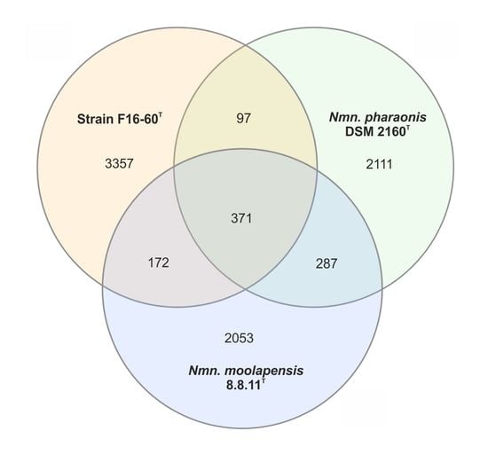
3.4. Genomic Characteristics

The draft genome of strain F16-60T was successfully sequenced and de novo assembled in a total of 54 contigs. The genome size was 4019787 bp and the sequencing depth 340.85X coverage of the entire genome. The DNA G+C content was 68.0 mol%. Additional genomic characteristics are shown in Table 3. All genomic characteristics are in accordance with the Minimal Standards established for the use of genomic data in prokaryotic taxonomy [14].
Additionally, to determine the number of genes shared between strain F16-60T and the two closest related species, Natronomonas pharaonis and Natronomonas moolapensis, a Venn Diagram was constructed (Figure S4).

3.5. Metagenomic Fragment Recruitment Analysis

To assess the presence in hypersaline habitats from different locations and the geographical distribution of haloarchaeal strains related at species level to strain F16-60T, fragment recruitments with several environmental metagenomic datasets (Table S1) were performed. Results are shown in Figure 5 and in Figure S5.
Figure 5 represents recruitment plots from three different metagenomic datasets obtained from different samples, ordered by salinity gradient, corresponding to two different hypersaline environments, Santa Pola saltern, located in East Spain, and Lake Meyghan in Iran. In both environments, the presence and abundance of strain F16-60T in the metagenomes increases with the salinity, indicating that strain F16-60T is a halophilic archaeon which prefers to inhabit ecosystems with moderate to high salinities.
Additionally, data indicated that even strain F16-60T is not highly abundant in most hypersaline environments studied, it is widely distributed, as it is present in different geographically remote habitats (Figure 5 and Figure S5). On the other hand, the abundance of reads above 95% similarity against SS37, R, W, and S7 metagenomic datasets (Figure 5 and Figure S5), also suggests the existence of additional species of the genus Haloglomus or at least closely related, which are present in these habitats but have not yet been isolated.

3.6. Description of Haloglomus gen. nov.

Haloglomus (Ha.lo.glo´mus. Gr. masc. n. hals, halos salt; L. neut. n. glomus a ball; N.L. neut. n. Haloglomus, a salt ball).
Cells are Gram-stain-negative, non-motile, and pleomorphic, curved to irregular shaped. Red-pigmented colonies. Extremely halophilic. Strictly aerobic. Catalase positive but oxidase negative. The polar lipids are phosphatidylglycerol (PG), phosphatidylglycerol phosphate methyl ester (PGP-Me), phosphatidylglycerol sulfate (PGS), and one glycolipid chromatographically identical to sulfated mannosyl glucosyl diether (S-DGD-1).
The type species is Haloglomus irregulare. The DNA G + C content is 68.0 mol%. Phylogenetically affiliated to the family Haloarculaceae, within the order Halobacteriales, and distantly related to the genus Natronomonas (≤ 92.9% sequence similarity on the basis of 16S rRNA sequence analysis). The recommended three-letter abbreviation: Hgl.

3.7. Description of Haloglomus irregulare sp. nov.

Haloglomus irregulare (ir.re.gu.la´re. L. neut. adj. irregulare, irregular cells).
Cells are non-motile, irregular pleomorphic shaped (0.6 × 3 µm) (Figure S3) and Gram-stain negative. Colonies are red pigmented, entire, round, 0.5 mm of diameter in a modified DBCM2 medium after 20 days of incubation at 37 °C. Extremely halophilic and neutrophilic. Cells require 20%–35% (w/v) NaCl, grow at pH 6.5–9.0, and at 30–45 °C. Mg2+ is not required for growth. Optimal growth occurs at 30% (w/v) NaCl, pH 7.5 and 37 °C. Strictly aerobic. Anaerobic growth does not occur in the presence of arginine, KNO3 or DMSO. Catalase positive and oxidase negative. Positive for nitrate and nitrite reduction without gas production, gelatin hydrolysis and weakly for methyl red test, but negative for Voges–Proskauer, indole production, citrate, urease and H2S production. Does not hydrolyse DNA or Tween 80. Acid is produced from D-arabinose, D-cellobiose, citruline, fructose, D-glucose, D-ribose and D-xylose, but not from D-amygdalin, arbutine, D-dulcitol, D-galactose, glycerol, maltose, D-mannitol, D-mannose, D-melezitose, D-raffinose, sucrose, sorbitol, D-trehalose, or xylitol. Utilizes D-glucose, D-melibiose, D-raffinose, L-alanine, L-cysteine, and pyruvate as sole carbon and energy source, but does not utilize D-arabinose, D-cellobiose, fructose, D-galactose, lactose, maltose, D-mannose, D-ribose, sucrose, D-trehalose, D-xylose, D-melezitose, salicin, buthanol, dulcitol, ethanol, glycerol, D-mannitol, sorbitol, xylitol, methanol, L-arginine, glutamine, L-methionine, L-glycine, L-lysine, isoleucine, valine, benzoate, citrate, formiate, fumarate, propionate, valerate, hippurate, malate, or tartrate. Polar lipids include phosphatidylglycerol (PG), phosphatidylglycerol phosphate methyl ester (PGP-Me), phosphatidylglycerol sulfate (PGS), and one glycolipid chromatographically identical to sulfated mannosyl glucosyl diether (S-DGD-1). The DNA G + C content is 68.0 mol% (genome).
The type strain is F16-60T (= CECT 9635T = JCM 33318T), isolated from a marine saltern located in Isla Cristina, Huelva, Spain.
The GenBank/EMBL/DDBJ accession number for the 16S rRNA and rpoB´ gene sequences of Haloglomus irregulare F16-60T are MH424600 and MK182272, respectively, and that of the genome is QMDX00000000.

4. Conclusions

Considering that recent studies were carried out on hypersaline environments have brought to light that a huge number of haloarchaea remain uncultured, we designed new strategies to isolate them. During the course of studies at Isla Cristina marine saltern in Spain, a new extremely halophilic and neutrophilic archaeon, designated strain F16-60T was isolated in pure culture and selected for further analyses. Therefore, the taxonomic study, based on a combination of phylogenetic, chemotaxonomic, and phenotypic analysis, as well as the comparative genomic study, supports the classification of strain F16-60T within a separate genus and species, for which we propose the new designation Haloglomus irregulare gen. nov., sp. nov. Metagenomic fragment recruitments indicated that this new taxon prefers to inhabit environments with intermediate to high salinities.

Supplementary Materials

The following are available online at https://www.mdpi.com/2076-2607/8/2/206/s1. Table S1: Features of the different databases from hypersaline habitats used for metagenomic fragment recruitments; Figure S1: High performance thin layer chromatography (HPTLC) of polar lipids extracted from strain F16-60T and some other haloarchaeal species; Figure S2: High performance thin layer chromatography (HPTLC) of phospholipids extracted from strain F16-60T and some other haloarchaeal species; Figure S3: Photomicrograph of cells of strain F16-60T observed under a phase-contrast microscope (1000X, immersion oil), cultured in a liquid medium under optimal conditions; Figure S4: Venn diagram showing the number of genes shared between the genome of strain F16-60T and closest related species Natronomonas pharaonis DSM 2160T and Natronomonas moolapensis 8.8.11T; Figure S5: Metagenomic fragment recruitment plots of strain F16-60T against different metagenomic datasets.

Author Contributions

A.D.-V. performed the isolation of the strain, the taxonomic characterization, and the genomic analyses. A.D.-V. prepared the manuscript, tables, and figures. C.S.-P. and A.V. designed the study and revised the manuscript. All authors read and approved the final manuscript.

Funding

This study was supported by the Spanish Ministry of Economy and Competitiveness (MINECO) through project CGL2017-83385-P and by Junta de Andalucía (Spain) (BIO-213, US-1263771), all including European (FEDER) funds.

Acknowledgments

A. Durán-Viseras was the recipient of a predoctoral fellowship from the Ministry of Education, Culture and Sports, Spain. We thank A. Oren for his help on the nomenclature of the new genus and species.

Conflicts of Interest

The authors declare no conflict of interest. The funders had no role in the design of the study; in the collection, analyses, or interpretation of data; in the writing of the manuscript, or in the decision to publish the results.

References

  1. Oren, A.; Ventosa, A.; Kamekura, M. Halobacteria. In Bergey’s Manual of Systematics of Archaea and Bacteria; John Wiley & Sons, Inc.: West Sussex, UK, 2017. [Google Scholar]
  2. Gupta, R.S.; Naushad, S.; Baker, S. Phylogenomic analyses and molecular signatures for the class Halobacteria and its two major clades: A proposal for division of the class Halobacteria into an emended order Halobacteriales and two new orders, Haloferacales ord. nov. and Natrialbales ord. nov. Int. J. Syst. Evol. Microbiol. 2015, 65, 1050–1069. [Google Scholar] [PubMed]
  3. Gupta, R.S.; Naushad, S.; Fabros, R.; Adeolu, M. A phylogenomic reappraisal of family-level divisions within the class Halobacteria: Proposal to divide the order Halobacteriales into the families Halobacteriaceae, Haloarculaceae fam. nov., and Halococcaceae fam. nov., and the order Haloferacales into the families, Haloferacaceae and Halorubraceae fam. nov. Antonie van Leeuwenhoek 2016, 109, 565–587. [Google Scholar] [PubMed]
  4. Amoozegar, M.A.; Siroosi, M.; Atashgahi, S.; Smidt, H.; Ventosa, A. Systematics of haloarchaea and biotechnological potential of their hydrolytic enzymes. Microbiology 2017, 163, 623–645. [Google Scholar] [CrossRef] [PubMed] [Green Version]
  5. Oren, A. The Order Halobacteriales. In The Prokaryotes; Springer: Berlin, Germany, 2006; pp. 113–164. [Google Scholar]
  6. Ventosa, A.; Fernández, A.B.; León, M.J.; Sánchez-Porro, C.; Rodriguez-Valera, F. The Santa Pola saltern as a model for studying the microbiota of hypersaline environments. Extremophiles 2014, 18, 811–824. [Google Scholar] [CrossRef] [PubMed]
  7. Fernández, A.B.; Vera-Gargallo, B.; Sánchez-Porro, C.; Ghai, R.; Papke, R.T.; Rodriguez-Valera, F.; Ventosa, A. Comparison of prokaryotic community structure from Mediterranean and Atlantic saltern concentrator ponds by a metagenomic approach. Front. Microbiol. 2014, 5, 196. [Google Scholar] [CrossRef]
  8. Ventosa, A.; de la Haba, R.R.; Sánchez-Porro, C.; Papke, R.T. Microbial diversity of hypersaline environments: A metagenomic approach. Curr. Opin. Microbiol. 2015, 25, 80–87. [Google Scholar] [CrossRef]
  9. León, M.J.; Fernández, A.B.; Ghai, R.; Sánchez-Porro, C.; Rodriguez-Valera, F.; Ventosa, A. From metagenomics to pure culture: Isolation and characterization of the moderately halophilic bacterium Spiribacter salinus gen. nov., sp. nov. Appl. Environ. Microbiol. 2014, 80, 3850–3857. [Google Scholar] [CrossRef] [Green Version]
  10. León, M.J.; Vera-Gargallo, B.; Sánchez-Porro, C.; Ventosa, A. Spiribacter roseus sp. nov., a moderately halophilic species of the genus Spiribacter from salterns. Int. J. Syst. Evol. Microbiol. 2016, 66, 4218–4224. [Google Scholar] [CrossRef]
  11. León, M.J.; Martínez-Checa, F.; Ventosa, A.; Sánchez-Porro, C. Idiomarina aquatica sp. nov., a moderately halophilic bacterium isolated from salterns. Int. J. Syst. Evol. Microbiol. 2015, 65, 4595–4600. [Google Scholar] [CrossRef]
  12. León, M.J.; Sánchez-Porro, C.; Ventosa, A. Marinobacter aquaticus sp. nov., a moderately halophilic bacterium from a solar saltern. Int. J. Syst. Evol. Microbiol. 2017, 67, 2622–2627. [Google Scholar] [CrossRef]
  13. López-Hermoso, C.; de la Haba, R.R.; Sánchez-Porro, C.; Ventosa, A. Salinivibrio kushneri sp. nov., a moderately halophilic bacterium isolated from salterns. Syst. Appl. Microbiol. 2018, 41, 159–166. [Google Scholar] [CrossRef] [PubMed]
  14. Durán-Viseras, A.; Ventosa, A.; Sánchez-Porro, C. Halonotius aquaticus sp. nov., a new haloarchaeon isolated from a marine saltern. Int. J. Syst. Evol. Microbiol. 2019, 69, 1306–1312. [Google Scholar] [CrossRef] [PubMed]
  15. Durán-Viseras, A.; Andrei, A.-S.; Ghai, R.; Sánchez-Porro, C.; Ventosa, A. new Halonotius species provide genomics-based insights into cobalamin synthesis in Haloarchaea. Front. Microbiol. 2019, 10, 1928. [Google Scholar] [CrossRef] [PubMed]
  16. Durán-Viseras, A.; Sánchez-Porro, C.; Ventosa, A. Halorientalis pallida sp. nov., an extremely halophilic archaeon isolated from a marine saltern. Int. J. Syst. Evol. Microbiol. 2019, 69, 3636–3643. [Google Scholar] [CrossRef]
  17. Oren, A.; Ventosa, A.; Grant, W.D. Proposed minimal standards for description of new taxa in the Order Halobacteriales. Int. J. Syst. Bacteriol. 1997, 47, 233–238. [Google Scholar] [CrossRef] [Green Version]
  18. Chun, J.; Oren, A.; Ventosa, A.; Christensen, H.; Arahal, D.R.; da Costa, M.S.; Rooney, A.P.; Yi, H.; Xu, X.-W.; De Meyer, S.; et al. Proposed minimal standards for the use of genome data for the taxonomy of prokaryotes. Int. J. Syst. Evol. Microbiol. 2018, 68, 461–466. [Google Scholar] [CrossRef]
  19. Marmur, J. A procedure for the isolation of deoxyribonucleic acid from micro-organisms. J. Mol. Biol. 1961, 3, 208–218. [Google Scholar] [CrossRef]
  20. Sambrook, J.; Russell, D.W. Molecular Cloning: A Laboratory Manual, III; Cold Spring Harbor Laboratory Press: Cold Spring Harbor, NY, USA, 2001; ISBN 9781936113415. [Google Scholar]
  21. DeLong, E.F. Archaea in coastal marine environments. Proc. Natl. Acad. Sci. USA 1992, 89, 5685–5689. [Google Scholar] [CrossRef] [Green Version]
  22. Arahal, D.R.; Dewhirst, F.E.; Paster, B.J.; Volcani, B.E.; Ventosa, A. Phylogenetic analyses of some extremely halophilic archaea isolated from dead sea water, determined on the basis of their 16S rRNA sequences. Appl. Environ. Microbiol. 1996, 62, 3779–3786. [Google Scholar] [CrossRef] [Green Version]
  23. Fullmer, M.S.; Soucy, S.M.; Swithers, K.S.; Makkay, A.M.; Wheeler, R.; Ventosa, A.; Gogarten, J.P.; Papke, R.T. Population and genomic analysis of the genus Halorubrum. Front. Microbiol. 2014, 5, 140. [Google Scholar] [CrossRef] [Green Version]
  24. Bankevich, A.; Nurk, S.; Antipov, D.; Gurevich, A.A.; Dvorkin, M.; Kulikov, A.S.; Lesin, V.M.; Nikolenko, S.I.; Pham, S.; Prjibelski, A.D.; et al. SPAdes: A new genome assembly algorithm and its applications to single-cell sequencing. J. Comput. Biol. 2012, 19, 455–477. [Google Scholar] [CrossRef] [PubMed] [Green Version]
  25. Parks, D.H.; Imelfort, M.; Skennerton, C.T.; Hugenholtz, P.; Tyson, G.W. CheckM: Assessing the quality of microbial genomes recovered from isolates, single cells, and metagenomes. Genome Res. 2015, 25, 1043–1055. [Google Scholar] [CrossRef] [PubMed] [Green Version]
  26. Gurevich, A.; Saveliev, V.; Vyahhi, N.; Tesler, G. QUAST: Quality assessment tool for genome assemblies. Bioinformatics 2013, 29, 1072–1075. [Google Scholar] [CrossRef] [PubMed]
  27. Tatusova, T.; DiCuccio, M.; Badretdin, A.; Chetvernin, V.; Nawrocki, E.P.; Zaslavsky, L.; Lomsadze, A.; Pruitt, K.D.; Borodovsky, M.; Ostell, J. NCBI prokaryotic genome annotation pipeline. Nucleic Acids Res. 2016, 44, 6614–6624. [Google Scholar] [CrossRef]
  28. Yoon, S.H.; Ha, S.M.; Kwon, S.; Lim, J.; Kim, Y.; Seo, H.; Chun, J. Introducing EzBioCloud: A taxonomically united database of 16S rRNA gene sequences and whole-genome assemblies. Int. J. Syst. Evol. Microbiol. 2017, 67, 1613–1617. [Google Scholar] [CrossRef]
  29. Felsenstein, J. Confidence limits on phylogenies: An approach using the bootstrap. Evolution 1985, 39, 783–791. [Google Scholar] [CrossRef] [PubMed]
  30. Saitou, N.; Nei, M. The neighbor-joining method: A new method for reconstructing phylogenetic trees. Mol. Biol. Evol. 1987, 4, 406–425. [Google Scholar]
  31. Fitch, W.M. Toward defining the course of evolution: Minimum change for a specific tree topology. Syst. Biol. 1971, 20, 406–416. [Google Scholar] [CrossRef]
  32. Felsenstein, J. Evolutionary trees from DNA sequences: A maximum likelihood approach. J. Mol. Evol. 1981, 17, 368–376. [Google Scholar] [CrossRef]
  33. Ludwig, W.; Strunk, O.; Westram, R.; Richter, L.; Meier, H.; Yadhukumar; Buchner, A.; Lai, T.; Steppi, S.; Jobb, G.; et al. ARB: A software environment for sequence data. Nucleic Acids Res. 2004, 32, 1363–1371. [Google Scholar] [CrossRef] [Green Version]
  34. Tamura, K.; Stecher, G.; Peterson, D.; Filipski, A.; Kumar, S. MEGA6: Molecular Evolutionary Genetics Analysis version 6.0. Mol. Biol. Evol. 2013, 30, 2725–2729. [Google Scholar] [CrossRef] [PubMed] [Green Version]
  35. Jukes, T.H.; Cantor, C.R. Evolution of protein molecules. In Mammalian Protein Metabolism; Elsevier: Amsterdam, The Netherlands, 1969; pp. 21–132. [Google Scholar]
  36. Altschul, S.F.; Gish, W.; Miller, W.; Myers, E.W.; Lipman, D.J. Basic local alignment search tool. J. Mol. Biol. 1990, 215, 403–410. [Google Scholar] [CrossRef]
  37. Edgar, R.C. MUSCLE: A multiple sequence alignment method with reduced time and space complexity. BMC Bioinf. 2004, 5, 113. [Google Scholar] [CrossRef] [PubMed] [Green Version]
  38. Heberle, H.; Meirelles, G.V.; da Silva, F.R.; Telles, G.P.; Minghim, R. InteractiVenn: A web-based tool for the analysis of sets through Venn diagrams. BMC Bioinf. 2015, 16, 169. [Google Scholar] [CrossRef] [PubMed]
  39. Lee, I.; Ouk Kim, Y.; Park, S.-C.; Chun, J. OrthoANI: An improved algorithm and software for calculating average nucleotide identity. Int. J. Syst. Evol. Microbiol. 2016, 66, 1100–1103. [Google Scholar] [CrossRef] [PubMed]
  40. Rodriguez-R, L.M.; Konstantinidis, K.T. The enveomics collection: A toolbox for specialized analyses of microbial genomes and metagenomes. PeerJ Prepr. 2016, 4, e1900v1. [Google Scholar]
  41. Meier-Kolthoff, J.P.; Auch, A.F.; Klenk, H.-P.; Göker, M. Genome sequence-based species delimitation with confidence intervals and improved distance functions. BMC Bioinf. 2013, 14, 60. [Google Scholar] [CrossRef] [Green Version]
  42. Auch, A.F.; Klenk, H.-P.; Göker, M. Standard operating procedure for calculating genome-to-genome distances based on high-scoring segment pairs. Stand. Genomic Sci. 2010, 2, 142–148. [Google Scholar] [CrossRef] [Green Version]
  43. Corcelli, A.; Lobasso, S. 25 Characterization of lipids of halophilic Archaea. Methods Microbiol. 2006, 35, 585–613. [Google Scholar]
  44. Angelini, R.; Corral, P.; Lopalco, P.; Ventosa, A.; Corcelli, A. Novel ether lipid cardiolipins in archaeal membranes of extreme haloalkaliphiles. Biochim. Biophys. Acta Biomembr. 2012, 1818, 1365–1373. [Google Scholar] [CrossRef] [Green Version]
  45. Corral, P.; Gutierrez, M.C.; Castillo, A.M.; Dominguez, M.; Lopalco, P.; Corcelli, A.; Ventosa, A. Natronococcus roseus sp. nov., a haloalkaliphilic archaeon from a hypersaline lake. Int. J. Syst. Evol. Microbiol. 2013, 63, 104–108. [Google Scholar] [CrossRef] [PubMed]
  46. Kates, M. Techniques of Lipidology: Isolation, Analysis, and Identification of Lipids; Elsevier: Amsterdam, The Netherlands, 1986; ISBN 0444807322. [Google Scholar]
  47. Dussault, H.P. An improved technique for staining red halophilic bacteria. J. Bacteriol. 1955, 70, 484–485. [Google Scholar] [CrossRef] [PubMed] [Green Version]
  48. Barrow, G.I.; Feltham, R.K.A. Cowan and Steel’s Manual for the Identification of Medical Bacteria; Cambridge University Press: Cambridge, UK, 2003; ISBN 9780521543. [Google Scholar]
  49. Kovacs, N. Identification of Pseudomonas pyocyanea by the oxidase reaction. Nature 1956, 178, 703. [Google Scholar] [CrossRef] [PubMed]
  50. Gerhardt, P.; Murray, R.G.; Wood, W.A.; Krieg, N. Methods for General and Molecular Bacteriology; American Society for Microbiology: Washington, DC, USA, 1994. [Google Scholar]
  51. Clarke, P.H. Hydrogen sulphide production by bacteria. J. Gen. Microbiol. 1953, 8, 397–407. [Google Scholar] [CrossRef] [PubMed] [Green Version]
  52. Smibert, R.M.; Krieg, N.R. General characterization. In Manual of Methods for General Bacteriology; American Society for Microbiology: Washington, DC, USA, 1981; pp. 409–443. [Google Scholar]
  53. Ventosa, A.; Quesada, E.; Rodriguez-Valera, F.; Ruiz-Berraquero, F.; Ramos-Cormenzana, A. Numerical taxonomy of moderately halophilic gram-negative rods. J. Gen. Microbiol. 1982, 128, 1959–1968. [Google Scholar] [CrossRef] [Green Version]
  54. Boucher, Y.; Douady, C.J.; Sharma, A.K.; Kamekura, M.; Doolittle, W.F. Intragenomic heterogeneity and intergenomic recombination among haloarchaeal rRNA genes. J. Bacteriol. 2004, 186, 3980–3990. [Google Scholar] [CrossRef] [PubMed] [Green Version]
  55. Sun, D.-L.; Jiang, X.; Wu, Q.L.; Zhou, N.-Y. Intragenomic heterogeneity of 16S rRNA genes causes overestimation of prokaryotic diversity. Appl. Environ. Microbiol. 2013, 79, 5962–5969. [Google Scholar] [CrossRef] [Green Version]
  56. Konstantinidis, K.T.; Tiedje, J.M. Trends between gene content and genome size in prokaryotic species with larger genomes. Proc. Natl. Acad. Sci. USA 2004, 101, 3160–3165. [Google Scholar] [CrossRef] [Green Version]
  57. Richter, M.; Rosselló-Móra, R. Shifting the genomic gold standard for the prokaryotic species definition. Proc. Natl. Acad. Sci. USA 2009, 106, 19126–19131. [Google Scholar] [CrossRef] [Green Version]
  58. Stackebrandt, E.; Goebel, B.M. Taxonomic note: A place for DNA-DNA reassociation and 16S rRNA sequence analysis in the present species definition in Bacteriology. Int. J. Syst. Evol. Microbiol. 1994, 44, 846–849. [Google Scholar] [CrossRef] [Green Version]
  59. Rodríguez-R, L.M.; Konstantinidis, K.T. Bypassing cultivation to identify bacterial species. Microbe 2014, 9, 111–118. [Google Scholar] [CrossRef]
  60. Klappenbach, J.A.; Goris, J.; Vandamme, P.; Coenye, T.; Konstantinidis, K.T.; Tiedje, J.M. DNA–DNA hybridization values and their relationship to whole-genome sequence similarities. Int. J. Syst. Evol. Microbiol. 2007, 57, 81–91. [Google Scholar]
  61. Konstantinidis, K.T.; Rosselló-Móra, R.; Amann, R. Uncultivated microbes in need of their own taxonomy. ISME J. 2017, 11, 2399–2406. [Google Scholar] [CrossRef] [PubMed]
  62. Burns, D.G.; Janssen, P.H.; Itoh, T.; Minegishi, H.; Usami, R.; Kamekura, M.; Dyall-Smith, M.L. Natronomonas moolapensis sp. nov., non-alkaliphilic isolates recovered from a solar saltern crystallizer pond, and emended description of the genus Natronomonas. Int. J. Syst. Evol. Microbiol. 2010, 60, 1173–1176. [Google Scholar] [CrossRef] [PubMed]
Figure 1. Maximum-likelihood phylogenetic tree based on 16S rRNA gene sequence of strain F16-60T and related members of the order Halobacteriales. Filled circles indicate branches that were supported by the neighbour-joining, maximum-parsimony, and maximum-likelihood algorithms. Bootstrap values higher than 70% are indicated at branch-points. Sequence accession numbers are shown in parentheses. Bar: 0.01 substitutions per nucleotide position. The species Haloferax volcanii NCIMB 2012T was used as an outgroup.
Microorganisms 08 00206 g001
Figure 2. Phylogenetic reconstruction based on the comparison of the rpoB’ gene of strain F16-60T and related members of the order Halobacteriales based on the maximum-likelihood algorithm. Filled circles indicate branches that were also supported by the neighbour-joining algorithm. Bootstrap values higher than 70% are indicated at branch-points. Sequence accession numbers are shown in parentheses. Bar: 0.02 substitutions per nucleotide position. The species Haloferax volcanii JCM 8879T was used as an outgroup.
Microorganisms 08 00206 g002
Figure 3. Maximum-likelihood core gene phylogenetic tree including 11 genomes of members of the order Halobacteriales. This tree was based on the Jukes–Cantor distance calculated from the alignment of 170 shared orthologous single-copy genes of these genomes. Filled circles indicate branches that were also supported by the neighbour-joining algorithm. Bootstrap values higher than 60% are indicated at branch-points. Bar: 0.05 substitutions per nucleotide position.
Microorganisms 08 00206 g003
Figure 4. Average Nucleotide Identity (ANI) and in silico DNA–DNA hybridization (DDH) percentages between strain F16-60T and other species and genera from the order Halobacteriales. Average Nucleotide Identity (ANI) and in silico DNA–DNA hybridization (DDH) are represented by heat maps, where similarity percentages are represented by the color key histograms on the upper panel. Strains: 1. Halosimplex carlsbadense 2-9-1T (GCF_000337455); 2. Halorhabdus utahensis DSM 12940T (GCF_000023945); 3. Halorientalis regularis IBRC-M 10760T (GCF_900102305); 4. Haloarcula vallismortis ATCC 29715T (GCF_000337775); 5. Natronomonas pharaonis DSM 2160T (GCF_000026045); 6. Halobacterium salinarum NRC-1 (GCF_000006805); 7. Halomicrobium mukohatei DSM 12286T (GCF_000023965); 8. Natronomonas moolapensis 8.8.11T (GCF_000591055); 9. Halapricum salinum CBA 1105T (GCF_000755225); 10. Strain F16-60T (QMDX00000000); 11. Halomarina oriensis SPP-AMP-1T (GCA_003862495).
Microorganisms 08 00206 g004
Figure 5. Metagenomic fragment recruitment plots of strain F16-60T against the metagenomic datasets: (A) SS13, SS19, and SS37, from Santa Pola saltern; (B) G, R, and W from Lake Meyghan (Iran). In each panel the Y-axis represents the identity percentage and X-axis represents the genome length. A restrictive cut-off 95% of nucleotide identity in at least 30 bp of the metagenomic read was used. The black dashed line shows the threshold for the presence of the same species (95% identity). Abbreviations: SS13: Metagenome from Santa Pola saltern (Spain) with 13% salinity; SS19: Metagenome from Santa Pola saltern (Spain) with 19% salinity; SS37: Metagenome from Santa Pola saltern (Spain) with 37% salinity; G: Metagenome from Lake Meyghan (Iran) with 5% salinity; R: Metagenome from Lake Meyghan (Iran) with 18% salinity; W: Metagenome from Lake Meyghan (Iran) with 30% salinity.
Microorganisms 08 00206 g005
Table 1. Average Amino Acid Identity (AAI) percentages, highlighted in bold, between strain F16-60T and other species and genera from the order Halobacteriales.
AAI Values1234567891011
1-64.065.265.262.861.665.962.465.162.561.3
2--64.063.962.760.864.462.266.361.861.1
3---65.364.062.165.663.865.364.062.9
4----62.561.568.762.965.463.161.7
5-----62.763.373.063.264.962.4
6------61.961.261.361.460.6
7-------63.465.962.961.9
8--------63.064.662.5
9---------62.462.0
10----------62.0
11-----------
Strains: 1. Halosimplex carlsbadense 2-9-1T (GCF_000337455); 2. Halorhabdus utahensis DSM 12940T (GCF_000023945); 3. Halorientalis regularis IBRC-M 10760T (GCF_900102305); 4. Haloarcula vallismortis ATCC 29715T (GCF_000337775); 5. Natronomonas pharaonis DSM 2160T (GCF_000026045); 6. Halobacterium salinarum NRC-1 (GCF_000006805); 7. Halomicrobium mukohatei DSM 12286T (GCF_000023965); 8. Natronomonas moolapensis 8.8.11T (GCF_000591055); 9. Halapricum salinum CBA 1105T (GCF_000755225); 10. Strain F16-60T (QMDX00000000); 11. Halomarina oriensis SPP-AMP-1T (GCA_003862495).
Table 2. Differential characteristics of strain F16-60T and Natronomonas moolapensis CECT 7526T.
Characteristic12
MorphologyIrregular, pleomorphicRods or pleomorphic *
Motility+ *
Cell size (µm)0.6–3.00.7 × 1.7 *
Colony size (mm)0.50.5–1.0 *
Colony-pigmentationRedPink *
NaCl Range (%, w/v)25–3514–36 *
NaCl optimum (%, w/v)3018–20 *
Temperature range for growth (°C)25–4525–45 *
pH range6.5–9.05.5–8.5 *
pH optimum7.57–7.5 *
Nitrite reduction+
Hydrolisis of gelatin+
Utilization as sole carbon and energy source of:
d-glucose+
d-melibiose+
d-raffinose+
 Glycerol+
l-cysteine+
l-glycine+
l-lysine+
 Isoleucine+
 Valine+
 Fumarate+
 Malate+
 Pyruvate+
Strains: 1. Haloglomus irregulare F16-60T; 2. Natronomonas moolapensis CECT 7526T. All data from this study, except * which were obtained from the original description [62]. +: Positive; −: negative.
Table 3. General features of the sequenced genome.
FeatureStrain F16-60T
Size (bp)4019787
Contigs54
Completeness (%)97.6
G + C (mol%)68.0
N50 (bp)274728
Total genes3922
Protein coding genes3673
rRNA4
tRNA53
Accession numberQMDX00000000

