Effect of Different Laser Wavelengths on Periodontopathogens in Peri-Implantitis: A Review of In Vivo Studies


Abstract
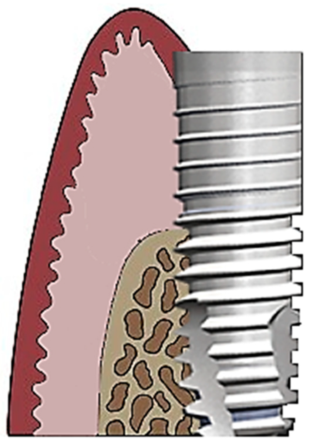
:1. Introduction
2. Materials and Methods
2.1. Focused Question
2.2. Protocol
2.3. Eligibility Criteria
- Studies involving human subjects;
- Patients with peri-implantitis;
- Surgical or non-surgical use of dental lasers in the treatment of peri-implantitis;
- Evaluated changes in specified oral bacterial profiles before and after the laser treatment;
- Prospective case series;
- Non-randomized controlled clinical trials (NRS); and
- Randomized controlled clinical trials (RCT).
- Animal studies;
- In vitro studies;
- Review articles;
- No full-text accessible; or
- Duplicated publications.
2.4. Information Sources, Search Strategy, and Study Selection
2.5. Data Collection Process, Data Items
2.6. Risk of Bias in Individual Studies
2.7. Quality Assessment
2.8. Risk of Bias Across Studies
3. Results
3.1. Study Selection
3.2. General Characteristics of the Included Studies
3.3. Results of Individual Studies
3.4. Synthesis of Results
3.5. Risk of Bias Across Studies
4. Discussion
5. Conclusions
Author Contributions
Funding
Conflicts of Interest
References
- Matys, J.; Dominiak, M. Assessment of Pain When Uncovering Implants with Er:YAG Laser or Scalpel for Second Stage Surgery. Adv. Clin. Exp. Med. 2016, 25, 1179–1184. [Google Scholar] [PubMed] [Green Version]
- Matys, J.; Świder, K.; Flieger, R.; Dominiak, M. Assessment of the primary stability of root analog zirconia implants designed using cone beam computed tomography software by means of the Periotest® device: An ex vivo study. A preliminary report. Adv. Clin. Exp. Med. 2017, 26, 803–809. [Google Scholar] [CrossRef] [PubMed]
- Matys, J.; Botzenhart, U.; Gedrange, T.; Dominiak, M. Thermodynamic effects after Diode and Er:YAG laser irradiation of grade IV and V titanium implants placed in bone–an ex vivo study. Preliminary report. Biomed. Tech. (Berl). 2016, 61, 499–507. [Google Scholar] [CrossRef] [PubMed]
- Aglietta, M.; Siciliano, V.I.; Rasperini, G.; Cafiero, C.; Lang, N.P.; Salvi, G.E. A 10-year retrospective analysis of marginal bone-level changes around implants in periodontally healthy and periodontally compromised tobacco smokers. Clin. Oral Implant. Res. 2011, 22, 47–53. [Google Scholar] [CrossRef] [PubMed]
- Roccuzzo, M.; De Angelis, N.; Bonino, L.; Aglietta, M. Ten-year results of a three-arm prospective cohort study on implants in periodontally compromised patients. Part 1: Implant loss and radiographic bone loss. Clin. Oral Implant. Res. 2010, 21, 490–496. [Google Scholar] [CrossRef] [PubMed]
- Roccuzzo, M.; Bonino, F.; Aglietta, M.; Dalmasso, P. Ten-year results of a three arms prospective cohort study on implants in periodontally compromised patients. Part 2: Clinical results. Clin. Oral Implant. Res. 2012, 23, 389–395. [Google Scholar] [CrossRef] [PubMed]
- Mombelli, A.; Lang, N.P. The diagnosis and treatment of peri-implantitis. Periodontol 2000, 17, 63–76. [Google Scholar] [CrossRef]
- Yeh, H.C.; Lu, J.J.; Chang, S.C.; Ge, M.C. Identification of microbiota in peri-implantitis pockets by matrix-assisted laser desorption/ionization time-of-flight mass spectrometry. Sci. Rep. 2019, 9, 774. [Google Scholar] [CrossRef]
- Lang, N.P.; Wilson, T.G.; Corbet, E.F. Biological complica- tions with dental implants: Their prevention, diagnosis and treatment. Clin. Oral Implant. Res. 2000, 11, 146–155. [Google Scholar] [CrossRef]
- Dennison, D.K.; Huerzeler, M.B.; Quinones, C.; Caffesse, R.G. Contaminated implant surfaces: An in vitro comparison of implant surface coating and treatment modalities for decontamination. J. Periodontol. 1994, 65, 942–948. [Google Scholar] [CrossRef]
- Augthun, M.; Tinschert, J.; Huber, A. In vitro studies on the effect of cleaning methods on different implant surfaces. J. Periodontol. 1998, 69, 857–864. [Google Scholar] [CrossRef] [PubMed]
- Schar, D.; Ramseier, C.A.; Eick, S.; Arweiler, N.B.; Sculean, A.; Salvi, G.E. Anti-infective therapy of peri-implantitis with adjunctive local drug delivery or photodynamic therapy: Six-month outcomes of a prospective randomized clinical trial. Clin. Oral Implant. Res. 2013, 24, 104–110. [Google Scholar] [CrossRef] [PubMed]
- Gosau, M.; Hahnel, S.; Schwarz, F.; Gerlach, T.; Reichert, T.E.; Bürgers, R. Effect of six different peri-implantitis disinfection methods on in vivo human oral biofilm. Clin. Oral Implant. Res. 2010, 21, 866–872. [Google Scholar]
- Bassetti, M.; Schär, D.; Wicki, B.; Eick, S.; Ramseier, C.A.; Arweiler, N.B.; Sculean, A.; Salvi, G.E. Anti-infective therapy of peri-implantitis with adjunctive local drug delivery or photodynamic therapy: 12-month outcomes of a randomized controlled clinical trial. Clin. Oral Implant. Res. 2014, 25, 279–287. [Google Scholar] [CrossRef] [PubMed]
- Pick, R.M.; Pecaro, B.C.; Silberman, C.J. The laser gingivetomy. The use of the CO2 laser for the removal of phnytoin hyperplasia. J. Periodontol. 1985, 56, 492–496. [Google Scholar] [CrossRef] [PubMed]
- Folwaczny, M.; Aggstaller, H.; Mehl, A.; Hickel, R. Removal of bacterial endotoxin from root surface with Er:YAG laser. Am. J. Dent. 2003, 16, 3–5. [Google Scholar] [PubMed]
- Grzech-Leśniak, K.; Sculean, A.; Gašpirc, B. Laser reduction of specific microorganisms in the periodontal pocket using Er:YAG and Nd:YAG lasers: A randomized controlled clinical study. Lasers Med. Sci. 2018, 33, 1461–1470. [Google Scholar] [CrossRef]
- Grzech-Leśniak, K. Making use of lasers in periodontal treatment: A new gold standard? Photomed. Laser Surg. 2017, 35, 513–514. [Google Scholar] [CrossRef]
- Vohra, F.; Al-Rifaiy, M.Q.; Lillywhite, G.; Abu Hassan, M.I.; Javed, F. Efficacy of mechanical debridement with adjunct antimicrobial photodynamic therapy for the management of peri-implant diseases: A systematic review. Photochem. Photobiol. Sci. 2014, 13, 1160–1168. [Google Scholar] [CrossRef]
- Grzech-Leśniak, K.; Matys, J.; Jurczyszyn, K.; Ziółkowski, P.; Dominiak, M.; Brugnera Junior, A., Jr.; Romeo, U. Histological and Thermometric Examination of Soft Tissue De-Epithelialization Using Digitally Controlled Er:YAG Laser Handpiece: An Ex Vivo Study. Photomed. Laser Surg. 2018, 36, 313–319. [Google Scholar] [CrossRef]
- Coluzzi, D.J.; Goldstein, A.J. Lasers in dentistry. An overview. Dent. Today. 2004, 23, 120–127. [Google Scholar] [CrossRef] [PubMed]
- Matys, J.; Dominiak, M.; Flieger, R. Energy and power density: A key factor in lasers studies. J. Clin Diag Res. 2015, 9, ZL01. [Google Scholar] [CrossRef] [PubMed]
- Matys, J.; Flieger, R.; Dominiak, M. Assessment of Temperature Rise and Time of Alveolar Ridge Splitting by Means of Er:YAG Laser, Piezosurgery, and Surgical Saw: An Ex Vivo Study. Biomed. Res Int. 2016, 2016. [Google Scholar] [CrossRef] [PubMed]
- Matys, J.; Flieger, R.; Tenore, G.; Grzech-Leśniak, K.; Romeo, U.; Dominiak, M. Er:YAG laser, piezosurgery, and surgical drill for bone decortication during orthodontic mini-implant insertion: Primary stability analysis—an animal study. Lasers Med. Sci. 2018, 33, 489–495. [Google Scholar] [CrossRef] [PubMed]
- Matys, J.; Swider, K.; Flieger, R. Laser instant implant impression method: A case presentation. Dent. Med. Probl. 2017, 54, 101–106. [Google Scholar] [CrossRef] [Green Version]
- Schwarz, F.; Rothamel, D.; Sculean, A.; Georg, T.; Scherbaum, W.; Becker, J. Effects of an Er:YAG laser and the Vector ultrasonic system on the biocompatibility of titanium implants in cultures of human osteoblast-like cells. Clin. Oral Implant. Res. 2003, 14, 784–792. [Google Scholar] [CrossRef]
- Friedmann, A.; Antic, L.; Bernimoulin, J.P.; Purucker, P. In vitro attachment of osteoblasts on contaminated rough titanium surfaces treated by Er:YAG laser. J. Biomed. Mater. Res. A. 2006, 79, 53–60. [Google Scholar] [CrossRef] [PubMed]
- Moher, D.; Liberati, A.; Tetzlaff, J.; Altman, D.G.; PRISMA, Group. Pre-ferred reportingitems for systematic reviews andmeta-analyses: The PRISMA statement. J. Clin. Epidemiol. 2009, 62, 1006–1012. [Google Scholar] [CrossRef]
- Higgins, J.P.T.; Green, S. Cochrane Handbook for Systematic Reviews of Interventions Version 5.1.0. The Cochrane Collaboration, 2011. Available online: http://handbook.cochrane.org (accessed on 2 April 2019).
- Birang, E.; Talebi Ardekani, M.R.; Rajabzadeh, M.; Sarmadi, G.; Birang, R.; Gutknecht, N. Evaluation of Effectiveness of Photodynamic Therapy With Low-level Diode Laser in Nonsurgical Treatment of Peri-implantitis. J. Lasers Med. Sci. 2017, 8, 136–142. [Google Scholar] [CrossRef] [Green Version]
- Caccianiga, G.; Rey, G.; Baldoni, M.; Paiusco, A. Clinical, Radiographic and Microbiological Evaluation of High Level Laser Therapy, a New Photodynamic Therapy Protocol, in Peri-Implantitis Treatment; a Pilot Experience. Biomed. Res. Int. 2016, 2016, 6321906. [Google Scholar] [CrossRef]
- Persson, G.R.; Roos-Jansåker, A.M.; Lindahl, C.; Renvert, S. Microbiologic results after non-surgical erbium-doped:yttrium, aluminum, and garnet laser or air-abrasive treatment of peri-implantitis: A randomized clinical trial. J. Periodontol. 2011, 82, 1267–1278. [Google Scholar] [CrossRef] [PubMed]
- Arısan, V.; Karabuda, Z.C.; Arıcı, S.V.; Topçuoğlu, N.; Külekçi, G. A randomized clinical trial of an adjunct diode laser application for the nonsurgical treatment of peri-implantitis. Photomed Laser Surg. 2015, 33, 547–554. [Google Scholar] [CrossRef] [PubMed]
- Yoshino, T.; Yamamoto, A.; Ono, Y. Innovative regeneration technology to solve peri-implantitis by Er:YAG laser based on the microbiologic diagnosis: A case series. Int. J. Periodontics Restor. Dent. 2015, 35, 67–73. [Google Scholar] [CrossRef] [PubMed]
- Salaria, S.K.; Sharma, I.; Brar, N.K.; Kaur, S. Diode Laser and Periodontal Regeneration-Assisted Management of Implant Complications in Anterior Maxilla. Contemp. Clin. Dent. 2018, 9, 114–119. [Google Scholar] [PubMed]
- Karimi, M.R.; Hasani, A.; Khosroshahian, S. Efficacy of Antimicrobial Photodynamic Therapy as an Adjunctive to Mechanical Debridement in the Treatment of Peri-implant Diseases: A Randomized Controlled Clinical Trial. J. Lasers Med. Sci. 2016, 7, 139–145. [Google Scholar] [CrossRef] [PubMed] [Green Version]
- Schwarz, F.; Sahm, N.; Iglhaut, G.; Becker, J. Impact of the method of surface debridement and decontamination on the clinical outcome following combined surgical therapy of peri-implantitis: A randomized controlled clinical study. J. Clin. Periodontol. 2011, 38, 276–284. [Google Scholar] [CrossRef] [PubMed]
- Schwarz, F.; John, G.; Schmucker, A.; Sahm, N.; Becker, J. Combined surgical therapy of advanced peri-implantitis evaluating two methods of surface decontamination: A 7-year follow-up observation. J. Clin. Periodontol. 2017, 44, 337–342. [Google Scholar] [CrossRef] [PubMed]
- Schwarz, F.; John, G.; Hegewald, A.; Becker, J. Non-surgical treatment of peri-implant mucositis and peri-implantitis at zirconia implants: A prospective case series. J. Clin. Periodontol. 2015, 42, 783–788. [Google Scholar] [CrossRef]
- Schwarz, F.; Bieling, K.; Sculean, A.; Herten, M.; Becker, J. Treatment of periimplantitis with laser or ultrasound. A review of the literature. Schweiz Mon. Zahnmed. 2004, 114, 1228–1235. [Google Scholar]
- Scarano, A.; Nardi, G.; Murmura, G.; Rapani, M.; Mortellaro, C. Evaluation of the Removal Bacteria on Failed Titanium Implants After Irradiation With Erbium-Doped Yttrium Aluminium Garnet Laser. J. Craniofac. Surg. 2016, 27, 1202–1204. [Google Scholar] [CrossRef] [PubMed]
- Pommer, B.; Haas, R.; Mailath-Pokorny, G.; Fürhauser, R.; Watzek, G.; Busenlechner, D.; Müller-Kern, M.; Kloodt, C. Periimplantitis Treatment: Long-Term Comparison of Laser Decontamination and Implantoplasty Surgery. Implant. Dent. 2016, 25, 646–649. [Google Scholar] [CrossRef] [PubMed]
- Norton, M.R. Efficacy of Er:YAG Laser in the Decontamination of Peri-implant Disease: A One-Year Prospective Closed Cohort Study. Int. J. Periodontics Restor. Dent. 2017, 37, 781–788. [Google Scholar] [CrossRef] [PubMed] [Green Version]
- Lerario, F.; Roncati, M.; Gariffo, A.; Attorresi, E.; Lucchese, A.; Galanakis, A.; Palaia, G.; Romeo, U. Non-surgical periodontal treatment of peri-implant diseases with the adjunctive use of diode laser: Preliminary clinical study. Lasers Med. Sci. 2016, 31, 1–6. [Google Scholar] [CrossRef] [PubMed]
- Hegazy, S.; Elmekawy, N.; Emera, R.M. Peri-implant Outcomes with Laser vs Nanosurface Treatment of Early Loaded Implant-Retaining Mandibular Overdentures. Int. J. Oral Maxillofac. Implant. 2016, 31, 424–430. [Google Scholar] [CrossRef] [PubMed]
- John, G.; Becker, J.; Schmucker, A.; Schwarz, F. Non-surgical treatment of peri-implant mucositis and peri-implantitis at two-piece zirconium implants: A clinical follow-up observation after up to 3 years. J. Clin. Periodontol. 2017, 44, 756–761. [Google Scholar] [CrossRef] [PubMed]
- Valente, N.A.; Andreana, S. Treatment of Peri-implantitis Using a Combined Decontaminative and Regenerative Protocol: Case Report. Compend. Contin. Educ. Dent. 2018, 39, 96–101. [Google Scholar] [PubMed]
- Romeo, U.; Nardi, G.M.; Libotte, F.; Sabatini, S.; Palaia, G.; Grassi, F.R. The Antimicrobial Photodynamic Therapy in the Treatment of Peri-Implantitis. Int. J. Dent. 2016, 2016, 7692387. [Google Scholar] [CrossRef] [PubMed]
- Al Amri, M.D.; Kellesarian, S.V.; Ahmed, A.; Al-Kheraif, A.A.; Romanos, G.E.; Javed, F. Efficacy of periimplant mechanical debridement with and without adjunct antimicrobial photodynamic therapy in patients with type 2 diabetes mellitus. Photodiagnosis Photodyn. 2016, 14, 166–169. [Google Scholar] [CrossRef] [PubMed]
- Abduljabbar, T.; Javed, F.; Kellesarian, S.V.; Vohra, F.; Romanos, G.E. Effect of Nd:YAG laser-assisted non-surgical mechanical debridement on clinical and radiographic peri-implant inflammatory parameters in patients with peri-implant disease. J. Photochem Photobiol B. 2017, 168, 16–19. [Google Scholar] [CrossRef] [PubMed]
- Larsen, O.I.; Enersen, M.; Kristoffersen, A.K.; Wennerberg, A.; Bunæs, D.F.; Lie, S.A.; Leknes, K.N. Antimicrobial Effects of Three Different Treatment Modalities on Dental Implant Surfaces. J. Oral Implantol. 2017, 43, 429–436. [Google Scholar] [CrossRef]
- Nicholson, D.; Blodgett, K.; Beaverton, O.R.; Braga, C.; Finkbeiner, L.; Fourrier, J.; George, J.; Gregg, R.; Honigman, A.; Houser, B.; et al. Pulsed Nd:YAG Laser Treatment for Failing Dental Implants Due to Peri-implantitis. Proc. SPIE 2014, 8929. [Google Scholar]
- Spadari, F.; Bombeccari, G.P.; Bosotti, B.; Marino, R. Photodynamic therapy on peri-implantitis: Comparative effectiveness - an in vivo trial. Oral Dis. 2010, 16, 553. [Google Scholar]
- Bombeccari, G.P.; Guzzi, G.; Gualini, F.; Gualini, S.; Santoro, F.; Spadari, F. Photodynamic therapy to treat periimplantitis. Implant. Dent. 2013, 22, 631–638. [Google Scholar] [CrossRef] [PubMed]
- Chambrone, L.; Wang, H.L.; Romanos, G.E. Antimicrobial photodynamic therapy for the treatment of periodontitis and peri-implantitis: An American Academy of Periodontology best evidence review. J. Periodontol. 2018, 89, 783–803. [Google Scholar] [PubMed]
- Esposito, M.; Grusovin, M.G.; Coulthard, P.; Worthington, H.V. The efficacy of interventions to treat peri-implantitis: A Cochrane systematic review of randomised controlled clinical trials. Eur. J. Oral Implant. 2008, 1, 111–125. [Google Scholar]
- Ashnagar, S.; Nowzari, H.; Nokhbatolfoghahaei, H.; Yaghoub Zadeh, B.; Chiniforush, N.; Choukhachi Zadeh, N. Laser treatment of peri-implantitis: A literature review. J. Lasers Med. Sci. 2014, 5, 153–162. [Google Scholar]
- Papadopoulos, C.A.; Vouros, I.; Menexes, G.; Konstantinidis, A. The utilization of a diode laser in the surgical treatment of peri-implantitis. A randomized clinical trial. Clin. Oral Investig. 2015, 19, 1851–1860. [Google Scholar] [CrossRef]
- Renvert, S.; Roos-Jansåker, A.M.; Claffey, N. Non-surgical treatment of peri-implant mucositis and peri-implantitis: A literature review. J. Clin. Periodontol. 2008, 35, 305–315. [Google Scholar] [CrossRef]
- Renvert, S.; Lindahl, C.; Roos Jansåker, A.M.; Persson, G.R. Treatment of peri-implantitis using an Er:YAG laser or an air-abrasive device: A randomized clinical trial. J. Clin. Periodontol. 2011, 38, 65–73. [Google Scholar] [CrossRef]
- Yan, M.; Liu, M.; Wang, M.; Yin, F.; Xia, H. The effects of Er:YAG on the treatment of peri-implantitis: A meta-analysis of randomized controlled trials. Lasers Med. Sci. 2015, 30, 1843–1853. [Google Scholar] [CrossRef]
- Natto, Z.S.; Aladmawy, M.; Levi, P.A., Jr.; Wang, H.L. Comparison of the efficacy of different types of lasers for the treatment of peri-implantitis: A systematic review. Int. J. Oral Maxillofac. Implant. 2015, 30, 338–345. [Google Scholar] [CrossRef] [PubMed]
- Smeets, R.; Henningsen, A.; Jung, O.; Heiland, M.; Hammächer, C.; Stein, J.M. Definition, etiology, prevention and treatment of peri-implantitis--a review. Head Face Med. 2014, 10, 34. [Google Scholar] [CrossRef] [PubMed]
- Kotsakis, G.A.; Konstantinidis, I.; Karoussis, I.K.; Ma, X.; Chu, H. Systematic review and meta-analysis of the effect of various laser wavelengths in the treatment of peri-implantitis. J. Periodontol. 2014, 85, 1203–1213. [Google Scholar] [CrossRef] [PubMed]
- Figuero, E.; Graziani, F.; Sanz, I.; Herrera, D.; Sanz, M. Management of peri-implant mucositis and peri-implantitis. Periodontol 2000, 66, 255–273. [Google Scholar] [CrossRef] [PubMed]
- Suárez-López Del Amo, F.; Yu, S.H.; Wang, H.L. Non-Surgical Therapy for Peri-Implant Diseases: A Systematic Review. J. Oral Maxillofac Res. 2016, 7, e13. [Google Scholar] [CrossRef] [PubMed]
- Alshehri, F.A. The role of lasers in the treatment of peri-implant diseases: A review. Saudi Dent. J. 2016, 28, 103–108. [Google Scholar] [CrossRef] [PubMed]
- Ghanem, A.; Pasumarthy, S.; Ranna, V.; Kellesarian, S.V.; Abduljabbar, T.; Vohra, F.; Malmstrom, H. Is mechanical curettage with adjunct photodynamic therapy more effective in the treatment of peri-implantitis than mechanical curettage alone? Photodiagnosis Photodyn. 2016, 15, 191–196. [Google Scholar] [CrossRef]
- Mizutani, K.; Aoki, A.; Coluzzi, D.; Yukna, R.; Wang, C.Y.; Pavlic, V.; Izumi, Y. Lasers in minimally invasive periodontal and peri-implant therapy. Periodontol 2000, 71, 185–212. [Google Scholar] [CrossRef]
- Mahato, N.; Wu, X.; Wang, L. Management of peri-implantitis: A systematic review, 2010-2015. Springerplus 2016, 5, 105. [Google Scholar] [CrossRef]
- Al Habashneh, R.; Asa’ad, F.A.; Khader, Y. Photodynamic therapy in periodontal and peri-implant diseases. Quintessence Int. 2015, 46, 677–690. [Google Scholar]
- Subramani, K.; Wismeijer, D. Decontamination of titanium implant surface and re-osseointegration to treat peri-implantitis: A literature review. Int. J. Oral Maxillofac. Implant. 2012, 27, 1043–1054. [Google Scholar]
- Rajesh, S.; Koshi, E.; Philip, K.; Mohan, A. Antimicrobial photodynamic therapy: An overview. J. Indian Soc Periodontol. 2011, 15, 323–327. [Google Scholar] [PubMed]
- Gonçalves, F.; Zanetti, A.L.; Zanetti, R.V.; Martelli, F.S.; Avila-Campos, M.J.; Tomazinho, L.F.; Granjeiro, J.M. Effectiveness of 980-mm diode and 1064-nm extra-long-pulse neodymium-doped yttrium aluminum garnet lasers in implant disinfection. Photomed Laser Surg. 2010, 28, 273–280. [Google Scholar] [CrossRef] [PubMed]
- Kotsovilis, S.; Karoussis, I.K.; Trianti, M.; Fourmousis, I. Therapy of peri-implantitis: A systematic review. J. Clin. Periodontol. 2008, 35, 621–629. [Google Scholar] [CrossRef] [PubMed]
- Dörtbudak, O.; Haas, R.; Bernhart, T.; Mailath-Pokorny, G. Lethal photosensitization for decontamination of implant surfaces in the treatment of peri-implantitis. Clin. Oral Implant. Res. 2001, 12, 104–108. [Google Scholar] [CrossRef] [Green Version]
- Grzech-Leśniak, K.; Nowicka, J.; Pajączkowska, M.; Matys, J.; Szymonowicz, M.; Kuropka, P.; Rybak, Z.; Dobrzyński, M.; Dominiak, M. Effects of Nd:YAG laser irradiation on the growth of Candida albicans and Streptococcus mutans: In vitro study. Lasers Med. Sci. 2019, 34, 129–137. [Google Scholar] [CrossRef] [PubMed]
- Matys, J.; Flieger, R.; Dominiak, M. Effect of diode lasers with wavelength of 445 and 980 nm on a temperature rise when uncovering implants for second stage surgery: An ex-vivo study in pigs. Dent. Med. Probl. 2017, 26, 687–693. [Google Scholar] [CrossRef]
- Grzech-Leśniak, K.; Matys, J.; Dominiak, M. Comparison of the clinical and microbiological effects of antibiotic therapy in periodontal pockets following laser treatment: An in vivo study. Adv. Clin. Exp. Med. 2018, 27, 1263. [Google Scholar] [CrossRef]


| Focused Question | What is the Effect of Different Laser Wavelengths on Oral Bacteria that Cause Peri-Implantitis? |
|---|---|
| Search strategy | |
| Population | Patients diagnosed with peri-implantitis |
| Intervention or exposure | Surgical or non-surgical laser treatment |
| Comparison | Changes in oral bacterial profiles before and after treatment |
| Outcome | Changed oral bacterial profiles or the number of specified bacteria |
| Search combination | (peri-implantitis OR periimplantitis) OR/AND (microbial OR microbiologic) AND (laser OR Er:YAG OR erbium OR diode OR Nd:YAG OR neodymium-doped OR Er,Cr:YSGG OR chromium-doped) |
| Electronic database search | PubMed, Cochrane Central Register of Controlled Trials (CENTRAL) |
| Selection criteria | |
| Inclusion criteria |
|
| Exclusion criteria |
|
| Criteria | First Author | |||||
|---|---|---|---|---|---|---|
| Birang et al. [30] | Caccianiga et al. [31] | Persson et al. [32] | Arisan et al. [33] | Yoshino et al. [34] | Bassetti et al. [14] | |
| Population representativeness in the treatment group (average of the population) | 1 | 1 | 1 | 1 | 0 | 1 |
| Comparability of the baseline and the outcome parameters | 1 | 1 | 1 | 1 | 1 | 1 |
| Accuracy of the microbial genome evaluation technique | 1 | 1 | 0 | 0 | 0 | 1 |
| Randomization | 1 | 0 | 1 | 0 | 0 | 0 |
| Adequate follow-up (for outcomes to occur) | 1 | 1 | 1 | 1 | 1 | 1 |
| Acceptable follow-up loss (complete follow-up, subjects lost to follow-up unlikely to introduce bias) | 1 | 1 | 1 | 1 | 1 | 1 |
| Total | 6 | 5 | 5 | 4 | 3 | 5 |
| First Author | Year of Publication | Reason for Exclusion |
|---|---|---|
| Salaria et al. [35] | 2018 | No bacterial profile evaluated |
| Karimi et al. [36] | 2016 | No bacterial profile evaluated |
| Schwarz et al. [37] | 2011 | No bacterial profile evaluated |
| Schwarz et al. [38] | 2017 | No bacterial profile evaluated |
| Schwarz et al. [39] | 2015 | No bacterial profile evaluated |
| Schwarz et al. [40] | 2004 | Systematic review |
| Scarano et al. [41] | 2016 | An in vitro study |
| Pommer et al. [42] | 2016 | No bacterial profile evaluated |
| Norton [43] | 2017 | No bacterial profile evaluated |
| Lerario et al. [44] | 2016 | No bacterial profile evaluated |
| Hegazy et al. [45] | 2016 | No bacterial profile evaluated |
| John et al. [46] | 2017 | No bacterial profile evaluated |
| Valente et al. [47] | 2018 | No bacterial profile evaluated |
| Romeo et al. [48] | 2016 | No bacterial profile evaluated |
| Al Amri et al. [49] | 2016 | No bacterial profile evaluated |
| Abduljabbar et al. [50] | 2017 | No bacterial profile evaluated |
| Larsen et al. [51] | 2017 | An in vitro study |
| Nicholson et al. [52] | 2017 | No bacterial profile evaluated |
| Spadari et al. [53] | 2010 | No full-text accessible |
| Bombeccari et al. [54] | 2013 | No bacterial profile evaluated |
| Chambrone et al. [55] | 2018 | Systematic review |
| Esposito et al. [56] | 2008 | Systematic review |
| Ashnagar et al. [57] | 2014 | Systematic review |
| Papadopoulos et al. [58] | 2015 | No bacterial profile evaluated |
| Renvert et al. [59] | 2008 | Systematic review |
| Renvert et al. [60] | 2011 | No bacterial profile evaluated |
| Yan et al. [61] | 2015 | Systematic review |
| Natto et al. [62] | 2015 | Systematic review |
| Smeets et al. [63] | 2014 | Systematic review |
| Kotsakis et al. [64] | 2014 | Systematic review |
| Figuero et al. [65] | 2000 | Systematic review |
| Suárez-López Del Amo et al. [66] | 2016 | Systematic review |
| Alshehri et al. [67] | 2016 | Systematic review |
| Ghanem et al. [68] | 2016 | Systematic review |
| Mizutani et al. [69] | 2000 | Systematic review |
| Mahato et al. [70] | 2016 | Systematic review |
| Al Habashneh et al. [71] | 2015 | Systematic review |
| Subramani et al. [72] | 2012 | Systematic review |
| Rajesh et al. [73] | 2011 | Systematic review |
| Gonçalves at al. [74] | 2010 | Systematic review |
| Kotsovilis at al. [75] | 2008 | Systematic review |
| First Author | Study Design (No. of Subjects) | Laser Type | Laser Parameters | Evaluated Bacteria |
|---|---|---|---|---|
| Birang et al. [30] | RCT | diode | 810 nm, 300 mW, 30 s per site, large-area handpiece (transgingival) or bulb fiber (intra-pocket) or bare fiber (granulation tissues), irradiation repeated after 2 weeks | Aggregatibacter actinomycetemcomitans, Porphyromonas gingivalis, Prevotella intermedia, Treponema denticola, Tannerella forsythia |
| Caccianiga et al. [31] | prospective case series | diode (aPDT) | 2.5 W, 0.5 W (mean power), 10 kHz, T-on 20 us, T-off 80 us, 60 s per site, 400 micron fiber, periodontal and peri-implant pocket site, irradiation repeated after 15 days and then for the next 3 months every 20 days, 3% hydrogen peroxide | Aggregatibacter actinomycetemcomitans, Porphyromonas gingivalis, Treponema denticola, Tannerella forsythia, Fusobacterium nucleatum, Campylobacter rectus, Eikenella corrodens |
| Persson et al. [32] | RCT | Er:YAG | 100 mJ/pulse, 10 Hz (12.7 J/cm), cone-shaped sapphire tip, parallel mode, pocket site | 74 specimens (Campylobacter showae, Capnocytophaga ochracea, P. melaninogenica, S. anaerobius, S. hae- molyticus, S.intermedius, and S. mutans) |
| Arisan et al. [33] | RCT | diode | 810 nm (energy density, 3 J/cm2; power density, 400 mW/cm2; energy 1.5 J; spot diameter, 1 mm), 60 s per site, pulsed mode, power level of 1 W, 400 um optical fiber tip, peri-implant pocket area | 20 specimen (Tannerella forsythia, Treponema denticola, Porphyromonas gingivalis, Campylobacter rectus, Prevotella intermedia, Peptostreptococcus micros, Fusobacterium nucleatum, Eubacterium nodatum, Streptococcus constellatus group, Campylobacter gracilis, Prevotella nigrescens) |
| Yoshino et al. [34] | prospective case series | Er:YAG | 150 mJ (10 ps), 40 mJ (10 pps), 70 mJ (25 pps), straight tip (bone area), side tip (implant area), straight-and-side (gingival sulcus area) | Aggregatibacter actinomycetemcomitans, Porphyromonas gingivalis, Prevotella intermedia, Treponema denticola, Tannerella forsythia |
| Bassetti et al. [14] | RCT | diode (aPDT) | 660 nm, 100 mW, 10 s per site, peri-implant pocket area, irradiation repeated after 1 week toluidine blue O dye (TBO) | Aggregatibacter actinomycetemcomitans, Porphyromonas gingivalis, Prevotella intermedia, Treponema denticola, Tannerella forsythia, Fusobacterium nucleatum, Campylobacter rectus, Capnocytophaga gingivalis, Parvimonas micra, Eubacterium nodatum, Eikenella corrodens |
| Dörtbudak et al. [76] | prospective case series | diode (aPDT) | 690 nm, 60 s per site, implant and per-implant pocket site TBO | Aggregatibacter actinomycetemcomitans, Porphyromonas gingivalis, Prevotella intermedia |
| First author | Subject Groups | Microbial Genome Evaluation | Follow-Up |
|---|---|---|---|
| Birang et al. [30] | Control (mechanical debridement + diode laser), test (mechanical debridement + diode laser EmunDo) | real-time polymerase chain reaction (RT-PCR) technique | 3 months |
| Caccianiga et al. [31] | Only one group (aPDT Oxylaser) | real-time polymerase chain reaction (RT-PCR) technique | 6 months |
| Persson et al. [32] | Test 1 (Er:YAG laser), test 2 (air-abrasive device) | DNA–DNA hybridization method | 6 months |
| Arisan et al. [33] | Control (mechanical debridement), test (mechanical debridement + diode laser) | polymerase chain reaction (PCR) technique | 6 months |
| Yoshino et al. [34] | Only one group (Er:YAG laser) | polymerase chain reaction (PCR) technique | 2 years |
| Bassetti et al. [14] | Control (mechanical debridement), test (mechanical debridement + aPDT) | real-time polymerase chain reaction (RT-PCR) technique | 12 months |
| Dörtbudak et al. [76] | Control (no treatment), test 1 (dye), test 2 (dye + laser—aPDT) | gram staining, colony morphology, positive catalase reaction, BANA, hydrolytic activity, a-glucosidase activity, ß-galactosidase, esculin hydrolysis and indole test | Right after the therapy |
| Periodontal Pathogens | Follow-Up | First Author | ||||||
|---|---|---|---|---|---|---|---|---|
| Birang et al. [30] | Caccianiga et al. [31] | Persson et al. [32] | Arisan et al. [33] | Yoshino et al. [34] | Bassetti et al. [14] | Dörtbudak et al. [76] | ||
| Aggregatibacter actinomycetemcomitans | Right after the therapy | ne | ne | ne | ne | ne | ne | L * |
| 1 month | ne | ne | H | L | ne | ne | ne | |
| 3 months | L * | ne | H | ne | ne | L * | ne | |
| 6 months | ne | L * | H | ne | ne | L * | ne | |
| 1 year | ne | ne | ne | ne | ne | L * | ne | |
| 2 years | ne | ne | ne | ne | L | ne | ne | |
| Porphyromonas gingivalis | Right after the therapy | ne | ne | ne | ne | ne | ne | L * |
| 1 month | ne | ne | H | L | ne | ne | ne | |
| 3 months | L * | ne | H | ne | ne | L * | ne | |
| 6 months | ne | L * | H | ne | ne | L * | ne | |
| 1 year | ne | ne | ne | ne | ne | L * | ne | |
| 2 years | ne | ne | ne | ne | L | ne | ne | |
| Prevotella intermedia | Right after the therapy | ne | ne | ne | ne | ne | ne | L * |
| 1 month | ne | ne | H | 0 | ne | ne | ne | |
| 3 months | L | ne | H | ne | ne | L * | ne | |
| 6 months | ne | L * | H | ne | ne | L * | ne | |
| 1 year | ne | ne | ne | ne | ne | L * | ne | |
| 2 years | ne | ne | ne | ne | L | ne | ne | |
| Treponema denticola | Right after the therapy | ne | ne | ne | ne | ne | ne | ne |
| 1 month | ne | ne | H | L | ne | ne | ne | |
| 3 months | L | ne | H | ne | ne | L * | ne | |
| 6 months | ne | L * | H | ne | ne | L * | ne | |
| 1 year | ne | ne | ne | ne | ne | L * | ne | |
| 2 years | ne | ne | ne | ne | L | ne | ne | |
| Tannerella forsythia | Right after the therapy | ne | ne | ne | ne | ne | ne | ne |
| 1 month | ne | ne | H | L | ne | ne | ne | |
| 3 months | L * | ne | H | ne | ne | L * | ne | |
| 6 months | ne | L * | H | ne | ne | L * | ne | |
| 1 year | ne | ne | ne | ne | ne | L * | ne | |
| 2 years | ne | ne | ne | ne | L | ne | ne | |
| Fusobacterium nucleatum | Right after the therapy | ne | ne | ne | ne | ne | ne | ne |
| 1 month | ne | ne | L * | 0 | ne | ne | ne | |
| 3 months | ne | ne | H | ne | ne | L * | ne | |
| 6 months | ne | L * | H | ne | ne | L * | ne | |
| 1 year | ne | ne | ne | ne | ne | L * | ne | |
| 2 years | ne | ne | ne | ne | ne | ne | ne | |
| Campylobacter rectus | Right after the therapy | ne | ne | ne | ne | ne | ne | ne |
| 1 month | ne | ne | H | L | ne | ne | ne | |
| 3 months | ne | ne | H | ne | ne | L * | ne | |
| 6 months | ne | L * | H | ne | ne | L | ne | |
| 1 year | ne | ne | ne | ne | ne | L | ne | |
| 2 years | ne | ne | ne | ne | ne | ne | ne | |
| Eikenella corrodens | Right after the therapy | ne | ne | ne | ne | ne | ne | ne |
| 1 month | ne | ne | H | L | ne | ne | ne | |
| 3 months | ne | ne | H | ne | ne | L * | ne | |
| 6 months | ne | H | H | ne | ne | L | ne | |
| 1 year | ne | ne | ne | ne | ne | L | ne | |
| 2 years | ne | ne | ne | ne | ne | ne | ne | |
| First Author | Randomization | Blinding Examiner | Blinding Patients | Statistical Methods |
|---|---|---|---|---|
| Birang et al. [30] | Program software | yes | yes | SPSS 20, Kruskal–Wallis test, Friedman’s and Wilcoxon tests, Wilcoxon test |
| Caccianiga et al. [31] | ||||
| Persson et al. [32] | Program software | yes | yes | Kruskal–Wallis test, Mann–Whitney U tests, Wilcoxon test, Spearman rank correlation, x2 analysis |
| Arisan et al. [33] | Not described | Not described | Not described | D’Agastino Pearson Omnibus Normality test, Sidak’s test, Fisher’s exact test, McNemar Test |
| Yoshino et al. [34] | ||||
| Bassetti et al. [14] | Not described | yes | Not described | SD, Student‘s t-test, Wilcoxon test, Chi-square test, Mann–Whitney U-test, Fisher’s exact test |
| Dörtbudak et al. [76] | Tukey Student test |
© 2019 by the authors. Licensee MDPI, Basel, Switzerland. This article is an open access article distributed under the terms and conditions of the Creative Commons Attribution (CC BY) license (http://creativecommons.org/licenses/by/4.0/).
Share and Cite
Świder, K.; Dominiak, M.; Grzech-Leśniak, K.; Matys, J. Effect of Different Laser Wavelengths on Periodontopathogens in Peri-Implantitis: A Review of In Vivo Studies. Microorganisms 2019, 7, 189. https://doi.org/10.3390/microorganisms7070189
Świder K, Dominiak M, Grzech-Leśniak K, Matys J. Effect of Different Laser Wavelengths on Periodontopathogens in Peri-Implantitis: A Review of In Vivo Studies. Microorganisms. 2019; 7(7):189. https://doi.org/10.3390/microorganisms7070189
Chicago/Turabian StyleŚwider, Katarzyna, Marzena Dominiak, Kinga Grzech-Leśniak, and Jacek Matys. 2019. "Effect of Different Laser Wavelengths on Periodontopathogens in Peri-Implantitis: A Review of In Vivo Studies" Microorganisms 7, no. 7: 189. https://doi.org/10.3390/microorganisms7070189