Article

Isolation and Identification of Fusarium proliferatum Associated with Fusarium Wilt of Phalaenopsis and Screening of Endophytic Fungi with Biocontrol Potential


1
The Innovation and Application Engineering Technology Research Center of Ornamental Plant Germplasm Resources in Fujian Province, National Long Term Scientific Research Base for Fujian Orchid Conservation, College of Landscape Architecture and Art, Fujian Agriculture and Forestry University, Fuzhou 350002, China
2
Fuzhou Sanjiangkou Botanical Garden Research Institute, Fuzhou 350007, China
3
Fuzhou Garden Center, Fuzhou 350007, China
*
Authors to whom correspondence should be addressed.
Microorganisms 2026, 14(8), 1628; https://doi.org/10.3390/microorganisms14081628
Submission received: 13 June 2026 / Revised: 18 July 2026 / Accepted: 20 July 2026 / Published: 26 July 2026
(This article belongs to the Section Plant Microbe Interactions)

Abstract

Fusarium wilt of Phalaenopsis spp., caused by Fusarium species, is a major soil-borne disease that severely threatens the yield and ornamental quality of Phalaenopsis. Endophytic fungi can serve as natural biocontrol agents against soil-borne pathogens and have potential applications in biological control. In this study, a pathogenic isolate, HT-1, was obtained from Fusarium-wilted Phalaenopsis plants, and its pathogenicity was confirmed through inoculation assays on detached leaves and intact plants. Morphological observations, together with internal transcribed spacer (ITS) and translation elongation factor 1-α (TEF-1α) sequence analyses, identified HT-1 as Fusarium proliferatum. Seven endophytic fungi with strong antagonism against F. proliferatum were screened using a dual-culture assay. All seven isolates consistently inhibited five representative plant pathogens in vitro, showing broad-spectrum and promising biocontrol potential. Detached leaf inoculation assays showed that both Trichoderma virens and Trichoderma asperellum significantly reduced lesion area and disease severity caused by F. proliferatum, indicating potential application in disease management. In summary, this study identified seven endophytic fungi from Phalaenopsis with broad-spectrum antagonistic potential against multiple plant pathogenic fungi. Meanwhile, the protective effects of T. virens and T. asperellum against F. proliferatum were preliminarily evaluated using detached leaves. These findings provide theoretical reference and fungal resources for screening candidate biocontrol strains against Phalaenopsis Fusarium wilt and related plant diseases, and lay a foundation for future studies on biocontrol mechanisms, whole-plant efficacy verification, and practical application.

1. Introduction

Phalaenopsis spp. are perennial orchids in the family Orchidaceae and are widely regarded as among the world’s top ornamental orchids, owing to their unique flower shapes, diverse colors, and long blooming periods [1]. Phalaenopsis spp. are major commodities in international trade and are highly valued as both cut flowers and potted plants, making them an economically important ornamental crop [2,3]. In 2020, the market sales of Phalaenopsis in the Netherlands reached 117 million plants, valued at 422 million Euros [4]. However, with the expansion of Phalaenopsis cultivation, the issue of plant diseases has become increasingly severe. Fusarium wilt, commonly referred to as “black head disease” or “yellow leaf disease,” is a prevalent and devastating soil-borne fungal disease in Phalaenopsis horticulture. The disease can affect up to 30–60% of susceptible varieties [5]. It typically occurs during high-temperature summer months, with symptoms including yellowing of leaves, black lesions at the base of the leaf, leaf drop, and ultimately plant wilt and death. This disease has caused significant economic losses and has become one of the most devastating diseases in the industrial cultivation of Phalaenopsis [6].
The genus Fusarium comprises some of the most important phytopathogens responsible for wilt diseases in numerous economically important agricultural and horticultural crops, including banana, cotton, and chrysanthemum [7,8,9]. These fungi are widely distributed in soil ecosystems and are characterized by long-term survival, strong pathogenicity, and wide dissemination potential [7,10]. It has been reported that F. oxysporum, F. proliferatum, F. solani, and F. subglutinans can cause multiple organ diseases, including root rot, stem rot, and leaf spot, in orchid plants such as Dendrobium spp. and Cymbidium spp. [11,12]. These diseases lead to reduced propagation efficiency and deterioration of flower quality, posing significant challenges to the production and commercialization of Orchidaceae plants [13]. In addition, F. solani, F. oxysporum, and F. proliferatum have been identified as the causal agents of Phalaenopsis Fusarium wilt, posing severe threats to plants at all developmental stages [14,15]. Currently, the management of Fusarium wilt in Phalaenopsis mainly relies on substrate disinfection, breeding of resistant cultivars, and chemical control measures [16,17,18]. However, long-term application of chemical fungicides often results in increased pathogen resistance, environmental contamination, and potential risks to human health [19,20]. In contrast, biological control, which is cost-effective and environmentally friendly, provides an efficient and sustainable strategy for the management of Phalaenopsis diseases [21].
Endophytic fungi, as natural resources for biological control, can colonize healthy plant tissues without causing apparent disease symptoms [22]. They suppress pathogens through multiple mechanisms, thereby enhancing the host plant’s resilience to both biotic and abiotic stresses and ultimately promoting plant growth [23,24]. In recent years, endophytic fungi have demonstrated great potential as biological control agents against a wide range of plant diseases [25]. For instance, five strains of endophytic fungi were isolated from Lilium brownii that exhibited over 30% inhibition against F. oxysporum, a soil-borne pathogen. Among them, Penicillium crustosum displayed the best inhibitory effect, with a suppression rate of 74% [26]. Additionally, T. koningiopsis has been shown to effectively inhibit F. oxysporum, activate antioxidant mechanisms in plants, enhance plant resistance, and promote plant growth. Notably, it also provides long-term protection against Fusarium wilt [27]. These studies underscore the significant role of endophytic fungi in enhancing host plant disease resistance and promoting the green control of Fusarium wilt, making them an invaluable resource for sustainable agricultural practices.
Endophytic fungi are widely recognized as important contributors to the growth and development of Orchidaceae species, including the promotion of seed germination [28], the establishment of mutualistic associations with seedlings, and the regulation of growth performance and disease resistance in mature plants [29]. Studies have shown that a diverse range of non-mycorrhizal endophytic fungi (ONF) in the roots of cultivated Phalaenopsis have potential applications in promoting orchid growth and enhancing disease resistance. Therefore, biological control strategies based on endophytic fungi are considered an important direction for the green control of Phalaenopsis Fusarium wilt and other Orchidaceae diseases. At present, research on Phalaenopsis Fusarium wilt primarily focuses on pathogen identification, the evaluation of resistant cultivars, and rapid disease detection technologies [13,18,30]. However, studies on the use of Phalaenopsis endophytic fungi for Fusarium wilt control are still limited. Only a few studies have shown that Ceratorhiza sp. and Epulorhiza sp., two endophytes from Phalaenopsis, have some inhibitory effects against F. solani. However, their antagonistic spectrum and potential applications have not been systematically evaluated [31]. This limitation has, to some extent, hindered the application of Phalaenopsis endophytic fungi in Fusarium wilt management and highlights the necessity of conducting systematic research on pathogen identification and biocontrol fungal screening.
In this study, diseased Phalaenopsis plants affected by Fusarium wilt were used as experimental materials. The causal pathogen of Phalaenopsis Fusarium wilt was first isolated and identified to clarify the pathogenic species. Subsequently, a dual-culture assay was performed to evaluate the antagonistic activity of 13 endophytic fungal strains previously isolated from healthy Phalaenopsis plants against the pathogen, and the most effective strains were selected as dominant biocontrol candidates. Furthermore, five representative plant pathogenic fungi commonly encountered in horticultural production were used to assess the antagonistic spectrum of the selected strains, aiming to explore their potential broad-spectrum antifungal activity. Finally, detached leaf inoculation assays were conducted to verify the protective effects of representative strains against Fusarium wilt. The results provide novel microbial resources and scientific evidence for the biological control of Phalaenopsis Fusarium wilt and contribute to sustainable disease management strategies.

2. Materials and Methods

2.1. Sampling and Isolation of the Pathogen

Diseased Phalaenopsis plants exhibiting typical Phalaenopsis Fusarium wilt symptoms were collected from Zhangzhou Xinzhenyu Biotechnology Co., Ltd. (Zhangzhou, China). The pathogen was isolated from the boundary regions between healthy and diseased tissues using the tissue isolation method. The samples were first cleaned to remove surface debris and air-dried. Under aseptic conditions, the tissues were cut into approximately 5 mm × 5 mm segments. The segments were sequentially surface-sterilized by immersion in 75% (v/v) ethanol for 30 s and 2% (w/v) sodium hypochlorite for 3 min, followed by rinsing with sterile distilled water five to seven times. Excess moisture was removed using sterilized filter paper, and the treated segments were placed on Potato Dextrose Agar (PDA) medium and maintained at 25 °C under dark conditions. The cultures were examined daily for fungal growth. Once hyphal growth was observed, mycelial pieces were picked from the colony margins and transferred onto fresh PDA for purification. This procedure was repeated until morphologically stable single colonies were obtained. The purified fungal strains were maintained in 20% (v/v) glycerol and kept at −80 °C for further experiments.

2.2. Pathogenicity Tests

Pathogenicity assays were performed on healthy Phalaenopsis plants exhibiting normal growth. Fungal isolates were revived on PDA and cultured at 25 °C in darkness for 7 days. Actively growing mycelial plugs (6 mm in diameter) were excised at the colony margins using a sterile cork borer. Uniformly growing Phalaenopsis plants were used as inoculation material. Leaf surfaces were initially cleaned under running water to remove debris, treated with 75% ethanol for disinfection, and subsequently washed several times using sterile distilled water. After air-drying, one inoculation site was selected on each side of the main leaf vein. The inoculation sites were wounded 6–8 times using a sterile needle, with the wounded area smaller than the size of the mycelial plug. Pathogenicity assays were performed using both detached-leaf inoculation and whole-plant inoculation methods. Mycelial plugs of the fungal isolates were placed onto the wounded leaf surfaces with the mycelium side facing the leaf tissue. Sterile PDA plugs without mycelia were used as negative controls. During incubation, the inoculated leaves or plants were kept under high humidity by spraying sterile water and cultured at 25 °C in darkness. Each treatment was performed with three replicates. The time of symptom appearance and disease development were recorded. To fulfill Koch’s postulates, the pathogen was reisolated from symptomatic lesions after inoculation, and the morphological characteristics of the re-isolated fungi were compared with those of the original isolates.

2.3. Morphological Identification of the Pathogen

The purified pathogenic isolate HT-1 was inoculated onto PDA plates and incubated in the dark at 25 °C for 7 d. Colony characteristics, including morphology, colour, texture, and aerial mycelia, were observed and recorded daily. Micromorphological characteristics, including hyphae, macroconidia, and microconidia, were examined using a light microscope at 200× magnification for hyphae and at 400× magnification for macroconidia and microconidia, and photomicrographs were taken. Following the method described by Xu et al. [32], 30 randomly selected microconidia were measured. In addition, the morphological characteristics of macroconidia were recorded to provide morphological evidence for subsequent identification in combination with molecular analyses.

2.4. DNA Extraction, PCR Amplification, and Sequencing

Genomic DNA was extracted from the pathogenic fungal isolate using an OMEGA fungal DNA extraction kit (SP Fungal DNA Kit D5542-01, Omega Bio-tek, Norcross, GA, USA) according to the manufacturer’s instructions. The internal transcribed spacer (ITS) region and the translation elongation factor 1-α (TEF-1α) gene were amplified using the primer pairs ITS1/ITS4 and EF1H/EF2T, respectively (Table S1). ITS was selected as a universal fungal barcode marker, while TEF-1α was included because of its higher resolution for species-level identification within Fusarium [33]. PCR amplification was carried out in a 50 μL reaction mixture containing 25 μL of 2× SanTaq Fast PCR Mix (with blue dye), 2.5 μL of each forward and reverse primer, 2 μL of template DNA, and 18 μL of ddH2O. The PCR amplification conditions were as follows: an initial denaturation at 94 °C for 5 min; followed by 35 cycles of denaturation at 94 °C for 10 s, annealing at 55–57 °C for 20 s, and extension at 72 °C for 30 s; with a final extension at 72 °C for 5 min. PCR products were verified by electrophoresis on a 1% agarose gel using 5 μL of each PCR product. Qualified PCR products were sequenced by Shanghai Biotechnology Co., Ltd. (Shanghai, China).

2.5. Phylogenetic Analysis

The sequences obtained in this study (Table S2) were used as queries for BLAST (https://blast.ncbi.nlm.nih.gov/Blast.cgi, accessed on 1 March 2025) searches against reference sequences in the GenBank database to assess sequence similarity and homology. Related reference sequences were downloaded from GenBank based on BLAST results and taxonomic relevance for phylogenetic analysis. Detailed information on all fungal strains included in the phylogenetic analysis is provided in Table S3. For phylogenetic reconstruction, the ITS and TEF-1α gene sequences of the target isolate and the reference strains were concatenated in the order ITS–TEF-1α to generate a combined dataset. The concatenated sequences were aligned using MEGA version 7.0. Phylogenetic trees were constructed using the Neighbor-Joining (NJ) method, and branch support was evaluated with 1000 bootstrap replications.

2.6. Antagonistic Activity of Endophytic Fungi Against the Wilt Pathogen

The antagonistic activity of endophytic fungi against the Phalaenopsis Fusarium wilt pathogen was assessed using a dual-culture assay on PDA. Thirteen endophytic fungal isolates were selected for the assay. These isolates were previously obtained from the roots of healthy Phalaenopsis plants using the tissue isolation method and were repeatedly purified on PDA to obtain pure cultures. The isolates were identified based on morphological characteristics and ITS sequence analysis. The 13 tested endophytic fungi included Trichoderma virens, Trichoderma koningiopsis, Trichoderma asperellum, Trichoderma hamatum, Talaromyces aculeatus, Penicillium janthinellum, Talaromyces pinophilus, Arcopilus aureus, Penicillium ochrochloron, Talaromyces ruber, Chaetomium nigricolor, Arxotrichum gangligerum, and Acephala sp.
Both the endophytic fungi and the F. proliferatum isolate causing Fusarium wilt of Phalaenopsis were pre-cultured on PDA plates prior to the antagonistic assay. A 6 mm mycelial plug of the pathogen was placed at the center of each PDA plate, while four equally sized plugs of an endophytic fungus were positioned 2.5 cm away from the pathogen plug in four directions. Plates inoculated with the pathogen alone served as the control. All treatments were performed in triplicate and incubated at 25 °C in the dark for 7 days. The colony diameter of the pathogen was measured using the cross method, and the inhibition rate (IR) was calculated as IR (%) = [(D1 − D2)/D1] × 100, where D1 represents the colony diameter of the pathogen in the control group, and D2 represents that in the treatment group [34].

2.7. Antifungal Spectrum Assay of Biocontrol Fungi

To evaluate the broad-spectrum antifungal activity of the biocontrol fungi, five plant pathogenic fungi preserved previously in our laboratory were used as indicator pathogens, including Colletotrichum truncatum isolated from Passiflora edulis, Cladosporium fulvum isolated from Solanum lycopersicum, Colletotrichum gloeosporioides isolated from Cymbidium ensifolium, Colletotrichum gloeosporioides isolated from Dendrobium officinale, and Fusarium solani isolated from Phalaenopsis. All fungal isolates were stored at −80 °C in a freezer prior to use.
Endophytic fungi that exhibited significant antagonistic activity against the Phalaenopsis Fusarium wilt pathogen in the dual-culture assay described in Section 2.6 were selected for further analysis. The antagonistic activity of these biocontrol fungi against different plant pathogenic fungi was assessed using the same dual-culture method on PDA plates. The inhibition rate of each biocontrol fungus against the tested pathogens was determined to assess the antifungal spectrum of the selected strains.

2.8. Control Efficacy of Trichoderma Strains on Phalaenopsis Detached Leaves

Based on the dual-culture results and previous reports on biocontrol potential, T. virens and T. asperellum were selected as representative Trichoderma candidates for the detached-leaf assay. Spore suspensions of T. virens and T. asperellum were prepared at a concentration of 1 × 10⁶ conidia mL−1. Healthy leaves of Phalaenopsis with uniform growth and no visible lesions were used as experimental materials. Leaves were sequentially washed with 75% ethanol and sterile water, air-dried, and then gently wounded with a sterile blade to create approximately 0.5 cm-long incisions. F. proliferatum was activated and cultured, and the resulting mycelial discs (6 mm in diameter) were reserved for inoculation. Biocontrol fungi were applied to the leaf surfaces using a spraying method. Six treatments were established: (1) sterile water as the blank control; (2) inoculation with T. virens only (biocontrol control); (3) inoculation with T. asperellum only (biocontrol control); (4) inoculation with F. proliferatum only (pathogen control); (5) inoculation with T. virens followed by F. proliferatum three days later; (6) inoculation with T. asperellum followed by F. proliferatum three days later. Each treatment consisted of three independent biological replicates, with three detached leaves included in each biological replicate as technical subsamples. The biological replicate was used as the experimental unit for statistical analysis. After inoculation, the leaves were placed in Petri dishes containing sterile, moist filter paper and incubated in the dark at 25 °C. Sterile water was sprayed regularly to maintain humidity. Disease development was assessed seven days post-inoculation, when symptoms were fully developed and suitable for endpoint quantitative evaluation. Lesion areas were measured using ImageJ software (version 1.54d), and the percentage of lesion area relative to the total leaf area was calculated to normalize differences in leaf size.

2.9. Data Analysis

All data were processed and statistically analyzed using IBM SPSS Statistics 26.0 (IBM Corp., Armonk, NY, USA). Before one-way analysis of variance (ANOVA), normality and homogeneity of variance were assessed using the Shapiro–Wilk test and Levene’s test, respectively. Values are expressed as mean ± standard error (SE). Duncan’s multiple range test was applied to evaluate the significance of differences among treatment groups at p < 0.05. Bar graphs were generated using Origin 2021 (OriginLab Corp., Northampton, MA, USA).

3. Results

3.1. Field Symptoms of Phalaenopsis Fusarium Wilt and Pathogen Isolation

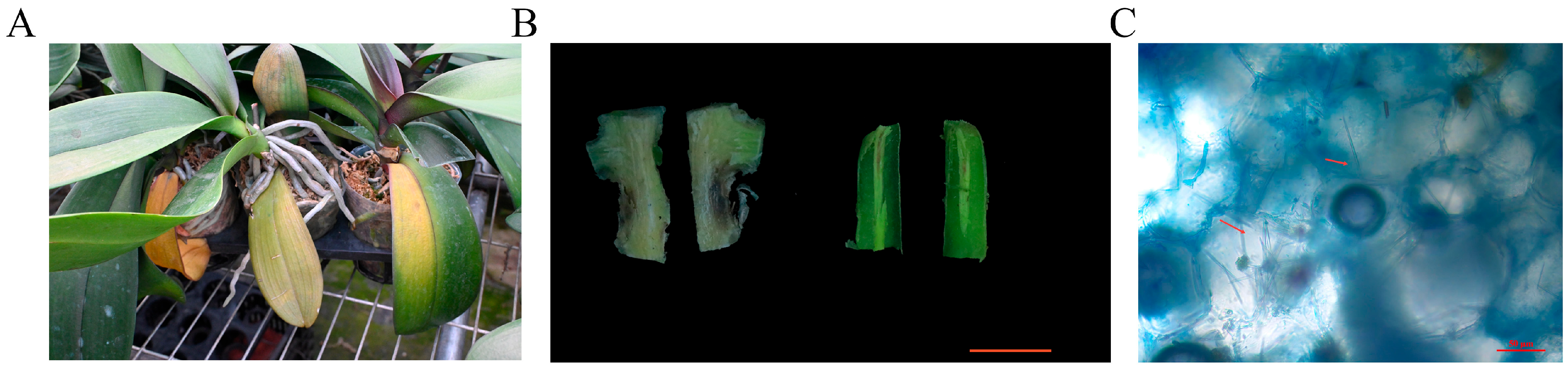
Plants showing symptoms of Phalaenopsis Fusarium wilt were collected from a nursery of Zhangzhou Xinzhenyu Biotechnology Co., Ltd., and the disease progression in the affected plants was observed and recorded. The results showed that, under greenhouse conditions, naturally infected plants mainly exhibited gradual chlorophyll loss, leaf yellowing, and wilting (Figure 1A). As the disease progressed, the yellowed leaves gradually abscised, and the plants showed overall decline, eventually leading to plant death in severe cases. After longitudinal sectioning of symptomatic basal stem and root tissues, obvious internal browning was observed in both tissues (Figure 1B). Further observation using lactophenol cotton blue staining revealed hyphal structures in the infected tissues of symptomatic basal stems (Figure 1C). To identify the causal agent of this disease, symptomatic plant samples were collected, and fungal isolation was performed from the junctions between diseased and healthy tissues. A representative pathogenic fungal isolate, designated HT-1, was obtained.

3.2. Pathogenicity Tests

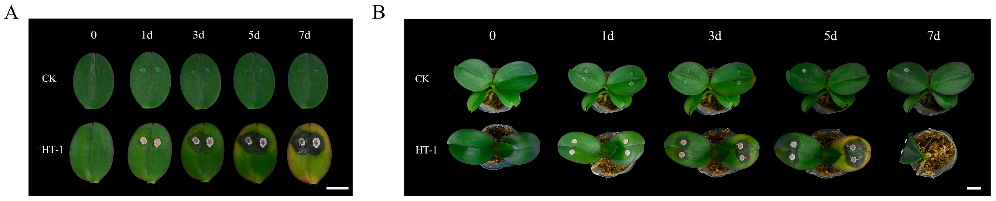
The pathogenicity of the purified isolate HT-1 was evaluated on detached leaves and intact plants of healthy Phalaenopsis. The results showed that isolate HT-1 induced obvious disease symptoms in Phalaenopsis under both inoculation methods. As shown in Figure 2, after inoculation with HT-1, disease symptoms on both detached leaves and intact plants gradually became more severe over time. At the initial stage, water-soaked lesions first appeared at the inoculation sites, and then the lesions gradually expanded and developed into necrotic spots. By 5 days post-inoculation, yellowing symptoms began to appear on both detached leaves and intact plants, followed by further aggravation of leaf yellowing and wilting. By 7 days post-inoculation, the inoculated intact plants showed yellowed leaf abscission, leaf wilting, yellow-brown discoloration of the basal stem, and black necrosis of the roots (Figure 2B). The symptom development was generally consistent with the leaf yellowing, wilting, discoloration of basal stem and root tissues, and plant decline observed in naturally infected plants in the field. In contrast, no obvious disease symptoms were observed in the control treatments.
In addition, fungi with characteristics consistent with those of the original inoculated isolate HT-1 were re-isolated from lesions produced on both detached leaves and intact plants, whereas no similar fungi were obtained from the control tissues. Based on the cultural characteristics and molecular confirmation of the re-isolated strains, they were confirmed to be consistent with the original inoculated isolate HT-1. These results demonstrated that isolate HT-1 caused disease symptoms on both detached leaves and intact Phalaenopsis plants and could be re-isolated from symptomatic tissues, thereby fulfilling Koch’s postulates.

3.3. Morphological Characteristics

The purified isolate HT-1 was cultured on PDA plates for morphological observation. Colonies produced abundant aerial mycelia and were white and cottony at the early stage, with regular margins. As incubation progressed, the colonies gradually turned purple, and purple pigmentation was observed on the reverse side of the colonies (Figure 3A). Microscopic observation showed hyaline, septate hyphae (Figure 3B). Microconidia were abundant, mostly aseptate, and oval to clavate, occurring singly or in small aggregates, and measured 4.53–14.23 × 2.26–5.48 μm (length × width) (Figure 3C). Macroconidia were slender, nearly straight to slightly curved, and septate (Figure 3D). Based on colony morphology and microscopic characteristics, isolate HT-1 was preliminarily identified as a member of the genus Fusarium.

3.4. Molecular Identification of the Pathogen

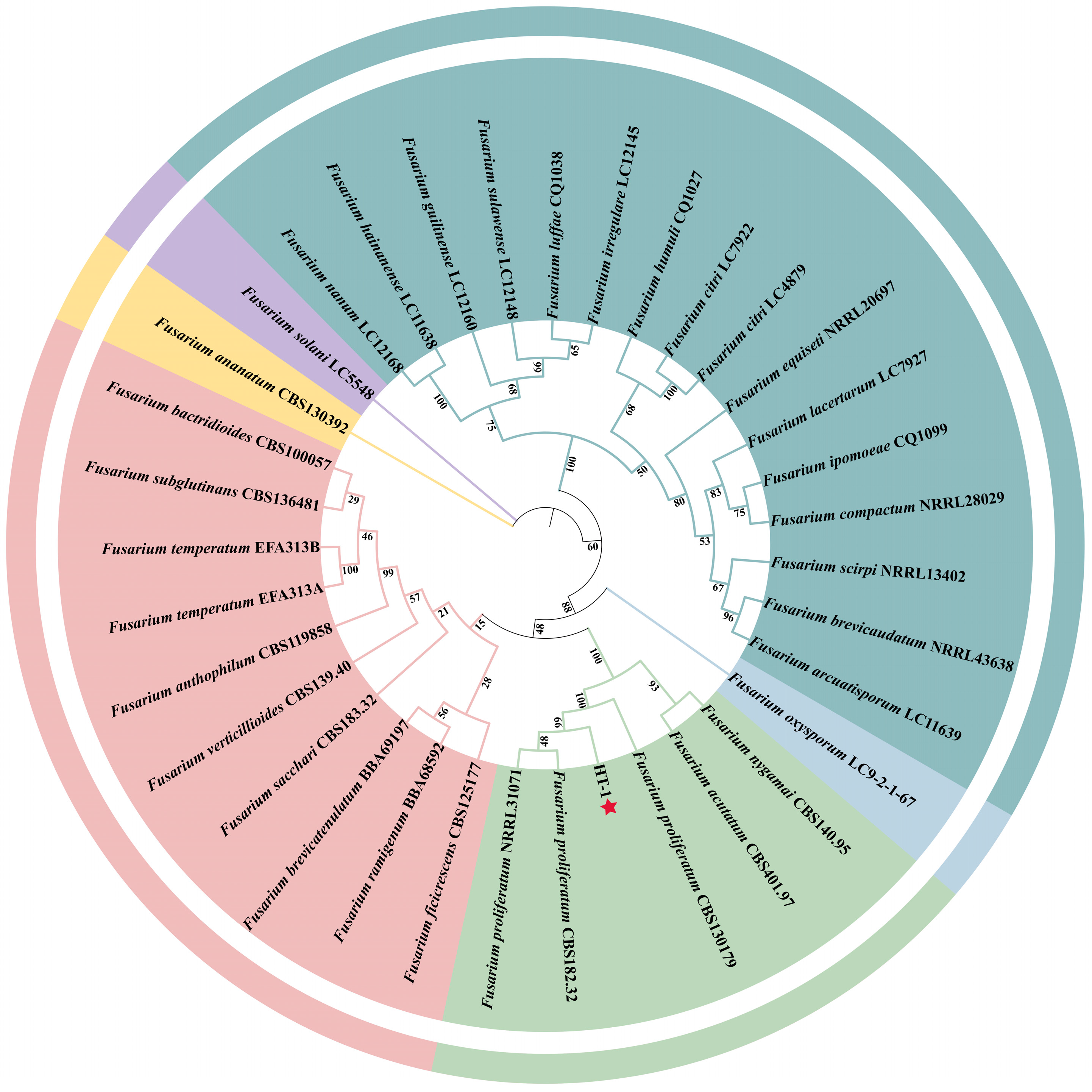
DNA was isolated from the HT-1 strain obtained from infected Phalaenopsis tissues, and PCR was used to amplify the ITS and TEF-1α regions. Agarose gel electrophoresis showed single and clear bands for both loci. The amplified fragments were 565 bp for the ITS region and 722 bp for the TEF-1α gene. The ITS and TEF-1α sequences of isolate HT-1 were generated in this study and analyzed by BLAST against reference sequences available in the GenBank database. The results indicated that both sequences showed the highest similarity (>99%) to F. proliferatum sequences archived in the NCBI database. Based on the ITS and TEF-1α sequences, a phylogenetic tree was inferred in MEGA version 7.0, and detailed information on the reference strains included in the analysis is provided in Table S3. Phylogenetic analysis revealed that isolate HT-1 clustered within the F. proliferatum clade with 100% bootstrap support (Figure 4). Combining morphological characteristics and molecular phylogenetic evidence, the pathogen causing Phalaenopsis Fusarium wilt was identified as F. proliferatum.

3.5. Screening of Biocontrol Fungi

3.5.1. In Vitro Evaluation of Endophytic Fungi for Suppression of the Fusarium Wilt Pathogen

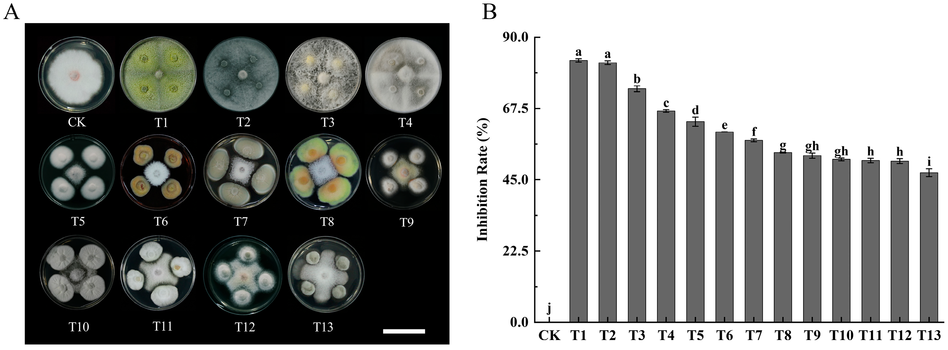
The antagonistic activity of 13 endophytic fungal isolates obtained from healthy Phalaenopsis plants was evaluated against the causal agent of Phalaenopsis Fusarium wilt, F. proliferatum, using a dual-culture assay. All tested endophytic fungi displayed varying degrees of suppression of F. proliferatum mycelial growth (Figure 5). Significant differences in inhibition rates were observed among different treatments (p < 0.05).
Among the tested isolates, T. virens, T. koningiopsis, T. asperellum, and T. hamatum showed the strongest antagonistic activity against F. proliferatum, with inhibition rates of 82.68%, 81.93%, 73.69%, and 66.66%, respectively. These four Trichoderma isolates exhibited rapid mycelial growth and formed a clear competitive advantage during confrontation, effectively overgrowing or markedly restricting the expansion of the pathogen mycelia (Figure 5A). Their inhibitory effects were significantly higher than those of the other endophytic fungi. In addition, T. aculeatus, A. aureus, and P. janthinellum also demonstrated strong antagonistic activity, with inhibition rates exceeding 57%. Clear inhibition zones were observed at the interaction interface between these isolates and F. proliferatum (Figure 5A). The remaining endophytic fungi, including T. pinophilus, T. ruber, P. ochrochloron, C. nigricolor, A. gangligerum, and Acephala sp., exhibited moderate inhibitory effects, with inhibition rates ranging from 47.25% to 53.61%.
Overall, based on colony interaction characteristics and inhibition rate analysis in the dual-culture assay, seven endophytic fungal isolates—T. virens, T. koningiopsis, T. asperellum, T. hamatum, T. aculeatus, A. aureus, and P. janthinellum—were identified as the most effective antagonists against the causal pathogen of Phalaenopsis Fusarium wilt, F. proliferatum, and were selected for further evaluation.

3.5.2. Broad-Spectrum Antagonistic Activity of Selected Endophytic Fungi

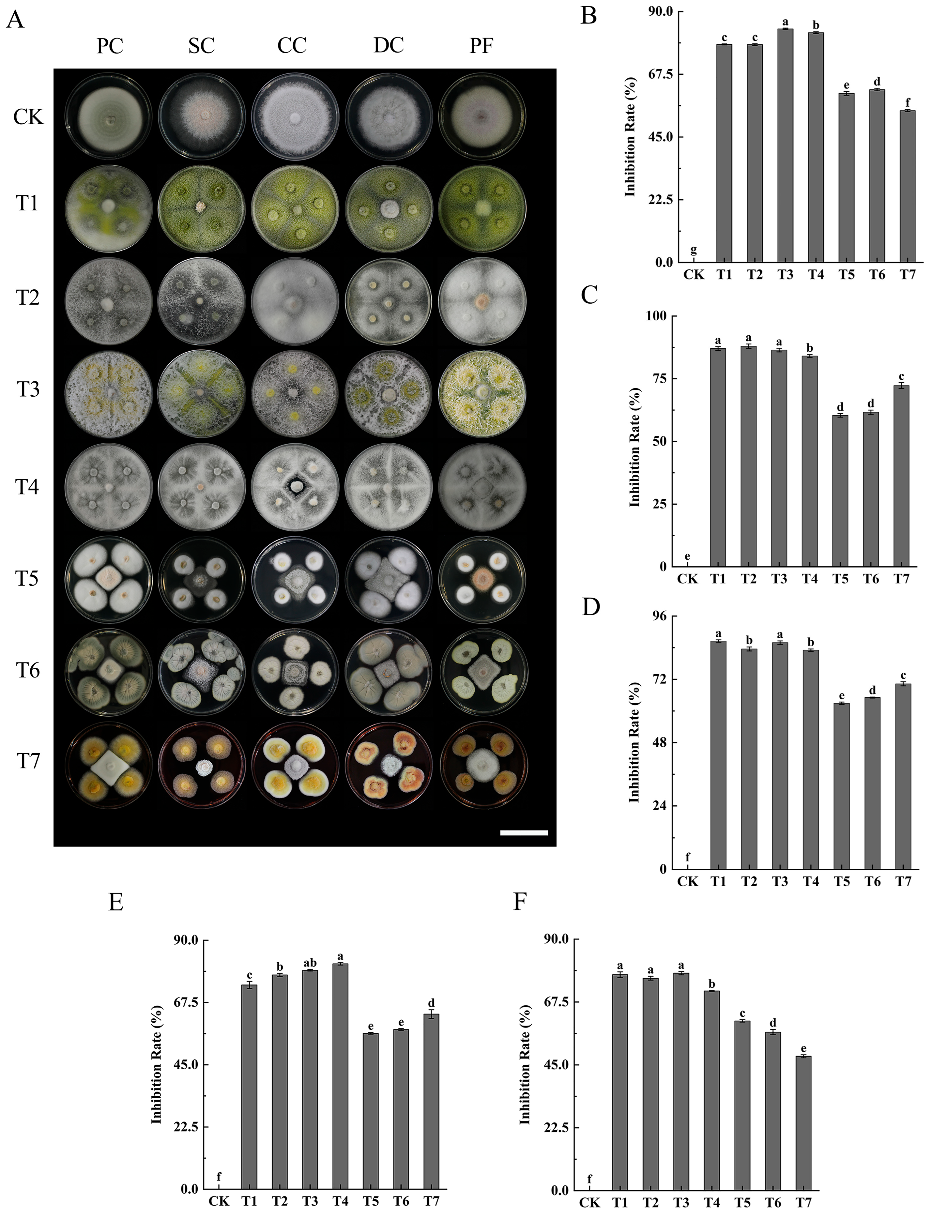
Seven endophytic fungal isolates selected from the dual-culture assays were further evaluated for their antifungal spectrum against five plant pathogenic fungi. The results showed that all tested endophytic fungi exhibited varying degrees of inhibitory activity against the pathogens, with overall inhibition rates ranging from 48.08% to 87.94% (Figure 6B–F), indicating a broad-spectrum antagonistic potential.
Among the tested endophytes, Trichoderma spp. exhibited particularly strong inhibitory effects against different plant pathogens. T. virens, T. koningiopsis, T. asperellum, and T. hamatum showed consistently high inhibition against all five pathogenic fungi, with inhibition rates generally exceeding 71%. In contrast, T. aculeatus, A. aureus, and P. janthinellum displayed relatively weaker antagonistic activity; however, they still exerted consistent inhibitory effects against all tested pathogens, with inhibition rates remaining above 48%.
Overall, all seven endophytic fungi demonstrated broad-spectrum inhibitory activity against plant pathogenic fungi. Among them, Trichoderma spp. showed a stronger capacity for competition for space and nutrients, resulting in higher and more consistent inhibition compared with other treatments. Although the antagonistic effects of Talaromyces spp., A. aureus, and Penicillium spp. were comparatively weaker than those of Trichoderma spp., distinct inhibition zones were consistently observed at the confrontation interface with the pathogens (Figure 6A), suggesting that these endophytes also possess considerable biocontrol potential.

3.5.3. Control Efficacy of Trichoderma Strains on Phalaenopsis Detached Leaves

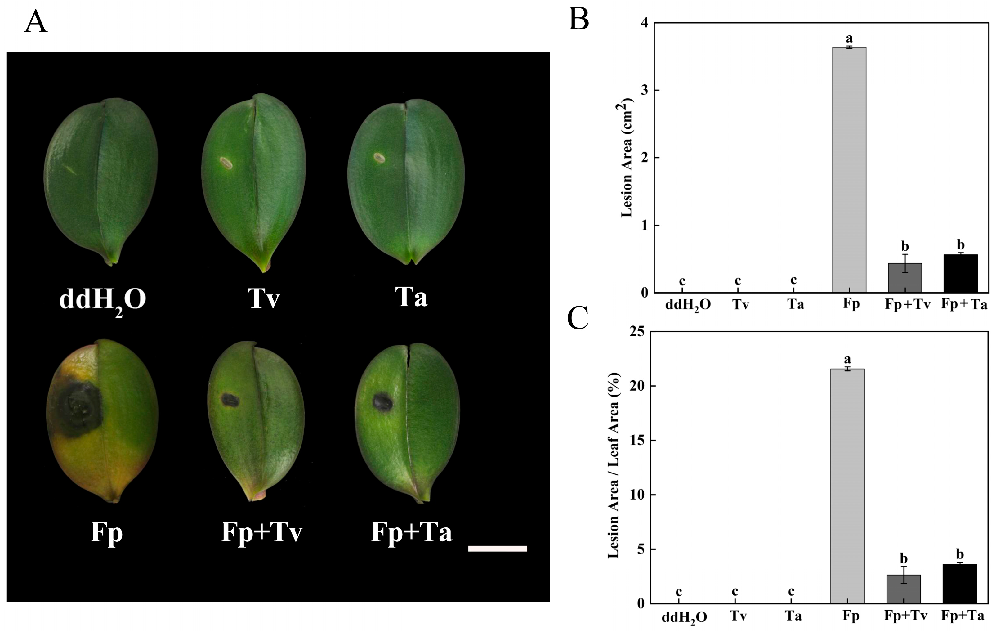
The biocontrol effect of T. virens and T. asperellum was evaluated using detached Phalaenopsis leaves. Disease development was assessed 7 days after inoculation. The results showed that leaves treated with sterile water, T. virens alone, or T. asperellum alone showed no obvious lesions, indicating that the biocontrol fungi themselves were not pathogenic to Phalaenopsis. Compared with the pathogen control inoculated only with F. proliferatum, pretreatment with T. virens or T. asperellum significantly reduced the lesion area and disease severity caused by F. proliferatum on detached Phalaenopsis leaves (Figure 7).
In this study, leaves inoculated only with F. proliferatum showed the most severe disease symptoms, with a lesion area of 3.6367 cm2 and a lesion-to-leaf area ratio of 21.57%. When the biocontrol fungi were pre-inoculated 3 days before pathogen inoculation, the lesion area was significantly reduced. Specifically, leaves pretreated with T. virens developed a lesion area of 0.4363 cm2, with a lesion-to-leaf area ratio of 2.63%, whereas leaves pretreated with T. asperellum developed a lesion area of 0.5660 cm2, with a lesion-to-leaf area ratio of 3.60%. No significant difference was observed between the two Trichoderma treatments, but both were significantly lower than the pathogen control (p < 0.05). These results indicate that T. virens and T. asperellum significantly suppressed lesion expansion caused by F. proliferatum at the detached-leaf level and showed preliminary disease-suppression potential.

4. Discussion

Fusarium wilt has emerged as a major limiting factor in the industrial-scale cultivation of Phalaenopsis, with multiple members of the genus Fusarium being implicated as the etiological pathogens responsible for this disease. To date, the reported pathogens associated with Phalaenopsis Fusarium wilt mainly include F. solani, F. oxysporum, and F. proliferatum [15,35]. A pathogenic fungal isolate, designated HT-1, was obtained from diseased Phalaenopsis tissues. Pathogenicity assays on both detached leaves and intact plants demonstrated that isolate HT-1 consistently induced typical symptoms of Phalaenopsis Fusarium wilt (Figure 2), confirming its stable pathogenicity. Morphological characteristics, together with multilocus sequence analysis of ITS and TEF-1α regions, classified the isolate as F. proliferatum (Figure 4), suggesting that this species represents a key causal agent of Phalaenopsis Fusarium wilt in the study area. In Gyeonggi Province, South Korea, F. solani is the predominant pathogen, frequently accompanied by F. oxysporum and F. proliferatum [14], whereas in Taiwan, F. solani has been reported as the main causal agent [15]. Notably, only F. proliferatum was identified in this study, indicating that host cultivar, cultivation methods, and geographic location may influence the composition of Fusarium pathogens responsible for Phalaenopsis Fusarium wilt. Given the pathogenic importance and wide distribution of F. proliferatum, its potential risks also warrant attention. In addition to its pathogenicity toward plants, F. proliferatum has also been reported to produce various mycotoxins, highlighting the potential risks associated with this species [36].
Endophytic fungi are natural microbial resources residing within plant tissues and represent a potential green strategy for controlling Phalaenopsis Fusarium wilt. To date, a limited number of endophytic fungi from Phalaenopsis are known to possess biocontrol potential against Fusarium wilt, and systematic investigations of their broad-spectrum antagonistic activity remain scarce [31]. In this study, dual-culture assays demonstrated that T. virens, T. asperellum, T. koningiopsis, and T. hamatum consistently inhibited all tested pathogenic fungi more effectively than other treatments, indicating their consistent and broad-spectrum biocontrol potential. Trichoderma species are widely recognized as biocontrol organisms targeting various plant pathogens [37], and their antagonistic mechanisms include competition for space and nutrients, antibiosis, mycoparasitism, and the induction of systemic resistance in host plants [38]. These mechanisms likely account for the strong competitive advantage of Trichoderma observed in this study. Previous research has shown that T. asperellum, T. hamatum, and T. virens can significantly inhibit F. proliferatum, F. solani, and F. oxysporum, with inhibition rates around 70% [39]. Moreover, T. koningiopsis has demonstrated strong antagonistic activity against multiple plant pathogenic fungi, including F. proliferatum [40], which is consistent with the broad-spectrum activity observed in this study. Overall, the strong inhibitory effects of Trichoderma isolates observed in this study are consistent with those reported for effective biocontrol Trichoderma strains in previous studies.
Endophytic fungi coexist with their host plants in complex interactions, and those inhabiting different ecological niches may suppress diverse pathogenic fungi through multiple mechanisms, including the induction of systemic resistance in the host plant [41]. In the present study, three endophytic isolates—T. aculeatus, A. aureus, and P. janthinellum—exhibited strong inhibitory effects against all tested pathogenic fungi, as evidenced by distinct inhibition zones. Unlike Trichoderma species, which primarily antagonize pathogens through rapid colonization and competition for space and nutrients, T. aculeatus, A. aureus, and P. janthinellum are more likely to suppress pathogens via chemical antagonism, highlighting their potential utility in multi-strain biocontrol consortia. For example, Penicillium species have been shown to secrete a range of antifungal enzymes and metabolites with suppressive effects on plant pathogenic fungi [42]. Similarly, organic solvent extracts derived from the fermentation broth of A. aureus effectively restrained the mycelial growth of F. fujikuroi and several other phytopathogens [34]. Although there are currently no reports demonstrating direct inhibition of F. proliferatum by these three isolates, previous studies have indicated that Talaromyces spp., A. aureus, and Penicillium spp. possess potential for controlling soil-borne fungal diseases caused by Fusarium species [43,44,45]. These findings underscore the potential utility of these endophytic fungi as components of integrated biocontrol strategies against Fusarium-related wilt diseases in Phalaenopsis.
Although Trichoderma spp. have been widely applied for the biological control of various plant diseases and have shown promising potential against Fusarium spp., research on their application in controlling Phalaenopsis Fusarium wilt remains relatively limited, particularly regarding F. proliferatum. In this study, a detached-leaf assay was used to evaluate the suppressive effect of T. virens and T. asperellum against F. proliferatum. The results showed that both Trichoderma strains significantly reduced lesion area and disease severity on detached leaves, indicating that they had preliminary disease-suppression potential at the detached-leaf level. Pre-inoculation of Phalaenopsis leaves with T. virens or T. asperellum may allow the biocontrol strains to establish preferential colonization, thereby gaining a competitive advantage in space and nutrient acquisition, restricting pathogen invasion, and delaying disease progression. Consistent with our findings, pre-treatment of Stevia rebaudiana with T. asperellum has been reported to markedly alleviate Fusarium-induced wilt [46]. Moreover, T. virens exhibits strong antagonistic activity against F. proliferatum, and its fermentation products can effectively suppress pathogen growth and reduce its abundance in the rhizosphere, while promoting plant growth [47]. Collectively, these results indicate that Trichoderma fungi represent important candidate resources for the biological control of Phalaenopsis Fusarium wilt. Although T. koningiopsis and T. hamatum were not included in the detached-leaf assay, their marked antagonistic activity in vitro suggests that they also represent promising candidates for future whole-plant biocontrol evaluation.
In summary, this study investigated Phalaenopsis Fusarium wilt from the perspective of endophytic fungi and screened seven endogenous biocontrol fungal isolates that showed significant antagonistic activity against F. proliferatum, the causal agent of Phalaenopsis Fusarium wilt, under in vitro conditions. Among them, T. virens, T. koningiopsis, T. asperellum, and T. hamatum mainly exhibited antagonistic effects through rapid competition, whereas T. aculeatus, A. aureus, and P. janthinellum showed complementary potential in chemical antagonism, providing potential value for the development of combined biocontrol systems. On this basis, pretreatment of detached Phalaenopsis leaves with T. virens or T. asperellum significantly reduced lesion area and disease severity, indicating that these two strains could serve as candidate biocontrol agents for Phalaenopsis Fusarium wilt. However, because the disease-suppression evaluation in this study was mainly based on an endpoint assessment of a short-term detached-leaf assay at 7 days post-inoculation, disease progression analyses at multiple time points, whole-plant experiments, and cultivation-condition trials are still needed to verify the control stability and practical efficacy of these biocontrol strains.
Consistent with the results of this study, F. proliferatum has been reported to infect multiple orchid species, including Cymbidium, Dendrobium and Cattleya [35,48,49], underscoring its broad host range, strong pathogenicity, and the necessity for effective control strategies. Collectively, the endophytic fungal isolates identified in this study provide valuable candidate biocontrol resources and a theoretical basis for the development of sustainable management strategies against Fusarium wilt in Phalaenopsis and other economically and horticulturally important crops, while laying a foundation for future studies on their antagonistic mechanisms and practical applications.

5. Conclusions

In this study, F. proliferatum was identified as one of the main causal agents of Phalaenopsis Fusarium wilt in the study area, based on pathogenicity tests using detached leaves and intact plants, combined with morphological characteristics and multi-gene phylogenetic analysis of ITS and TEF-1α sequences. Seven fungal isolates showing significant inhibitory effects against F. proliferatum were initially screened through dual-culture assays. Further evaluation showed that these seven endophytic fungi also exhibited consistent and broad-spectrum inhibitory activity against five additional plant pathogenic fungi. Detached-leaf assays indicated that T. virens and T. asperellum significantly reduced the lesion area and disease severity caused by F. proliferatum, showing preliminary disease-suppression potential and suggesting that they could serve as candidate biocontrol strains for Phalaenopsis Fusarium wilt. Overall, the findings of this study provide fungal resources for screening candidate biocontrol strains against Phalaenopsis Fusarium wilt and other plant fungal diseases, and lay a foundation for future studies on biocontrol mechanisms, whole-plant efficacy verification, and field application.

Supplementary Materials

The following supporting information can be downloaded at: https://www.mdpi.com/article/10.3390/microorganisms14081628/s1, Table S1. Information on primer sequences used in this study. Table S2. ITS and TEF-1α nucleotide sequences of isolate HT-1. Table S3. Fungal strains and gene information used in the phylogenetic analysis.

Author Contributions

Conceptualization, D.P. and Y.G.; methodology, Y.D. (Yanru Duan) and L.X.; software, Y.D. (Yanru Duan), Q.Z., S.G. and Y.P.; validation, Y.D. (Yanru Duan), Z.W., S.T. and J.X.; formal analysis, Y.D. (Yanru Duan), F.L. and Y.D. (Yan Deng); investigation, Y.D. (Yanru Duan) and L.X.; resources, S.L., X.J. and Y.Z.; data curation, Y.D. (Yanru Duan), and Q.Z.; writing—original draft preparation, Y.D. (Yanru Duan); writing—review and editing, Y.D. (Yanru Duan) and Y.G.; visualization, Y.D. (Yanru Duan); supervision, Y.G.; project administration, D.P.; funding acquisition, D.P. All authors have read and agreed to the published version of the manuscript.

Funding

This research was funded by the National Key Research and Development Program of China (2023YFD230090404), the Fujian Provincial Natural Science Foundation General Project (2025J01597), and the Forestry Science and Technology Research Project of Fujian Province (2025FKJ43).

Institutional Review Board Statement

Not applicable.

Informed Consent Statement

Not applicable.

Data Availability Statement

The original contributions presented in this study are included in the article/Supplementary Materials. Further inquiries can be directed to the corresponding authors.

Acknowledgments

We acknowledge the technical support of the laboratory staff during the laboratory experiments.

Conflicts of Interest

The authors declare no conflicts of interest.

References

  1. Han, C.; Dong, F.; Qi, Y.; Wang, Y.; Zhu, J.; Li, B.; Zhang, L.; Lv, X.; Wang, J. The Breeding, Cultivation, and Potential Applications of Ornamental Orchids with a Focus on Phalaenopsis—A Brief Review. Plants 2025, 14, 1689. [Google Scholar] [CrossRef] [PubMed]
  2. Liang, C.Y.; Rengasamy, K.P.; Huang, L.M.; Hsu, C.C.; Jeng, M.F.; Chen, W.H.; Chen, H.H. Assessment of violet-blue color formation in Phalaenopsis orchids. BMC Plant Biol. 2020, 20, 212. [Google Scholar] [CrossRef] [PubMed]
  3. Hsing, H.-X.; Lin, Y.-J.; Tong, C.-G.; Li, M.-J.; Chen, Y.-J.; Ko, S.-S. Efficient and heritable transformation of Phalaenopsis orchids. Bot. Stud. 2016, 57, 30. [Google Scholar] [CrossRef] [PubMed]
  4. Iiyama, C.M.; Vilcherrez-Atoche, J.A.; Germanà, M.A.; Vendrame, W.A.; Cardoso, J.C. Breeding of ornamental orchids with focus on Phalaenopsis: Current approaches, tools, and challenges for this century. Heredity 2024, 132, 163–178. [Google Scholar] [CrossRef] [PubMed]
  5. Su, J.; Lee, Y.; Chen, C.; Hsieh, T.-F.; Huang, J. Sheath and root rot of Phalaenopsis caused by Fusarium solani. Acta Hortic. 2010, 878, 389–394. [Google Scholar] [CrossRef]
  6. Kim, J.-W.; Chun, S.-C. Root and Basal Stem Rot of Moth Orchid (Phalaenopsis spp.), Pung-nan (Neofinetia falcata) and Nadopung-nan (Aerides japonicum) Caused by Fusarium spp. Res. Plant Dis. 2007, 13, 6–14. [Google Scholar] [CrossRef]
  7. Jin, L.; Huang, R.; Zhang, J.; Li, Z.; Li, R.; Li, Y.; Kong, G.; Xi, P.; Jiang, Z.; Li, M. Identification and Characterization of Endophytic Fungus DJE2023 Isolated from Banana (Musa sp. cv. Dajiao) with Potential for Biocontrol of Banana Fusarium Wilt. J. Fungi 2024, 10, 877. [Google Scholar] [CrossRef] [PubMed]
  8. Cianchetta, A.N.; Davis, R. Fusarium wilt of cotton: Management strategies. Crop Prot. 2015, 73, 40–44. [Google Scholar] [CrossRef]
  9. Balamurugan, A.; Ashajyothi, M.; Shanu, K.; Charishma, K.; Varun, H.; Gunjeet, K.; Kumar, A. Chrysanthemum wilt caused by Fusarium incarnatum: Etiology unveiled through polyphasic taxonomic methods. Physiol. Mol. Plant Pathol. 2024, 129, 102214. [Google Scholar] [CrossRef]
  10. Dean, R.; Van Kan, J.A.; Pretorius, Z.A.; Hammond-Kosack, K.E.; Di Pietro, A.; Spanu, P.D.; Rudd, J.J.; Dickman, M.; Kahmann, R.; Ellis, J.; et al. The Top 10 fungal pathogens in molecular plant pathology. Mol. Plant Pathol. 2012, 13, 414–430. [Google Scholar] [CrossRef] [PubMed]
  11. Han, K.-S.; Park, J.-H.; Back, C.-G.; Park, M.-J. First report of Fusarium subglutinans causing leaf spot disease on Cymbidium orchids in Korea. Mycobiology 2015, 43, 343–346. [Google Scholar] [CrossRef] [PubMed]
  12. Swett, C.; Uchida, J.Y. Characterization of Fusarium diseases on commercially grown orchids in Hawaii. Plant Pathol. 2015, 64, 648–654. [Google Scholar] [CrossRef]
  13. Shih, M.-S.; Chang, K.-C.; Chou, S.-A.; Liu, T.-S.; Ouyang, Y.-C. The automated detection of Fusarium wilt on Phalaenopsis using VIS-NIR and SWIR hyperspectral imaging. Remote Sens. 2023, 15, 4174. [Google Scholar] [CrossRef]
  14. Kim, W.G.; Lee, B.D.; Kim, W.S.; Cho, W.D. Root Rot of Moth Orchid Caused by Fusarium spp. Plant Pathol. J. 2002, 18, 225–227. [Google Scholar] [CrossRef]
  15. Chung, W.; Chen, L.; Huang, J.; Huang, H.; Chung, W. A new ‘forma specialis’ of Fusarium solani causing leaf yellowing of Phalaenopsis. Plant Pathol. 2011, 60, 244–252. [Google Scholar] [CrossRef]
  16. Gullino, M.L.; Minuto, A.; Gilardi, G.; Garibaldi, A. Efficacy of azoxystrobin and other strobilurins against Fusarium wilts of carnation, cyclamen and Paris daisy. Crop Prot. 2002, 21, 57–61. [Google Scholar] [CrossRef]
  17. Chen, S.-Y.; Wu, Y.-J.; Hsieh, T.-F.; Su, J.-F.; Shen, W.-C.; Lai, Y.-H.; Lai, P.-C.; Chen, W.-H.; Chen, H.-H. Develop an efficient inoculation technique for Fusarium solani isolate “TJP-2178-10” pathogeny assessment in Phalaenopsis orchids. Bot. Stud. 2021, 62, 4. [Google Scholar] [CrossRef] [PubMed]
  18. Yeh, Y.-C.; Lin, P.-C.; Yang, T.-C.; Su, J.-F.; Chang, Y.-C.A. ‘Hualien No. 1-Pink Apple’: A Leaf-yellowing Disease–resistant Phalaenopsis Cultivar. HortScience 2025, 60, 738–740. [Google Scholar] [CrossRef]
  19. Reis, A.; Costa, H.; Boiteux, L.S.; Lopes, C.A. First report of Fusarium oxysporum f. sp. lycopersici race 3 on tomato in Brazil. Fitopatol. Bras. 2005, 30, 426–428. [Google Scholar] [CrossRef]
  20. Rani, L.; Thapa, K.; Kanojia, N.; Sharma, N.; Singh, S.; Grewal, A.S.; Srivastav, A.L.; Kaushal, J. An extensive review on the consequences of chemical pesticides on human health and environment. J. Clean. Prod. 2021, 283, 124657. [Google Scholar] [CrossRef]
  21. Pandit, M.A.; Kumar, J.; Gulati, S.; Bhandari, N.; Mehta, P.; Katyal, R.; Rawat, C.D.; Mishra, V.; Kaur, J. Major biological control strategies for plant pathogens. Pathogens 2022, 11, 273. [Google Scholar] [CrossRef] [PubMed]
  22. Baron, N.C.; Rigobelo, E.C. Endophytic fungi: A tool for plant growth promotion and sustainable agriculture. Mycology 2022, 13, 39–55. [Google Scholar] [CrossRef] [PubMed]
  23. Etesami, H. Bacterial mediated alleviation of heavy metal stress and decreased accumulation of metals in plant tissues: Mechanisms and future prospects. Ecotoxicol. Environ. Saf. 2018, 147, 175–191. [Google Scholar] [CrossRef] [PubMed]
  24. Santoyo, G.; Moreno-Hagelsieb, G.; del Carmen Orozco-Mosqueda, M.; Glick, B.R. Plant growth-promoting bacterial endophytes. Microbiol. Res. 2016, 183, 92–99. [Google Scholar] [CrossRef] [PubMed]
  25. Abro, M.A.; Sun, X.; Li, X.; Jatoi, G.H.; Guo, L.-D. Biocontrol potential of fungal endophytes against Fusarium oxysporum f. sp. cucumerinum causing wilt in cucumber. Plant Pathol. J. 2019, 35, 598. [Google Scholar] [CrossRef] [PubMed]
  26. Sun, B.; Wang, X.; Lv, Y.; Da, X.; Zhou, M.; Kang, J.; He, H.; Qin, C.; Wang, X.; Yang, L.; et al. Screening of biocontrol agents against lily bulb rot caused by F. oxysporum and research on their fermentation process. Bioresour. Technol. Rep. 2025, 31, 102267. [Google Scholar] [CrossRef]
  27. Luo, M.; Chen, Y.; Huang, Q.; Huang, Z.; Song, H.; Dong, Z. Trichoderma koningiopsis Tk905: An efficient biocontrol, induced resistance agent against banana Fusarium wilt disease and a potential plant-growth-promoting fungus. Front. Microbiol. 2023, 14, 1301062. [Google Scholar] [CrossRef] [PubMed]
  28. Alghamdi, S.A. Influence of mycorrhizal fungi on seed germination and growth in terrestrial and epiphytic orchids. Saudi J. Biol. Sci. 2019, 26, 495–502. [Google Scholar] [CrossRef] [PubMed]
  29. Li, T.; Wu, S.; Yang, W.; Selosse, M.-A.; Gao, J. How mycorrhizal associations influence orchid distribution and population dynamics. Front. Plant Sci. 2021, 12, 647114. [Google Scholar] [CrossRef] [PubMed]
  30. Tsao, W.-C.; Li, Y.-H.; Tu, Y.-H.; Nai, Y.-S.; Lin, T.-C.; Wang, C.-L. Identification and molecular detection of the pathogen of Phalaenopsis leaf yellowing through genome analysis. Front. Microbiol. 2024, 15, 1431813. [Google Scholar] [CrossRef] [PubMed]
  31. Setiani, Y.; Kasiamdari, R.S. In Vitro Antagonism Test between Fusarium solani Caused Fusarium Wilt on Orchid Phalaenopsis Taida Salu by Using Endophytic Fungi. In Proceedings of the International Conference on Bioscience and Biotechnology 2011, Yogyakarta, Indonesia, 11–12 October 2011. [Google Scholar]
  32. Xu, X.; Zhang, L.; Yang, X.; Cao, H.; Li, J.; Cao, P.; Guo, L.; Wang, X.; Zhao, J.; Xiang, W. Alternaria spp. associated with leaf blight of maize in Heilongjiang Province, China. Plant Dis. 2022, 106, 572–584. [Google Scholar] [CrossRef] [PubMed]
  33. Erima, S.; Nyine, M.; Edema, R.; Nkuboye, A.; Habiba, N.; Candiru, A.; Paparu, P. Molecular Characterisation of Fusarium Species Causing Common Bean Root Rot in Uganda. J. Fungi 2025, 11, 283. [Google Scholar] [CrossRef] [PubMed]
  34. Xu, R.; Song, S.; Wu, G.; Hu, B.; Xie, J.; Wang, X.; Xia, Y.; Zheng, S.; Peng, S.; Yuan, Y.; et al. Biocontrol ability of Arcopilus aureus YZXR against Fusarium fujikuroi causing leaf spot on Polygonatum odoratum. Phytopathol. Res. 2024, 6, 68. [Google Scholar] [CrossRef]
  35. Wang, C.J.; Chen, Y.J.; Jain, Y.C.; Chung, W.C.; Wang, C.L.; Chung, W.H. Identification of Fusarium proliferatum causing leaf spots on Cymbidium orchids in Taiwan. J. Phytopathol. 2018, 166, 675–685. [Google Scholar] [CrossRef]
  36. Waskiewicz, A.; Golinski, P.; Karolewski, Z.; Irzykowska, L.; Bocianowski, J.; Kostecki, M.; Weber, Z. Formation of fumonisins and other secondary metabolites by Fusarium oxysporum and F. proliferatum: A comparative study. Food Addit. Contam. Part A 2010, 27, 608–615. [Google Scholar] [CrossRef] [PubMed]
  37. Mukherjee, P.K.; Mendoza-Mendoza, A.; Zeilinger, S.; Horwitz, B.A. Mycoparasitism as a mechanism of Trichoderma-mediated suppression of plant diseases. Fungal Biol. Rev. 2022, 39, 15–33. [Google Scholar] [CrossRef]
  38. Sharma, V.; Salwan, R.; Sharma, P. The comparative mechanistic aspects of Trichoderma and probiotics: Scope for future research. Physiol. Mol. Plant Pathol. 2017, 100, 84–96. [Google Scholar] [CrossRef]
  39. Liu, R.; Chen, M.; Gao, J.; Luo, M.; Wang, G. Identification of antagonistic fungi and their antifungal activities against aconite root rot pathogens. Plant Signal. Behav. 2023, 18, 2211852. [Google Scholar] [CrossRef] [PubMed]
  40. Azuddin, N.F.; Mohd, M.H.; Rosely, N.F.N.; Mansor, A.; Zakaria, L. Molecular phylogeny of endophytic fungi from rattan (Calamus castaneus Griff.) spines and their antagonistic activities against plant pathogenic fungi. J. Fungi 2021, 7, 301. [Google Scholar] [CrossRef] [PubMed]
  41. Strobel, G.; Daisy, B. Bioprospecting for microbial endophytes and their natural products. Microbiol. Mol. Biol. Rev. 2003, 67, 491–502. [Google Scholar] [CrossRef] [PubMed]
  42. Win, T.; Bo, B.; Malec, P.; Fu, P. The effect of a consortium of Penicillium sp. and Bacillus spp. in suppressing banana fungal diseases caused by Fusarium sp. and Alternaria sp. J. Appl. Microbiol. 2021, 131, 1890–1908. [Google Scholar] [CrossRef] [PubMed]
  43. Waing, K.; Abella, E.; Kalaw, S.; Waing, F.; Galvez, C. Antagonistic interactions among different species of leaf litter fungi of Central Luzon State University. Plant Pathol. Quar. 2015, 5, 122–130. [Google Scholar] [CrossRef]
  44. Yuan, X.; Wang, B.; Hong, S.; Xiong, W.; Shen, Z.; Ruan, Y.; Li, R.; Shen, Q.; Dini-Andreote, F. Promoting soil microbial-mediated suppressiveness against Fusarium wilt disease by the enrichment of specific fungal taxa via crop rotation. Biol. Fertil. Soils 2021, 57, 1137–1153. [Google Scholar] [CrossRef]
  45. Sun, D.; Li, F.; Wang, L.; Chen, R.; Liu, F.; Guo, L.; Li, N.; Zhang, F.; Lei, L. Identification and application of an endophytic fungus Arcopilus aureus from Panax notoginseng against crop fungal disease. Front. Plant Sci. 2024, 15, 1305376. [Google Scholar] [CrossRef] [PubMed]
  46. Carol, D.-G.; Catalina, A.; Mercè, L.; Charlotte, P.; Soledad, M.; Carlos, P. Trichoderma asperellum as a preventive and curative agent to control Fusarium wilt in Stevia rebaudiana. Biol. Control 2021, 155, 104537. [Google Scholar] [CrossRef]
  47. Wang, H.; Tang, W.; Mao, Y.; Ma, S.; Chen, X.; Shen, X.; Yin, C.; Mao, Z. Isolation of Trichoderma virens 6PS-2 and its effects on Fusarium proliferatum f. sp. Malus domestica MR5 related to apple replant disease in China. Hortic. Plant J. 2024, 10, 1291–1308. [Google Scholar] [CrossRef]
  48. Zakaria, L.; Zain, N.H.M.; Salleh, B.; Zakaria, M. Morphological and RAPD analysis of Fusarium species associated with root and stem rot of Dendrobium orchid in Northern Peninsula Malaysia. HAYATI J. Biosci. 2009, 16, 64–68. [Google Scholar] [CrossRef]
  49. Tsavkelova, E.; Kolomeitseva, G. Fusarium–orchid interactions under greenhouse conditions. S. Afr. J. Bot. 2022, 146, 889–896. [Google Scholar] [CrossRef]
Figure 1. Field symptoms, internal tissue browning, and fungal-like hyphal structures in symptomatic Phalaenopsis plants. (A) Naturally infected Phalaenopsis plant under greenhouse conditions; (B) Longitudinal sections of symptomatic basal stem and root tissues showing internal browning. Scale bar = 1 cm; (C) Lactophenol cotton blue-stained microscopic image of internal tissues from symptomatic basal stems. Arrows indicate hyphal structures. Scale bar = 50 μm.
Microorganisms 14 01628 g001
Figure 2. Pathogenicity assay of isolate HT-1 on detached leaves and intact plants of Phalaenopsis. (A) Symptoms on detached leaves inoculated with isolate HT-1. Scale bar = 2 cm. (B) Symptoms on intact Phalaenopsis plants inoculated with isolate HT-1. Scale bar = 2 cm.
Microorganisms 14 01628 g002
Figure 3. Morphological characteristics of isolate HT-1. (A) Colony morphology on PDA, scale bar = 2 cm. (B) Hyphal morphology observed under a light microscope, scale bar = 50 µm. (C) Microconidial morphology observed under a light microscope, scale bar = 25 µm. (D) Macroconidial morphology observed under a light microscope, scale bar = 25 µm.
Microorganisms 14 01628 g003
Figure 4. Phylogenetic tree inferred by the NJ method from the concatenated ITS and TEF-1α sequences. The star (★) indicates isolate HT-1.
Microorganisms 14 01628 g004
Figure 5. Antagonistic effects of 13 endophytic fungi isolated from Phalaenopsis against Fusarium proliferatum. (A) Dual-culture assays showing the inhibitory effects of 13 endophytic fungi on the mycelial growth of F. proliferatum; CK: F. proliferatum inoculated alone; T1: T. virens; T2: T. koningiopsis; T3: T. asperellum; T4: T. hamatum; T5: T. aculeatus; T6: A. aureus; T7: P. janthinellum; T8: T. pinophilus; T9: T. ruber; T10: P. ochrochloron; T11: C. nigricolor; T12: A. gangligerum; T13: Acephala sp.; scale bar = 5 cm. (B) Inhibition rates of F. proliferatum by the 13 endophytic fungal isolates. Values are presented as mean ± standard error (n = 3). Different lowercase letters indicate significant differences among treatments according to Duncan’s multiple range test (p < 0.05). Other abbreviations are the same as in panel (A). Note: The control (CK) was inoculated with F. proliferatum alone, and the inhibition rate was set to 0.
Microorganisms 14 01628 g005
Figure 6. Inhibitory effects of strains T1–T7 on five plant pathogenic fungi on PDA plates. (A) Dual-culture assay showing the inhibitory effects of strains T1–T7 on five plant pathogenic fungi; PC: P. edulis C. truncatum; SC: S. lycopersicum C. fulvum; CC: C. ensifolium C. gloeosporioides; DC: D. officinale C. gloeosporioides; PF: P. F. solani; T1: T. virens; T2: T. koningiopsis; T3: T. asperellum; T4: T. hamatum; T5: T. aculeatus; T6: P. janthinellum; T7: A. aureus; scale bar = 5 cm. (BF) Inhibition rates of strains T1–T7 against PC, SC, CC, DC, and PF, respectively. CK: pathogen inoculated alone; T1–T7: corresponding to the strains shown in panel (A). Values represent the mean ± standard error (n = 3). Different letters indicate significant differences among treatments within each pathogen (p < 0.05, Duncan’s test). Note: The control group was inoculated with the pathogen and not inoculated with endophytic fungi, and the inhibition rate was 0%.
Microorganisms 14 01628 g006
Figure 7. Biocontrol efficacy of T. virens and T. asperellum on detached Phalaenopsis leaves. (A) Disease symptoms on detached leaves under different treatments, scale bar = 2 cm; (B) Lesion area under different treatments; (C) Lesion area as a proportion of leaf area under different treatments. Note: ddH2O: sterile distilled water; Tv: inoculation with T. virens alone; Ta: inoculation with T. asperellum alone; Fp: inoculation with F. proliferatum alone; Fp + Tv: inoculation with T. virens 3 d prior to F. proliferatum inoculation; Fp + Ta: inoculation with T. asperellum 3 d prior to F. proliferatum inoculation. No visible lesions were observed in ddH2O, Tv, and Ta treatments. Values represent the mean ± standard error (n = 3). Different letters indicate significant differences among treatments (p < 0.05, Duncan’s test).
Microorganisms 14 01628 g007
Disclaimer/Publisher’s Note: The statements, opinions and data contained in all publications are solely those of the individual author(s) and contributor(s) and not of MDPI and/or the editor(s). MDPI and/or the editor(s) disclaim responsibility for any injury to people or property resulting from any ideas, methods, instructions or products referred to in the content.