Article

Toxoplasma gondii in Egyptian Blood Donors and Livestock: Seroprevalence, Risk Factors, and One Health Implications

by
Marwa A. Gouda
1,
Sahar M. Selim
1,
Eman Fathi Fadel
2,
Eman Attia Elmorsy
3,
Aya Abdallah Seleem
4,
Hany M. Ibrahim
5,
Reda Abdel Latif Ibrahem
6,
Yomna Mohamed Hegazy
1,
Kholoud Adel Alsawy
6,
Mostafa Adel Ahmed Younis
7,
Khaled A. Abd El-Razik
8,
Ehab Ali Fouad
9,
Eman L. Shehata
10,
Tameem M. A. Mohammed
10,
Shrouq Moustafa Tolba
11,
Ann Hegazy
11,
Mona Wagdy Ayad
12 and
Shaimaa A. Farag
1,*
1
Clinical and Molecular Parasitology Department, National Liver Institute, Menoufia University, Shebin El-Kom 32511, Egypt
2
Medical Parasitology Department, Faculty of Medicine, Sohag University, Sohag 82524, Egypt
3
Medical Parasitology Department, Faculty of Medicine, Alexandria University, Alexandria 21526, Egypt
4
Zoonoses Department, Faculty of Veterinary Medicine, Cairo University, Giza 12613, Egypt
5
Zoology Department, Faculty of Science, Menoufia University, Shebin El-Kom 32511, Egypt
6
Public Health and Community Medicine Department, Faculty of Medicine, Menoufia University, Shebin El-Kom 32511, Egypt
7
Clinical Pathology Department, Faculty of Medicine, Sohag University, Sohag 82524, Egypt
8
Department of Animal Reproduction, Veterinary Research Institute, National Research Centre, Dokki 12622, Egypt
9
Department of Zoonosis, Veterinary Research Institute, National Research Centre, Dokki 12622, Egypt
10
Clinical Pathology Department, National Liver Institute, Menoufia University, Shebin El-Kom 32511, Egypt
11
Department of Clinical Pathology, Faculty of Medicine, Suez Canal University, Ismailia 41522, Egypt
12
Clinical Pathology Department, Faculty of Medicine, Alexandria University, Alexandria 21526, Egypt
*
Author to whom correspondence should be addressed.
Microorganisms 2026, 14(8), 1627; https://doi.org/10.3390/microorganisms14081627
Submission received: 25 June 2026 / Revised: 17 July 2026 / Accepted: 24 July 2026 / Published: 26 July 2026
(This article belongs to the Section Medical Microbiology)

Abstract

Toxoplasma gondii is a globally prevalent zoonotic parasite with major public health and veterinary impacts. In Egypt, One Health data on its distribution among blood donors and livestock are limited. This multicentric cross-sectional study aimed to determine the seroprevalence of Toxoplasma gondii among asymptomatic blood donors and livestock across six different Egyptian governorates and to identify the associated risk factors. Serum samples of 2000 asymptomatic blood donors and 500 animals (cattle and buffaloes) were tested for anti-Toxoplasma gondii IgG and IgM antibodies using ELISA. Sociodemographic and behavioral data were collected using a structured questionnaire. Overall IgG seroprevalence in blood donors was 41.8%, with IgM seropositivity at 2.1%. Significant regional variation was observed (p < 0.001), ranging from 66.5% in Alexandria to 16% in Cairo. The most significant risk factors were consumption of undercooked meat, contact with cats, and engagement in agricultural activities (p < 0.001). Among livestock, IgG and IgM seroprevalence were 7.2% and 5.2%, respectively, indicating active parasite circulation. The presence of seropositivity among apparently healthy donors raises concerns regarding transfusion safety. These findings highlight substantial and regionally heterogeneous Toxoplasma gondii exposure in Egypt. A One Health approach integrating public health education, food safety measures, and targeted screening of high-risk populations and animals is essential to reduce disease burden.

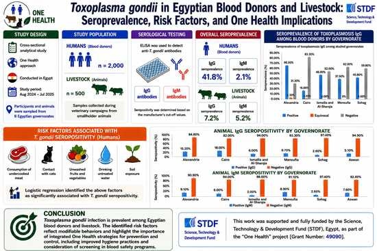
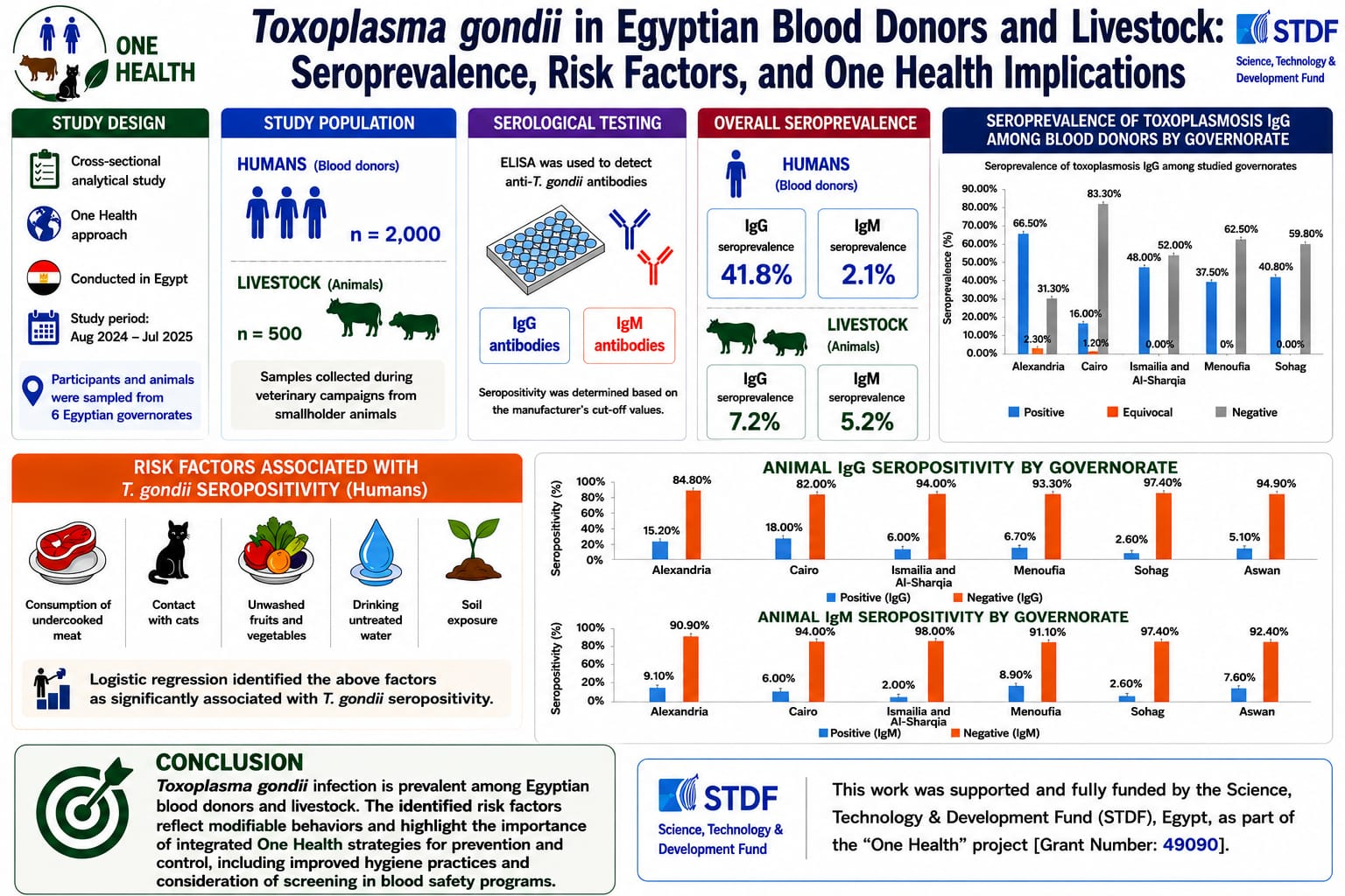
Graphical Abstract

1. Introduction

The One Health framework recognizes the intrinsic interconnection between human, animal, and environmental health and requires collaborative action to manage complex zoonotic risks. This framework is actively supported by international organizations, such as the World Health Organization (WHO), the Food and Agriculture Organization (FAO), and the World Organization for Animal Health (OIE), which collaborate to implement global health initiatives. The success of this approach in controlling several zoonotic epidemics highlights its relevance to diseases with multi-host transmission dynamics. Toxoplasma gondii (T. gondii), an obligate intracellular protozoan, causes toxoplasmosis, a zoonotic disease found worldwide across terrestrial and marine environments, making it an ideal model for the application of the One Health approach [1].
The host range of T. gondii is exceptionally broad, encompassing nearly all warm-blooded animals, including humans, livestock, and avian species. It remains a major public health and veterinary concern because of its association with congenital infections and detrimental effects on animal productivity [2]. It is estimated that over one billion people worldwide are infected with T. gondii. Felids, particularly domestic cats, serve as definitive hosts responsible for environmental contamination, whereas humans and other animals function as intermediate hosts, sustaining transmission cycles [3]. In Egypt, domestic and stray cats are ubiquitous in both urban and rural settings, contributing to widespread environmental contamination with T. gondii oocysts and facilitating transmission to humans and livestock [4,5].
Human toxoplasmosis is mainly acquired through the oral intake of either tissue cysts in inadequately cooked meat or oocysts contaminating food, water, and soil. Ingestion of contaminated milk is a less common but notable route of infection. Less commonly, transmission may occur via organ transplantation or blood transfusion from infected donors [6]. Globally, seroprevalence estimates vary widely, ranging from 10% to 80%, with marked variations across continents [7]. A high prevalence of toxoplasmosis has been reported in Africa, Southeast Asia, the Middle East, Central/Eastern Europe, and Latin America. Variable prevalence rates have been reported in Asia (13.3−85.3%), Europe (40−76%), Africa (21.74−74.8%), and North America (7.3−26.5%) [8,9], reflecting differences in environmental conditions, dietary habits, and socio-cultural practices [7]. Although infection is typically asymptomatic in immunocompetent individuals, it can lead to severe and potentially fatal outcomes in immunocompromised patients and developing fetuses [10].
Blood transfusion is an essential medical intervention, with approximately 92 million donations collected annually worldwide; however, approximately 1.6 million units are discarded due to infectious risks [11]. Current screening protocols primarily target viral and bacterial pathogens, including the human immunodeficiency virus (HIV), hepatitis B and C viruses, and Treponema pallidum. However, increasing evidence underscores the need to consider T. gondii as a potential transfusion-transmitted infection (TTI) [12]. Notably, tachyzoites can survive in refrigerated blood for prolonged periods, raising concerns about transmission through stored blood products, particularly those rich in leukocytes [13]. Given the high prevalence of asymptomatic infections, the absence of routine screening for toxoplasmosis may represent an underrecognized risk factor for transfusion safety [14,15].
From a veterinary and economic perspective, toxoplasmosis is a major cause of reproductive failure in livestock, resulting in substantial financial loss. Ruminants, in particular, play a key role in human infection through the consumption of undercooked meat or contaminated milk [5,16]. Globally, the disease is responsible for abortions, stillbirths, and neonatal mortality, with estimated annual losses in the sheep and goat industries reaching millions of dollars [17,18]. In cattle and buffaloes, although clinical disease is less frequent, subclinical infection can still affect productivity and serve as a potential source of human exposure via the same foodborne routes [19].
Despite numerous studies in Egypt addressing T. gondii seroprevalence and associated risk factors, the existing evidence remains fragmented, with marked regional variability and limited integration of human and animal data [2]. Furthermore, studies specifically targeting blood donors, a critical population for transfusion safety, are scarce [20,21,22].
Previous studies investigating the seroprevalence of T. gondii in Egypt have primarily focused on single governorates or single-center designs, limiting their ability to capture regional variability and broader transmission patterns in Egypt. Moreover, these studies rarely incorporated an integrated One Health approach that considers both human and animal populations. To address these gaps, the present study adopted a multicentric design encompassing multiple Egyptian governorates and included both human blood donors and livestock. This comprehensive approach provides a broader epidemiological perspective by enabling the assessment of regional heterogeneity and integrated human–livestock transmission patterns within a One Health framework. While individual studies have investigated T. gondii in specific livestock species or single regions, to the best of our knowledge, no previous Egyptian study has simultaneously investigated both human and animal populations across multiple governorates using a unified One Health protocol. The current study was designed to provide a direct comparison of seroprevalence and risk factors across six geographically distinct regions, with the aim of generating robust evidence for region-specific control strategies and addressing the fragmentation that has characterized Egyptian toxoplasmosis research to date.

2. Materials and Methods

2.1. Ethical Considerations

This study was approved by the Institutional Review Board of the National Liver Institute, Menoufia University (NLI IRB No. 00651/2024), and the Medical Research Ethics Committee of the Faculty of Medicine, Sohag University (IRB No. SohMed24806PD). Written informed consent was obtained from all human participants after explanation of study objectives. Confidentiality was maintained through anonymized coded identifiers. Animal procedures complied with Egyptian regulations governing research and publication ethics.

2.2. Study Design and Setting

This multi-centric cross-sectional analytical study was conducted between August 2024 and July 2025 across six Egyptian governorates representing major geographic regions: Menoufia (Delta region, north of Cairo), Cairo (Greater Cairo, the capital), Sohag (Upper Egypt, southern Nile Valley), Alexandria (North Coast, Mediterranean Sea), Al-Sharqia, and Ismailia (Canal region, adjacent to the Suez Canal). Five centers were selected to cover these governorates.

2.3. Study Population and Sampling

A total of 2000 asymptomatic human blood donors and 500 animals (cattle and buffaloes) were included in the study. From each center, 400 human and 100 animal serum samples were collected. Human participants were recruited from blood donation centers using predefined inclusion and exclusion criteria for the study. The inclusion criteria were age ≥ 18 years, provision of informed consent, and negative screening for hepatitis B surface antigen (HBsAg), hepatitis C virus (HCV), and HIV. The exclusion criteria were age < 18 years and a history of chronic disease. Animal samples were obtained from cows and buffaloes owned by smallholders during the veterinary campaigns.
A multistage random sampling technique was employed: governorates were randomly selected to represent geographic regions; within each governorate, the main blood donation center was chosen by simple random sampling; and individual participants were recruited according to eligibility criteria. Animal samples were collected concurrently from the same governorates to ensure a balanced distribution.

2.4. Sample Size Calculation

For humans, the sample size was estimated based on a previously reported seroprevalence of 59.6% among Egyptian blood donors [20]. Assuming a 95% confidence interval and a 5% acceptable error margin, we calculated the minimum sample size as 400 individuals per center based on the standard formula:
n = Z2 × p (1 − p)/e2
In this equation, n denotes the required sample size, Z represents the standard normal deviate, p stands for the anticipated prevalence proportion, and e indicates the tolerable sampling error. To enhance geographic representativeness, the sample size was expanded across five centers to reach a total of 2000 participants.
For animals, the sample size was based on reported seroprevalence rates of 9.7% [23] and 7.4% [24]. Using the same formula, the minimum required sample size was approximately 470. To ensure statistical power and account for potential exclusions, 500 samples were collected from five centers.

2.5. Sample Collection and Processing

Approximately 3 mL of venous blood was aseptically collected from each participant. For the animals, 10 mL of blood was obtained via jugular venipuncture. Animal samples were collected from cows and buffaloes owned by smallholders during veterinary campaigns, using a convenience-based approach.
Blood samples were centrifuged at 3000 rpm for 10 min, and the sera were separated and stored at −20 °C until analysis. Available data on animal characteristics were recorded.

2.6. Laboratory/Serological Analysis

All serum samples were processed at the Parasitology Laboratory, Clinical and Molecular Parasitology Department, National Liver Institute, Menoufia University.
Human serum samples were screened for specific IgG and IgM antibodies against T. gondii using commercially available ELISA kits (PerkinElmer Health Sciences, Shelton, CT, USA; catalog numbers 10234 for IgG and 10235 for IgM). All procedures strictly adhered to the manufacturer’s protocols.
Animal sera were tested using ELISA kits (BT LAB, Shanghai, China) for the qualitative detection of anti-T. gondii IgG (Cat. No. ED0056Bo) and IgM (Cat. No. ED0063Bo), according to the manufacturer’s protocols.
All assays were performed in duplicate, and positive and negative controls were included in each run to ensure the reliability and reproducibility of the assay.
Molecular detection of T. gondii by conventional PCR was performed on a subset of samples as part of a parallel investigation; however, these data fall outside the scope of the present study and will be presented separately.

2.7. Data Collection

A structured questionnaire was administered to human participants to collect sociodemographic data (age, sex, residence, education, income, marital status, occupation, and crowding index). The crowding index was defined as the ratio of household members to the number of rooms, excluding kitchens and bathrooms. Following the Egyptian Demographic and Health Survey and validated socioeconomic scales for Egypt, households were classified as follows: no crowding (<1 person per room), crowding (=1 person per room), and severe crowding (>1 person per room) [25,26]. The potential risk factors assessed included dietary habits (undercooked meat and unwashed fruits/vegetables), cat contact, agricultural activities, soil exposure, hand hygiene, and water source. For animals, data on species, sex, and season were also recorded.
Participants were also provided with an informational brochure to raise awareness of toxoplasmosis, covering its transmission, clinical features, diagnosis, and prevention.

2.8. Statistical Analysis

Data were analyzed using SPSS version 26 (SPSS Inc., Chicago, IL, USA). Categorical variables are expressed as frequencies and percentages. The associations between variables were assessed using the Pearson chi-square (χ2) test. Binary logistic regression analysis was performed to identify the independent predictors of T. gondii IgG seropositivity. A p value of <0.05 was considered statistically significant.

3. Results

This study recruited 2000 participants. The majority (94.4%) were males, with the remaining 5.6% being females. More than half of the participants were under the age of 29 (53.3%), lived in rural areas (50.9%), were married (64.8%), had a secondary level of education (42.7%), had a sufficient income (64.4%), and had a severe crowding index (57.2%). In terms of toxoplasmosis risk factors, 28.7% of the participants consumed undercooked meat; 24.6% consumed unwashed raw vegetables and fruits; 35.4% reported being involved in gardening or agricultural practices; 55.4% reported hand washing before eating; 23.5% had contact with cats; 38.9% reported working with soil; 25.6% reported drinking untreated water; and only 12.8% had a history of blood transfusion (Table 1).
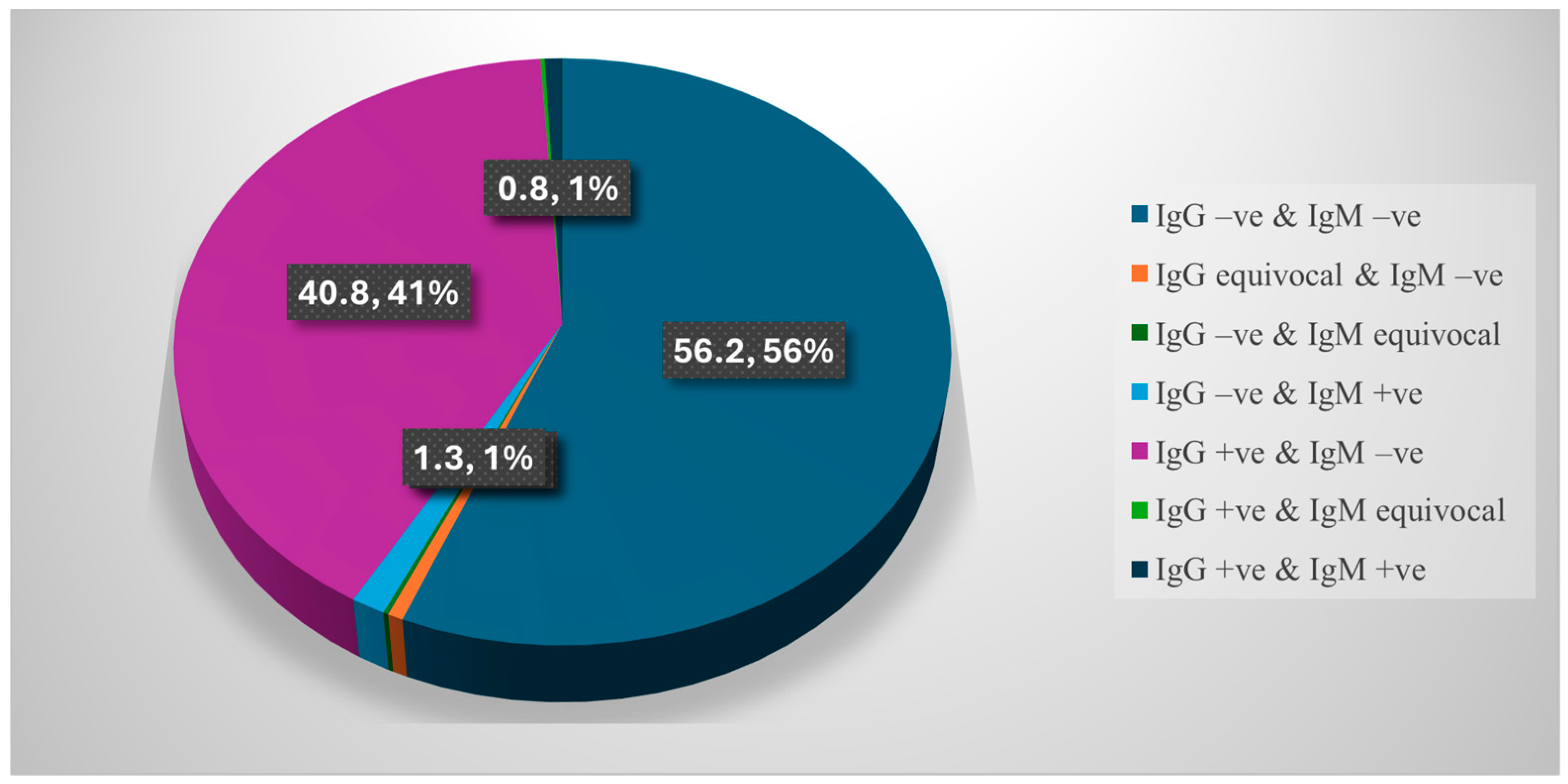
Toxoplasmosis IgM seroprevalence was positive in 2.1% of the study participants, while the seroprevalence of IgG was positive in 41.8%. Of this seroprevalence, 0.8% were positive for both IgM and IgG, 1.3% were positive for IgM and negative for IgG, and 40.8% were positive for IgG only (Table 2, Figure 1). Statistically significant geographical heterogeneity in T. gondii seroprevalence was observed across Egyptian governorates for both IgM and IgG (p < 0.001 for both). The highest IgM seroprevalence was in Cairo (6.5%), while the lowest was in Menoufia (0.3%). The highest IgG seroprevalence was in Alexandria (66.5%), while the lowest was in Cairo (16%) (Table 2).
Toxoplasmosis IgG seropositivity was statistically more prevalent among participants who consumed undercooked meat and raw vegetables and fruits; engaged in agricultural practices; had contact with cats; and dealt with soil (p < 0.05) (Table 3).
Binary logistic regression was performed to detect relevant risk factors for toxoplasmosis IgG seropositivity. On univariable analysis, undercooked meat, raw vegetables, agricultural activity, contact with cats, and dealing with soil were significant independent risk factors for toxoplasmosis IgG seropositivity; therefore, they were included in the multivariable analysis, which highlighted undercooked meat, agricultural activity, and contact with cats as significant independent risk factors for toxoplasmosis IgG seropositivity (p < 0.05) (Table 4).
The percentages for the three smallest categories, not individually labeled in the chart due to space, are: IgG equivocal & IgM negative, 0.6%; IgG negative & IgM equivocal, 0.2%; IgG positive & IgM equivocal, 0.2% (together with the four labeled categories, totaling 100%).
Regarding the seroprevalence of toxoplasmosis among the studied animals, the overall IgM seroprevalence was 5.2%, with the highest prevalence detected in the Sohag governorate (10%), while the lowest was in the Ismailia and Al-Sharqia governorates (2%). The overall seroprevalence of IgG was 7.2%, with the highest prevalence detected in the Cairo governorate, and the lowest was in the Ismailia and Al-Sharqia governorates, where none of the animals showed IgG seropositivity (0%) (Table 5).

4. Discussion

Toxoplasmosis is one of the most widespread zoonotic infections worldwide, posing considerable challenges to both human and animal health [2]. To our knowledge, this is one of the largest multicentric One Health studies in Egypt, integrating human and animal seroprevalence across geographically distinct regions. In this multicentric study, the overall seroprevalence of Toxoplasma gondii IgG antibodies among asymptomatic blood donors was 41.8%, reflecting a substantial level of prior exposure within the studied population. This relatively high prevalence suggests sustained environmental exposure and ongoing transmission cycles, reinforcing the endemicity of toxoplasmosis in Egypt.
Significant geographical heterogeneity in T. gondii seroprevalence was observed across Egyptian governorates, ranging from 66.5% in Alexandria to 16% in Cairo (p < 0.001). This statistically significant variation indicates genuine regional differences rather than sampling fluctuations. The observed patterns are likely shaped by a combination of environmental, climatic, and socioeconomic factors. For example, Alexandria’s coastal climate may enhance oocyst survival, while regional variations in diet, sanitation, and livestock contact could further influence transmission. These results highlight the focal nature of toxoplasmosis endemicity in Egypt and support region-specific rather than nationally uniform control strategies. This finding has important public health implications, as it suggests that interventions should be tailored to the epidemiological profiles of each region.
The present findings should also be interpreted in the context of previously published studies from Egypt, which have largely been limited to single-center or single governorate designs. In contrast to earlier work, including a prior study conducted in Menoufia Governorate by the same first author [21], the current study expands the epidemiological perspective through its multicentric design and inclusion of multiple geographically distinct regions. Moreover, the integration of human and animal data within a One Health framework provides additional insights into potential transmission pathways and environmental exposure, offering a more comprehensive understanding of the epidemiology of T. gondii in Egypt.
These findings aligned closely with earlier Egyptian studies, which reported seroprevalence rates of 65.3% among Alexandrian donors [27], 59.6% in Mansoura [20], and 45% in Fayoum [22]. In contrast, lower rates had been reported in Menoufia (~11%) [21,28], suggesting possible temporal changes in exposure. Similar variability had been documented across Arab countries, with seroprevalence estimates of 19.5% in Makkah Al Mukarramah, 33.5% in Tripoli, and 44% in Tunisia [5,29,30]. Globally, the findings of the present study fall within the intermediate range, comparable to reports from Greece (33%) [31] and Romania (45.9%) [32], but lower than those observed in northern Iran (73.5%) [33] and higher than estimates from Serbia, Taiwan, and China [3,34,35]. Collectively, these comparisons highlight the strong influence of geography, climate, and cultural practices on toxoplasmosis epidemiology.
No significant associations were observed between T. gondii seropositivity and sociodemographic characteristics such as age, sex, residence, marital status, educational level, income, or crowding index. This suggests that exposure to infection is widespread across the studied population rather than being confined to specific demographic groups.
These results were consistent with those of Sarkari et al. [36], who reported no association between sex and T. gondii seropositivity, attributing this to the predominance of male participants in blood donor populations, a profile similar to that of the current study. The predominance of male participants (94.4%) in this study reflects the demographic profile of blood donors in Egypt, where males constitute the vast majority of voluntary blood donors due to cultural and social factors. Blood donation campaigns in Egypt primarily target males, as females are often underrepresented due to cultural norms, a higher prevalence of anemia, and pregnancy-related deferrals. This gender distribution is consistent with previous studies on blood donors in Egypt and other Middle Eastern countries [20,21,28]. In contrast, Gouda et al. [21] stated that no significant associations were observed between T. gondii seropositivity and sociodemographic characteristics, except for sex, as 15.4% of males and 50% of females were positive, indicating that female sex was a major risk factor in this study. Moreover, Henin et al. [22] found that the prevalence of IgG seropositivity increased in those aged ≥ 30 years, and there was a significant correlation between toxoplasmosis and rural residence (86.2%). The results of the current study can be explained by the absence of age-related differences, which may reflect early-life exposure and cumulative infection over time. The lack of association with sex is likely influenced by the predominance of male participants, reflecting the demographic profile of blood donors rather than true biological differences [36]. Similarly, the non-significant associations with residence and socioeconomic indicators indicate that transmission is more strongly driven by environmental and behavioral factors than by demographic characteristics.
The prevalence of anti-T. gondii IgM antibodies was relatively low (2.1%), indicating that most infections in this population are likely chronic rather than recent. Nevertheless, significant regional variation was observed (p < 0.001), with the highest IgM prevalence in Cairo (6.5%) and the lowest in Menoufia (0.3%), suggesting possible differences in recent exposure across different settings. Comparably low IgM seroprevalence has been reported in other studies, including 0.5% and 0.41% [3,37], whereas slightly higher rates have been documented in Menoufia (5.5% and 5.9%) [21,28]. These variations may reflect differences in the study populations, environmental exposure, and diagnostic approaches. Although IgM positivity was uncommon, its presence remains clinically relevant, particularly in the context of transfusion safety, as it may indicate recent or potentially transmissible infections.
Several behavioral and environmental factors were significantly associated with seropositivity. Undercooked meat consumption was the most potent independent risk factor, reinforcing the established understanding that tissue cyst ingestion is the primary transmission route. Engagement in agricultural activities and contact with cats were also identified as independent risk factors, highlighting the importance of environmental exposure and the role of felids in environmental contamination with oocysts. Although the consumption of raw vegetables and contact with soil were significant in the univariate analysis, they did not remain independent predictors after adjustment, suggesting possible confounding or shared exposure pathways that require further investigation. These findings underscore the substantial contributions of both foodborne and environmental transmission routes to the transmission dynamics in the studied population. These results were consistent with those of Elsheikha et al. [20], Gouda et al. [21], and Ibrahem et al. [28].
From a One Health perspective, the inclusion of animal data provides important insights into the transmission dynamics of T. gondii infection. The observed seroprevalence among cattle and buffaloes (7.2% for IgG and 5.2% for IgM) confirmed the circulation of the parasite within livestock populations. The concordance between human and animal findings supports the existence of shared environmental sources of infection and ongoing zoonotic transmission of the disease. These results were consistent with studies from the Sohag (7%) [38], Kafr El Sheikh, Gharbia, and Qalyubia governorates in the Nile Delta of Egypt (7.4%) [24] and Qena (9.1%) [23], although lower (3%) [39] and considerably higher rates in other regions, such as Aswan and Assiut, have also been reported [5,40]. Such variability likely reflects differences in environmental conditions, animal husbandry practices, and levels of exposure to feline contamination.
Global prevalence estimates vary widely (0–87.79%) depending on the regional parasite genotypes [41]. These findings fall within this range and align with studies from China (7.5%) [42] and Iraq (7.4%) [43].
In Egypt, the endemicity of toxoplasmosis is driven by a combination of ecological and socioeconomic factors, including the widespread presence of stray and domestic cats, traditional farming systems, and close human–animal interactions [2,44,45]. While small ruminants are traditionally considered the primary reservoirs, increasing evidence suggests that cattle and buffaloes may also contribute to the transmission dynamics [2,46].
From a transfusion medicine perspective, the relatively high IgG seroprevalence among apparently healthy donors is noteworthy. Although IgG positivity reflects past infection, the potential for transient parasitemia raises concerns about transfusion-transmitted toxoplasmosis, particularly in immunocompromised recipients. The absence of routine screening for T. gondii in blood donation programs may represent an under-recognized gap in blood safety protocols [12].
Despite its strengths, including the large sample size and multicentric design, this study has some limitations. The cross-sectional design limited the ability to establish causal relationships between identified risk factors and T. gondii infection. In addition, the use of serological assays does not allow precise differentiation between recent and chronic infections. Although molecular analysis was performed on a subset of samples, the present study focused on serological findings; integration of molecular data will be addressed in future investigations. Furthermore, the study population, comprising blood donors and animals from selected sites, may not fully represent the general population, which could limit the generalizability of these findings. Finally, potential clustering effects across the study locations were not explicitly accounted for, which may have influenced regional comparisons.

5. Conclusions

In summary, this study provides robust epidemiological evidence of active T. gondii circulation in Egypt and underscores the critical value of a One Health strategy. The high seroprevalence among asymptomatic blood donors reflects widespread exposure, with undercooked meat consumption, agricultural activities, and contact with cats identified as key drivers of infection, emphasizing both foodborne and environmental transmission pathways. The detection of infection in livestock further supports the shared human–animal transmission cycle. These findings underscore the need for integrated control strategies, including public health education, food safety measures, veterinary surveillance, and targeted screening of high-risk groups, to enhance transfusion safety. Future studies incorporating molecular approaches and longitudinal designs are warranted to better understand transmission dynamics and inform effective prevention strategies.

Author Contributions

Conceptualization, M.A.G. and S.M.S.; methodology, all authors; validation, all authors; formal analysis, all authors; investigation, all authors; resources, all authors; data curation, R.A.L.I. and K.A.A.; writing—original draft preparation, S.A.F., A.A.S. and E.F.F.; writing—review and editing, S.A.F. and M.A.G.; supervision, M.A.G. and S.M.S.; project administration, M.A.G.; funding acquisition, S.M.S. All authors have read and agreed to the published version of the manuscript.

Funding

This work was supported and fully funded by the Science, Technology & Development Fund (STDF), Egypt, as part of the ‘One Health’ project [Grant Number: 49090]. The authors would like to express their sincere gratitude to STDF for financial support, which made this research possible.

Institutional Review Board Statement

This study was approved by the Institutional Review Board of the National Liver Institute, Menoufia University (NLI IRB No. 00651/2024), and the Medical Research Ethics Committee of the Faculty of Medicine, Sohag University (IRB No. SohMed24806PD).

Informed Consent Statement

Informed consent was obtained from all subjects involved in the study.

Data Availability Statement

The data supporting the findings of this study are contained within the manuscript. The raw data are available from the corresponding author upon request.

Acknowledgments

The authors would like to sincerely thank all the staff of the different blood banks in all governorates included in the study for their cooperation during data collection. The authors appreciate the blood donors who participated in this research.

Conflicts of Interest

The authors declare no competing interests.

Abbreviations

The following abbreviations are used in this manuscript:
WHOWorld Health Organization
OIEWorld Organization for Animal Health
FAOFood and Agriculture Organization
T. gondiiToxoplasma gondii
TTITransfusion-transmitted infection
HBsAgHepatitis B surface antigen
HCVHepatitis C virus
HIVHuman immunodeficiency virus
ELISAEnzyme-linked immunosorbent assay
IgGImmunoglobulin G
IgMImmunoglobulin M

References

  1. İnci, A.; Sohel, M.H.; Babür, C.; Uslu, S.; Karademir, G.K.; Yürk, M.; Düzlü, Ö.; Kızgın, A.D.; Yıldırım, A. An overview of one health concept focusing on toxoplasmosis. Turk. Parazitol. Derg. 2023, 47, 256–274. [Google Scholar] [CrossRef]
  2. Barghash, S.; Abdel-Baky, S.M.; Abou Elnaga, T.R.; Balata, M.E.; Yassin, S.; Khattab, R.; Khidr, R.E.; Mohsen, A.S. Epidemiological insights into toxoplasmosis in Egypt: A review of transmission, risk factors, diagnostics, and molecular diversity of Toxoplasma gondii in humans and livestock animals. Egypt. J. Camel Sci. 2025, 3, 13–25. [Google Scholar] [CrossRef]
  3. Wang, X.; Ruan, W.; Xu, W.; Chen, H.; Zhang, J.; Zhang, X.; Yu, K.; Lu, Q.; Wang, Z.; Sun, J. Different patterns of Toxoplasma gondii infection epidemiology in the general population, animal contact workers, and blood donors in southeastern China between 2019 and 2023: A cross-sectional study. Parasite 2025, 32, 68. [Google Scholar] [CrossRef] [PubMed]
  4. Elmonir, W.; Mabrouk, I.; Mohamed, R.A.; El-Tras, W.F.; Hegazy, Y. Prevalence and Risk Factors for Toxoplasma gondii Infection at the Cat-Human Interface in Gharbia Governorate, Nile Delta, Egypt. Egypt. J. Vet. Sci. 2023, 54, 273–284. [Google Scholar] [CrossRef]
  5. Dyab, A.K.; Osman, H.E.; Desoky, R.M.; Nageeb, H.M.; Abdelhamid, M. Seroprevalence of toxoplasmosis in reproductive-age women and animals with first molecular characterization of Toxoplasma gondii isolated from cats in Aswan governorate, Egypt. Assiut Vet. Med. J. 2025, 71, 92–102. [Google Scholar] [CrossRef]
  6. Nayeri, T.; Moosazadeh, M.; Dalimi Asl, A.; Ghaffarifar, F.; Sarvi, S.; Daryani, A. Toxoplasma gondii infection and ABO blood groups: A systematic review and meta-analysis. Trans. R. Soc. Trop. Med. Hyg. 2024, 118, 234–246. [Google Scholar] [CrossRef] [PubMed]
  7. Lachkhem, A.; Lahmar, I.; Galal, L.; Babba, O.; Mezhoud, H.; Hassine, M.; Lachkhem, A.; Dardé, M.L.; Mercier, A.; Babba, H. Seroprevalence of Toxoplasma gondii among healthy blood donors in two locations in Tunisia and associated risk factors. Parasite 2020, 27, 51. [Google Scholar] [CrossRef] [PubMed]
  8. Bisetegn, H.; Debash, H.; Ebrahim, H.; Mahmood, N.; Gedefie, A.; Tilahun, M.; Alemayehu, E.; Mohammed, O.; Feleke, D.G. Global seroprevalence of Toxoplasma gondii infection among patients with mental and neurological disorders: A systematic review and meta-analysis. Health Sci. Rep. 2023, 6, e1319. [Google Scholar] [CrossRef] [PubMed]
  9. Zhao, X.Y.; Ewald, S.E. The molecular biology and immune control of chronic Toxoplasma gondii infection. J. Clin. Investig. 2020, 130, 3370–3380. [Google Scholar] [CrossRef] [PubMed]
  10. Abbas, I.E.; Villena, I.; Dubey, J.P. A review on toxoplasmosis in humans and animals from Egypt. Parasitology 2020, 147, 135–159. [Google Scholar] [CrossRef] [PubMed]
  11. Alharazi, T.; Alzubiery, T.K.; Alcantara, J.C.; Qanash, H.; Bazaid, A.S.; Altayar, M.A.; Aldarhami, A. Prevalence of transfu-sion transmitted infections (HCV, HIV, syphilis and malaria) in blood donors: A large-scale cross-sectional study. Pathogens 2022, 11, 726. [Google Scholar] [CrossRef] [PubMed]
  12. Wesołowski, R.; Pawłowska, M.; Mila-Kierzenkowska, C. The medical relevance of Toxoplasma infections in terms of the safety of blood recipients under immunosuppression—A meta-analysis. Microorganisms 2023, 11, 1980. [Google Scholar] [CrossRef] [PubMed]
  13. Baloochi, M.; Amini Fard, M.R.; Ordoni, R.; Pendar, A.; Vafae Eslahi, A.; Badri, M.; Dezhkam, A.; Kalkali, M.; Rahdar, H.A.; Mohammadi, M.; et al. Seroepidemiology of Toxoplasma gondii infection among patients with beta-thalassemia major: A case-control study in Southeastern Iran. BMC Infect. Dis. 2025, 25, 867. [Google Scholar] [CrossRef] [PubMed]
  14. Foroutan-Rad, M.; Majidiani, H.; Dalvand, S.; Daryani, A.; Kooti, W.; Saki, J.; Hedayati-Rad, F.; Ahmadpour, E. Toxoplasmosis in blood donors: A systematic review and meta-analysis. Transfus. Med. Rev. 2016, 30, 116–122. [Google Scholar] [CrossRef] [PubMed]
  15. Foroutan, M.; Majidiani, H.; Hassanipour, S.; Badri, M. Toxoplasma gondii seroprevalence in the Iranian blood donors: A systematic review and meta-analysis. Heliyon 2024, 10, e28013. [Google Scholar] [CrossRef] [PubMed]
  16. Omonijo, A.O.; Kalinda, C.; Mukaratirwa, S. Toxoplasma gondii infections in animals and humans in Southern Africa: A systematic review and meta-analysis. Pathogens 2022, 11, 183. [Google Scholar] [CrossRef] [PubMed]
  17. Stelzer, S.; Basso, W.; Silván, J.B.; Ortega-Mora, L.M.; Maksimov, P.; Gethmann, J.; Conraths, F.J.; Schares, G. Toxoplasma gondii infection and toxoplasmosis in farm animals: Risk factors and economic impact. Food Waterborne Parasitol. 2019, 15, e00037. [Google Scholar] [CrossRef] [PubMed]
  18. Shapiro, K.; Bahia-Oliveira, L.; Dixon, B.; Dumètre, A.; de Wit, L.A.; VanWormer, E.; Villena, I. Environmental transmission of Toxoplasma gondii: Oocysts in water, soil and food. Food Waterborne Parasitol. 2019, 15, e00049. [Google Scholar] [CrossRef] [PubMed]
  19. Dubey, J.P. Toxoplasmosis of Animals and Humans, 3rd ed.; CRC Press: Boca Raton, FL, USA, 2022. [Google Scholar]
  20. Elsheikha, H.M.; Azab, M.S.; Abousamra, N.K.; Rahbar, M.H.; Elghannam, D.M.; Raafat, D. Seroprevalence of and risk factors for Toxoplasma gondii antibodies among asymptomatic blood donors in Egypt. Parasitol. Res. 2009, 104, 1471–1476. [Google Scholar] [CrossRef] [PubMed]
  21. Gouda, M.A.; Saied, S.A.; Ashry, W.M.O.; Abd-Eltwab, R.A.R.; Aldesoky, M.M.; El-Dydamoni, O.A.; Yousef, M.; El-Derbawy, M.M. Trends of Toxoplasma gondii and common transfusable venereal infections among blood donors in Menoufia Province, Egypt. Sci. Rep. 2024, 14, 20920. [Google Scholar] [CrossRef] [PubMed]
  22. Henin, R.W. Evaluation of donated blood for toxoplasmosis using ELISA and PCR in Fayoum Governorate. J. Egypt. Soc. Parasitol. 2019, 49, 653–660. [Google Scholar] [CrossRef]
  23. Fereig, R.M.; El-Alfy, E.S.; Alharbi, A.S.; Abdelraheem, M.Z.; Almuzaini, A.M.; Omar, M.A.; Kandil, O.M.; Frey, C.F. Seroprevalence of Toxoplasma gondii in cattle in Southern Egypt: Do milk and serum samples tell the same story? Animals 2024, 14, 3122. [Google Scholar] [CrossRef] [PubMed]
  24. Selim, A.; Alshammari, A.; Gattan, H.S.; Alruhaili, M.H.; Rashed, G.A.; Shoulah, S. Seroprevalence and associated risk factors for Toxoplasma gondii in water buffaloes (Bubalus bubalis) in Egypt. Comp. Immunol. Microbiol. Infect. Dis. 2023, 101, 102058. [Google Scholar] [CrossRef] [PubMed]
  25. El-Gilany, A.; El-Wehady, A.; El-Wasify, M. Updating and validation of the socioeconomic status scale for health research in Egypt. East. Mediterr. Health J. 2012, 18, 962–968. [Google Scholar] [CrossRef] [PubMed]
  26. Ministry of Health and Population [Egypt]; El-Zanaty and Associates; ICF. Egypt Demographic and Health Survey 2021; Ministry of Health and Population: Cairo, Egypt; ICF: Rockville, MD, USA, 2022. [Google Scholar]
  27. El-Geddawi, O.A.; El-Sayad, M.H.; Sadek, N.A.; Hussien, N.A.; Ahmed, M.A. Detection of T. gondii infection in blood donors in Alexandria, Egypt, using serological and molecular strategies. Parasitol. United J. 2016, 9, 24–30. [Google Scholar] [CrossRef]
  28. Ibrahem, R.A.L.; Abdelaty, N.B.; Barakat, A.M.; Abdelwanees, S.A. Seroprevalence and risk factors of Toxoplasma gondii among blood donors in Menoufia Governorate, Egypt. Egypt. J. Community Med. 2024, 42, 131–138. [Google Scholar]
  29. Mohamed, K.; Zamzami, H.; Deqnah, N.; Al Malki, A.; Alzayer, S.; Al-Gethamy, M.; Al-Pakistani, H.; Al-Harbi, W. Seroprevalence of Toxoplasma gondii infection in blood donors in Makkah Al Mukarramah. Asian J. Epidemiol. 2019, 12, 25–31. [Google Scholar] [CrossRef]
  30. Alawaini, K.A.; Albhlool, S.O.; Shaife, E.O.; Qreiwa, H.A.; Abodena, M.K. Prevalence of toxoplasmosis antibodies in blood donors in Tripoli area. World J. Adv. Res. Rev. 2021, 10, 169–175. [Google Scholar] [CrossRef]
  31. Kantzanou, M.; Kostares, E.; Kostare, G.; Papagiannopoulou, E.; Kostares, M.; Tsakris, A. Prevalence of Toxoplasma gondii infection among blood donors: A systematic review and meta-analysis. Cureus 2025, 17, e85403. [Google Scholar] [CrossRef] [PubMed]
  32. Lupu, M.A.; Lighezan, R.; Paduraru, A.A.; Dragomir, A.; Pavel, R.; Grada, S.; Mihu, A.G.; Ursoniu, S.; Olariu, T.R. Seroepidemiology of Toxoplasma gondii infection in blood donors from western Romania. Microorganisms 2022, 10, 973. [Google Scholar] [CrossRef] [PubMed]
  33. Hosseini, S.A.; Golchin, E.; Sharif, M.; Sarvi, S.; Ahmadpour, E.; Rostamian, A.; Gholami, S.; Amouei, A.; Daryani, A. A serological investigation and genotyping of Toxoplasma gondii among Iranian blood donors indicates threat to health of blood recipients. Transfus. Apher. Sci. 2020, 59, 102723. [Google Scholar] [CrossRef] [PubMed]
  34. Stopić, M.; Štajner, T.; Marković-Denić, L.; Nikolić, V.; Djilaš, I.; Srzentić, S.J.; Djurković-Djakovć, O.; Bobić, B. Epidemiology of toxoplasmosis in Serbia: A cross-sectional study on blood donors. Microorganisms 2022, 10, 492. [Google Scholar] [CrossRef] [PubMed]
  35. Chiang, T.Y.; Hsieh, H.H.; Kuo, M.C.; Chiu, K.T.; Lin, W.C.; Fan, C.K.; Fang, C.T.; Ji, D.D. Seroepidemiology of Toxoplasma gondii infection among healthy blood donors in Taiwan. PLoS ONE 2012, 7, e48139. [Google Scholar] [CrossRef] [PubMed]
  36. Sarkari, B.; Shafiei, R.; Zare, M.; Sohrabpour, S.; Kasraian, L. Seroprevalence and molecular diagnosis of Toxoplasma gondii infection among blood donors in southern Iran. J. Infect. Dev. Ctries. 2014, 8, 543–547. [Google Scholar] [CrossRef] [PubMed]
  37. Saki, J.; Foroutan, M.; Khodkar, I.; Khodadadi, A.; Nazari, L. Seroprevalence and molecular detection of Toxoplasma gondii in healthy blood donors in southwest Iran. Transfus. Apher. Sci. 2019, 58, 79–82. [Google Scholar] [CrossRef] [PubMed]
  38. Ali, A.O.; Qossa, W.; Khalifa, F.A.; Frey, C.F.; Fereig, R.M. Seroprevalence and risk factors of Toxoplasma gondii infection in buffalo (Bubalus bubalis) from Sohag, Egypt. Vet. Sci. 2026, 13, 166. [Google Scholar] [CrossRef] [PubMed]
  39. Hussein, D.E.; Abou Khashaba, A.M.; Kamar, A.M. Advanced studies on Toxoplasma in buffalo meat. Alex. J. Vet. Sci. 2023, 77, 9–16. [Google Scholar] [CrossRef]
  40. Kuraa, H.M.; Malek, S.S. Seroprevalence of Toxoplasma gondii in ruminants by using latex agglutination test (LAT) and en-zyme-linked immunosorbent assay (ELISA) in Assiut governorate. Assiut Vet. Med. J. 2016, 62, 90–97. [Google Scholar]
  41. De Barros, L.D.; Garcia, J.L.; Bresciani, K.D.S.; Cardim, S.T.; Storte, V.S.; Headley, S.A. A review of toxoplasmosis and neosporosis in water buffalo (Bubalus bubalis). Front. Vet. Sci. 2020, 7, 455. [Google Scholar] [CrossRef] [PubMed]
  42. Zou, F.; Yu, X.; Yang, Y.; Hu, S.; Chang, H.; Yang, J.; Duan, G. Seroprevalence and risk factors of Toxoplasma gondii infection in buffaloes, sheep and goats in Yunnan Province, Southwestern China. Iran. J. Parasitol. 2015, 10, 648–651. [Google Scholar]
  43. Kazem, R.J.; Ali, I.K. Seropositivity and other determinants associated with toxoplasmosis in local buffalo in Iraq. Open J. Vet. Med. 2024, 14, 1–9. [Google Scholar] [CrossRef]
  44. Ibrahim, H.M.; Mohamed, A.H.; El-Sharaawy, A.A.; El-Shqanqery, H.E. Molecular and serological prevalence of Toxoplasma gondii in pregnant women and sheep in Egypt. Asian Pac. J. Trop. Med. 2017, 10, 996–1001. [Google Scholar] [CrossRef] [PubMed]
  45. Fereig, R.M.; Wareth, G.; Abdelbaky, H.H.; Mazeed, A.M.; El-Diasty, M.; Abdelkhalek, A.; Mahmoud, H.Y.; Ali, A.O.; El-Tayeb, A.; Alsayeqh, A.F.; et al. Seroprevalence of specific antibodies to Toxoplasma gondii, Neospora caninum, and Brucella spp. in sheep and goats in Egypt. Animals 2022, 12, 3327. [Google Scholar] [CrossRef] [PubMed]
  46. Zeedan, G.S.; Abdalhamed, A.M.; Shaapan, R.M.; El-Namaky, A.H. Rapid diagnosis of Toxoplasma gondii using loop-mediated isothermal amplification assay in camels and small ruminants. Beni-Suef Univ. J. Basic Appl. Sci. 2022, 11, 1. [Google Scholar] [CrossRef]
Figure 1. Toxoplasmosis serology (IgG and IgM) among the studied cases.
Microorganisms 14 01627 g001
Table 1. Sociodemographic profile and risk factors of the study participants (N = 2000).
Variable N (%)
Age (Years)Mean ± SD30.32 ± 9.40
Range 18–66
Age (Years)≤29 years1065 (53.3)
>29 years935 (46.8)
SexMale1887 (94.4)
Female113 (5.6)
ResidenceRural1019 (50.9)
Urban981 (49.1)
Marital statusSingle704 (35.2)
Married1296 (64.8)
Educational levelIlliterate169 (8.5)
Basic390 (19.5)
Secondary854 (42.7)
University587 (29.4)
IncomeLess than sufficient546 (27.3)
Sufficient1287 (64.4)
More than sufficient167 (8.4)
Crowding index<1 (no crowding)217 (10.9)
=1 (crowding)640 (32.0)
>1 (severe crowding)1143 (57.2)
Consumption of undercooked meat 574 (28.7)
Consumption of unwashed raw vegetables & fruits 492 (24.6)
Gardening or practice of agriculture 708 (35.4)
Hand washing before eating 1108 (55.4)
Contact with cats 469 (23.5)
Dealing with soil 778 (38.9)
Drinking untreated water 511 (25.6)
History of blood transfusion 256 (12.8)
Table 2. Seroprevalence of toxoplasmosis in the different Egyptian governorates studied.
VariableAlex
(N = 400)
Cairo (N = 400)Ismailia
and Al-Sharqia (N = 400)
Menoufia (N = 400)Sohag (N = 400)Total (N = 2000)
N (%)N (%)N (%)N (%)N (%)N (%)
Toxoplasmosis IgM
Positive 9 (2.3)26 (6.5)2 (0.5)1 (0.3)3 (0.8)41 (2.1)
Equivocal4 (1.0)0 (0.0)1 (0.3)0 (0.0)2 (0.5)7 (0.4)
Negative387 (96.8)374 (93.5)397 (99.3)399 (99.8)395 (98.8)1952 (97.6)
χ2 (p value)62.10 (<0.001) *
Toxoplasmosis IgG
Positive266 (66.5)64 (16.0)192 (48.0)150 (37.5)163 (40.8)835 (41.8)
Equivocal9 (2.3)3 (0.8)0 (0.0)0 (0.0)0 (0.0)12 (0.6)
Negative125 (31.3)333 (83.3)208 (52.0)250 (62.5)237 (59.8)1153 (57.7)
χ2 (p value)251.14 (<0.001) *
* Statistically significant; χ2: chi-squared test.
Table 3. Relationship between toxoplasmosis IgG seropositivity and sociodemographic, clinical data, and risk factors among the participants (N = 2000).
VariableToxoplasmosis IgGχ2p Value
Negative and Equivocal
(N = 1165)
Positive
(N = 835)
N (%)N (%)
Sex
Male 1108 (95.1)779 (93.3)3.000.083
Female 57 (4.9)56 (6.7)
Age (Years)
≤29 years 618 (53.0)447 (53.5)0.050.830
>29 years 547 (47.0)388 (46.5)
Residence
Rural 614 (52.7)405 (48.5)3.440.064
Urban 551 (47.3)430 (51.5)
Marital status
Single 412 (35.4)292 (35.0)0.030.855
Married 753 (64.6)543 (65.0)
Educational level
Illiterate 94 (8.1)75 (9.0)4.570.207
Basic 240 (20.6)150 (18.0)
Secondary 479 (41.1)375 (44.9)
University 352 (30.2)235 (28.1)
Income
Less than sufficient 302 (25.9)244 (29.2)3.140.208
Sufficient 768 (65.9)519 (62.2)
More than sufficient 95 (8.2)72 (8.6)
Crowding index
<1 (no crowding)126 (10.8)91 (10.9)0.370.831
=1 (crowding)379 (32.5)261 (31.3)
>1 (severe crowding)660 (56.7)483 (57.8)
Undercooked meat 237 (20.3)337 (40.4)95.23<0.001 *
Raw vegetables & fruits 267 (22.9)225 (26.9)4.250.039 *
Agricultural activity342 (29.4)366 (43.8)44.57<0.001 *
Hand washing 628 (53.9)480 (57.5)2.520.112
Contact with cats 227 (19.5)242 (29.0)24.44<0.001 *
Dealing with soil 418 (35.9)360 (43.1)10.710.001 *
Untreated water300 (25.8)211 (25.3)0.060.808
Blood transfusion 136 (11.7)120 (14.4)3.170.075
* Statistically significant; χ2: chi-squared test. Bold values indicate statistical significance (p < 0.05).
Table 4. Logistic regression for relevant risk factors for toxoplasmosis IgG seropositivity.
VariableUnivariableMultivariable
OR95% CIp ValueOR95% CIp Value
LowerUpper LowerUpper
Undercooked meat
Yes2.652.173.23<0.001 *2.492.023.06<0.001 *
No1.01.0
Raw vegetables & fruits
Yes1.241.011.520.039 *0.880.701.100.262
No1.01.0
Agricultural activity
Yes1.881.562.26<0.001 *1.741.412.14<0.001 *
No1.01.0
Contact with cats
Yes1.691.372.08<0.001 *1.531.231.90<0.001 *
No1.01.0
Dealing with soil
Yes1.351.131.630.001 *1.010.821.240.933
No1.01.0
* Statistically significant; OR: odds ratio; CI: confidence interval. Bold values indicate statistical significance (p < 0.05).
Table 5. Characteristics and toxoplasmosis seroprevalence of the studied animals.
VariableAlex
(N = 100)
Cairo (N = 100)Ismailia and Al-Sharqia (N = 100)Menoufia (N = 100)Sohag (N = 100)
N (%)N (%)N (%)N (%)N (%)
Animal type
Buff 0 (0.0)100 (100.0)0 (0.0)22 (22.0)100 (100.0)
Cow100 (100.0)0 (0.0)100 (100.0)78 (78.0)0 (0.0)
Animal sex
Male 16 (16)42 (42.0)0 (0.0)0 (0.0)97 (97.0)
Female 84 (84)58 (58.0)100 (100.0)100 (100.0)3 (3.0)
Season
Winter0 (0.0)43 (43.0)0 (0.0)22 (22.0)0 (0.0)
Summer100 (100.0)57 (57.0)100 (100.0)78 (78.0)100 (100.0)
IgM
Positive 3 (3.0)7 (7.0)2 (2.0)4 (4.0)10 (10.0)
Negative97 (97.0)93 (93.0)98 (98.0)96 (96.0)90 (90.0)
IgG
Positive 2 (2.0)13 (13.0)0 (0.0)11 (11.0)10 (10.0)
Negative 98 (98.0)87 (87.0)100 (100.0)89 (89.0)90 (90.0)
Disclaimer/Publisher’s Note: The statements, opinions and data contained in all publications are solely those of the individual author(s) and contributor(s) and not of MDPI and/or the editor(s). MDPI and/or the editor(s) disclaim responsibility for any injury to people or property resulting from any ideas, methods, instructions or products referred to in the content.