


Abstract
Lipids (sphingolipids, fatty acids, phospholipids, and lipoproteins) are vital to intestinal barrier integrity, as precursors for pro-inflammatory and pro-resolving mediators and undergo remodelling through host microbiome interactions. Accumulating evidence implicates the Western diet—high in long-chain saturated and omega-6 polyunsaturated fatty acids and low in omega-3—in both the onset and progression of IBD. In contrast, microbiota derived lipid metabolites, including short-chain fatty acids and secondary bile acids, contribute to mucosal homeostasis and immune regulation. This review is structured around three interconnected pillars. First, we classified lipidomic alterations in IBD across major lipid classes: sphingolipids, fatty acids, phospholipids, and lipoproteins by integrating host, dietary, and microbiome contributions. Second, we examined the potential of lipidomics in IBD as a source of prognostic, diagnostic and therapy response biomarkers. Third, we critically assessed the challenges that currently limit clinical implementation including analytical variability, pre-analytical confounding, small cohort sizes, and the lack of prospective validation. Addressing these barriers will be essential to fully realise the potential of lipidomics in advancing personalised care for patients with IBD.
1. Introduction
Inflammatory bowel disease (IBD) encompasses a group of chronic, immune-mediated conditions affecting the gastrointestinal tract, including Crohn’s disease (CD), ulcerative colitis (UC), and less-defined entities such as IBD-unclassified (IBD-U) and indeterminate colitis [1,2]. Both CD and UC share a multifactorial pathogenesis rooted in the interaction between genetic predisposition, environmental exposures, gut microbiota composition, and immune dysregulation, ultimately producing a state of unresolved mucosal inflammation and tissue injury [1,2].
Although several medications are now available, IBD diagnosis relies on a composite of clinical, biochemical and imaging parameters while standardised molecular biomarkers are still lacking. Omics technologies offer a systematic means of characterising the molecular landscape of disease at scale [3,4]. While genetic and transcriptomic studies have been explored extensively in IBD, lipidomics seems to capture the dynamic interplay between metabolism, immunity, and the microbiome. Lipidomics employs mass spectrometry to systematically profile lipid species and their associated metabolic pathways, using three principal analytical approaches: targeted lipidomics, which quantifies predefined lipid classes using high-sensitivity instruments such as triple-quadrupole mass spectrometers; shotgun lipidomics, in which complex lipid extracts are analysed directly, preferably on high-resolution platforms such as Orbitrap instruments, without prior chromatographic separation; imaging lipidomics, which maps lipid distributions across tissue sections through surface ionisation [4].
Lipids constitute structural components of the intestinal barrier, serve as precursors to pro-inflammatory and pro-resolving mediators, and are subject to extensive remodelling by the gut microbiota. Emerging evidence links the Western dietary lipid pattern to IBD risk and progression, a relationship developed mechanistically in Section 3.6 [5,6]. Whereas specific microbially derived lipid metabolites such as short-chain fatty acids (SCFAs) and secondary bile acids, exert protective effects on mucosal homeostasis [7,8].
The implementation of lipidomics into the understanding of IBD pathogenesis could shed new light on traditional IBD classification. Beyond cataloguing these associations, we argue that lipidomic data may ultimately support a biochemical reclassification of IBD—defining mechanistic endotypes that cut across the conventional Crohn’s disease and ulcerative colitis division. We develop this framework in the Discussion, while delineating the validation it would require before it could inform clinical practice. This could lead to an improved comprehension of the disease that could help define disease trajectories, predict response to therapies and favour more personalised medicine.
This review aims to draw attention to lipidomics as an underexplored yet promising field in IBD research, given the growing evidence implicating lipids and their associated pathways in disease pathogenesis, including their potential role in disease discrimination, monitoring of mucosal inflammation, prediction of therapeutic response, and refinement of treat-to-target strategies.
2. Materials and Methods
This work is a structured narrative review rather than a systematic review; it was designed to deliver a critical, mechanistically organised synthesis of lipidomics in IBD rather than an exhaustive enumeration of every primary study. A structured search was performed in PubMed/MEDLINE, Embase, Scopus, and Web of Science for articles published between January 2002 and December 2025, supplemented by landmark older references where foundational. Search terms combined controlled-vocabulary and free-text terms for inflammatory bowel disease, ulcerative colitis, and Crohn’s disease with terms for lipidomics and the individual lipid classes discussed herein (sphingolipids, glycerophospholipids, glycolipids, glycerolipids, fatty acids, and lipoproteins), as well as specialised pro-resolving mediators, bile acids, biomarkers, and therapeutic response. Original human studies, multi-cohort validation studies, Mendelian randomisation analyses, and mechanistic work in validated murine colitis models were prioritised; narrative and systematic reviews were cited only when they synthesised primary evidence not otherwise available, and methodological papers on mass-spectrometry lipidomics, pre-analytical variability, and biomarker reproducibility were included to frame the translational barriers discussed in Section 5 and Section 7. Studies were excluded if not published in English, if they lacked human or IBD-relevant preclinical data, or if they did not report lipid-specific measurements. Reference lists of included articles were hand-searched for additional citations. Study selection and synthesis were guided by topical relevance and expert appraisal rather than by formal systematic screening or dual independent review; accordingly, the absence of a PRISMA-based selection process and formal risk-of-bias assessment is acknowledged as a limitation, and the cited literature should be read as a representative critical synthesis.
3. Alterations in Lipid Profiles in IBD
Lipids are a structurally and functionally heterogeneous class of hydrophobic molecules. In intestinal physiology, they cover a broad spectrum of bioactive species, including dietary-derived fatty acids, microbially synthesised sphingolipids, and phospholipid-derived signalling mediators, each of which may be uniquely involved in the pathogenesis and progression of IBD [4,9,10,11].
3.1. Sphingolipids
Sphingolipids are fundamental constituents of the cell membrane architecture and include two broad subclasses: complex sphingolipids such as sphingomyelin, cerebrosides, and gangliosides; and simple sphingolipids such as ceramide and sphingosine, together with their phosphorylated forms, sphingosine-1-phosphate (S1P) and ceramide-1-phosphate (C1P), which are pivotal regulators of cell survival, apoptosis, and inflammatory signalling [10,11].
The relationship between sphingolipid species and IBD activity has been characterised through correlation with established inflammatory markers. Cer 18:1;O2/24:0 and HexCer 18:1;O2/22:0, 23:0, and 24:0 are inversely correlated with both C-reactive protein (CRP) and faecal calprotectin. Ceramide 18:1;O2/26:0 shows a negative correlation with CRP alone while ceramide 18:1;O2/23:0 and 18:1;O2/26:0 do not associate with calprotectin, suggesting that distinct ceramide species track different dimensions of mucosal and systemic inflammation [10].
Elger et al. demonstrated that an elevated ratio of long-chain (LC) to very-long-chain ceramides (VLC) in serum is associated with disease severity in IBD and primary sclerosing cholangitis (PSC). Notably, this LC/VLC ceramide ratio is elevated in IBD, PSC-IBD, and PSC compared to healthy controls thus establishing it as a possible quantitative index of disease severity [10].
The pattern of sphingolipid alteration differs substantially according to treatment status and disease subtype. Moe et al. characterised the mucosal lipid profile across treatment-refractory and treatment-naïve UC patients and demonstrated that refractory UC patients exhibit pronounced decreases in ceramides, hexosylceramides, sphingomyelins, glycerophosphocholines (PC), glycerophosphoethanolamines (PE), and glycerophosphoserines (PS) relative to both treatment-naïve patients and healthy controls [11].However, treatment-naïve UC patients show elevated ceramide and sphingomyelin levels compared to controls, whereas remission profiles approach those of healthy controls [11]. In CD, refractory patients showed major alterations in PE, SM, PS, HexCer, and PC compared to controls, but no consistent differences emerged between treatment-naïve CD patients and controls, nor between refractory and naïve CD patients, a pattern that may reflect the greater heterogeneity in CD location and transmural extent [11].
3.2. Glycolipids
Glycolipids comprise glycosphingolipids, glucosyl and galactosylceramides, lactosylceramide, and the sialylated gangliosides together with glyceroglycolipids. Glucosylceramide metabolism is tied to mucosal immune regulation [12]. In IBD patients, UDP-glucose ceramide glucosyltransferase (UGCG) the glycosphingolipid-synthesising enzyme is downregulated, and this loss is reproduced in dextran sulphate sodium (DSS) colitis, with reduced UGCG and glucosylceramide content in mesenteric lymph nodes and splenic T cells [12]. The downstream consequences for the regulatory T cell compartment, and the protective effect of glucosylceramide repletion, are preclinical (murine) and are developed alongside the immune-homeostasis literature in Section 4.2; glucosylceramide thus behaves as a regulator of mucosal tolerance rather than a passive membrane constituent.
The same enzymatic node likely links inflammation to neoplastic progression. Disrupted sphingolipid metabolism promotes inflammation in IBD and fosters a microenvironment permissive to colorectal cancer, with glucosylceramide synthase activity and high tumoural UGCG expression associated with worse outcomes in established disease [13]. Dedicated glycolipid profiling in human IBD cohorts remains limited relative to the phospholipid and sphingolipid literature, and most available data derives from animal models or from glycosphingolipid species captured incidentally within broader lipidomic panels. This gap could be closed by targeted glycosphingolipid lipidomics, including ganglioside and hexosylceramide speciation, thus establishing whether the depletion seen in refractory disease is cause or consequence of impaired barrier and immune function.
3.3. Glycerolipids (Triglycerides)
Glycerolipids, mono-, di-, and triacylglycerols are the principal neutral and storage lipids of the lipidome [14]. Unlike the sphingolipid and phospholipid classes, triglycerides show no clear quantitative signature in IBD: a systematic review and meta-analysis of 53 studies did not identify triglycerides among the serum lipid fractions consistently altered in IBD relative to healthy controls, with findings varying by disease activity, nutritional status, and treatment across cohorts [14]. This heterogeneity indicates that the total triglyceride pool is a poor discriminator of disease state, and that any informative glycerolipid signal is more likely to reside at the level of individual molecular species than in bulk concentration [14].
Species-resolved genetic approaches support this. In a bidirectional two-sample Mendelian randomisation analysis of the plasma lipidome, diacylglycerol was causally associated with an increased risk of Crohn’s disease (OR 1.21), placing a specific glycerolipid intermediate, rather than total triglyceride, within the mechanism of the disease [15]. Diacylglycerols occupy a branch point between triglyceride storage and phospholipid synthesis and act as second messengers in protein kinase C signalling [15], offering a plausible route by which glycerolipid flux could shape epithelial and immune responses. Beyond this genetic signal, dedicated acylglycerol lipidomics in IBD tissue remains sparse as most triglyceride data derives from routine serum measurement rather than untargeted profiling. Future studies would benefit from studying species-level glycerolipid remodelling and its relationship to the mucosal phospholipid and sphingolipid shifts.
3.4. Phospholipids
Phospholipids are major structural components of the intestinal epithelial membrane and play active roles in barrier function, vesicle trafficking, and PI3K-dependent signalling [16]. In CD, phosphatidylinositol abnormalities are observed with relative preservation of ceramide, phosphatidylcholine, and phosphatidylserine composition, indicating selective phospholipid dysregulation affecting specific intracellular signalling pathways [17]. Intestinal phospholipid disequilibrium can initiate endoplasmic reticulum (ER) stress responses that drive goblet cell necroptosis and spontaneous colitis, as demonstrated in murine models by Kennelly et al. (2021) [16]. Spatial imaging of UC biopsies by MALDI-MSI added another layer. Dysregulated phosphatidylinositols, phosphatidylethanolamines, and PE plasmalogens were found to change in a gradient along the colonic crypt axis, mirroring disrupted colonocyte differentiation. This reveals a phospholipidome that is highly cell-type-specific able to distinguish inflammatory, malignant, and differentiating epithelial states from one another [18].
3.5. High-Density Lipoprotein Cholesterol and Lipoprotein Remodelling
High-density lipoprotein cholesterol (HDL-C) is well recognised for its cardiovascular protective and systemic anti-inflammatory properties, and emerging Mendelian randomisation evidence suggests a causal role in IBD susceptibility [19]. Tao et al. demonstrated a causal association between elevated HDL-C levels and a reduced risk of IBD, including both UC and CD, implicating HDL-C in the regulation of mucosal immune homeostasis [19]. The anti-inflammatory mechanism involves interaction with cholesteryl ester transfer protein (CETP): CETP inhibition, which raises HDL-C while reducing transfer of cholesteryl esters to LDL and VLDL, is associated with a reduced risk of developing CD [19].
IBD patients also exhibit a disease activity-dependent compositional shift in LDL particle size from large, atheroprotective subclasses (LDL-1 to LDL-3) toward smaller, denser, more atherogenic species (LDL-4 to LDL-6), while ApoA1 and ApoA2, which are the primary anti-inflammatory constituents of HDL, decline progressively with increasing disease activity [12,20]. These systemic lipoprotein alterations simultaneously position IBD patients at elevated cardiovascular risk independently from conventional lipid fractions.
3.6. Fatty Acids and Derived Lipid Mediators
Fatty acids are a class of dietary lipids implicated in immune regulation, gut microbiota modulation, and intestinal barrier maintenance [5,7]. They are classified by carbon chain length into very-long-chain (VLCFAs), long-chain (LCFAs), medium-chain (MCFAs), and short-chain fatty acids (SCFAs), each with distinct immunological profiles [5].
SCFAs, produced by microbial fermentation of dietary fibre in the colon, exert pronounced anti-inflammatory and immunoregulatory effects. They promote CD4+ T cell and innate lymphoid cell (ILC) production of IL-22 through upregulation of the aryl hydrocarbon receptor (AhR) and hypoxia-inducible factor 1-alpha (HIF-1α), thereby maintaining intestinal homeostasis [7]. Among SCFAs, butyrate exerts the most potent anti-inflammatory activity: its interaction with the G protein-coupled receptor GPR109A drives regulatory T cell (Treg) differentiation and IL-10 production [7,8]. The dominant butyrate-producing species, Eubacterium, Faecalibacterium, and Roseburia, belonging to Clostridium clusters IV and XIVa of the phylum Firmicutes, are consistently depleted in IBD, with Faecalibacterium prausnitzii and the Clostridium coccoides/Eubacterium rectale group reduced in luminal fractions and Roseburia spp. reduced in mucosal fractions of UC samples [8,21].
Long-chain fatty acids (LCFAs) encompass saturated fatty acids (SFAs), monounsaturated fatty acids (MUFAs), and polyunsaturated fatty acids (PUFAs), with divergent immunological effects. Omega-3 PUFAs, derived from eicosapentaenoic acid (EPA) and docosahexaenoic acid (DHA), exert anti-inflammatory effects and modulate the gut microbiota by suppressing Enterobacteria and promoting Bifidobacteria growth [5]. Omega-6 fatty acids, as precursors of arachidonic acid, have the potential to generate both pro-inflammatory and anti-inflammatory metabolites depending on the enzymatic pathway engaged. An imbalanced omega-6/omega-3 ratio, as characterised by the Western diet, where this ratio may reach approximately 20:1, predisposes to IBD onset and progression [5]. Saturated and trans-unsaturated fatty acids promote inflammation, whereas oleic acid and omega-3 PUFAs display anti-inflammatory actions [5,7,8,21]. Ananthakrishnan et al. confirmed diet as a modifiable risk factor for IBD in a prospective study of women, demonstrating that high dietary intake of long-chain omega-3 PUFAs is associated with a reduced risk of UC, while high trans-unsaturated fat intake is associated with an increased risk [5].
Lipid mediators derived from polyunsaturated fats, including eicosanoids and oxylipins, exert both pro-inflammatory and pro-resolving functions in IBD [22,23,24]. Prostaglandins and leukotrienes derived from arachidonic acid are pro-inflammatory mediators operative at the onset of inflammation, while lipoxin A4 (LXA4), LXB4, and omega-3-derived mediators act as pro-resolving signals during the resolution phase [24]. Masoodi et al. demonstrated in a case–control study that UC colonic mucosal inflammation is characterised by significant elevations in PGE2, PGD2, TXB2, 5-HETE, 15-HETE, 12-HETE, and 11-HETE, with the profile of these lipid mediators correlating with histological severity, supporting the deployment of eicosanoid profiling as a quantitative tool for grading mucosal inflammation [24].
4. Lipid-Driven Pathogenic Pathways
4.1. Specialised Pro-Resolving Mediators and Resolution of Inflammation
Specialised pro-resolving mediators (SPMs) are endogenous bioactive lipids biosynthesised from omega-3 PUFAs through lipoxygenase-dependent enzymatic pathways. The principal SPM families, resolvins, protectins, and maresins, actively orchestrate resolution of inflammation by reprogramming immune cells, reducing cytokine production, enhancing clearance of cellular debris, and restoring tissue homeostasis without suppressing the immune response [22,23]. E-series resolvins (RvE) are derived from EPA, while D-series resolvins, maresins, and protectins arise from DHA; their receptor-mediated signalling operates through stereoselective activation of G protein-coupled receptors at nanomolar to picomolar concentrations [25]. RvE1, for example, activates ChemR23 while also serving as a partial agonist at the leukotriene B4 receptor BLT1, allowing simultaneous attenuation of pro-inflammatory signalling and promotion of resolution programmes [25].
Preclinical evidence supports a therapeutic role for SPMs in experimental colitis. Arita et al. showed that resolvin E1 protected mice from TNBS-induced colitis, with improvement in metabolic and histological parameters, reduction in TNF-α, IL-1β, IL-12, and NO, and limitation of leukocyte trafficking [22]. Bento et al. demonstrated that RvD2, AT-RvD1, and 17R-hydroxydocosahexaenoic acid improved the clinical profile and histopathological lesions of DSS- and TNBS-induced experimental colitis, reducing NF-κB expression and adhesion molecule levels [23]. Building on these findings, Chaim et al. showed that RvD2 and its omega-3 precursor reduce the disease activity index, colonic damage, and CXCL1 expression while increasing IL-10 in DSS-induced colitis in C57BL/6J mice, suggesting synergy with anti-TNF therapy and supporting RvD2 as a possible adjunct therapeutic in IBD [26].
Since omega-3 fatty acids are precursors of SPMs, their dietary deficiency may contribute to unresolved mucosal inflammation, providing a mechanistic rationale for the omega-3 supplementation strategies discussed in Section 6 [22,23,27,28].
4.2. Sphingolipid Dysregulation and Immune Homeostasis
Sphingolipid metabolism constitutes a critical node in the IBD pathogenic network. As the most reproducible class-level alteration noted in Section 3, it has both host-derived and microbiome-derived sphingolipids contributing to disease perpetuation or protection. Untargeted lipidomic analysis of UC mucosal biopsies revealed discriminant lipid signatures between treatment-naïve patients, deep-remission patients, and healthy controls, characterised by decreased phosphatidylcholine and ceramide species in inflamed mucosa [29].
Colonisation of mice with sphingolipid biosynthesis-deficient Bacteroides fragilis resulted in increased colonic invariant natural killer T cells, elevated IL-4 and IL-13 secretion, and exacerbated oxazolone-induced colitis compared to wild-type B. fragilis colonisation, demonstrating that commensal sphingolipids directly restrict pro-inflammatory ILC responses [30]. Across Bacteroides species, dihydroceramide phosphoethanolamine is the most abundant sphingolipid (constituting 19–29% of total bacterial lipids), and fluorescent labelling experiments confirmed its migration to the liver, colon, ileum, brain, and skin via intestinal epithelial transport, suggesting systemic participation in host-microbe sphingolipid homeostasis [31].
At the host immune level, glucosylceramide, synthesised by UGCG, is essential for maintaining the Th17/Treg balance. In murine DSS colitis, UGCG knockdown reduced regulatory T cell populations and increased CD4 effector cells, while intravenous nanoparticle-delivered glucosylceramide improved colitis and restored splenic and colonic Treg populations [12]. Whether this axis is therapeutically tractable in human IBD is currently untested. Atypical intestinal sphingolipid metabolism also shapes gut microbiome composition by selectively increasing Firmicutes and Verrucomicrobia while promoting conjugated bile acid accumulation; in IBD patients, elevated colonocyte sphingosine kinase 2 (SphK2) has been associated with the development of metabolic dysfunction-associated steatotic liver disease, extending the consequences of intestinal sphingolipid dysregulation to systemic metabolic outcomes [32]. Figure 1 depicts the lipid immune gut axis and its alterations in IBD.
Figure 1.
The lipid–immune gut axis in inflammatory bowel disease. The illustration summarises the three interconnected pathophysiological compartments through which lipid dysregulation contributes to persistent mucosal inflammation in IBD: the gut lumen and microbiota, the intestinal epithelial barrier, and the mucosal immune system. In the luminal compartment, microbial dysbiosis, characterised by depletion of beneficial Firmicutes and expansion of Proteobacteria, is associated with reduced short-chain fatty acid production, altered bile acid metabolism, and increased exposure to microbial products such as lipopolysaccharide. At the epithelial interface, impaired barrier integrity, increased intestinal permeability, mitochondrial dysfunction, inflammasome activation, apoptosis, and disruption of the ceramide/S1P balance amplify epithelial injury and pro-inflammatory signalling. Within the mucosal immune compartment, Th17/Treg imbalance, IL-17/IL-23-driven cytokine responses, S1P-mediated lymphocyte trafficking, and defective specialised pro-resolving mediator activity sustain immune-cell recruitment and chronic inflammation. Red arrows indicate pathological or pro-inflammatory pathways, green arrows protective or anti-inflammatory mechanisms, black arrows metabolic consequences or directional flow, and dashed arrows bidirectional feedback. Together, these processes create a self-reinforcing lipid–microbiota–barrier–immune loop that perpetuates intestinal inflammation in IBD.
The main lipid-targeted and microbiota-directed therapeutic strategies that may contribute to mucosal immune restoration in IBD are summarised in Figure 2.
Figure 2.
Lipid-targeted and microbiota-directed therapeutic strategies for restoring mucosal homeostasis in inflammatory bowel disease. The figure illustrates dietary and microbial interventions, including omega-3 PUFA/SPM precursors, SCFA/butyrate supplementation, probiotics, and faecal microbiota transplantation, alongside lipid-targeted mechanisms such as S1P receptor modulation and ApoA1-mimetic peptide signalling.
4.3. Phospholipid Remodelling and Barrier Dysfunction
Phospholipid metabolism is an active mediator of host-microbe crosstalk and barrier integrity [33]. Gut microbiome metabolism of dietary phosphatidylcholine generates lysophosphatidylcholine (LPC) and lysophosphatidic acid (LPA), which are elevated in the faeces of IBD patients and induce visceral hypersensitivity through TRPC5 and LPAR1/LPAR3-dependent mechanisms, directly connecting microbial lipid metabolism to the visceral pain experienced by IBD patients [34].Simultaneously, a suppressed one-carbon cycle in IBD patient tissues limits choline availability, impairing the interconversion of phospholipids and sphingolipids and producing a state of metabolic inflexibility characteristic of established disease [33].
Treatment-refractory IBD patients with adequate biological therapy trough levels but persistent non-response exhibit marked mucosal lipid shifts relative to treatment-naïve patients who subsequently achieved remission, including alterations in very-long-chain fatty acid metabolism (ELOVL3-related pathways) and distinct phospholipid species [29]. This intrinsic mucosal lipid dysregulation may contribute to biological therapy resistance independently of drug pharmacokinetics, identifying phospholipid profiles as possible predictive biomarkers for refractoriness.
4.4. The Bile Acid–Microbiome Axis
Bile acid metabolism exemplifies the bidirectional lipid-microbe relationship in IBD. IBD patients exhibit depletion of Firmicutes bacteria involved in bile acid biotransformation and enrichment of Proteobacteria, leading to impaired secondary bile acid production and accumulation of primary and conjugated bile acids [35]. The abundance of the bile acid-inducible (bai) operon in metagenomic samples is highly predictive of the secondary bile acid metabolic state, with bai genes from Clostridium species more prevalent in IBD patients than controls, thereby establishing specific bacterial genetic signatures as potential biomarkers for predicting metabolic dysregulation [36].
4.5. Immunometabolic Reprogramming
The immunometabolic landscape of IBD is characterised by the reprogramming of energy substrate utilisation across immune cell subsets. Effector T cells rely on TCR/mTOR-driven de novo lipogenesis to support proliferation and cytokine secretion, while regulatory T cells depend on fatty acid oxidation (FAO)-derived metabolites to sustain Foxp3 expression and suppressive function [37]. M1 macrophages utilise glycolysis and de novo lipogenesis, whereas M2 macrophages activate PPAR-γ and LXR pathways, promoting FAO and anti-inflammatory activity [37]. In IBD, the butyrate depletion described above (Section 3.6) compromises colonocyte energetics and Treg support, while triglyceride and phospholipid alterations are closely linked to pathogenesis and treatment response [37].
Cross-cohort integrative analysis of nine metagenomic and four metabolomics cohorts identified three organisms, Asaccharobacter celatus, Gemmiger formicilis, and Erysipelatoclostridium ramosum, with genes in two-component system pathways linked to faecal calprotectin [38].
5. Lipids as Emerging Biomarkers in IBD
Before surveying individual candidates, it is important to define the evidential lens applied throughout this section. We distinguish four tiers:
- discovery-stage markers identified in a single, usually small, cross-sectional cohort without replication;
- replicated markers showing a consistent direction of effect across two or more independent cohorts but without prospective or interventional testing;
- mechanistically grounded candidates supported by model-system biology yet clinically unproven in humans;
- clinically actionable markers that have been externally validated, benchmarked head-to-head against established tools such as CRP and faecal calprotectin, and shown to alter a management decision.
At present no lipidomic biomarker in IBD has reached this final tier, and the candidates below—cross-referenced to their validation status in Table 1 (Panel F)—sit predominantly at the discovery or replication stage. This distinction is maintained deliberately to avoid conflating biological plausibility with clinical readiness.
Table 1.
The clinical pipeline of lipidomics in IBD: from biological sample to clinical decision-making. This multi-panel table maps the translational pipeline from sample collection through analytical interrogation, lipid class identification, clinical application, and barriers to implementation. Panel A: Sample Types. Panel B: Analytical Platforms. Panel C: Lipid Classes Detected. Panel D: Clinical Applications. Panel E: Barriers to Clinical Implementation. Panel F: Key Lipid Biomarker Candidates. Panel F summarises key biomarker candidates with their current validation status.
5.1. Diagnostic Biomarkers
The identification of blood or stool-based lipidomic signatures capable of accurately distinguishing IBD from non-IBD conditions has become a major investigative focus. The landmark contribution in this area is the study by Salihovic et al. (2024), which identified and validated a two-lipid blood-based diagnostic signature for paediatric IBD across three independent Scandinavian inception cohorts [9]. The signature comprised elevated lactosylceramide [LacCer] and reduced ether-linked phosphatidylcholine [PC], two species that are not broad markers of inflammation but appear specific to IBD. Across all three cohorts, this two-analyte signature outperformed high-sensitivity CRP and achieved diagnostic performance comparable to faecal calprotectin without requiring stool collection. LacCer is recognised by innate immune receptors including natural killer T cells and toll-like receptors, and its elevation reflects innate immune activation concurrent with disrupted glycosphingolipid catabolism at the inflamed intestinal epithelium. Conversely, depletion of ether-linked PC reflects impaired peroxisomal biogenesis and oxidative phospholipid metabolism [9]. The multi-cohort inception design employed by Salihovic et al. is the methodological reference point for this field, and is examined as such in Section 7.4 [9].
Tews et al. (2024) performed 1H-NMR metabolomic and lipidomic profiling in the largest NMR-based IBD cohort to date (CD n = 55, UC n = 34, healthy controls n = 40), achieving cross-validated AUROC values exceeding 0.90 for IBD versus healthy controls [20]. Key discriminating features included perturbations in VLDL and LDL subclass triglyceride and phospholipid fractions, and decreased ApoA1 and ApoA2. This study demonstrated that IBD-associated lipidomic perturbations carry dual utility as both diagnostic and cardiovascular risk biomarkers [20].
Guan et al. (2020), applying UPLC-QTOF-MS untargeted plasma lipidomics, identified 55 significantly altered metabolites in IBD versus healthy controls across five lipid classes (fatty acyls, glycerophospholipids, prenol lipids, sphingolipids, and sterol lipids) achieving sensitivity and specificity above 80% in an independent validation cohort [39]. Across these three diagnostic studies the evidential weight is uneven as only Salihovic et al. provides genuine external validation across independent inception cohorts, whereas the metrics of Tews et al. rest on internal cross-validation and those of Guan et al. on a single validation set. Their reported accuracies should therefore be read as replication and discovery-grade signals of feasibility rather than as established diagnostic performance.
5.2. Lipidomic Differentiation Between CD and UC
Accurate sub-classification of IBD into CD and UC carries fundamental therapeutic consequences, as approximately 10% of IBD cases cannot be definitively classified at diagnosis. A methodologically rigorous study that focused on CD-specific characterisation was reported by Ferru-Clément et al. (2023), who applied untargeted high-resolution LC-QTOF-MS covering 25 lipid subclasses to serum from 600 CD patients and 600 matched controls across two independent cohorts [40]. Over 70 lipid features were strongly associated with CD, and classification models achieved AUROC values from 0.84 to 0.97 using as few as 5–9 lipid species. The most discriminating features reflected mechanistically distinct pathological processes: a phosphatidylethanolamine ether species indicating peroxisomal plasmalogen dysfunction; a sphingomyelin bearing an odd-chain fatty acid reflecting reduced microbial propionate availability; a cholesterol ester reflecting systemic lipid transport dysfunction; a very-long-chain dicarboxylic acid reflecting peroxisomal β-oxidation capacity; and sitosterol sulphate reflecting intestinal sterol absorption impairment [40].
Tews et al. (2024) provided clinically actionable inter-subtype discrimination through NMR-based lipoprotein subclass analysis, demonstrating significantly elevated VLDL-5 triglycerides and phospholipids in CD relative to UC, and significantly reduced LDL-2 phospholipids, ApoB, and LDL-2 particle number in CD compared to UC, thereby reflecting the more extensive metabolic disruption in CD secondary to small bowel involvement and fat malabsorption [20]. The persistence of elevated phosphatidylserine in CD patients both during active disease and remission, as demonstrated by Iwatani et al. (2020), represents a disease-state-independent CD-specific marker not observed at equivalent levels in UC, and may reflect ongoing subclinical immune cell turnover in the mucosa [41]. They profiled 698 molecular species across 22 lipid classes and demonstrated that lysophosphatidic acid, lysophosphatidylserine and S1P are elevated in UC, while LPC and PC are decreased in CD, thus providing both shared and subtype-specific diagnostic features [41]. Multi-sample profiling across serum, faeces, and colonic mucosa showed that UC in remission with IBS-like symptoms is lipidomically distinct from diarrhoea-predominant IBS [42]. Lipidomic signatures therefore carry biological specificity beyond clinical phenotype, even when symptom burdens overlap.
Studies supporting lipidomic differentiation between CD and UC are summarised in Table 2.
Table 2.
Lipidomic differentiation between Crohn’s disease and ulcerative colitis: study design, discriminating lipid features, comparative patterns, and mechanistic interpretation. The arrows indicate changes relative to the reference value, with upward arrows denoting increased values and downward arrows denoting decreased values.
5.3. Prediction of Onset, Disease Activity & Relapse
Beyond cross-sectional diagnosis, lipidomic biomarkers have demonstrated capacity to track mucosal inflammation quantitatively, predict relapse in quiescent disease, and serve as pharmacodynamic markers of therapeutic response.
Using pre-diagnostic profiling of prospectively collected biobank samples, Hua et al. (2023) showed that IBD-associated lipid-pathway disruptions are measurable in blood several years before clinical diagnosis [43]. This places lipid dysregulation within the pre-diagnostic pathogenic cascade and suggests a window for early interception through lipidomic screening in high-risk populations [43].
Mendelian randomisation by Long et al. (2024) [44] linked genetically determined sterol esters, phosphatidylcholines, sphingomyelins, and phosphatidylethanolamines to IBD risk, with the associations mediated by circulating inflammatory proteins such as CD6 and CCL4. Notably, elevated sphingomyelin was causally protective against UC [44]. A complementary 2025 Mendelian randomisation study identified PD-L1 as a mediator of plasma lipid effects on UC risk, suggesting that lipid-mediated immune checkpoint regulation represents a novel mechanism linking metabolic and immune dysregulation [45]. Yin et al. (2025) further confirmed statistically significant causal effects of lysophosphatidylcholine and phosphatidylcholine on UC risk through inverse-variance weighted analysis [46].
Tews et al. (2025) reported a significant decline in serum LPC species specifically in patients with severe IBD, reflecting heightened phospholipase A2 (PLA2) activity rerouting membrane phospholipids toward eicosanoid synthesis, thereby creating a pharmacodynamic lipid fingerprint of severe mucosal inflammation that may function as a continuous severity scale from remission to severe active disease [47].
Yang et al. (2022) [48] profiled plasma lipidomics in CD patients before and after exclusive enteral nutrition (EEN), identifying seven lipid classes altered in active CD, four of which, PC, PS, phosphatidylinositol (PI), and simple glucosylceramide, normalised to control levels following clinical remission. Pre-treatment lipid class levels correlated significantly with the Crohn’s Disease Activity Index (CDAI) and systemic inflammatory markers including hs-CRP [48].
Bjerrum et al. (2022) demonstrated through serial colonic biopsy lipidomics that distinct phospholipid and oxylipin trajectories characterise delayed mucosal wound healing even in clinically and endoscopically quiescent UC, identifying a subset of patients at elevated relapse risk not captured by colonoscopy alone [49]. This is relevant given the known dissociation between clinical, endoscopic, and histological remission in UC, and the unpredictable nature of the disease course [49].
5.4. Prognostic Biomarkers and Therapy Response Prediction
The clinical imperative for pre-treatment therapy response biomarkers in IBD is quantified by PANTS cohort data: Chanchlani et al. (2024), reporting three-year outcomes from 1610 anti-TNF-naïve CD patients initiating infliximab or adalimumab across 92 UK sites, found that only 43.0% achieved remission at week 54 and that loss of response occurred at 19.5% per year in initial responders [50]. These prospective data frame the therapeutic window that lipidomic predictive biomarkers could address.
The proof-of-concept lipidomic investigation of biological therapy response is the pilot study by Rioux et al. (2024), which performed serum lipidomics across over 1100 lipid entities in 92 IBD patients at baseline and 14 weeks after initiating vedolizumab, identifying baseline lipidomic features differentiating subsequent responders from non-responders and on-treatment lipid changes most pronounced in clinical responders [51].
Ferru-Clément et al. (2023) demonstrated that CD-specific lipid classifiers are differentially associated with disease behaviour, specifically the B2 (stricturing) and B3 (penetrating) phenotypes of the Montreal classification, independently of disease location [40]. Sphingomyelins bearing very-long-chain fatty acids were specifically depleted in complicated disease behaviour, implicating impaired ceramide synthase 2 (CerS2) activity or peroxisomal dysfunction as drivers of CD complications, and raising the prospect of lipidomic-guided early escalation to advanced therapies [40].
Diab et al. (2025) demonstrated that treatment-refractory UC patients show marked mucosal lipid shifts compared to all control groups, with significant decreases in ceramides, sphingomyelins, PC, and PE compared to treatment-naïve patients; while IBD patients in biological remission showed mucosal lipid profiles comparable to healthy controls, establishing remission as a state of mucosal lipid normalisation and nominating mucosal lipidomics as a potential pre-treatment predictor of biological refractoriness [29].
6. Future Translational Perspectives
The mechanistic insights emerging from lipidomic research in IBD suggest several translational directions, though further investigation is needed before therapeutic application. However, the strategies below are pending interventional human evidence in IBD: the supporting data are genetic, preclinical, or observational, and these interventions remain hypotheses for testing which in time could become approaches for clinical deployment.
6.1. Lipoprotein-Directed Strategies
Genetic and experimental evidence implicates lipoprotein metabolism as a modifiable axis in IBD pathogenesis. CETP inhibition with evacetrapib attenuated experimental colitis in a murine model through HDL-C elevation, without affecting LDL-C or triglycerides [52,53], and Mendelian randomisation data suggest that CETP suppression may reduce CD risk, whereas PCSK9 inhibition may paradoxically increase IBD susceptibility implying that the direction and mechanism of lipoprotein modulation matter considerably [19]. Retrospective clinical data have also associated statin use with reduced IBD onset risk [27]. Whether these observations translate into viable therapeutic strategies will depend on prospective trials in IBD populations, which have not yet been undertaken.
6.2. ApoA1 Mimetic Peptides
Short ApoA1 mimetic peptides (4F, Tg6F) restate the anti-inflammatory functions of HDL-associated apolipoprotein A-I and have shown efficacy in the Cox2-MKO/CCHF model of CD-like ileocolitis, reducing intestinal inflammation and improving histopathological indices even when administered after disease establishment [53]. These findings provide proof-of-concept that HDL-mimetic approaches can target intestinal lipid peroxidation and inflammatory signalling, but translation to human IBD remains distant, as no clinical trials have been initiated and the pharmacokinetics of oral peptide delivery require optimisation.
6.3. Short-Chain Fatty Acid Supplementation
SCFA supplementation represents perhaps the most clinically advanced lipid-based intervention in IBD. Meta-analytic data suggest that SCFA enemas may contribute to remission in UC, potentially synergising with 5-ASA and oral supplementation over 6–12 weeks has been associated with improved inflammatory biomarkers in active UC [28,54]. However, the independent therapeutic contribution of SCFAs has not been isolated from combination regimens, and optimal formulation, dosing, and patient selection criteria remain undefined. These are critical gaps that future randomised controlled trials will need to address.
6.4. Sphingolipid-Axis Modulation
The sphingolipid axis demonstrates that lipid-signalling pathways are inducible in IBD. Ozanimod, a selective sphingosine-1-phosphate (S1P) receptor 1 and 5 modulator, achieved induction and maintenance efficacy in moderately-to-severely active ulcerative colitis in the phase 3 True North trial and is now approved for this indication [55]. Mechanistically, it acts by internalising S1P1 receptors on lymphocytes and restricting their egress from lymphoid tissue into inflamed mucosa, rather than by correcting a measured lipidomic abnormality [55].
It is therefore important to frame ozanimod accurately: it was developed through lymphocyte-trafficking immunology, not through lipidomic biomarker discovery. Nonetheless, its success is conceptually significant for this review, as it confirms that a node of the sphingolipid rheostat repeatedly implicated in IBD lipidomic studies is not merely a correlative marker but a tractable point of intervention. Whether upstream elements of the same axis, such as the glucosylceramide-dependent regulation of mucosal tolerance discussed above [31], can be exploited therapeutically in humans remains untested, but the clinical arrival of an S1P-directed agent provides a strong rationale for pursuing them.
The current evidence supporting proposed lipid-based therapeutic strategies in IBD, ranging from preclinical models to human interventional data, is summarized in Table 3.
Table 3.
Evidence tiers for proposed lipid-based therapeutic strategies in IBD. Dashes denote no direct evidence identified; “clinical readiness” reflects the highest tier reached in human IBD.
7. Challenges and Reproducibility in IBD Lipidomics
Despite the compelling accumulation of lipidomic evidence in IBD, significant methodological, biological, and translational barriers currently prevent the deployment of lipid biomarker panels in routine clinical practice. Table 4 summarizes the key methodological limitations currently constraining reproducibility and clinical translation in IBD lipidomics, together with practical recommendations for their mitigation.
Table 4.
Overview of the key methodological limitations reported in lipidomics studies and corresponding recommendations for their mitigation.
7.1. Analytical Variability and Inter-Platform Reproducibility
The reproducibility of lipidomic identifications across analytical platforms is a fundamental and insufficiently appreciated challenge. Von Gerichten et al. (2024) quantified this directly in a head-to-head comparison of two widely used software platforms, MS-DIAL (v4.9.221218) and Lipostar (v2.1.4) (Table 2): identical LC-MS data yielded poor inter-platform agreement in lipid identifications, improved only modestly by tandem MS confirmation [56]. The root causes are algorithmic: differences in spectral processing, database coverage and annotation stringency, adduct modelling, and peak alignment meaning that two laboratories can derive divergent identifications from the same raw spectra. For closely related species such as regioisomers and double-bond positional isomers, particularly prevalent among glycerophospholipids and ether lipids, these algorithmic differences generate fundamentally divergent identifications from equivalent raw data.
7.2. Pre-Analytical Variability and Sample Matrix Effects
The quantitative lipidome is exquisitely sensitive to pre-analytical conditions. Reis et al. (2021) demonstrated that storage even at 4 °C produces progressive lipid concentration changes within 3 days, while benchtop or heated storage rapidly amplifies degradation of free fatty acids, lysophospholipids, and eicosanoids, the precise lipid classes most frequently reported as IBD biomarkers [57]. A single freeze–thaw cycle produces measurable effects on LPC and diacylglycerol concentrations; comparisons between studies using serum versus EDTA-plasma are further complicated by LPC and diacylglycerol elevation from platelet activation during serum clotting [57]. Drug effects represent an additional source of pre-analytical noise: Li et al. (2023) demonstrated that thiopurine-induced leukopenia is associated with a distinct plasma lipidomic perturbation profile, directly attributable to drug-induced leukocyte depletion rather than disease activity, thus providing direct evidence that concomitant immunomodulator use can generate false-positive disease associations in biomarker discovery analyses [59].
7.3. Isobaric Overlap and Quantification Challenges
Intrinsic mass spectrometric challenges substantially limit confident lipid identification and quantification. Höring et al. (2021) systematically demonstrated that co-occurrence of multiple lipid species at identical or near-identical m/z values introduces systematic quantification errors of clinically relevant magnitude across glycerophospholipid and sphingolipid classes without dedicated isotope correction algorithms [58]. Adduct formation variability, whereby individual lipid species generate multiple ionic adducts ([M+H]+, [M+Na]+, [M+NH4]+) with relative proportions varying by up to 70% depending on molecular structure and ionisation conditions, exacerbates quantification inaccuracy in the absence of class-specific isotope-labelled internal standards [58].
7.4. Small Cohort Sizes and Absence of Independent Validation
The majority of published IBD lipidomic studies report discovery findings from cohorts of 20–200 patients without independent external validation in geographically or ethnically distinct populations. Small discovery cohorts are inherently susceptible to overfitting: untargeted lipidomic workflows generate thousands of candidate features tested against limited sample sizes, producing discriminant statistics that reflect sample-specific noise rather than generalisable disease biology. The study by Salihovic et al. (2024), having validated its two-lipid signature across three independent inception cohorts with consistent performance; by contrast, the 20–50 patient cohorts typical of most published IBD lipidomic studies cannot support complex statistical models that capture the heterogeneous IBD spectrum, including the effects of disease location, duration, prior therapy, and concomitant medications, all of which interact with the circulating lipidome in disease-specific ways [9,59].
7.5. Confounding by Diet, BMI, and Biological Variables
Plasma fatty acid and phospholipid profiles are directly influenced by dietary fat composition, the omega-3/omega-6 ratio, and total caloric intake. IBD patients frequently adopt therapeutic exclusion diets or enteral nutrition interventions that alter habitual lipid intake: a confound demonstrated directly by Yang et al. (2022), who observed normalisation of four plasma lipid classes after EEN induction [48]. Geographic and ethnic variation in dietary patterns may substantially contribute to inter-cohort lipidomic heterogeneity. Tews et al. (2024) further confirmed that sex, age, and BMI exert significant effects on lipoprotein subclass compositions and apolipoprotein levels in IBD cohorts, confirming that these biological variables must be systematically covaried in any multivariate lipidomic model intended for clinical translation [20].
Because habitual diet is among the strongest determinants of the circulating and faecal lipidome, dietary status should be treated as a measured covariate rather than background noise. Future IBD lipidomic studies should, at minimum, capture habitual intake with a validated instrument. This can be achieved by a food frequency questionnaire or multi-day food diary. Quantifying the omega-6/omega-3 ratio while also recording all exclusion diets, enteral or parenteral nutrition, and lipid-relevant supplements (fish oil, fibre, probiotics); standardise and document fasting duration and sampling time to limit diurnal and post-prandial variation; and enter dietary descriptors as explicit covariates in multivariable models. Where feasible, a short standardised-diet run-in before sampling might further reduce diet-driven between-subject variance and improve the interpretability of disease-associated signals.
7.6. Lack of Longitudinal, Endoscopy-Integrated Study Designs
The vast majority of published IBD lipidomic studies are cross-sectional in design, capturing a single lipid snapshot at one disease state without temporal tracking across disease phases. Clinical-grade IBD biomarker validation requires demonstration that a candidate marker correlates prospectively with endoscopically assessed mucosal inflammation, precedes clinical relapse, and changes with therapeutic intervention, a standard met by only a small fraction of existing studies. Future IBD lipidomic investigations must be designed prospectively, with multiple sampling time points aligned to pre-specified endoscopic outcomes, across geographically diverse multicentre cohorts, and with rigorous pre-analytical standardisation. Only through such designs can lipidomic biomarkers achieve the clinical-grade evidence required for regulatory approval and routine implementation in IBD management.
7.7. Minimum Validation Criteria for Clinically Oriented Lipidomic Biomarkers
The barriers above can be reframed constructively as a checklist that a candidate lipid biomarker should satisfy before it is proposed for clinical use. We suggest the following minimum criteria:
- (i)
- independent external validation in at least one cohort distinct from the discovery population, ideally of differing geography and ethnicity
- (ii)
- standardised, pre-registered sample handling with full pre-analytical reporting (tube type, processing time, storage temperature, freeze–thaw count)
- (iii)
- explicit correction for diet, body mass index, medication, and disease activity
- (iv)
- a predefined, locked lipid panel and analytic pipeline rather than post hoc feature selection
- (v)
- demonstrated reproducibility across platforms and laboratories, using class-specific isotope-labelled internal standards and MS/MS-level confirmation
- (vi)
- head-to-head comparison against established biomarkers such as CRP and faecal calprotectin
- (vii)
- clinically meaningful, prospectively defined cut-offs rather than cohort-optimised thresholds
- (viii)
- evidence that applying the biomarker changes a clinical decision or outcome. Criteria (i)–(v) establish analytical and statistical credibility, whereas (vi)–(viii) establish clinical value. A marker meeting only the former remains a discovery tool, and only one satisfying the latter approaches actionable status. Against this framework, the paediatric signature of Salihovic et al. currently advances furthest, while most reported candidates satisfy few of these conditions.
8. Discussion
This review supports the concept that lipidomics may contribute to a broader transition in IBD from conventional clinical classification toward biochemical disease characterisation. Current disease labels, including CD, UC, IBD-unclassified, disease location, behaviour, and activity scores, remain essential for clinical communication and treatment decisions. However, they only partially capture the biological heterogeneity that drives disease onset, progression, complications, and response to therapy. The evidence summarised in this review indicates that lipidomic signatures may reflect several biologically relevant dimensions of IBD, including epithelial barrier disruption, immune activation, failed resolution of inflammation, microbiome-derived metabolic dysfunction, bile acid perturbation, and systemic lipoprotein remodelling. This biochemical reclassification could be particularly relevant across distinct disease phenotypes. In early disease, lipidomic perturbations detected before or around diagnosis may help identify patients with an active preclinical inflammatory trajectory, opening a potential window for early interception and risk-stratified monitoring. In late or long-standing disease, persistent lipid abnormalities may reflect cumulative tissue remodelling, chronic epithelial injury, metabolic inflexibility, or incomplete resolution despite apparent clinical control. In perianal disease, where current biomarkers often fail to adequately represent local tissue inflammation and fistula biology, lipidomic profiling may offer a complementary approach to characterise immune-metabolic pathways involved in penetrating complications, although dedicated studies in this phenotype are still lacking. In difficult-to-treat or treatment-refractory disease, mucosal and circulating lipidomic signatures may help distinguish patients with pharmacokinetic failure from those with intrinsic biological refractoriness, thereby supporting more rational therapeutic sequencing. Similarly, complicated Crohn’s disease phenotypes, including stricturing and penetrating behaviour, may be better understood through lipidomic markers of sphingolipid, phospholipid, and peroxisomal dysfunction rather than through morphology alone.
The findings reviewed here are consistent with this vision. Diagnostic studies have identified lipidomic signatures capable of discriminating IBD from healthy controls and, in some cases, differentiating CD from UC. Other studies suggest that lipidomic alterations correlate with disease activity, endoscopic severity, mucosal healing, relapse risk, and response to therapy. Particularly relevant is the observation that lipid pathway disruptions may be detectable before clinical diagnosis, suggesting that metabolic remodelling is not merely a consequence of established inflammation but may participate in the earliest phases of disease development. Likewise, the association between specific lipid profiles and treatment-refractory disease supports the hypothesis that lipidomics may help identify disease biology that is not captured by CRP, faecal calprotectin, endoscopy, or traditional clinical indices.
Several limitations still prevent lipidomics from modifying routine IBD classification and the methodological constraints detailed earlier (Section 7; Table 2) apply with equal force to any reclassification scheme: most studies remain cross-sectional, single-centre, and modestly powered, lipidomic profiles are strongly shaped by diet and other biological and analytical covariates that are often incompletely controlled, and few candidate markers have been validated against hard clinical endpoints. The specific requirement for an endotype framework is more stringent as it must be shown that lipid-defined subgroups are reproducible across platforms and populations and that they predict disease course or treatment response independently of, and ideally better than, conventional classification.
Establishing this will require prospective, multi-cohort studies spanning the disease phenotypes above, with serial sampling aligned to the clinical, endoscopic, dietary, and microbiome measures already outlined as future priorities (Section 7). Lipidomic models also need to be tested head-to-head against existing biomarkers. Critically, reclassification is unlikely to emerge from lipidomics in isolation: biologically coherent subtypes such as failed pro-resolving mediator production, sphingolipid-driven immune activation, bile acid–microbiome disruption, phospholipid-mediated barrier dysfunction, or systemic lipoprotein remodelling will be defined only by integrating lipidomic data with transcriptomic, proteomic, metagenomic, and immunophenotypic profiles. Such molecularly defined subgroups may ultimately prove more informative than conventional labels in predicting disease course and guiding treatment selection.
9. Conclusions
Lipidomics offers a promising route toward a biochemical characterisation of IBD that could complement and eventually reshape traditional classification systems. At present, its greatest value lies in revealing disease mechanisms and generating candidate biomarkers for diagnosis, prognosis, and therapeutic stratification. Its future clinical impact will depend on rigorous standardisation, multicentre validation, longitudinal study designs, and integration with other omics and clinical datasets. If these challenges are addressed, lipidomics may contribute to a new taxonomy of IBD in which patients are classified not only by disease location, behaviour, and symptoms, but by the molecular pathways that drive their individual disease trajectory.
Author Contributions
Conceptualisation, P.P., F.D.V., C.P. (Christopher Patteril) and C.P. (Chiara Pezzella); methodology, L.L.; software, D.N.; validation, A.G., A.P. and F.S.; investigation P.P., F.D.V., C.P. (Christopher Patteril) and C.P. (Chiara Pezzella); resources, G.C.; data curation, C.P. (Christopher Patteril) and C.P. (Chiara Pezzella); writing—original draft preparation, C.P. (Christopher Patteril) and C.P. (Chiara Pezzella); writing—review and editing, P.P. and F.D.V.; visualisation, A.P., G.C., D.N., L.L. and L.R.L.; supervision, A.G., G.C., A.P. and F.S.; project administration, F.S.; funding acquisition, F.S. All authors have read and agreed to the published version of the manuscript.
Funding
This research received no external funding.
Institutional Review Board Statement
Not applicable.
Informed Consent Statement
Not applicable.
Data Availability Statement
No new data were created or analysed in this study. Data sharing is not applicable to this article.
Acknowledgments
We thank Fondazione Roma for its continuous support of our scientific research. During the preparation of this manuscript, the authors used ChatGPT-5 (OpenAI) and Claude Sonnet 4.6 to support language editing and text refinement.
Conflicts of Interest
The authors declare no conflicts of interest.
Abbreviations
The following abbreviations are used in this manuscript:
| IBD | Inflammatory Bowel Disease |
| CD | Crohn’s Disease |
| UC | Ulcerative Colitis |
| IBD-U | IBD-unclassified |
| PSC | Primary Sclerosing Cholangitis |
| IBS | Irritable Bowel Syndrome |
| D-IBS | Diarrhoea-predominant IBS |
| SCFA/SCFAs | Short-Chain Fatty Acid(s) |
| MCFA | Medium-Chain Fatty Acids |
| LCFA | Long-Chain Fatty Acids |
| VLCFA | Very-Long-Chain Fatty Acids |
| SFA | Saturated Fatty Acids |
| MUFA | Monounsaturated Fatty Acids |
| PUFA | Polyunsaturated Fatty Acids |
| EPA | Eicosapentaenoic Acid |
| DHA | Docosahexaenoic Acid |
| SPM/SPMs | Specialised Pro-Resolving Mediator(s) |
| S1P | Sphingosine-1-phosphate |
| C1P | Ceramide-1-phosphate |
| Cer | Ceramide |
| HexCer | Hexosylceramide |
| SM | Sphingomyelin |
| PC | Phosphatidylcholine |
| PE | Phosphatidylethanolamine |
| PS | Phosphatidylserine |
| PI | Phosphatidylinositol |
| LPC | Lysophosphatidylcholine |
| LPE | Lysophosphatidylethanolamine |
| LPA | Lysophosphatidic Acid |
| LacCer | Lactosylceramide |
| PGE2 | Prostaglandin E2 |
| PGD2 | Prostaglandin D2 |
| TXB2 | Thromboxane B2 |
| HETE | Hydroxyeicosatetraenoic acid |
| LXA4/LXB4 | Lipoxin A4/B4 |
| RvE1/RvD1/RvD2 | Resolvins |
| LTB4 | Leukotriene B4 |
| CRP | C-reactive protein |
| IL | Interleukin (es. IL-10, IL-22) |
| TNF-α | Tumour Necrosis Factor alpha |
| NF-κB | Nuclear Factor kappa B |
| Treg | Regulatory T cells |
| ILC | Innate Lymphoid Cells |
| AhR | Aryl hydrocarbon receptor |
| HIF-1α | Hypoxia-inducible factor 1-alpha |
| HDL-C | High-Density Lipoprotein Cholesterol |
| LDL | Low-Density Lipoprotein |
| VLDL | Very Low-Density Lipoprotein |
| ApoA1/ApoA2 | Apolipoproteins |
| CETP | Cholesteryl Ester Transfer Protein |
| LC-MS/MS | Liquid Chromatography–Tandem Mass Spectrometry |
| GC-MS | Gas Chromatography–Mass Spectrometry |
| MALDI-MSI | Matrix-Assisted Laser Desorption/Ionisation Mass Spectrometry Imaging |
| DIMS | Direct Infusion Mass Spectrometry |
| NMR | Nuclear Magnetic Resonance |
| UPLC-QTOF-MS | Ultra-Performance Liquid Chromatography Quadrupole Time-of-Flight Mass Spectrometry |
| RCT | Randomised Controlled Trial |
| AUROC | Area Under the Receiver Operating Characteristic curve |
| CDAI | Crohn’s Disease Activity Index |
| SOP | Standard Operating Procedure |
| IS | Internal Standard |
| ER | Endoplasmic Reticulum |
| FAO | Fatty Acid Oxidation |
References
- Lewis, J.D.; Parlett, L.E.; Jonsson Funk, M.L.; Brensinger, C.; Pate, V.; Wu, Q.; Dawwas, G.K.; Weiss, A.; Constant, B.D.; McCauley, M.; et al. Incidence, Prevalence, and Racial and Ethnic Distribution of Inflammatory Bowel Disease in the United States. Gastroenterology 2023, 165, 1197–1205.e2. [Google Scholar] [CrossRef] [Scilit] [PubMed]
- Caviglia, G.P.; Garrone, A.; Bertolino, C.; Vanni, R.; Bretto, E.; Poshnjari, A.; Tribocco, E.; Frara, S.; Armandi, A.; Astegiano, M.; et al. Epidemiology of inflammatory bowel diseases: A population-based study in a healthcare district of North-West Italy. J. Clin. Med. 2023, 12, 641. [Google Scholar] [CrossRef] [Scilit] [PubMed]
- Fiocchi, C. Omics and Multi-Omics in IBD: No Integration, No Breakthroughs. Int. J. Mol. Sci. 2023, 24, 14912. [Google Scholar] [CrossRef] [Scilit] [PubMed]
- Titz, B.; Gadaleta, R.M.; Lo Sasso, G.; Elamin, A.; Ekroos, K.; Ivanov, N.V.; Peitsch, M.C.; Hoeng, J. Proteomics and Lipidomics in Inflammatory Bowel Disease Research: From Mechanistic Insights to Biomarker Identification. Int. J. Mol. Sci. 2018, 19, 2775. [Google Scholar] [CrossRef] [Scilit] [PubMed]
- Ananthakrishnan, A.N.; Khalili, H.; Konijeti, G.G.; Higuchi, L.M.; de Silva, P.; Fuchs, C.S.; Willett, W.C.; Richter, J.M.; Chan, A.T. Long-term intake of dietary fat and risk of ulcerative colitis and Crohn’s disease. Gut 2014, 63, 776–784. [Google Scholar] [CrossRef] [Scilit] [PubMed]
- Cabré, E.; Mañosa, M.; Gassull, M.A. Omega-3 fatty acids and inflammatory bowel diseases—A systematic review. Br. J. Nutr. 2012, 107, S240–S252. [Google Scholar] [CrossRef] [Scilit] [PubMed]
- Yang, W.; Yu, T.; Huang, X.; Bilotta, A.J.; Xu, L.; Lu, Y.; Sun, J.; Pan, F.; Zhou, J.; Zhang, W.; et al. Intestinal microbiota-derived short-chain fatty acids regulation of immune cell IL-22 production and gut immunity. Nat. Commun. 2020, 11, 4457. [Google Scholar] [CrossRef] [Scilit] [PubMed]
- Pryde, S.E.; Duncan, S.H.; Hold, G.L.; Stewart, C.S.; Flint, H.J. The microbiology of butyrate formation in the human colon. FEMS Microbiol. Lett. 2002, 217, 133–139. [Google Scholar] [CrossRef] [PubMed]
- Salihovic, S.; Nyström, N.; Mathisen, C.B.; Kruse, R.; Olbjørn, C.; Andersen, S.; Noble, A.J.; Dorn-Rasmussen, M.; Bazov, I.; Perminow, G.; et al. Identification and validation of a blood-based diagnostic lipidomic signature of pediatric inflammatory bowel disease. Nat. Commun. 2024, 15, 4567. [Google Scholar] [CrossRef] [Scilit] [PubMed]
- Elger, T.; Huss, M.; Liebisch, G.; Höring, M.; Loibl, J.; Kandulski, A.; Müller, M.; Tews, H.C.; Buechler, C. Elevated long-to-very-long-chain ceramide ratio correlates with disease severity in inflammatory bowel disease and primary sclerosing cholangitis. Sci. Rep. 2025, 15, 20294. [Google Scholar] [CrossRef] [Scilit] [PubMed]
- Moe, Ø.K.; Gao, Q.; Geng, D.; Jensen, E.; Goll, R.; Nestegard, O.; Gundersen, M.D.; Florholmen, J.R.; Moritz, T. Marked mucosal lipid shifts in treatment refractory inflammatory bowel disease: A lipidomic study. BMC Gastroenterol. 2025, 25, 389. [Google Scholar] [CrossRef] [Scilit] [PubMed]
- Komuro, M.; Nagane, M.; Endo, R.; Nakamura, T.; Miyamoto, T.; Niwa, C.; Fukuyama, T.; Harashima, H.; Aihara, N.; Kamiie, J.; et al. Glucosylceramide in T cells regulates the pathology of inflammatory bowel disease. Biochem. Biophys. Res. Commun. 2022, 599, 24–30. [Google Scholar] [CrossRef] [Scilit] [PubMed]
- Espinoza, K.S.; Snider, A.J. Therapeutic potential for sphingolipids in inflammatory bowel disease and colorectal cancer. Cancers 2024, 16, 789. [Google Scholar] [CrossRef] [Scilit] [PubMed]
- Chen, H.; Li, W.; Hu, J.; Xu, F.; Lu, Y.; Zhu, L.; Shen, H. Association of serum lipids with inflammatory bowel disease: A systematic review and meta-analysis. Front. Med. 2023, 10, 1198988. [Google Scholar] [CrossRef] [Scilit] [PubMed]
- Lei, H.; Jiang, Y.; Chen, Z.; Yao, J.; Ma, W.; Huang, Y.; Zhang, P.; Xie, Z.; Zhu, L.; Tang, W. Unveiling the influence of lipidomes on inflammatory bowel disease: A bidirectional Mendelian randomization study. BMC Gastroenterol. 2025, 25, 247. [Google Scholar] [CrossRef] [Scilit] [PubMed]
- Kennelly, J.P.; Carlin, S.; Ju, T.; van der Veen, J.N.; Nelson, R.C.; Buteau, J.; Thiesen, A.; Richard, C.; Willing, B.P.; Jacobs, R.L. Intestinal Phospholipid Disequilibrium Initiates an ER Stress Response That Drives Goblet Cell Necroptosis and Spontaneous Colitis in Mice. Cell. Mol. Gastroenterol. Hepatol. 2021, 11, 999–1021. [Google Scholar] [CrossRef] [Scilit] [PubMed]
- Sewell, G.W.; Hannun, Y.A.; Han, X.; Koster, G.; Bielawski, J.; Goss, V.; Smith, P.J.; Rahman, F.Z.; Vega, R.; Bloom, S.L.; et al. Lipidomic profiling in Crohn’s disease: Abnormalities in phosphatidylinositols, with preservation of ceramide, phosphatidylcholine and phosphatidylserine composition. Int. J. Biochem. Cell Biol. 2012, 44, 1839–1846. [Google Scholar] [CrossRef] [Scilit] [PubMed]
- Maimó-Barceló, A.; Martín-Saiz, L.; Barceló-Nicolau, M.; Salivo, S.; Pérez-Romero, K.; Rodriguez, R.M.; Martín, J.; Martínez, M.A.; García, M.; Amengual, I.; et al. Lipid signature associated with chronic colon inflammation reveals a dysregulation in colonocyte differentiation process. Biochim. Biophys. Acta Mol. Cell Biol. Lipids 2024, 1869, 159528. [Google Scholar] [CrossRef] [Scilit] [PubMed]
- Tao, H.; Yu, Z.; Dong, Y.; Liu, L.; Peng, L.; Chen, X. Lipids, lipid-lowering agents, and inflammatory bowel disease: A Mendelian randomization study. Front. Immunol. 2023, 14, 1160312. [Google Scholar] [CrossRef] [Scilit] [PubMed]
- Tews, H.C.; Schmelter, F.; Kandulski, A.; Büchler, C.; Schmid, S.; Schlosser, S.; Elger, T.; Loibl, J.; Sommersberger, S.; Fererberger, T.; et al. Unique Metabolomic and Lipidomic Profile in Serum from Patients with Crohn’s Disease and Ulcerative Colitis Compared with Healthy Control Individuals. Inflamm. Bowel Dis. 2024, 30, 2405–2417. [Google Scholar] [CrossRef] [Scilit] [PubMed]
- Louis, P.; Flint, H.J. Diversity, metabolism and microbial ecology of butyrate-producing bacteria from the human large intestine. FEMS Microbiol. Lett. 2009, 294, 1–8. [Google Scholar] [CrossRef] [Scilit] [PubMed]
- Arita, M.; Yoshida, M.; Hong, S.; Tjonahen, E.; Glickman, J.N.; Petasis, N.A.; Blumberg, R.S.; Serhan, C.N. Resolvin E1, an endogenous lipid mediator derived from omega-3 eicosapentaenoic acid, protects against 2,4,6-trinitrobenzene sulfonic acid-induced colitis. Proc. Natl. Acad. Sci. USA 2005, 102, 7671–7676. [Google Scholar] [CrossRef] [Scilit] [PubMed]
- Bento, A.F.; Claudino, R.F.; Dutra, R.C.; Marcon, R.; Calixto, J.B. Omega-3 fatty acid-derived mediators 17(R)-hydroxy docosahexaenoic acid, aspirin-triggered resolvin D1 and resolvin D2 prevent experimental colitis in mice. J. Immunol. 2011, 187, 1957–1969. [Google Scholar] [CrossRef] [Scilit] [PubMed]
- Masoodi, M.; Pearl, D.S.; Eiden, M.; Shute, J.K.; Brown, J.F.; Calder, P.C.; Trebble, T.M. Altered colonic mucosal Polyunsaturated Fatty Acid (PUFA) derived lipid mediators in ulcerative colitis: New insight into relationship with disease activity and pathophysiology. PLoS ONE 2013, 8, e76532. [Google Scholar] [CrossRef] [Scilit] [PubMed]
- Serhan, C.N.; Chiang, N.; Dalli, J. New pro-resolving n-3 mediators bridge resolution of infectious inflammation to tissue regeneration. Mol. Asp. Med. 2018, 64, 1–17. [Google Scholar] [CrossRef] [Scilit] [PubMed] [PubMed Central]
- Chaim, F.H.M.; Pascoal, L.B.; de Castro, M.M.; Palma, B.B.; Rodrigues, B.L.; Fagundes, J.J.; Milanski, M.; Lopes, L.R.; Leal, R.F. The resolvin D2 and omega-3 polyunsaturated fatty acid as a new possible therapeutic approach for inflammatory bowel diseases. Sci. Rep. 2024, 14, 28698. [Google Scholar] [CrossRef] [Scilit] [PubMed]
- Ungaro, R.; Chang, H.L.; Côté-Daigneault, J.; Mehandru, S.; Atreja, A.; Colombel, J.F. Statins Associated with Decreased Risk of New Onset Inflammatory Bowel Disease. Am. J. Gastroenterol. 2016, 111, 1416–1423, Erratum in Am. J. Gastroenterol. 2016, 111, 1370. [Google Scholar] [CrossRef] [Scilit] [PubMed]
- Serrano-Fernández, V.; Carmona-Torres, J.M.; López-Fernández-Roldán, Á.; Castillo-Hermoso, M.I.; Rodríguez-Cañamero, S.; Molina-Madueño, R.M.; Laredo-Aguilera, J.A. Efficacy of Short-Chain Fatty Acids in the Treatment of Inflammatory Bowel Disease: A Systematic Review and Meta-analysis. Inflamm. Res. 2025, 74, 142. [Google Scholar] [CrossRef] [Scilit] [PubMed]
- Diab, J.; Al-Mahdi, R.; Gouveia-Figueira, S.; Hansen, T.; Jensen, E.; Goll, R.; Moritz, T.; Florholmen, J.; Forsdahl, G. A Quantitative Analysis of Colonic Mucosal Oxylipins and Endocannabinoids in Treatment-Naïve and Deep Remission Ulcerative Colitis Patients and the Potential Link with Cytokine Gene Expression. Inflamm. Bowel Dis. 2019, 25, 490–497. [Google Scholar] [CrossRef] [Scilit] [PubMed]
- An, D.; Oh, S.F.; Olszak, T.; Neves, J.F.; Avci, F.Y.; Erturk-Hasdemir, D.; Lu, X.; Zeissig, S.; Blumberg, R.S.; Kasper, D.L. Sphingolipids from a symbiotic microbe regulate homeostasis of host intestinal natural killer T cells. Cell 2014, 156, 123–133. [Google Scholar] [CrossRef] [Scilit] [PubMed]
- Le, H.H.; Lee, M.T.; Besler, K.R.; Johnson, E.L. Host hepatic metabolism is modulated by gut microbiota-derived sphingolipids. Cell Host Microbe 2022, 30, 798–808.e7. [Google Scholar] [CrossRef] [Scilit] [PubMed]
- Miao, R.-R.; Zhan, S.; Cui, S.-X.; Qu, X.-J. Intestinal aberrant sphingolipid metabolism shaped-gut microbiome and bile acids metabolome in the development of hepatic steatosis. FASEB J. 2022, 36, e22398. [Google Scholar] [CrossRef] [Scilit] [PubMed]
- Taubenheim, J.; Kadibalban, A.S.; Zimmermann, J.; Taubenheim, C.; Tran, F.; Rosenstiel, P.; Aden, K.; Kaleta, C. Metabolic modeling reveals a multi-level deregulation of host-microbiome metabolic networks in IBD. Nat. Commun. 2025, 16, 5120, Erratum in Nat. Commun. 2025, 16, 8978. [Google Scholar] [CrossRef] [Scilit] [PubMed]
- Pujo, J.; De Palma, G.; Lu, J.; Rueda, G.H.; Vicentini, F.A.; Hall-Bruce, M.; Nardelli, A.; Verdu, E.; Reed, D.E.; Vanner, S.J.; et al. A1 bacterial lysophosphatidylcholine (LPC) and lysophosphatidic acid (LPA) induce visceral hypersensitivity through transient receptor potential canonical 5 (TRPC5) and lysophosphatidic acid receptor 1 (LPAR1) and 3 (LPAR3) dependent-mechanisms. J. Can. Assoc. Gastroenterol. 2025, 8, i1. [Google Scholar] [CrossRef] [Scilit]
- Yang, M.; Gu, Y.; Li, L.; Liu, T.; Song, X.; Sun, Y.; Cao, X.; Wang, B.; Jiang, K.; Cao, H. Bile Acid-Gut Microbiota Axis in Inflammatory Bowel Disease: From Bench to Bedside. Nutrients 2021, 13, 3143. [Google Scholar] [CrossRef] [Scilit] [PubMed]
- Peterson, D.; Weidenmaier, C.; Timberlake, S.; Gura Sadovsky, R. Depletion of key gut bacteria predicts disrupted bile acid metabolism in inflammatory bowel disease. Microbiol. Spectr. 2025, 13, e0199924. [Google Scholar] [CrossRef] [Scilit] [PubMed]
- Pompura, S.L.; Wagner, A.; Kitz, A.; LaPerche, J.; Yosef, N.; Dominguez-Villar, M.; Hafler, D.A. Oleic acid restores suppressive defects in tissue-resident FOXP3 Tregs from patients with multiple sclerosis. J. Clin. Investig. 2021, 131, e138519. [Google Scholar] [CrossRef] [Scilit] [PubMed]
- Ning, L.; Zhou, Y.L.; Sun, H.; Zhang, Y.; Shen, C.; Wang, Z.; Xuan, B.; Zhao, Y.; Ma, Y.; Yan, Y.; et al. Microbiome and metabolome features in inflammatory bowel disease via multi-omics integration analyses across cohorts. Nat. Commun. 2023, 14, 7135. [Google Scholar] [CrossRef] [Scilit] [PubMed]
- Guan, S.; Jia, B.; Chao, K.; Zhu, X.; Tang, J.; Li, M.; Wu, L.; Xing, L.; Liu, K.; Zhang, L.; et al. UPLC-QTOF-MS-Based Plasma Lipidomic Profiling Reveals Biomarkers for Inflammatory Bowel Disease Diagnosis. J. Proteome Res. 2020, 19, 600–609. [Google Scholar] [CrossRef] [Scilit] [PubMed]
- Ferru-Clément, R.; Boucher, G.; Forest, A.; Bouchard, B.; Bitton, A.; Lesage, S.; Schumm, P.; Lazarev, M.; Brant, S.; Duerr, R.H.; et al. Serum Lipidomic Screen Identifies Key Metabolites, Pathways, and Disease Classifiers in Crohn’s Disease. Inflamm. Bowel Dis. 2023, 29, 1024–1037. [Google Scholar] [CrossRef] [Scilit] [PubMed]
- Iwatani, S.; Iijima, H.; Otake, Y.; Amano, T.; Tani, M.; Yoshihara, T.; Tashiro, T.; Tsujii, Y.; Inoue, T.; Hayashi, Y.; et al. Novel mass spectrometry-based comprehensive lipidomic analysis of plasma from patients with inflammatory bowel disease. J. Gastroenterol. Hepatol. 2020, 35, 1355–1364. [Google Scholar] [CrossRef] [Scilit] [PubMed]
- Chen, G.; Wu, X.; Zhu, H.; Li, K.; Zhang, J.; Sun, S.; Wang, H.; Wang, M.; Shao, B.; Li, H.; et al. Multisample lipidomic profiles of irritable bowel syndrome and irritable bowel syndrome-like symptoms in patients with inflammatory bowel disease: New insight into the recognition of the same symptoms in different diseases. J. Gastroenterol. 2024, 59, 1000–1010. [Google Scholar] [CrossRef] [Scilit] [PubMed]
- Hua, X.; Ungaro, R.C.; Petrick, L.M.; Chan, A.T.; Porter, C.K.; Khalili, H.; Ananthakrishnan, A.; Balasubramanian, R.; Burke, K.E.; Challa, P.; et al. Inflammatory Bowel Disease Is Associated with Prediagnostic Perturbances in Metabolic Pathways. Gastroenterology 2023, 164, 147–150.e2. [Google Scholar] [CrossRef] [Scilit] [PubMed]
- Long, X.; Zhang, Y.; Liu, M.; Liu, Z.; Xia, L.; Xu, X.; Wu, M. Causality of genetically determined blood metabolites on inflammatory bowel disease: A two-sample Mendelian randomization study. Sci. Rep. 2024, 14, 16361. [Google Scholar] [CrossRef] [Scilit] [PubMed]
- Li, P.; Wang, Y.; Hu, H.; Sun, B. Role of PD-L1 in mediating the effect of lipid on ulcerative colitis: A mediation Mendelian randomization study. Front. Genet. 2025, 16, 1390605. [Google Scholar] [CrossRef] [Scilit] [PubMed]
- Yin, L.; Zhu, Y.; Kong, F.; Tu, H.; Zhang, B. Causal Association of Plasma Lipidome with Inflammatory Bowel Diseases and Mediating Role of Circulating Inflammatory Proteins: A Mendelian Randomization Study. Food Sci. Nutr. 2025, 13, e70916. [Google Scholar] [CrossRef] [Scilit] [PubMed]
- Tews, H.C.; Elger, T.; Huss, M.; Loibl, J.; Kandulski, A.; Müller, M.; Höring, M.; Liebisch, G.; Buechler, C. Decline in Serum Lysophosphatidylcholine Species in Patients with Severe Inflammatory Bowel Disease. J. Clin. Med. 2025, 14, 5485. [Google Scholar] [CrossRef] [Scilit] [PubMed]
- Yang, Q.; Huang, Z.; Diao, N.; Tang, J.; Zhu, X.; Guo, Q.; Chao, K.; Gao, X. Lipidomics reveals significant alterations associated with exclusive enteral nutrition treatment in adult patients with active Crohn’s disease. Ann. Transl. Med. 2022, 10, 1062. [Google Scholar] [CrossRef] [Scilit] [PubMed]
- Bjerrum, J.T.; Wang, Y.; Zhang, J.; Riis, L.B.; Nielsen, O.H.; Seidelin, J.B. Lipidomic Trajectories Characterize Delayed Mucosal Wound Healing in Quiescent Ulcerative Colitis. Int. J. Biol. Sci. 2022, 18, 1813–1828. [Google Scholar] [CrossRef] [Scilit] [PubMed]
- Chanchlani, N.; Lin, S.; Bewshea, C.; Hamilton, B.; Thomas, A.; Smith, R.; Roberts, C.; Bishara, M.; Nice, R.; Lees, C.W.; et al. Mechanisms and management of loss of response to anti-TNF therapy for patients with Crohn’s disease: 3-year data from the prospective, multicentre PANTS cohort study. Lancet Gastroenterol. Hepatol. 2024, 9, 521–538. [Google Scholar] [CrossRef] [Scilit] [PubMed]
- Rioux, J.D.; Boucher, G.; Forest, A.; Bouchard, B.; Coderre, L.; Daneault, C.; Frayne, I.R.; Legault, J.T.; Bitton, A.; Ananthakrishnan, A.; et al. A pilot study to identify blood-based markers associated with response to vedolizumab in patients with inflammatory bowel disease. medRxiv 2024. [Google Scholar] [CrossRef] [Scilit] [PubMed]
- Wang, X.; Li, X.; Liu, K.; Yi, K.; Yang, Y.; Wu, D.; Liu, X. Targeting to high density lipoprotein cholesterol: New insights for inflammatory bowel disease treatment. J. Lipid Res. 2025, 66, 100836. [Google Scholar] [CrossRef] [Scilit] [PubMed]
- Meriwether, D.; Sulaiman, D.; Volpe, C.; Dorfman, A.; Grijalva, V.; Dorreh, N.; Solorzano-Vargas, R.S.; Wang, J.; O’connor, E.; Papesh, J.; et al. Apolipoprotein A-I mimetics mitigate intestinal inflammation in a COX2-dependent inflammatory disease model. J. Clin. Investig. 2019, 129, 3670–3685. [Google Scholar] [CrossRef] [Scilit] [PubMed]
- Vernia, P.; Marcheggiano, A.; Caprilli, R.; Frieri, G.; Corrao, G.; Valpiani, D.; DI Paolo, M.C.; Paoluzi, P.; Torsoli, A. Short-chain fatty acid topical treatment in distal ulcerative colitis. Aliment. Pharmacol. Ther. 1995, 9, 309–313. [Google Scholar] [CrossRef] [Scilit] [PubMed]
- Sandborn, W.J.; Feagan, B.G.; D’Haens, G.; Wolf, D.C.; Jovanovic, I.; Hanauer, S.B.; Ghosh, S.; Petersen, A.; Hua, S.Y.; Lee, J.H.; et al. True North Study Group. Ozanimod as Induction and Maintenance Therapy for Ulcerative Colitis. N. Engl. J. Med. 2021, 385, 1280–1291. [Google Scholar] [CrossRef] [Scilit] [PubMed]
- Von Gerichten, J.; Saunders, K.D.G.; Bailey, M.J.; Gethings, L.A.; Onoja, A.; Geifman, N.; Spick, M. Challenges in Lipidomics Biomarker Identification: Avoiding the Pitfalls and Improving Reproducibility. Metabolites 2024, 14, 461. [Google Scholar] [CrossRef] [Scilit] [PubMed]
- Reis, G.B.; Rees, J.C.; Ivanova, A.A.; Kuklenyik, Z.; Drew, N.M.; Pirkle, J.L.; Barr, J.R. Stability of lipids in plasma and serum: Effects of temperature-related storage conditions on the human lipidome. J. Mass Spectrom. Adv. Clin. Lab 2021, 22, 34–42. [Google Scholar] [CrossRef] [Scilit] [PubMed]
- Höring, M.; Ejsing, C.S.; Krautbauer, S.; Ertl, V.M.; Burkhardt, R.; Liebisch, G. Accurate quantification of lipid species affected by isobaric overlap in Fourier-Transform mass spectrometry. J. Lipid Res. 2021, 62, 100050. [Google Scholar] [CrossRef] [Scilit] [PubMed]
- Li, P.; Chao, K.; Hu, Z.; Qin, L.; Yang, T.; Mao, J.; Zhu, X.; Hu, P.; Wang, X.; Gao, X.; et al. Plasma lipidomic profiling of thiopurine-induced leukopenia after NUDT15 genotype-guided dosing in Chinese IBD patients. Front. Nutr. 2023, 10, 1138506. [Google Scholar] [CrossRef] [Scilit] [PubMed]
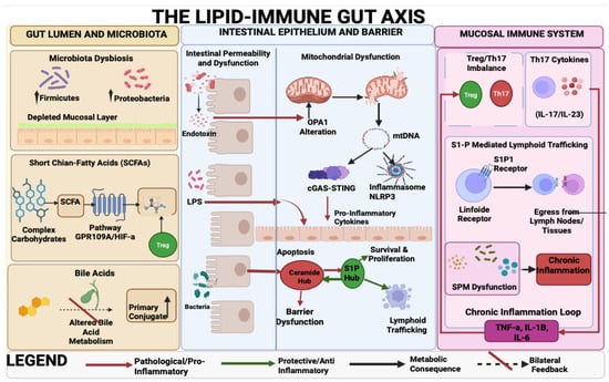
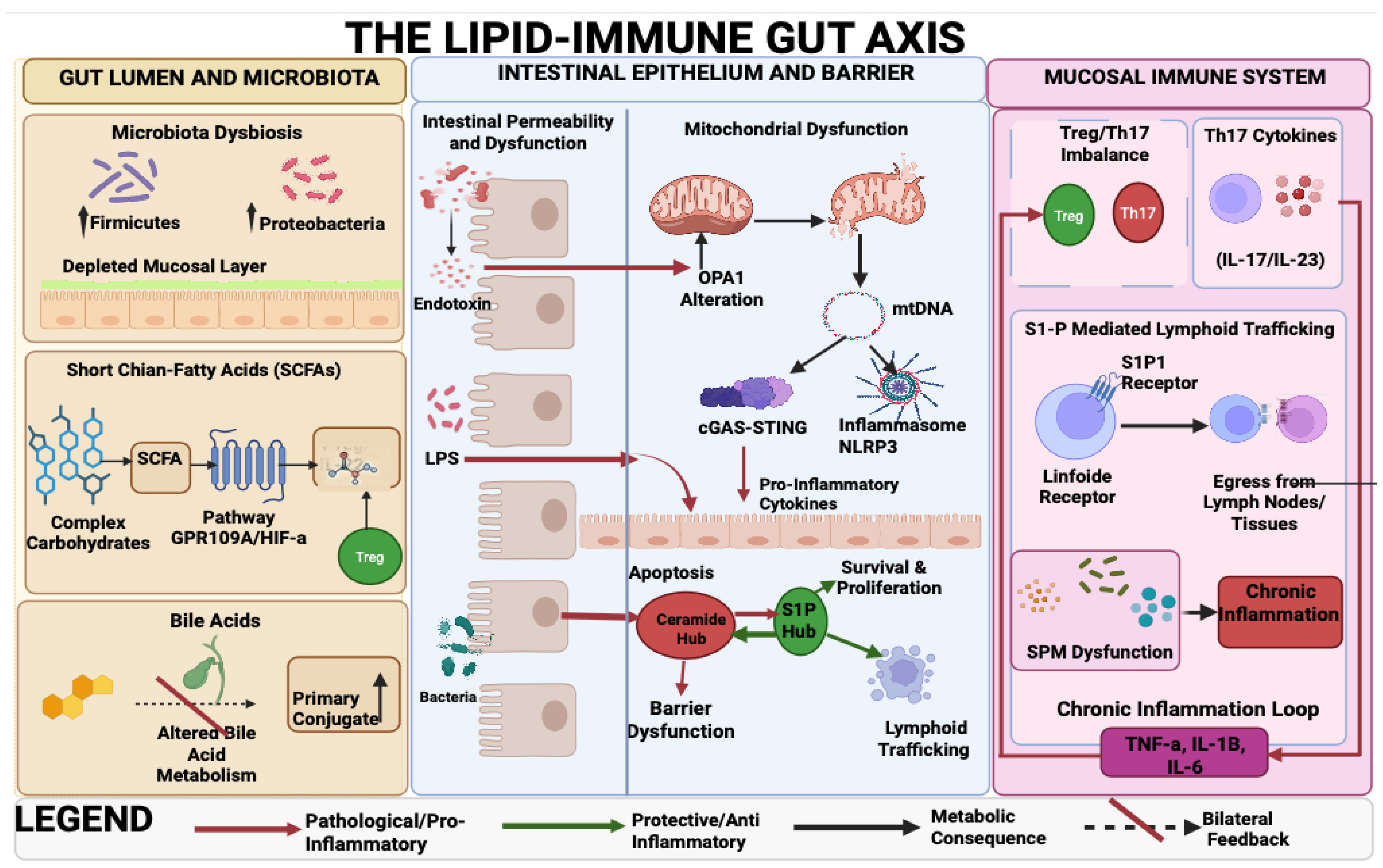
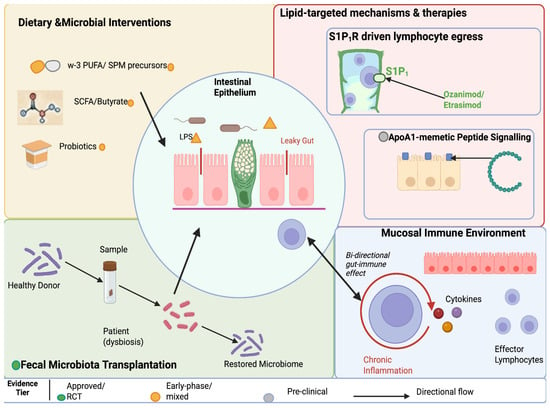
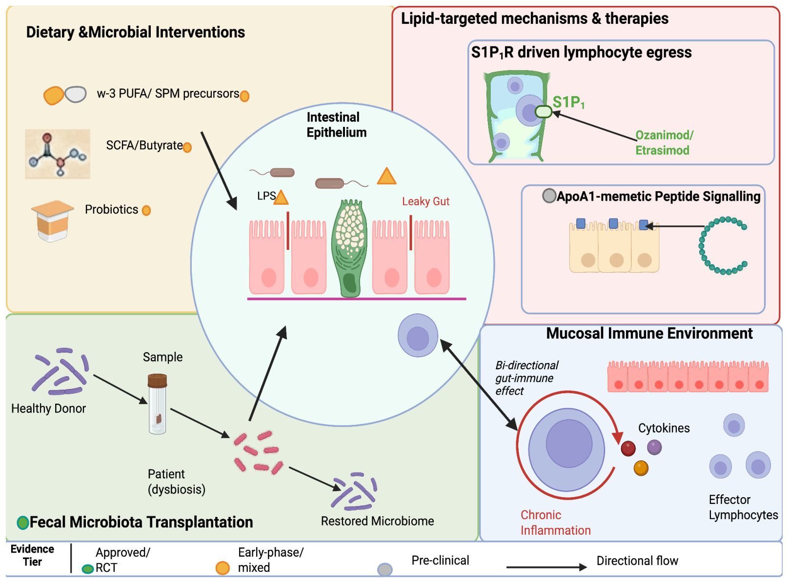

Disclaimer/Publisher’s Note: The statements, opinions and data contained in all publications are solely those of the individual author(s) and contributor(s) and not of MDPI and/or the editor(s). MDPI and/or the editor(s) disclaim responsibility for any injury to people or property resulting from any ideas, methods, instructions or products referred to in the content. |
© 2026 by the authors. Licensee MDPI, Basel, Switzerland. This article is an open access article distributed under the terms and conditions of the Creative Commons Attribution (CC BY) license.