Persistence and Risk Assessment of Biofilm-Forming MDR and XDR Bacteria on Non-Poultry Meat Contact Surfaces in Wah Cantt, Pakistan



Abstract
1. Impact Statement/Summary
2. Introduction
3. Materials and Methods
3.1. Study Area and Design
3.2. Sample Size and Collection
3.3. Sampling Technique
3.4. Sample Processing, Isolation, and Purification of Bacteria
3.5. Biofilm Formation Assays
- •
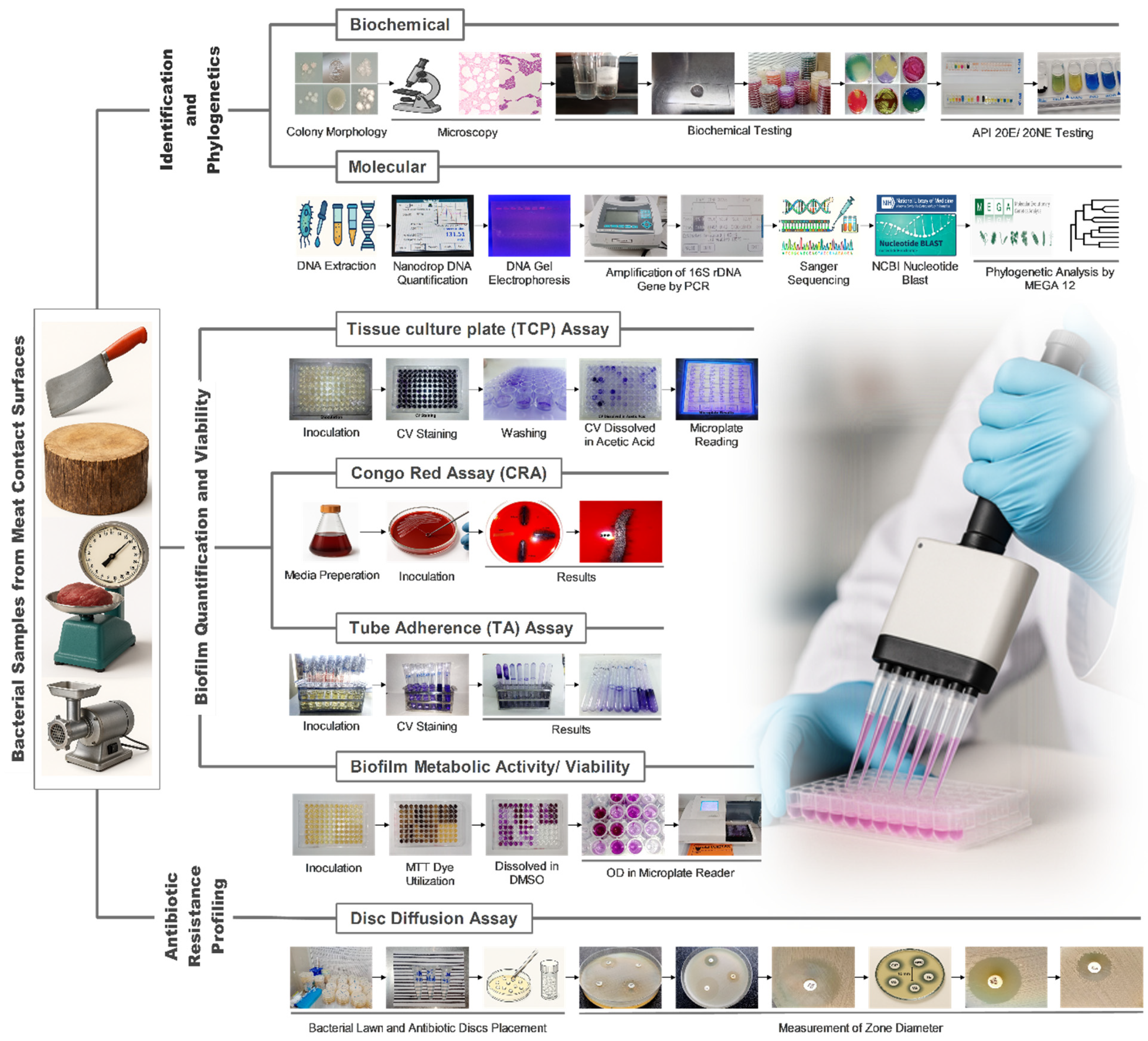
- Congo Red Agar Assay: Bacterial isolates were streaked onto Congo red agar plates composed of brain heart infusion agar supplemented with sucrose and Congo red dye. The inoculated plates were incubated at 37 °C for 24 h. Black colonies with a dry, crystalline appearance were interpreted as biofilm producers using the method described by Freeman et al. [21].
- •
- Tube Adherence Assay: Bacterial isolates were inoculated into sterile test tubes containing tryptic soy broth and incubated at 37 °C for 24 h. After incubation, the tubes were gently washed three times with phosphate-buffered saline to remove non-adherent cells, then stained with 0.1% crystal violet. Biofilm formation was evaluated qualitatively based on the presence and intensity of a visible film lining the inner walls and bottom of the tubes, as previously described by Reddy et al. [22].
- •
- Tissue Culture Plate Assay: Bacterial isolates were inoculated into sterile 96-well flat-bottom microtiter plates, with each well containing 200 µL of trypticase soy broth adjusted to the desired inoculum density (typically equivalent to a 0.5 McFarland standard). Plates were incubated statically at 37 °C for 24 h to allow biofilm formation. Following incubation, non-adherent cells were removed by washing the wells three times with 200 µL of phosphate-buffered saline. The plates were then air-dried and stained with 200 µL of 0.1% crystal violet for 15 min. Excess stain was gently rinsed off, and bound dye was subsequently solubilized with 200 µL of 30% acetic acid for 15 min. Biofilm biomass was quantified by measuring optical density at 570 nm with a microplate reader, using a cut-off OD (ODC), as described by Stepanović et al. [23].
3.6. Biochemical and Molecular Identification of Bacteria

3.7. Phylogenetic Analysis
3.8. Antibiotic Susceptibility Testing
3.9. Biofilm Biomass and Metabolic Activity Assessment
3.10. Statistical Methods
4. Results
4.1. Biofilm Formation and Resistance Correlation
4.2. Taxonomic Distribution and Source Characterization
- •
- a Richness (S): Total number of distinct bacterial families identified among the strong biofilm-producing isolates per surface.
- •
- b Shannon-Wiener Index (H′): Calculated as , representing taxonomic diversity within the biofilm-forming consortium.
- •
- c Pielou’s Evenness (J′): Calculated as . High evenness values (>0.75) indicate that resistance traits are not confined to a single family but are distributed across a balanced multi-family community.
4.3. Phylogenetic Relationships
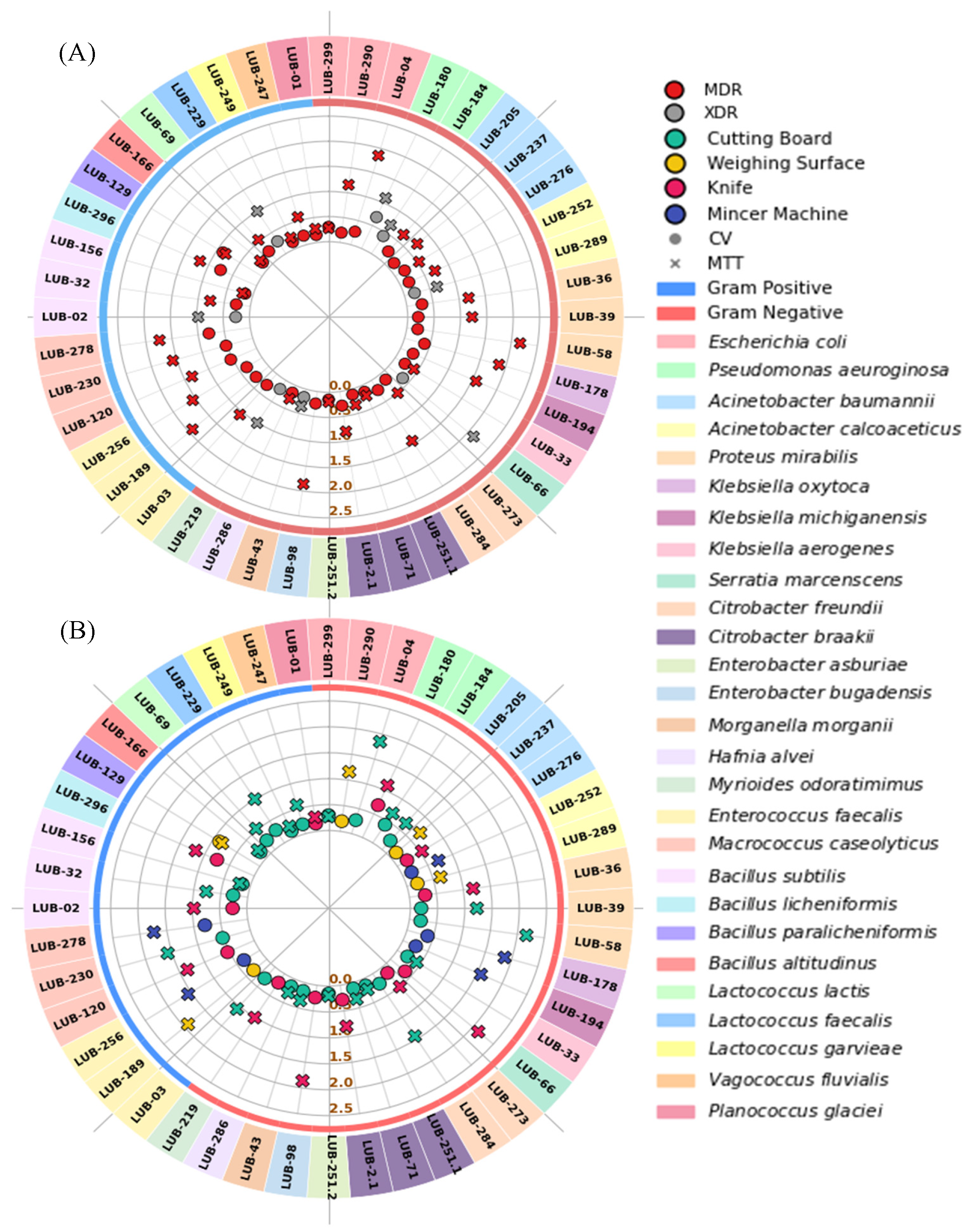
4.4. Antibiotic Resistance Polarity and Surface Hotspots
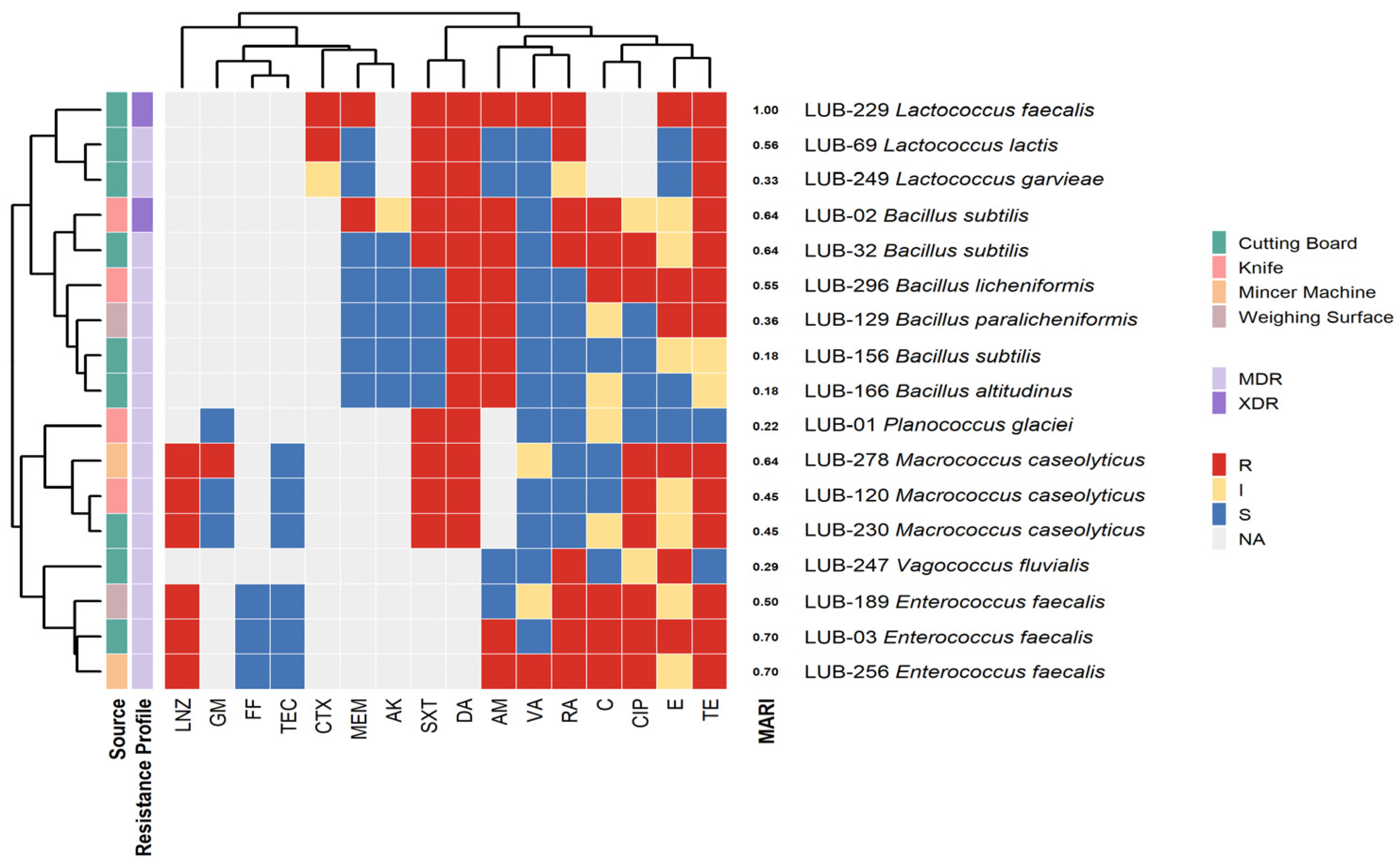
4.5. Clustering of Resistotypes and Critical Spatial AMR Signatures
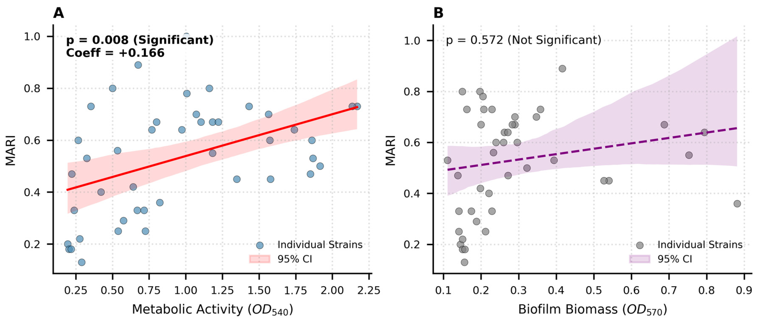
4.6. Quantitative Biofilm Dynamics: Biomass vs. Metabolic Activity

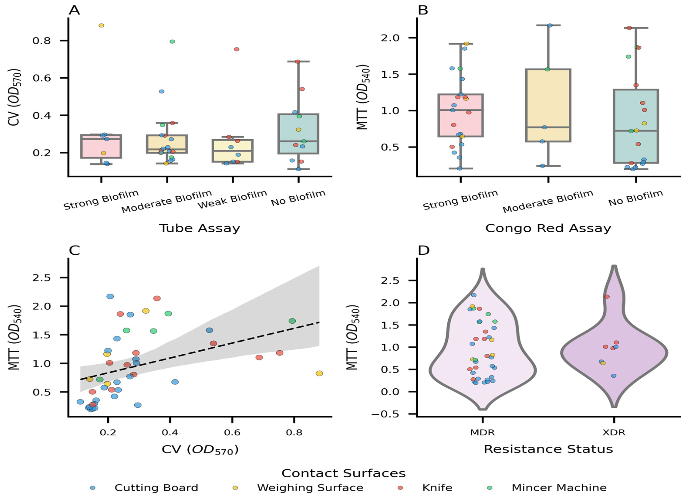
4.7. Integrated Analysis of Biofilm Phenotypes, Metabolic Fitness, and Antimicrobial Resistance Associations


5. Discussion
5.1. Phylogenetic Resolution and Taxonomic Diversity Analysis
5.2. The Biofilm-Resistance Nexus
5.3. Public Health Implications of XDR Reservoirs
5.4. Methodological and Genomic Limitations
6. Conclusions
Supplementary Materials
Author Contributions
Funding
Institutional Review Board Statement
Informed Consent Statement
Data Availability Statement
Acknowledgments
Conflicts of Interest
References
- Donlan, R.M.; Costerton, J.W. Biofilms: Survival Mechanisms of Clinically Relevant Microorganisms. Clin. Microbiol. Rev. 2002, 15, 167–193. [Google Scholar] [CrossRef] [PubMed]
- Srey, S.; Jahid, I.K.; Ha, S. Do Biofilm Formation in Food Industries: A Food Safety Concern. Food Control 2013, 31, 572–585. [Google Scholar] [CrossRef]
- Global Antimicrobial Resistance Surveillance System; World Health Organization. Global Antimicrobial Resistance and Use Surveillance System (GLASS) Report; World Health Organization: Geneva, Switzerland, 2022. [Google Scholar]
- Rahman, S.U.; Mohsin, M. The Under Reported Issue of Antibiotic-Resistance in Food-Producing Animals in Pakistan. Pak. Vet. J. 2019, 39, 323–328. [Google Scholar] [CrossRef]
- Wu, R.A.; Feng, J.; Yue, M.; Liu, D.; Ding, T. Overuse of Food-Grade Disinfectants Threatens a Global Spread of Antimicrobial-Resistant Bacteria. Crit. Rev. Food Sci. Nutr. 2024, 64, 6870–6879. [Google Scholar] [CrossRef]
- Van Boeckel, T.P.; Pires, J.; Silvester, R.; Zhao, C.; Song, J.; Criscuolo, N.G.; Gilbert, M.; Bonhoeffer, S.; Laxminarayan, R. Global Trends in Antimicrobial Resistance in Animals in Low- and Middle-Income Countries. Science 2019, 365, eaaw1944. [Google Scholar] [CrossRef]
- One Health Joint Plan of Action (2022–2026): Working Together for the Health of Humans, Animals, Plants and the Environment. Available online: https://www.who.int/publications/i/item/9789240059139 (accessed on 27 April 2026).
- Pinho, J.O.; Coelho, A.C.; Oliveira, P.; Figueiras, A.; Roque, F.; Herdeiro, M.T. Tackling Antibiotic Resistance Threat through the One Health Approach: A Focus on Veterinary Medicine. In Sustainable Animal Agriculture-Global Challenges and Practical Solutions; IntechOpen: Rijeka, Croatia, 2025; ISBN 978-0-85466-502-0. [Google Scholar]
- European Food Safety Authority (EFSA) and European Centre for Disease Prevention and Control (ECDC). The European Union Summary Report on Antimicrobial Resistance in Zoonotic and Indicator Bacteria from Humans, Animals and Food in 2021–2022. EFSA J. 2024, 22, e8583. [Google Scholar] [PubMed]
- Qiu, Y.; Ferreira, J.P.; Ullah, R.W.; Flanagan, P.; Zaheer, M.U.; Tahir, M.F.; Alam, J.; Hoet, A.E.; Song, J.; Akram, M. Assessment of the Implementation of Pakistan’s National Action Plan on Antimicrobial Resistance in the Agriculture and Food Sectors. Antibiotics 2024, 13, 206. [Google Scholar] [CrossRef]
- Fatima, A.; Saleem, M.; Nawaz, S.; Khalid, L.; Riaz, S.; Sajid, I. Prevalence and Antibiotics Resistance Status of Salmonella in Raw Meat Consumed in Various Areas of Lahore, Pakistan. Sci. Rep. 2023, 13, 22205. [Google Scholar] [CrossRef] [PubMed]
- Ali, N.H.; Farooqui, A.; Khan, A.; Khan, A.Y.; Kazmi, S.U. Microbial Contamination of Raw Meat and Its Environment in Retail Shops in Karachi, Pakistan. J. Infect. Dev. Ctries. 2010, 4, 382–388. [Google Scholar] [CrossRef]
- Pankratov, T.A.; Nikolaev, Y.A.; Yushina, Y.K.; Tikhonova, E.N.; El-Registan, G.I. Forms of Bacterial Survival in Model Biofilms. Coatings 2022, 12, 1913. [Google Scholar] [CrossRef]
- Sadiq, A.; Samad, M.; Saddam; Basharat, N.; Ali, S.; Roohullah; Saad, Z.; Khan, A.N.; Ahmad, Y.; Khan, A.; et al. Methicillin-Resistant Staphylococcus Aureus (MRSA) in Slaughter Houses and Meat Shops in Capital Territory of Pakistan During 2018–2019. Front. Microbiol. 2020, 11, 577707. [Google Scholar] [CrossRef] [PubMed]
- Antimicrobial Resistance in Biofilms: Executive Summary|Food Standards Agency. Available online: https://www.food.gov.uk/research/antimicrobial-resistance-in-biofilms-executive-summary (accessed on 26 April 2026).
- Pang, X.; Hu, X.; Du, X.; Lv, C.; Yuk, H.-G. Biofilm Formation in Food Processing Plants and Novel Control Strategies to Combat Resistant Biofilms: The Case of Salmonella spp. Food Sci. Biotechnol. 2023, 32, 1703–1718. [Google Scholar] [CrossRef] [PubMed]
- Al-Asmari, F.; Hamad, S.H.; Al Hashedi, S.A. Prevalence of Enterobacteriaceae in Camel, Cattle, and Sheep Carcasses at Slaughterhouses and Butcher Shops. Appl. Sci. 2023, 13, 11495. [Google Scholar] [CrossRef]
- Desalegn, S.; Abaya, G.; G’Yesus, T.; Admasu, D.; Mussema, A. Prevalence of Intestinal Parasites, Salmonella, Shigella and Antimicrobial Resistance of Bacterial Isolates among Food Handlers in Wachemo University Students’ Food Service Facility, Southcentral Ethiopia: A Cross-Sectional Study. IJID Reg. 2025, 15, 100617. [Google Scholar] [CrossRef] [PubMed]
- Wang, S.S.; Canida, T.A.; Ihrie, J.D.; Chirtel, S.J. Sample Size Determination for Food Sampling. J. Food Prot. 2023, 86, 100134. [Google Scholar] [CrossRef]
- Ahmed, S.K. How to Choose a Sampling Technique and Determine Sample Size for Research: A Simplified Guide for Researchers. Oral. Oncol. Rep. 2024, 12, 100662. [Google Scholar] [CrossRef]
- Freeman, D.J.; Falkiner, F.R.; Keane, C.T. New Method for Detecting Slime Production by Coagulase Negative Staphylococci. J. Clin. Pathol. 1989, 42, 872–874. [Google Scholar] [CrossRef]
- Reddy, K.R.M. Tube Adherence Test as a Screening Tool for Detection of Biofilm Formation Among Staphylococcus Aureus. Int. J. Curr. Microbiol. Appl. Sci. 2017, 6, 1325–1329. [Google Scholar] [CrossRef]
- Stepanović, S.; Vuković, D.; Hola, V.; Bonaventura, G.D.; Djukić, S.; Ćirković, I.; Ruzicka, F. Quantification of Biofilm in Microtiter Plates: Overview of Testing Conditions and Practical Recommendations for Assessment of Biofilm Production by Staphylococci. Apmis 2007, 115, 891–899. [Google Scholar] [CrossRef] [PubMed]
- Bergey, D.H.; Breed, R.S. Bergey’s Manual of Determinative Bacteriology, 9th ed.; Holt, J.G., Ed.; Williams & Wilkins Co.: Baltimore, MA, USA, 1994. [Google Scholar]
- Wilson, K. Preparation of Genomic DNA from Bacteria. Curr. Protoc. Mol. Biol. 2001, 56, 2.4.1–2.4.5. [Google Scholar] [CrossRef]
- Weisburg, W.G.; Barns, S.M.; Pelletier, D.A.; Lane, D.J. 16S Ribosomal DNA Amplification for Phylogenetic Study. J. Bacteriol. 1991, 173, 697–703. [Google Scholar] [CrossRef]
- Benov, L. Effect of Growth Media on the MTT Colorimetric Assay in Bacteria. PLoS ONE 2019, 14, e0219713. [Google Scholar] [CrossRef]
- Magiorakos, A.P.; Srinivasan, A.; Carey, R.B.; Carmeli, Y.; Falagas, M.E.; Giske, C.G.; Harbarth, S.; Hindler, J.F.; Kahlmeter, G.; Olsson-Liljequist, B.J.C.M.; et al. Multidrug-Resistant, Extensively Drug-Resistant and Pandrug-Resistant Bacteria: An International Expert Proposal for Interim Standard Definitions for Acquired Resistance. Clin. Microbiol. Infect. 2012, 18, 268–281. [Google Scholar] [CrossRef]
- Ullah, M.J.; Ahmad, K.; Khattak, F.; Inayat, F.; Huriya, S.; Menhaj, N. Prevalence of Multidrug-Resistant and Biofilm-Forming Salmonella Enterica in Beef from Peshawar. Maj. Kedokt. Bdg. 2026, 58, 1–9. [Google Scholar] [CrossRef]
- The WHO AWaRe (Access, Watch, Reserve) Antibiotic Book. Available online: https://iris.who.int/items/e3a52660-f2d4-4e3c-a311-e48418ad21e0 (accessed on 26 April 2026).
- Galié, S.; García-Gutiérrez, C.; Miguélez, E.M.; Villar, C.J.; Lombó, F. Biofilms in the Food Industry: Health Aspects and Control Methods. Front. Microbiol. 2018, 9, 315815. [Google Scholar] [CrossRef] [PubMed]
- AWaRe Classification of Antibiotics for Evaluation and Monitoring of Use. 2023. Available online: https://www.who.int/publications/i/item/WHO-MHP-HPS-EML-2023.04 (accessed on 28 April 2026).
- Detection of Biofilm Formation Among the Clinical Isolates of Staphylococci: An Evaluation of Three Different Screening Methods-PubMed. Available online: https://pubmed.ncbi.nlm.nih.gov/16505551/ (accessed on 26 April 2026).
- Prajapati, R.P.; Parul; Jain, U.; Sharma, B.; Mishra, R.P.; Singh, S.P.G.; Jeet, V.; Seth, K. Evaluation of Biofilm Production Capability of Shiga Toxin Producing Escherichia Coli Isolated from Milk, Milk Products and Hand Swabs. Asian J. Dairy Food Res. 2025, 1–6. [Google Scholar] [CrossRef]
- Almutairy, B. Extensively and Multidrug-Resistant Bacterial Strains: Case Studies of Antibiotics Resistance. Front. Microbiol. 2024, 15, 1381511. [Google Scholar] [CrossRef]
- Elshobary, M.E.; Badawy, N.K.; Ashraf, Y.; Zatioun, A.A.; Masriya, H.H.; Ammar, M.M.; Mohamed, N.A.; Mourad, S.; Assy, A.M. Combating Antibiotic Resistance: Mechanisms, Multidrug-Resistant Pathogens, and Novel Therapeutic Approaches: An Updated Review. Pharmaceuticals 2025, 18, 402. [Google Scholar] [CrossRef]
- Molecular Mechanisms of Biofilm-Based Antibiotic Resistance and Tolerance in Pathogenic Bacteria|FEMS Microbiology Reviews|Oxford Academic. Available online: https://academic.oup.com/femsre/article/41/3/276/3089981?login=false (accessed on 26 April 2026).
- Mirghani, R.; Saba, T.; Khaliq, H.; Mitchell, J.; Do, L.; Chambi, L.; Diaz, K.; Kennedy, T.; Alkassab, K.; Huynh, T.; et al. Biofilms: Formation, Drug Resistance and Alternatives to Conventional Approaches. AIMS Microbiol. 2022, 8, 239–277. [Google Scholar] [CrossRef]
- Frontiers|Synergistic Interactions in Multispecies Biofilm Combinations of Bacterial Isolates Recovered from Diverse Food Processing Industries. Available online: https://www.frontiersin.org/journals/microbiology/articles/10.3389/fmicb.2023.1159434/full (accessed on 26 April 2026).
- de Brito, F.A.; de Freitas, A.P.; Nascimento, M.S. Multidrug-Resistant Biofilms (MDR): Main Mechanisms of Tolerance and Resistance in the Food Supply Chain. Pathogens 2022, 11, 1416. [Google Scholar] [CrossRef] [PubMed]
- Bali, P.; Minhas, N. Isolation and Characterization of Multi-Drug Resistant (MDR) and Extensively Drug Resistant (XDR) Bacterial Pathogens from Diverse Environmental Niches of Shimla, Himachal Pradesh (India). Prepr. Res. Sq. 2023, 15, 398–408. [Google Scholar] [CrossRef]
- Sharland, M.; Gandra, S.; Huttner, B.; Moja, L.; Pulcini, C.; Zeng, M.; Mendelson, M.; Cappello, B.; Cooke, G.; Magrini, N.; et al. Encouraging AWaRe-Ness and Discouraging Inappropriate Antibiotic Use—The New 2019 Essential Medicines List Becomes a Global Antibiotic Stewardship Tool. Lancet Infect. Dis. 2019, 19, 1278–1280. [Google Scholar] [CrossRef]
- Global Action Plan on Antimicrobial Resistance. Available online: https://www.who.int/publications/i/item/9789241509763 (accessed on 27 April 2026).
- Founou, L.L.; Founou, R.C.; Essack, S.Y. Antibiotic Resistance in the Food Chain: A Developing Country-Perspective. Front. Microbiol. 2016, 7, 1881. [Google Scholar] [CrossRef] [PubMed]
- Brás, A.; Braz, M.; Martinho, I.; Duarte, J.; Pereira, C.; Almeida, A. Effect of Bacteriophages against Biofilms of Escherichia Coli on Food Processing Surfaces. Microorganisms 2024, 12, 366. [Google Scholar] [CrossRef]
- Sornchuer, P.; Saninjuk, K.; Amonyingcharoen, S.; Ruangtong, J.; Thongsepee, N.; Martviset, P.; Chantree, P.; Sangpairoj, K. Whole Genome Sequencing Reveals Antimicrobial Resistance and Virulence Genes of Both Pathogenic and Non-Pathogenic B. cereus Group Isolates from Foodstuffs in Thailand. Antibiotics 2024, 13, 245. [Google Scholar] [CrossRef]



| Surface Material | Swabs (N) | Total Recovered Isolates (n) | Strong Biofilm Formers | MDR (%) | XDR (%) | Mean MAR Index | Richness (S) a | Shannon (H′) b | Evenness (J′) c | Predominant Family (%) |
|---|---|---|---|---|---|---|---|---|---|---|
| Wood (Cutting Boards) | 30 | 97 | 22 | 86% | 14% | 0.51 | 7 | 1.53 | 0.79 | Enterobacteriaceae (50.0%) |
| Stainless Steel (Knives) | 30 | 96 | 10 | 70% | 30% | 0.6 | 5 | 1.36 | 0.84 | Enterobacteriaceae (50.0%) |
| Steel/Iron (Mincing Machines) | 30 | 34 | 5 | 100% | 0% | 0.56 | 4 | 1.33 | 0.96 | Enterobacteriaceae (40.0%) |
| Plastic/Mixed (Weighing Surface) | 30 | 73 | 5 | 80% | 20% | 0.45 | 4 | 1.33 | 0.96 | Moraxellaceae (40.0%) |
| Total/Overall | 120 | 300 | 42 | - | - | - | - | - | - | - |
| Surface Niche | Bacterial Family | Strong Biofilm Formers (n = 42) | Resistance Classes (n) | Critical Resistance Phenotype | Highest AWaRe Category a Compromised. | Mean MAR Index | Mean Biomass (OD570 ± SD) | Mean Viability (OD540 ± SD) |
|---|---|---|---|---|---|---|---|---|
| Wood (Cutting Boards) | Enterobacteriaceae | 11 | 10 | Polymyxins, Cephalosporins, Fluoroquinolones | Reserve | 0.51 | 0.19 ± 0.06 | 0.79 ± 0.73 |
| Bacillaceae | 3 | 7 | Fluoroquinolones | Watch | 0.33 | 0.19 ± 0.07 | 0.40 ± 0.32 | |
| Streptococcaceae | 3 | 9 | Carbapenems, Cephalosporins, Glycopeptides | Watch | 0.63 | 0.22 ± 0.08 | 0.59 ± 0.39 | |
| Enterococcaceae | 2 | 7 | Fluoroquinolones, Linezolid (Oxazolidinones), Macrolides | Reserve | 0.49 | 0.24 ± 0.07 | 0.82 ± 0.35 | |
| Moraxellaceae | 1 | 4 | Polymyxins, Cephalosporins, Fluoroquinolones | Reserve | 0.33 | 0.23 | 0.67 | |
| Pseudomonadaceae | 1 | 6 | Carbapenems, Polymyxins, Cephalosporins | Reserve | 0.89 | 0.42 | 0.68 | |
| Staphylococcaceae | 1 | 5 | Fluoroquinolones, Linezolid (Oxazolidinones) | Reserve | 0.45 | 0.53 | 1.58 | |
| Stainless Steel (Knives) | Enterobacteriaceae | 5 | 10 | Carbapenems, Polymyxins, Cephalosporins | Reserve | 0.69 | 0.26 ± 0.08 | 1.30 ± 0.69 |
| Bacillaceae | 2 | 9 | Carbapenems, Fluoroquinolones, Macrolides | Watch | 0.59 | 0.51 ± 0.35 | 1.08 ± 0.15 | |
| Moraxellaceae | 1 | 3 | Polymyxins, Fluoroquinolones | Reserve | 0.25 | 0.21 | 0.54 | |
| Pseudomonadaceae | 1 | 5 | Carbapenems, Polymyxins, Fluoroquinolones | Reserve | 0.67 | 0.69 | 1.1 | |
| Staphylococcaceae | 1 | 5 | Fluoroquinolones, Linezolid (Oxazolidinones) | Reserve | 0.45 | 0.54 | 1.35 | |
| Steel/Iron (Mincing Machines) | Enterobacteriaceae | 2 | 8 | Polymyxins, Cephalosporins, Fluoroquinolones | Reserve | 0.56 | 0.33 ± 0.09 | 1.72 ± 0.21 |
| Enterococcaceae | 1 | 7 | Polymyxins, Fluoroquinolones, Glycopeptides | Reserve | 0.7 | 0.35 | 1.56 | |
| Moraxellaceae | 1 | 4 | Polymyxins, Cephalosporins, Beta-Lactam/Inhibitors | Reserve | 0.33 | 0.17 | 0.72 | |
| Staphylococcaceae | 1 | 7 | Fluoroquinolones, Glycopeptides, Linezolid (Oxazolidinones) | Reserve | 0.64 | 0.79 | 1.74 | |
| Plastic/Mixed (Weighing Surface) | Moraxellaceae | 2 | 4 | Polymyxins, Cephalosporins, Aminoglycosides | Reserve | 0.33 | 0.17 ± 0.04 | 0.68 ± 0.06 |
| Bacillaceae | 1 | 4 | Macrolides | Watch | 0.36 | 0.88 | 0.82 | |
| Enterobacteriaceae | 1 | 9 | Carbapenems, Polymyxins, Cephalosporins | Reserve | 0.8 | 0.2 | 1.16 | |
| Enterococcaceae | 1 | 5 | Fluoroquinolones, Linezolid (Oxazolidinones) | Reserve | 0.5 | 0.32 | 1.92 |
Disclaimer/Publisher’s Note: The statements, opinions and data contained in all publications are solely those of the individual author(s) and contributor(s) and not of MDPI and/or the editor(s). MDPI and/or the editor(s) disclaim responsibility for any injury to people or property resulting from any ideas, methods, instructions or products referred to in the content. |
© 2026 by the authors. Licensee MDPI, Basel, Switzerland. This article is an open access article distributed under the terms and conditions of the Creative Commons Attribution (CC BY) license.
Share and Cite
Shakoor, L.; Naz, S.; Rashid, A.; Idrees, M. Persistence and Risk Assessment of Biofilm-Forming MDR and XDR Bacteria on Non-Poultry Meat Contact Surfaces in Wah Cantt, Pakistan. Microorganisms 2026, 14, 1051. https://doi.org/10.3390/microorganisms14051051
Shakoor L, Naz S, Rashid A, Idrees M. Persistence and Risk Assessment of Biofilm-Forming MDR and XDR Bacteria on Non-Poultry Meat Contact Surfaces in Wah Cantt, Pakistan. Microorganisms. 2026; 14(5):1051. https://doi.org/10.3390/microorganisms14051051
Chicago/Turabian StyleShakoor, Lubna, Shumaila Naz, Anas Rashid, and Muhammad Idrees. 2026. "Persistence and Risk Assessment of Biofilm-Forming MDR and XDR Bacteria on Non-Poultry Meat Contact Surfaces in Wah Cantt, Pakistan" Microorganisms 14, no. 5: 1051. https://doi.org/10.3390/microorganisms14051051
APA StyleShakoor, L., Naz, S., Rashid, A., & Idrees, M. (2026). Persistence and Risk Assessment of Biofilm-Forming MDR and XDR Bacteria on Non-Poultry Meat Contact Surfaces in Wah Cantt, Pakistan. Microorganisms, 14(5), 1051. https://doi.org/10.3390/microorganisms14051051