Article

A Retrospective Study on the Aetiology of Clinical Bovine Mastitis and Its Antibiotic Resistance Profiles in Western Australia Dairy Farms

1
Midwest Vet Centre, 117 Cathedral Avenue, Geraldton, WA 6530, Australia
2
College of Veterinary Medicine, Murdoch University, 90 South Street, Murdoch, WA 6150, Australia
3
Meat Livestock Australia Level 1, 40 Mount Street, North Sydney, NSW 2060, Australia
4
School of Veterinary Science, The University of Queensland, 5391 Warrego Highway, Gatton, QLD 4343, Australia
*
Author to whom correspondence should be addressed.
Microorganisms 2026, 14(1), 254; https://doi.org/10.3390/microorganisms14010254
Submission received: 6 November 2025 / Revised: 13 January 2026 / Accepted: 14 January 2026 / Published: 21 January 2026
(This article belongs to the Section Veterinary Microbiology)

Abstract

Clinical data on antimicrobial profiles are useful for dairy udder health treatment programmes and represents a component of antimicrobial stewardship. The study aimed to determine the bacterial aetiology of clinical mastitis in dairy herds in Western Australia and to evaluate their antibiotic resistance profiles. This retrospective study utilised clinical antimicrobial profile data from two referral diagnostic centres within the region of Western Australia. A total of 545 mastitic samples were submitted for antimicrobial culture and testing over a period of 10 years (2008–2018). Of these, 406 showed bacterial growth and 139 no bacterial growth was observed. The most common isolates were Streptococcus uberis (25.3%), Staphylococcus aureus (17.2%), and Escherichia coli (9.4%). No growth was identified in 25.5% of the mastitis milk samples. The antimicrobial profiles revealed high susceptibilities towards cefuroxime (95.7%), clavulox (89.4%), and oxytetracycline (89%), whilst showing high resistance towards novobiovin (70%). From this study, it is concluded that there was a decline in the resistance trends towards the isolates of both S. uberis and S. aureus over the 10-year period and contagious mastitis had a higher occurrence. There is a need to consider surveillance programmes that determine the patterns of on-farm antimicrobial usage and further characterise the pathogens based on the presence of resistance antimicrobial genes. Data on antimicrobial surveillance represent an important component of antimicrobial stewardship.

1. Introduction

Mastitis is a significant and costly disease in dairy herds across many countries [1,2,3]. It has been reported to be the second most common reason for involuntary culling after reproductive issues [4,5,6]. According to Countdown 2020 published by Dairy Australia, the average cost to treat a single cow for mastitis was estimated to be $270 [7]. It was also estimated that Australian farmers lose more than $150 million per annum through poor udder health of their dairy cows [7]. Major costs reported include milk production losses, discarded milk, veterinary services, drugs, labour, and premature culling [8,9,10]. Apart from causing significant economic losses, mastitis also has negative impacts on an animal’s welfare and is one of the main reasons for antimicrobial use in dairy herds [11,12,13]. Antibiotics play a significant role in supporting and improving both human and animal health and welfare through the treatment of pathogens [14]. Despite the advocacy for responsible use and good stewardship on the use of antibiotics in veterinary medicine, there has been an increasing trend of antimicrobial resistance at the global level [15,16]. Focusing on the dairy industry, methicillin-resistant S. aureus (MRSA) strains were recently isolated from bovine intramammary infections (IMIs) cases in Argentina and India [17,18]. Good antimicrobial stewardship can greatly reduce the rate of antimicrobial resistance through prudent use of existing antibiotics and replacement of antimicrobial agents with non-antimicrobial alternatives, wherever possible [19,20,21]. There are limited data on the aetiology and antimicrobial profile of clinical mastitis-causing pathogens in Western Australia (WA). Clinical mastitis can be caused by contagious and environmental bacterial pathogens described elsewhere [22]. The transmission and spread of mastitis is caused by several animal-, managemental-, environmental-, and pathogen-level factors described elsewhere [22].
Studies on mastitis in dairy farms in the south-west region of Western Australia did characterise the bacterial pathogens associated with clinical mastitis and subclinical mastitis cases and their associated antimicrobial susceptibility profiles identified the need for ongoing surveillance programmes and pattern profiles of resistance genes among cases of mastitis [23]. Understanding the trends, patterns, and surveillance of resistance against various antibiotics is an important and integral component of good antimicrobial stewardship [24]. The aim of this study was to document the pathogens isolated from clinical mastitis cases within dairy herds in WA over the past 10 years and to evaluate their antimicrobial resistance profile. It is essential to undertake such surveillance to document trends as a reference point for future interventions.

2. Materials and Methods

2.1. Study Design, Area, and Sample Collection

A retrospective study was conducted based on culture and sensitivity results provided by Vetpath, a privately run diagnostic service, and the diagnostic laboratory of the Department of Primary Industries and Regional Development (DPIRDs), Western Australia, formerly the Department of Agriculture Food Western Australia (DAFWA). There is potential bias regarding the type of laboratory centre that processed a clinical sample, possibly due to the preference by the clinician or close proximity to the dairy farms. The milk samples were all new cases and were collected aseptically to avoid the risk of contamination. This included wearing gloves throughout the collection process, fore-stripping of the quarters prior to sample collection, sterile preparation, and drying of teats. A case of clinical mastitis was characterised by physical changes in the milk colour or consistency. Onset of clinical mastitis was first identified via clinical presentation on the udder and teats including swelling, heat, hardness, redness and pain of the udder, and physical changes in the milk. To ensure that accurate results were obtained for culture and sensitivity testing, the milk samples were collected prior to antimicrobial treatment. Approximately 3 to 5 mL of mastitic milk was collected into each collection vial and labelled with the date and animal identification number. The milk samples were transported to the respective laboratories at 4 °C.

2.2. Data Collection, Storage, and Analysis

A total of 260 milk samples were submitted to Vetpath between January 2014 and June 2018 for culture and sensitivity testing. These milk samples were tested for susceptibility against the following eight antibiotics: cefuroxime (CM), clavulox (CX), cloxacillin (CN), lincomycin (L), neomycin (NE), novobiocin (NO), penicillin (P), and oxytetracycline (OX).
A total of 285 mastitic milk samples were submitted to DPIRD between 2008 and 2018 for culture and sensitivity testing. At DPIRD, the milk samples were tested for susceptibility against the following ten antibiotics: clavulox (CX), cloxacillin (CN), lincomycin (L), neomycin (NE), novobiocin (NO), penicillin (P), oxytetracycline (OX), oleandamycin (OL), tylosin (T), and cephalothin (CE).
The two sets of data from Vetpath and DPIRD were combined into one Excel spreadsheet (a total of 545 isolates) and were analysed together. Descriptive statistics (specifically measures of frequency) were used to analyse the isolate and culture and sensitivity test results submitted over the years. S. uberis, S. aureus, and non-haemolytic E. coli were analysed for their resistance profiles over multiple years.

2.3. Culture and Identification

Briefly, in both diagnostic centres, all milk samples were cultured on horse blood agar (HBA), Columbia naladixic acid (CNA), and MacConkey II agar; a Gram stain was performed on each sample. The HBA and CNA agar plates were incubated overnight at 37 °C in 5% carbon dioxide, whereas the MacConkey II agar plates were incubated at 37 °C in oxygen [25]. If growth was present after overnight culture, the bacterial colonies were speciated and tested for susceptibilities against different types of antibiotics.
Bacteria species were identified using biochemical methods including catalase, oxidase, indole and tube coagulase tests, chromogenic agars CPS and mannitol salt agar (MSA), and kit tests such as Streptococcus spp. grouping. A Vivek 2 compact analyser (bioMérieux, Marcy l’Etoile, France) was used to identify bacteria that could not be identified via biochemical methods as it contains a bigger range of biochemical tests. Streptococcus spp. often cannot be identified using the Vivek 2 (v8.01) compact analyser; thus, an API rapid ID strep kit was used if both methods previously mentioned failed to identify the bacterial isolate.
Inoculates were distributed onto Mueller–Hinton (MH) agars and antibiotic discs were placed on top of it. After incubation overnight at 37 °C, zone sizes were measured, and susceptibilities were determined according to CSLI guidelines [26].

3. Results

From the 545 mastitic milk samples submitted, 22 different pathogens (cases) were isolated (Table 1), of which S. uberis had the highest occurrence (138 cases, 25.3%). It was followed by S. aureus (94 cases, 17.2%); E. coli (51 cases, 9.4%); coagulase-negative Staphylococcus spp. (31 cases, 5.7%); Streptococcus dysgalactiae (16 cases, 2.94%); other Streptococcus spp. (15 cases, 2.75%); Enterobacter spp. (10 cases, 1.83%); Klebsiella spp. (8 cases, 1.47%); Serratia spp. (8 cases, 1.5%); and Bacillus spp. (5 cases, 0.92%), with a few cases (15) of minor pathogens. No growth was observed in 139 (25.5%) of the mastitic milk samples.
All the pathogens cultured from the milk samples were tested against the antimicrobial agents listed in Table 2. The isolates were most susceptible to cefuroxime (95.7%), followed by clavulox (89.4%), oxytetracycline (89%), cloxacillin (70.9%), and penicillin (70%). On the other hand, the highest level of resistance among the isolates was against novobiocin (70%), followed by oleandamycin (34.3%) and neomycin (28.6%).
In vitro susceptibility and resistance patterns of all the isolates are shown in Table 3 and Table 4. S. uberis showed complete susceptibility (100%) towards cefuroxime, cephalothin, oleandamycin, and tylosin, while having moderate to good susceptibility towards the remaining antibiotics. S. aureus isolates’ susceptibility patterns were similar to S. uberis. All the antibiotics, except for novobiocin and oleandamycin, were shown to be quite effective against S. aureus, with susceptibility levels above 80%. Novobiocin, however, was very ineffective, with a susceptibility level of only 4%. E. coli had similar susceptibility and resistance patterns towards the antimicrobial agents.
Some of the less frequently cultured bacteria such as Acinetobacter spp., Aerococcus spp., and Aeromonas spp. were demonstrated to be resistant towards all the antimicrobial agents tested in this study. In contrast, isolates including Histophilus somni, Leclercia adecarboxylata, Pantoea spp., Pseudomonas spp., and Sphingomonas paucimobilis were susceptible to all the antimicrobial agents except for novobiocin.
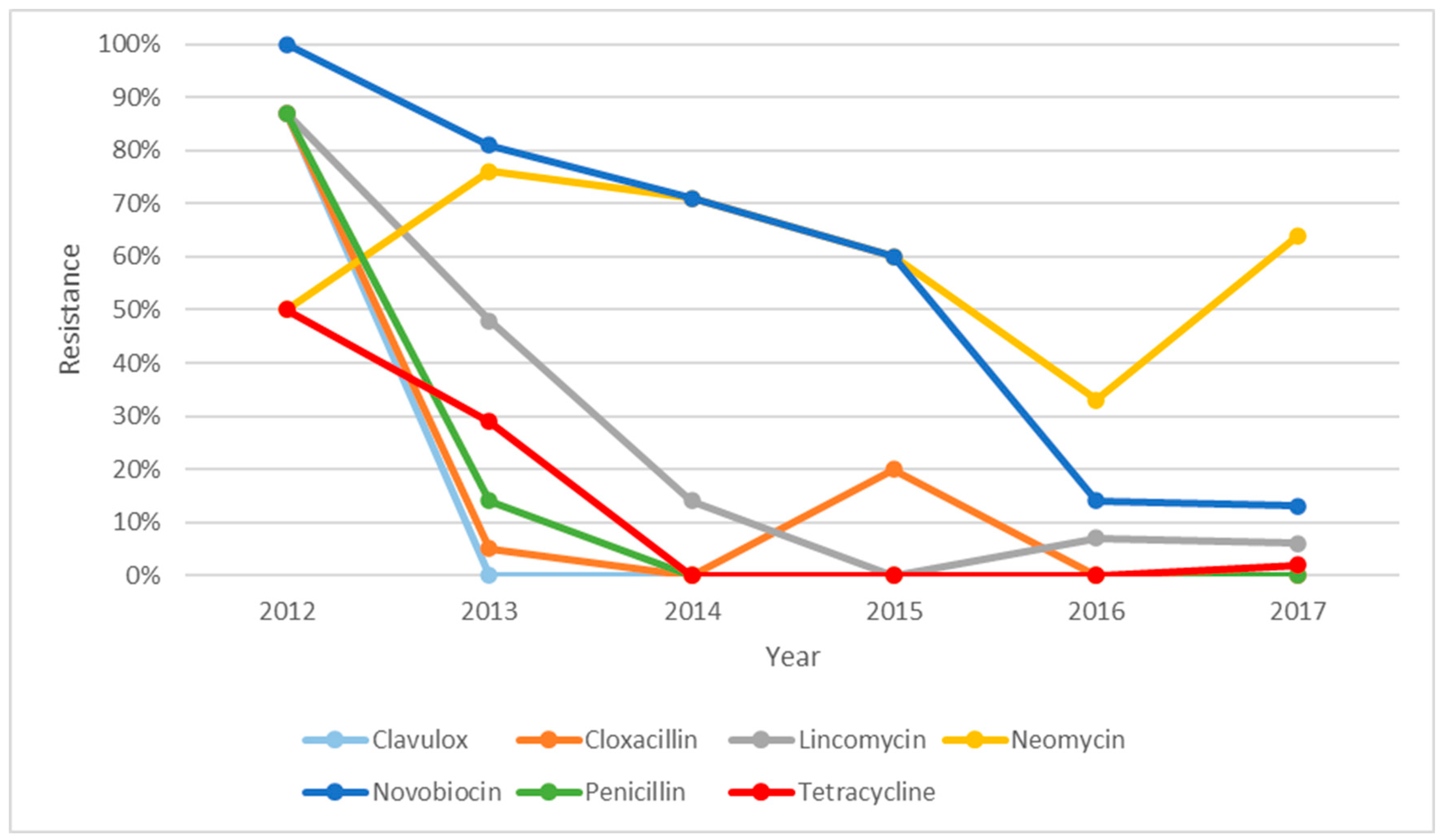
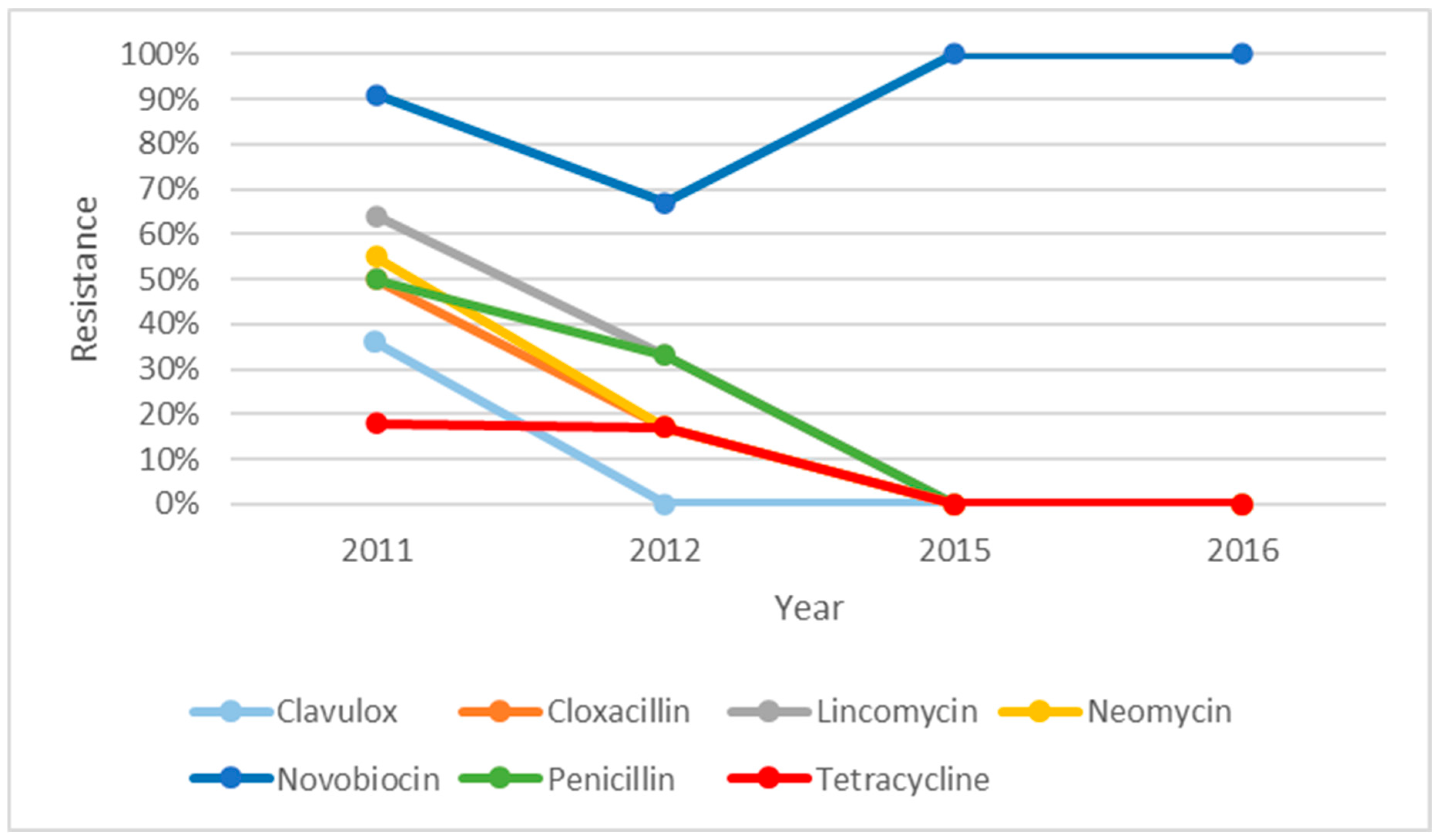
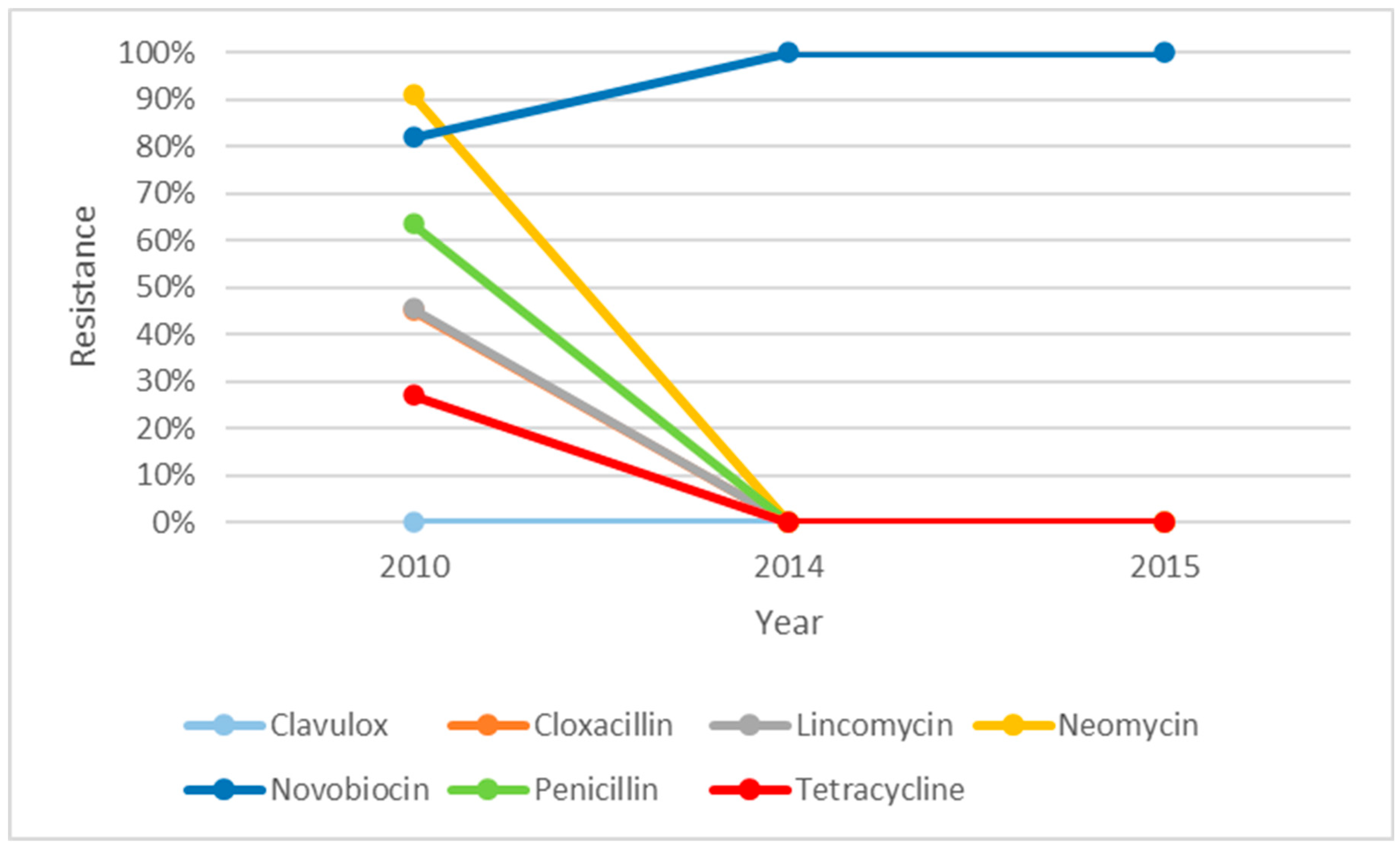
The resistance profile of S. uberis over five years, i.e., from 2012 to 2017, is illustrated in Figure 1. There were descending trends of resistance for all the antibiotics over five years except for neomycin. The resistance against neomycin was seen to be decreasing from 2013 but increased to 65% in 2017. There was an increase in resistance against cloxacillin in 2015; however, it dropped to zero in 2016. Figure 2 shows the resistance profile of S. aureus over four years (2011, 2012, 2015, and 2016). Novobiocin was the only antibiotic that displayed a different trend from the other antibiotics. The percentage of isolates that were resistant towards novobiocin seemed to decrease between 2011 and 2012 but changed to 100% resistance in 2015 and stayed at 100% in 2016. The S. aureus isolates were shown to have steady reduction in resistance towards all the other antibiotics and were zero in 2015 and 2016. As shown in Figure 3, non-haemolytic E. coli showed variable resistance towards all the antibiotics tested except for clavulox; however, it tapered off to no resistance in 2014 and 2015. This was an exception for novobiocin, which showed increased resistance over the years.

4. Discussion

Antimicrobial surveillance is an important component in dairy udder health programmes and in antimicrobial stewardship. The objectives of this study were to identify pathogens isolated from clinical mastitic samples and to evaluate their antimicrobial resistance profiles. Generally, S. uberis was the most common isolate recovered, followed by S. aureus. All the isolates demonstrated high susceptibilities towards cefuroxime, but they showed high resistance towards novobiovin (70%). A limitation to this study was the number of cows included in the analysis. The small sample size and lack of information on the geographical distribution of the mastitis cases across the regions are acknowledged as additional limitations of the study. Despite these limitations, the findings from this study provide a valuable insight into the occurrence and aetiology of mastitis cases and antibiotic resistance profile for S. aureus and S. uberis over the years in the Western Australian dairy industry.
In our study, S. uberis was the most common isolate cultured (34%) from the mastitic milk samples. This is consistent with the findings in other pasture-based systems [27,28] but contrary to the findings of other studies [23]. Despite its reputation as an environmental pathogen, S. uberis also has the potential to cause contagious mastitis and could be spread from cow to cow during milking [29,30]. High prevalence of environmental pathogens usually reflects managemental issues within dairy farms such as poor bedding management, overcrowding, and access to muddy areas [31]; however, this is more commonly seen in systems where dairy cattle are intensely managed or housed. In a pasture-based system, which is more common in Australia, attention to management practices plays an important role to reduce cow-to-cow transmission of contagious Streptococcus spp. [30].
S. aureus, the second most frequently isolated pathogen, is a contagious pathogen and can produce different types of enzymes and toxins that can evade tissue and eventually damage the mammary tissue. It is also known to be challenging to treat as it can survive and form biofilms within fibrous tissue of teat canals, where antibiotics cannot effectively penetrate [13,32]. Furthermore, it is becoming harder for antibiotics to cure mastitis caused by S. aureus as a lot of the strains have acquired the ability to produce beta-lactamase enzymes that could deactivate antibiotics like penicillin [33]. Due to its ability to survive in fibrous tissue and resist against some types of antibiotics, S. aureus has the tendency to cause chronic IMI, which could eventually become untreatable [34]. This reiterates the importance of identifying the aetiology of IMI and determining its susceptibility so that appropriate treatments can be given.
A total of 139 (25.5%) milk samples in our study yielded no growth. This is comparable with the percentage of culture-negative samples detected in other studies [35,36]. Possible reasons include low concentration of bacteria in the sample, improper sample handling and transport, inability of pathogens to grow in standard culture media, and interference from antibiotics or disinfectants [37,38]. A high proportion of culture-negative milk samples collected from cows showing IMI clinical signs can be significant, as identification of causative pathogens is crucial to the selection of effective antimicrobial treatments for the condition. It is important to bear in mind that Mycoplasma bovis and other related Mycoplasma spp. could be suspected of involvement in IMI; however, they are not routinely cultured from mastitis samples unless specifically requested. Recent studies within the dairying region of Western Australia showed a high seroprevalence of mycoplasma in dairy farms [39]. There is a need for surveillance for Mycoplasma in mastitis cases.
The results of this study will support aspects of antimicrobial stewardship in the dairying region in Western Australia. The Australian Veterinary Association (AVA) has urged veterinarians to choose first-line antibiotics, including tetracyclines, sulphonamides, and beta-lactams, for IMI treatments, as part of good antimicrobial stewardship practice [40]. According to the susceptibility profile generated in the study, these first-line antibiotics were proven to maintain their efficacies against common IMI pathogens. Cefuroxime and Clavulox showed the highest susceptibility levels among all the isolates and this is most likely due to their rare use in production animals. On the other hand, novobiocin was shown to be highly ineffective in treating IMI. Fujimoto-Nakamura et al. (2005) [41] suggested that the accumulation of mutant genes in Staph aureus is a factor leading to high-level resistance towards novobiocin. Commercially, novobiocin has been sold in combination with penicillin to increase its spectrum of action [42,43]. Alternative therapies for IMI have been suggested, including non-antimicrobial drug therapies (such as oxytocin) and pre-milking teat disinfection; however, they are less effective compared to aggressive antimicrobial treatments [44,45]. As incidence of IMI is influenced by factors such as management, treatment of clinical cases, stock replacement policy, dry-cow therapy and teat disinfection, alternative therapies, including improved management and husbandry, they might need to be used in conjunction with antimicrobial treatments to achieve the best therapeutic results.

5. Conclusions

This study described the aetiology and frequency of clinical mastitis-causing pathogens, with their antibiotic resistance profile over 10 years within dairy herds in Western Australia. The findings of the study are limited to the geographical location, nature of the retrospective study design, missing data points, as well as data sets such as the number of isolates within each milk sample. Resistance trends for the three major mastitis-causing pathogens (S. uberis, S. aureus, and non-haemolytic E. coli) were demonstrated to decline over the years. There is a need for continued surveillance programmes to determine the epidemiology of on-farm antimicrobial usage and further characterise the pathogens based on the presence of resistance genes.

Author Contributions

Conceptualization, J.W.A.; Methodology, H.C.; Formal analysis, H.C.; Investigation, J.W.A. and H.C.; Resources, J.W.A.; Writing—original draft, H.C.; Writing—review & editing, J.W.A. and M.L.; Supervision, J.W.A. and M.L.; Funding acquisition, J.W.A. All authors have read and agreed to the published version of the manuscript.

Funding

This study was funded by the Dermatology Chapter Australian and New Zealand College of Veterinary Scientists (ANZCVSs) to the recipient Josh Aleri (grant number 17811).

Institutional Review Board Statement

Not applicable.

Informed Consent Statement

Not applicable.

Data Availability Statement

The original contributions presented in this study are included in the article. Further inquiries can be directed to the corresponding author.

Acknowledgments

The authors would like to thank Vetpath and DPIRD for providing the data for this study. The grant recipient at the time was based at Murdoch University in the Department of Ruminant Medicine and Epidemiology. I thank the brilliant colleagues and many students that supported my research to this date. The legacy remains and as the saying goes forward and upwards is the attitude for great science. The authors are thankful to many farmers that submitted study samples to VethPath and DPIRD and laboratory staff members. The authors acknowledge Peter Irwin and Herb Rovay for their valuable critic in the study design. This study is a part of studies dedicated to the late Andrew Fisher.

Conflicts of Interest

Author Hilary Chok was employed by the Midwest Vet Centre, Michael Laurence was employed by the Meat Livestock Australia Level 1. The remaining author declares that the research was conducted in the absence of any commercial or financial relationships that could be construed as a potential conflict of interest.

References

  1. Kossaibati, M.A.; Esslemont, R.J. The costs of production diseases in dairy herds in England. Vet. J. 1997, 154, 41–51. [Google Scholar] [CrossRef]
  2. Sharifi, H.; Sardooei, M.A.; Abadi, M.B.; Babaei, H. Economic impact of mastitis in dairy cows: Case study of Tehran Province, Iran. Iran. J. Vet. Surg. 2014, 9, 39–44. [Google Scholar]
  3. Guimarães, J.L.B.; Brito, M.A.V.P.; Lange, C.C.; Silva, M.R.; Ribeiro, J.B.; Mendonça, J.F.M.; Mendonça, L.C.; Souza, G.N. Estimate of the economic impact of mastitis: A case study in a Holstein dairy herd under tropical conditions. Prev. Vet. Med. 2017, 142, 46–50. [Google Scholar] [CrossRef]
  4. Beaudeau, F.; Henken, A.; Fourichon, C.; Frankena, K.; Seegers, H. Associations between health disorders and culling of dairy cows: A review. Livest. Prod. Sci. 1993, 35, 213–236. [Google Scholar] [CrossRef]
  5. Esslemont, R.J.; Kossaibati, M.A. Culling in 50 dairy herds in England. Vet. Rec. 1997, 140, 36–39. [Google Scholar] [CrossRef]
  6. Petrovski, K.R.; Heuer, C.; Parkinson, T.J.; Williamson, N.B. The incidence and aetiology of clinical bovine mastitis on 14 farms in Northland, New Zealand. N. Z. Vet. J. 2009, 57, 109–115. [Google Scholar] [CrossRef]
  7. Dairy Australia. Countdown 2020: Farm Guidelines for Mastitis Control; Dairy Australia: Southbank, Australia, 2014. Available online: https://catalogue.nla.gov.au/catalog/6486570 (accessed on 8 December 2019).
  8. Seegers, H.; Fourichon, C.; Beaudeau, F. Production effects related to mastitis and mastitis economics in dairy cattle herds. Vet. Res. 2003, 34, 475–491. [Google Scholar] [CrossRef] [PubMed]
  9. Halasa, T.; Huijps, K.; Østerås, O.; Hogeveen, H. Economic effects of bovine mastitis and mastitis management: A review. Vet. Q. 2007, 29, 18. [Google Scholar] [CrossRef] [PubMed]
  10. Nielsen, C. Economic Impact of Mastitis in Dairy Cows. Ph.D. Thesis, Department of Animal Breeding and Genetics, Swedish University of Agricultural Sciences, Uppsala, Sweden, 2009. [Google Scholar]
  11. Mitchell, J.M.; Griffiths, M.W.; McEwen, S.A.; McNab, W.B.; Yee, A.J. Antimicrobial drug residues in milk and meat: Causes, concerns, prevalence, regulations, tests, and test performance. J. Food Prot. 1998, 61, 742. [Google Scholar] [CrossRef]
  12. Hawari, A.D.; Al-Dabbas, F. Prevalence and distribution of mastitis pathogens and their resistance against antimicrobial agents in dairy cows in Jordan. Am. J. Anim. Vet. Sci. 2008, 3, 36–39. [Google Scholar] [CrossRef]
  13. Côté-Gravel, J.; Malouin, F. Symposium review: Features of Staphylococcus aureus mastitis pathogenesis that guide vaccine development strategies. J. Dairy Sci. 2019, 102, 4727–4740. [Google Scholar] [CrossRef]
  14. Etebu, E.; Arikekpar, I. Antibiotics: Classification and mechanisms of action with emphasis on molecular perspectives. Int. J. Appl. Microbiol. Biotechnol. Res. 2016, 4, 90–101. [Google Scholar]
  15. Abraham, S.; Jordan, D.; Wong, H.S.; Johnson, J.R.; Toleman, M.A.; Wakeham, D.L.; Gordon, D.M.; Turnidge, J.D.; Mollinger, J.L.; Gibson, J.S.; et al. First detection of extended-spectrum cephalosporin- and fluoroquinolone-resistant Escherichia coli in Australian food-producing animals. J. Glob. Antimicrob. Resist. 2015, 3, 273–277. [Google Scholar] [CrossRef] [PubMed]
  16. Caniça, M.; Manageiro, V.; Abriouel, H.; Moran-Gilad, J.; Franz, C.M.A.P. Antibiotic resistance in foodborne bacteria. Trends Food Sci. Technol. 2019, 84, 41–44. [Google Scholar] [CrossRef]
  17. Nigam, R. Incidence and pattern of antibiotic resistance of Staphylococcus aureus isolated from clinical and subclinical mastitis in cattle and buffaloes. Asian J. Anim. Sci. 2015, 9, 100–109. [Google Scholar] [CrossRef]
  18. Srednik, M.E.; Crespi, E.; Testorelli, M.F.; Puigdevall, T.; Pereyra, A.M.D.; Rumi, M.V.; Caggiano, N.; Gulone, L.; Mollerach, M.; Gentilini, E.R. First isolation of a methicillin-resistant Staphylococcus aureus from bovine mastitis in Argentina. Vet. Anim. Sci. 2019, 7, 100043, Erratum in Vet. Anim. Sci. 2020, 9, 100156. [Google Scholar] [CrossRef]
  19. Pieterse, R.; Todorov, S.D. Bacteriocins—Exploring alternatives to antibiotics in mastitis treatment. Braz. J. Microbiol. 2010, 41, 542–562. [Google Scholar] [CrossRef] [PubMed]
  20. Szweda, P.; Schielmann, M.; Frankowska, A.; Kot, B.; Zalewska, M. Antibiotic resistance in Staphylococcus aureus strains isolated from cows with mastitis in Eastern Poland and analysis of susceptibility of resistant strains to alternative nonantibiotic agents: Lysostaphin, nisin and polymyxin B. J. Vet. Med. Sci. 2014, 76, 355–362. [Google Scholar] [CrossRef]
  21. Gomes, F.; Henriques, M. Control of bovine mastitis: Old and recent therapeutic approaches. Curr. Microbiol. 2016, 72, 377–382. [Google Scholar] [CrossRef]
  22. Parkinson, T.J.; Vermunt, J.J.; Malmo, J. Diseases of Cattle in Australasia: A Comprehensive Textbook; Massey University Press: Wellington, New Zealand, 2019. [Google Scholar]
  23. Chung, L.K.; Sahibzada, S.; Annandale, H.C.; Robertson, I.D.; Waichigo, F.W.; Tufail, M.S.; Aleri, J.A. Bacterial pathogens associated with clinical and subclinical mastitis in a Mediterranean pasture-based dairy production system of Australia. Res. Vet. Sci. 2021, 141, 103–109, Erratum in Res. Vet. Sci. 2022, 144, 77. [Google Scholar] [CrossRef]
  24. Weese, J.S.; Page, S.; Prescott, J. Antimicrobial stewardship in animals. In Antimicrobial Therapy in Veterinary Medicine, 5th ed.; Giguère, S., Prescott, J.F., Dowling, P.M., Eds.; Wiley Blackwell: Oxford, UK, 2013; pp. 117–132. [Google Scholar]
  25. Lagier, J.-C.; Edouard, S.; Pagnier, I.; Mediannikov, O.; Drancourt, M.; Raoult, D. Current and past strategies for bacterial culture in clinical microbiology. Clin. Microbiol. Rev. 2015, 28, 208–236. [Google Scholar] [CrossRef]
  26. Clinical and Laboratory Standards Institute. Performance Standards for Antimicrobial Disk Susceptibility Test: Approved Standard, 9th ed.; CLSI Document M2-A9; Clinical and Laboratory Standards Institute: Wayne, PA, USA, 2006. [Google Scholar]
  27. McDougall, S. Efficacy of two antibiotic treatments in curing clinical and subclinical mastitis in lactating dairy cows. N. Z. Vet. J. 1998, 46, 226–232. [Google Scholar] [CrossRef]
  28. Petrovski, K.R.; Williamson, N.B.; Lopez-Villalobos, N.; Parkinson, T.J.; Tucker, I.G. Culture results from milk samples submitted to veterinary diagnostic laboratories from August 2003 to December 2006 in New Zealand. N. Z. Vet. J. 2011, 59, 317–322. [Google Scholar] [CrossRef] [PubMed]
  29. Phuektes, P.; Mansell, P.D.; Dyson, R.S.; Hooper, N.D.; Dick, J.S.; Browning, G.F. Molecular epidemiology of Streptococcus uberis isolates from dairy cows with mastitis. J. Clin. Microbiol. 2001, 39, 1460–1466. [Google Scholar] [CrossRef]
  30. Zadoks, R.N.; Fitzpatrick, J.I. Changing trends in mastitis. Ir. Vet. J. 2009, 62, S59–S70. [Google Scholar] [CrossRef] [PubMed]
  31. Hogeveen, H. Mastitis in Dairy Production: Current Knowledge and Future Solutions; Wageningen Academic Publishers: Wageningen, The Netherlands, 2005. [Google Scholar]
  32. Alekish, M.; Al-Qudah, K.; Al-Saleh, A. Prevalence of antimicrobial resistance among bacterial pathogens isolated from bovine mastitis in northern Jordan. Rev. Méd. Vét. 2013, 164, 319–326. [Google Scholar]
  33. Barkema, H.; Schukken, Y.; Zadoks, R. Invited review: The role of cow, pathogen, and treatment regimen in the therapeutic success of bovine Staphylococcus aureus mastitis. J. Dairy Sci. 2006, 89, 1877–1895. [Google Scholar] [CrossRef]
  34. Andreotti, C.S.; Pereyra, E.A.; Baravalle, C.; Renna, M.S.; Ortega, H.H.; Calvinho, L.F.; Dallard, B.E. Staphylococcus aureus chronic intramammary infection modifies protein expression of transforming growth factor beta (TGF-β) subfamily components during active involution. Res. Vet. Sci. 2014, 96, 5–14. [Google Scholar] [CrossRef] [PubMed]
  35. Bradley, A.; Leach, K.; Breen, J.; Green, L.; Green, M. Survey of the incidence and aetiology of mastitis on dairy farms in England and Wales. Vet. Rec. 2007, 160, 253–258. [Google Scholar] [CrossRef]
  36. Koivula, M.; Pitkälä, A.; Pyörälä, S.; Mäntysaari, E.A. Distribution of bacteria and seasonal and regional effects in a new database for mastitis pathogens in Finland. Acta Agric. Scand. A 2007, 57, 89–96. [Google Scholar] [CrossRef]
  37. Taponen, S.; Salmikivi, L.; Simojoki, H.; Koskinen, M.; Pyörälä, S. Real-time polymerase chain reaction-based identification of bacteria in milk samples from bovine clinical mastitis with no growth in conventional culturing. J. Dairy Sci. 2009, 92, 2610–2617. [Google Scholar] [CrossRef]
  38. Koskinen, M.; Wellenberg, G.; Sampimon, O.; Holopainen, J.; Rothkamp, A.; Salmikivi, L.; Van Haeringen, W.; Lam, T.; Pyörälä, S. Field comparison of real-time polymerase chain reaction and bacterial culture for identification of bovine mastitis bacteria. J. Dairy Sci. 2010, 93, 5707–5715. [Google Scholar] [CrossRef]
  39. Gogoi-Tiwari, J.; Tiwari, H.K.; Wawegama, N.K.; Premachandra, C.; Robertson, I.D.; Fisher, A.D.; Waichigo, F.K.; Irons, P.; Aleri, J.W. Prevalence of Mycoplasma bovis infection in calves and dairy cows in Western Australia. Vet. Sci. 2022, 9, 351. [Google Scholar] [CrossRef] [PubMed]
  40. Australian Veterinary Association. Guidelines for Prescribing, Authorising and Dispensing Veterinary Medicines. In Prudent Use of Antibiotics—Global Basic Principles; Australian Veterinary Association Ltd.: Canberra, Australia, 2005. [Google Scholar]
  41. Fujimoto-Nakamura, M.; Ito, H.; Oyamada, Y.; Nishino, T.; Yamagishi, J.I. Accumulation of mutations in both gyrB and parE genes is associated with high-level resistance to novobiocin in Staphylococcus aureus. Antimicrob. Agents Chemother. 2005, 49, 3810–3815. [Google Scholar] [CrossRef] [PubMed][Green Version]
  42. Owens, W.E.; Nickerson, S.C. Treatment of Staphylococcus aureus mastitis with penicillin and novobiocin: Antibiotic concentrations and bacteriologic status in milk and mammary tissue. J. Dairy Sci. 1990, 73, 115–124. [Google Scholar] [CrossRef]
  43. Makovec, J.A.; Ruegg, P.L. Antimicrobial resistance of bacteria isolated from dairy cow milk samples submitted for bacterial culture: 8905 samples (1994–2001). J. Am. Vet. Med. Assoc. 2003, 222, 1582–1589. [Google Scholar] [CrossRef] [PubMed]
  44. Hillerton, J.E.; Kliem, K.E. Effective treatment of Streptococcus uberis clinical mastitis to minimize the use of antibiotics. J. Dairy Sci. 2002, 85, 1009–1014. [Google Scholar] [CrossRef]
  45. Rowe, S.; Tranter, W.; Laven, R. Effect of pre-milking teat disinfection on clinical mastitis incidence in a dairy herd in Northern Queensland, Australia. Aust. Vet. J. 2018, 96, 69–75. [Google Scholar] [CrossRef]
Figure 1. Resistance profile of S. uberis from clinical mastitic samples obtained from dairy farms in Western Australia, as observed from 2008 to 2018, from two diagnostic centres.
Microorganisms 14 00254 g001
Figure 2. Resistance profile of S. aureus from clinical mastitic samples obtained from dairy farms in Western Australia, as observed from 2008 to 2018, from two diagnostic centres.
Microorganisms 14 00254 g002
Figure 3. Resistance profile of non-haemolytic E. coli over 2010, 2014, and 2015 from clinical mastitic samples obtained from dairy farms in Western Australia, as observed from 2008 to 2018, from two diagnostic centres.
Microorganisms 14 00254 g003
Table 1. Frequency and occurrences of the isolated pathogens from clinical mastitic samples obtained from dairy farms in Western Australia, as observed from 2008 to 2018, from two diagnostic centres.
Bacteria IsolatedFrequencyPrevalence (%)
No growth13925.5
Streptococcus uberis13825.3
Staphylococcus aureus9417.2
Escherichia coli519.4
Coagulase-negative Staphylococcus spp.315.7
Streptococcus dysgalactiae162.94
Other Streptococcus spp.152.75
Enterobacter spp.101.83
Klebsiella spp.81.47
Serratia spp.81.5
Bacillus spp.50.92
Lactococcus spp.40.73
Sphingomonas paucimobilis40.73
Acinetobacter spp.30.55
Corynebacterium spp.30.55
Enterococcus spp.30.55
Leclercia adecarboxylata30.55
Pantoea spp.30.55
Aeromonas spp.20.37
Pseudomonas spp.20.37
Aerococcus viridians10.18
Histophilus somni10.18
Shewanella putrefaciens10.18
Total545100
Table 2. In vitro antimicrobial susceptibility test results of all mastitic milk isolates (545) obtained from dairy farms in Western Australia, as observed from 2008 to 2018, from two diagnostic centres.
AntibioticsSensitiveResistantNo Susceptibility Recorded
Cefuroxime (CM)247 (95.7%)11 (4.3%)0 (0%)
Clavulox (CX)363 (89.4%)35 (8.6%)8 (2.0%)
Cloxacillin (CN)288 (70.9%)73 (18%)45 (11.1%)
Lincomycin (L)264 (65%)96 (23.6%)46 (11.4%)
Neomycin (NE)200 (49.3%)116 (28.6%)90 (22.1%)
Novobiocin (NO)122 (30%)284 (70%)0 (0%)
Penicillin (P)284 (70%)75 (18.5%)47 (11.5%)
Oxytetracycline (OX)361 (89%)45 (11%)0 (0%)
Cephalothin (CP)118 (80.8%)28 (19.2)0 (0%)
Oleandamycin (OL)95 (65%)50 (34.3%)1 (0.7%)
Tylosin (T)110 (75.3%)34 (23.3%)2 (1.4%)
Table 3. In vitro susceptibility of the bacterial isolates from clinical mastitic samples obtained from dairy farms in Western Australia, as observed from 2008 to 2018, from two diagnostic centres.
Responses to Application of Antimicrobial Disc (Susceptibility in %)
IsolatesNo.CMCXCNLNENOPOXCPTOL
Acinetobacter spp.300000000---
Aerococcus spp.100000000---
Aeromonas spp.200000000---
Bacillus spp.508080200606080000
Corynebacterium spp.3100100000000---
Enterobacter spp.101001001006005080000
Enterococcus spp.3100100336710033100100000
Escherichia coli511009686887688294000
Histophilus somni11001001001001000100100---
Klebsiella spp.810010062503825621002900
Lactococcus spp.41007550505005010010000
Leclercia adecarboxylata31001001001001000100100---
Pantoea spp.31001001001001000100100---
Pseudomonas spp.21001001001001000100100---
Serratia spp.810010087.57550087.51001001000
Shewanella putrefaciens1-100000001001001000
Sphingomonas paucimobilis41001001001001000100100---
Staphylococcus aureus9410091.28782864869510010061
CNS311007458616135861100100100
Streptococcus dysgalactiae161009487.57550447594100100100
Streptococcus uberis13810088615114675992100100100
Other Strep spp.15100100877327407387100100100
Table 4. In vitro resistance of the bacterial isolates from clinical mastitic samples obtained from dairy farms in Western Australia, as observed from 2008 to 2018, from two diagnostic centres.
Responses to Application of Antimicrobial Disc (Resistance in %)
IsolatesNo.CMCXCNLNENOPOXCPTOL
Acinetobacter spp.3100100100100100100100100---
Aerococcus spp.1100100100100100100100100---
Aeromonas spp.2100100100100100100100100---
Bacillus spp.5100202080100404020100100100
Corynebacterium spp.300100100100100100100---
Enterobacter spp.100090100401005020100100100
Enterococcus spp.300673306700100100100
E. coli510414122492186100100100
Histophilus somni10000010000---
Klebsiella spp.8003850627538071100100
Lactococcus spp.40255050501005000100100
Leclercia adecarboxylata30000010000---
Pantoea spp.30000010000---
Pseudomonas spp.20000010000---
Serratia spp.80012.5255010012.5000100
Shewanella putrefaciens1-0100100100100100000100
Sphingomonas paucimobilis40000010000---
Staphylococcus aureus9408.8131814961450039
CNS31026423939974239000
Streptococcus dysgalactiae160612.5255056256000
Streptococcus uberis13805715283378000
Other Strep spp.150013273360213000
Disclaimer/Publisher’s Note: The statements, opinions and data contained in all publications are solely those of the individual author(s) and contributor(s) and not of MDPI and/or the editor(s). MDPI and/or the editor(s) disclaim responsibility for any injury to people or property resulting from any ideas, methods, instructions or products referred to in the content.