Article

The Role of Drug Resistance in Candida Inflammation and Fitness

1
Department of Surgical Sciences and Integrated Diagnostics (DISC), University of Genoa, 16126 Genoa, Italy
2
Unit of Microbiology, IRCCS Ospedale Policlinico San Martino, 16132 Genoa, Italy
3
Department of Experimental Medicine (DiMeS), University of Genoa, 16126 Genoa, Italy
*
Author to whom correspondence should be addressed.
Microorganisms 2025, 13(8), 1777; https://doi.org/10.3390/microorganisms13081777
Submission received: 25 June 2025 / Revised: 23 July 2025 / Accepted: 25 July 2025 / Published: 30 July 2025

Abstract

Drug resistance in Candida may result in either a fitness cost or a fitness advantage. Candida auris, whose intrinsic drug resistance remains unclear, has emerged as a significant human pathogen. We aimed to investigate whether Candida fitness, including early interaction with the host innate immune system, depends on the antifungal susceptibility phenotype and putative-associated resistance mutations. We compared interleukin-1β, interleukin-6, interleukin-8, and tumor necrosis factor α production by human colorectal adenocarcinoma cells stimulated by fluconazole-susceptible and fluconazole-resistant strains of Candida albicans, C. parapsilosis, C. tropicalis, and C. glabrata, as well as fluconazole-resistant C. auris strains. Sensitive Candida strains induced lower cytokine levels compared with C. auris and resistant strains, except for TNF a. Resistant strains induced cytokine levels like C. auris, except for higher IL-1β and lower TNF-α. Susceptible strains exhibited cytokine profiles distinct from those of resistant strains. C. auris induced cytokine levels comparable to resistant strains but displayed profiles resembling those of susceptible strains. This study highlights the relationship among antifungal susceptibility, fungal fitness and host early immunity. C. auris behavior appears to be between fluconazole-sensitive and fluconazole-resistant strains. Understanding these dynamics may enhance the knowledge of the survival and reproduction of resistant Candida and the epidemiology of fungal infections.

1. Introduction

Most fungal infections worldwide are superficial diseases of skin and mucosa in humans and affect patients in the community [1]. The most invasive and serious infections caused by yeasts, such as sepsis, occur in hospital settings and are caused by Candida species. These are commensal organisms and opportunistic pathogens whose invasive infections are strongly linked to the frailty of the host and life-threatening diseases [2].
Epidemiology studies in humans have demonstrated that the numerous virulence factors of Candida are effective in causing invasive infections only when host immune surveillance, typically maintained by the structural and immunological integrity of epithelial and mucosal barriers, is compromised [3,4]. Innate immunity is fundamental to maintaining the symbiotic relationship between the host and commensal microorganisms, among which yeasts are an integral component [5]. The protective response begins with the interaction between pattern recognition receptors (PRRs) of host cells and the conserved microbe-associated molecular patterns (MAMPs), leading to the production of inflammatory cytokines aimed at recruiting phagocytes and eliminating microorganisms [6,7]. Mannans, phospholipomannan, and β-glucans of the fungal cell wall have been identified as MAMPs, mediating the early stages of host–pathogen interactions in animals [8]. In addition, ergosterol of the fungal membrane has been identified as a MAMP, mediating the host–pathogen interactions in plants [9,10,11].
The antifungal treatment, which is limited by the evolutionary proximity of fungi to higher eukaryotes and the resulting scarcity of specific drug targets, is further hampered by the increasing phenomenon of resistance [12]. Fluconazole, a member of the azole class, has been, and remains, the most widely used therapeutic option [12]. Azoles inhibit the synthesis of ergosterol, a component specific to the fungal cell membrane, by targeting cytochrome P450-dependent lanosterol 14-α-demethylase and sterol Δ-5,6 desaturase [13]. Having no candidacidal activity, fluconazole is particularly prone to selecting inherently less susceptible species and promoting the emergence of new resistance mechanisms [14]. Several point mutations may be present alone or simultaneously in individual fluconazole resistant Candida isolates, resulting in a reduction in the intracellular concentration of fluconazole or a reduction in the affinity of the drug for the molecular target, or even resulting in the overexpression and increased function of enzymes in the metabolic pathway of ergosterol synthesis [15,16,17].
The ability of a microbial population to survive and reproduce is defined as fitness. Although viral, bacterial, and fungal genotypes that are resistant to drugs are expected to have a selective advantage over susceptible genotypes in the presence of antimicrobial agents, resistance may incur fitness costs [14,18,19,20]. The persistence and dissemination of drug-resistant organisms in nature depend on the relative fitness of drug-sensitive and drug-resistant genotypes [21]. The genomic plasticity and adaptation of yeasts, which exceed those of viruses and bacteria, make the problem of resistance and the associated fitness cost more difficult to interpret [14,22]. Several studies have reported divergent findings: both advantages and disadvantages in fungal fitness associated with resistance phenotypes, as measured in various functional experimental assays [13,21,23].
We aimed to investigate the fitness of Candida strains by examining potential differences between fluconazole-sensitive and fluconazole-resistant Candida strains in their interaction with cells involved in the early immune response, assessing the induction of pro-inflammatory cytokine.
In vitro we stimulated the human colorectal adenocarcinoma epithelial cell line DLD-1 with 91 Candida isolates, categorized as fluconazole-susceptible, fluconazole-resistant, and C. auris strains. The isolates belonged to C. albicans, C. parapsilosis, C. tropicalis, and C. glabrata species, in which azole resistance is known to develop through stepwise selection, and to the C. auris species, which is considered intrinsically multidrug-resistant [12]. Following the stimulation, we assessed pro-inflammatory cytokine production, i.e., the levels and profiles of interleukin-1β (IL-1β), interleukin-6 (IL-6), interleukin-8 (IL-8), and tumor necrosis factor-α (TNF-α).

2. Material and Methods

2.1. Candida Species Isolates

Candida isolates came from the collection of clinical strains, all kept frozen at −20 °C in 20% glycerol Luria broth (Sigma-Aldrich). We enrolled ninety-two Candida strains including eighteen C. albicans, thirty-nine C. parapsilosis, nineteen C. tropicalis, four C. glabrata, and twelve C. auris strains. All the strains were isolated from blood cultures.

2.2. Measure of Drug Susceptibility Phenotype of Candida Strains

The phenotype of susceptibility towards antifungal drugs was measured with a commercial microdilution assay (Sensitre YeastOne ITAMYUCC, by Thermo Fisher Scientific, Monza, Italy) following the manufacturer’s instructions and, with the exception of C. auris, compared with the clinical break point updated in 2022 by the CLSI reference study group [24]. The test evaluates the minimal inhibiting concentration (MIC) of polyenes (amphotericin B), azoles (isavuconazole, posaconazole, fluconazole, voriconazole, itraconazole), and echinocandins (micafungin, caspofungin, anidulafungin).

2.3. Cell Culture

DLD-1-CCL-221 (a colorectal adenocarcinoma cell line isolated from the large intestine of a colon adenocarcinoma patient expressing TLR-1, TLR-2, TLR-4, TLR-5, and TLR-8, in addition to other innate immune receptors, such as RIG-I-like, NOD-like, and C-type lectin receptors) was utilized for stimulation experiments and purchased from ATCC (https://www.atcc.org/, accessed on 5 May 2025). Cells were cultured in RPMI medium (Gibco) supplemented with 10% heat-inactivated fetal bovine serum (Sigma), free of antibiotics, at 37 °C in a humid atmosphere of 95% air and 5% CO2. Forty-eight hours before fungal stimulation, DLD-1 cells (1 mL) were seeded into 24-well plates at 1.5 × 10⁵ per well and allowed to grow to confluency over two days. On the day of the assay, RPMI on confluent cell monolayers was removed by suction, cells were washed with sterile phosphate-buffered saline (PBS), and 1 mL of fresh RPMI complete medium, as subsequently described, was applied.

2.4. DLD-1 Stimulation with Candida Strains

All clinical Candida strains, grown overnight in 5 mL Luria broth (Sigma-Aldrich, Milano, Italy), were assessed using a Vitek Densichek nephelometer (Biomerieux, Firenze, Italy) and diluted to a concentration of 0.5 McF (108 CFU mL−1) with PBS [25]. Then, 10 mL of each fungal suspension was added to each well containing DLD-1 cells to achieve a final concentration of 106 CFU mL−1. Uninfected cells were used as the baseline for the cytokine production assay. The fungi were spun onto the cells by a 3 min 400× g centrifugation and incubated for 6 and 24 h at 37 °C. The fungal concentration and the incubation time were chosen to minimize cytotoxicity to the colorectal adenocarcinoma cell line, which was checked by trypan blue staining, and to obtain an optimal fungal stimulation with respect to baseline cytokines and cytokine production. Briefly, cell death involved less than 2% of the cells at 6 h from the start of stimulation and less than 15% after 24 h. No significant differences in cell death were observed across experimental conditions, preventing a direct correlation between cytokine production and cell viability (Figure 1). After incubation, the supernatants were collected, centrifuged to remove fungi and cell debris, and stored at −80 °C until they were assayed using a human specific ELISA determination method.

2.5. Measurement of Cytokines Following DLD-1 Stimulation with Candida Strains

To quantify levels of IL-1β, IL-6, IL-8, and TNF-α after 6 and 24 h stimulation, culture supernatants obtained from duplicate experiments were analyzed by specific sandwich enzyme-linked immunosorbent assays (ELISAs). Once the collection of supernatants was cleared by centrifugation, cytokines were measured in triplicate using an ELISA kit (ImmunoTools GmbH, Friesoythe, Germany; IL-1β, catalogue number: 31670019; IL-6: 31670069U1; IL-8: 31670089U1; TNF-α: 31673019U1) according to the manufacturer’s instructions. The minimum detectable concentrations were 18 pg/mL for IL-1β, 6.1 pg/mL for IL-6, 2.6 pg/mL for IL-8, and 22 pg/mL for TNF-α, as reported by the manufacturer. The experiments were repeated three times, and the means ± standard deviations (SDs) were presented. For all the ELISA kits, the intra-assay precision was <8%, and the inter-assay precision was <10%.

2.6. Statistical Analyses

All the experiments in this study were performed three times. Experimental values were given as means ± SD. For statistical analysis, we considered the cumulative amount of each cytokine induced by a single species. The statistical significance of differences between the control values and the test values was evaluated using a one-way ANOVA. Normality assumptions were verified using the Shapiro–Wilk test. All the assumptions were fulfilled. For analyses of correlation between different cytokine levels referred to different Candida strains, we performed a Spearman test. p-values were declared significant at 0.05. Data were analyzed using the GraphPad Software version 10.5 (Boston, MA, USA).

3. Results

3.1. Candida Species, Drug Susceptibility Phenotypes, and Definitions

Table 1 shows the MICs of fluconazole and amphotericin B for ninety-two Candida strains (C. albicans, C. parapsilosis, C. tropicalis, C. glabrata, C. auris), as well as the MICs of those drugs, plus voriconazole and caspofungin for C. auris strains. Other drugs, not shown, including echinocandin, had an MIC under the breakpoint or the epidemiological cutoff value (for amphotericin B, whose MICs are not yet defined) for all strains of all species. Sixteen C. albicans were susceptible and two were resistant to fluconazole, while all had MIC of amphotericin B ≤ the epidemiological cutoff value. Eighteen C. parapsilosis strains were susceptible to fluconazole with an MIC of amphotericin B ≤ the epidemiological cutoff value; twenty-one strains were resistant to fluconazole, among which five had an MIC of amphotericin B > the epidemiological cutoff value (MICs 2 mg/L and 4 mg/L). The nineteen C. tropicalis and the four C. glabrata strains were susceptible to all antifungals tested. All fluconazole susceptible strains were susceptible to all the azoles. The interpretation of MICs values for C. auris is not yet defined [24]. C. auris is an emerging multidrug-resistant yeast, and its classification as resistant or sensitive is based on tentative MIC breakpoints, since no species-specific clinical breakpoints have been formally established by the Clinical and Laboratory Standards Institute [24]. We reported the MICs of fluconazole, amphotericin B, voriconazole, and caspofungin towards C. auris to allow comparison with breakpoints x or with epidemiological cut-off values, typically maintained by the structural and immunological integrity of epithelial and mucosal barriers [24]. All C. auris strains showed fluconazole MICs exceeding 256 mg/L (see Table 1 for MICs of other antifungals). The susceptible strains to all fungal drugs were classified as sensitive, whereas resistant strains to fluconazole were classified as resistant. Finally, C. auris strains were analyzed as a separate group due to their inherently uncertain drug resistance profile.

3.2. Quantity of Cytokines Induced over 6 and 24 h by Candida Strains According to the Global Fluconazole Susceptibility Phenotypes

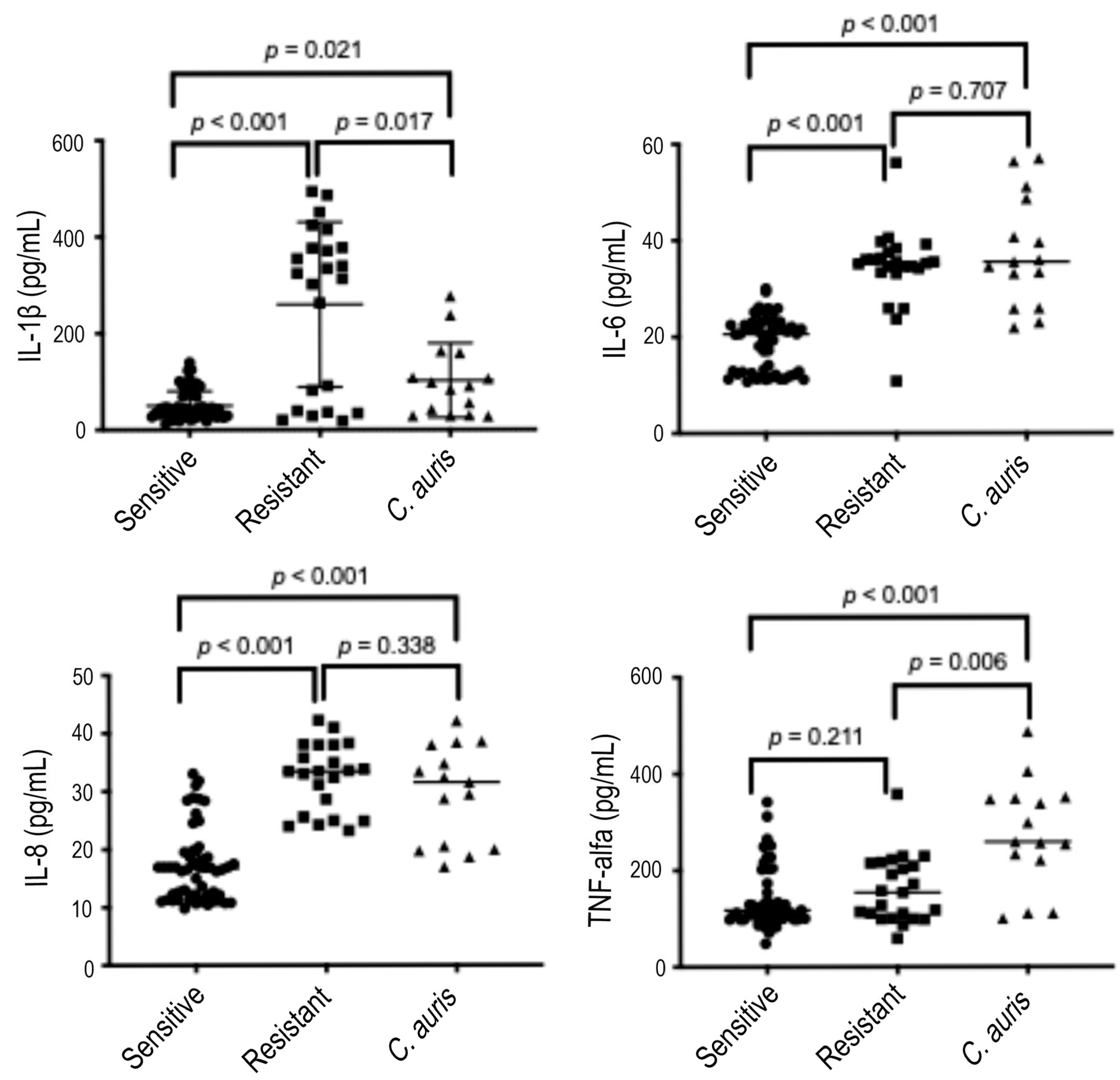
We stimulated DLD-1 cells with Candida strains from all species, grouped as ergosterol-susceptible (C. albicans, C. parapsilosis, C. tropicalis, and C. glabrata), ergosterol-resistant (C. albicans and C. parapsilosis), and C. auris strains. The induction of IL-1β, IL-6, and IL-8 by susceptible strains was lower than that induced by the resistant strains and by C. auris after both stimulation times points. Figure 2 summarizes the production of pro-inflammatory cytokines by DLD-1 cells after 6 hours of induction.
As can be depicted, the population of resistant strains exerts a dual effect on IL-1β production by DLD-1 cells after 6 h of stimulation. Accordingly, resistant Candida strains can be subdivided into two groups based on their ability to induce IL-1β production. Strains inducing low IL-1β levels (<100 pg/mL) behave similarly to susceptible strains and C. auris. Conversely, strains inducing high IL-1β production (>250 pg/mL) exhibit a distinct response compared with susceptible strains and C. auris. However, when considered as a unique group, resistant strains induce IL-1β production at levels that are statistically different from those induced by susceptible strains and by C. auris.
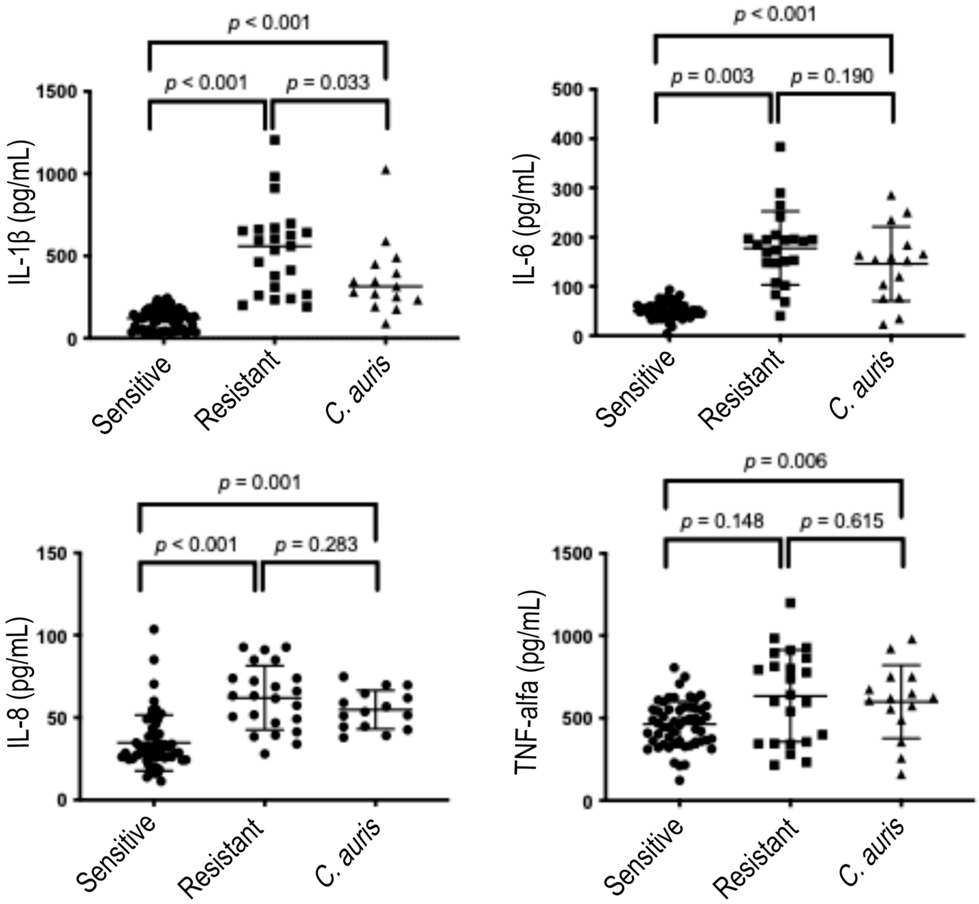
Finally, the heterogeneity in inducing IL-1β production suggests an early divergence in the inflammatory response. However, by 24 h, this distinction was no longer evident, as the populations appeared to converge into a single homogeneous group with no visibly discernible differences in IL-1β induction (compare Figure 2 to Figure 3).
IL-1β production induced by susceptible strains (53.93 pg/mL +/− 41.59) was significantly lower than by resistant ones (259.7 pg/mL +/− 170.8, p-value < 0.001) and by C. auris (102.3 pg/mL +/− 77.19, p-value = 0.021). IL-1β levels induced by resistant strains were significantly higher than those induced by C. auris (p-value = 0.017). The amount of IL-6 induced by susceptible strains (18.90 pg/mL +/− 5.53) was significantly lower than by resistant ones (34.42 pg/mL +/− 8.01, p-value < 0.001) and by C. auris (37.49 pg/mL +/− 11.45, p-value < 0.001). The inductions of IL-6 by resistant and C. auris strains were similar (p-value = 0.707). A similar trend was observed for IL-8: susceptible strains induced lower levels (17.19 ± 6.34 pg/mL) compared with resistant strains (32.41 ± 5.84 pg/mL, p < 0.001) and C. auris (29.56 ± 8.39 pg/mL, p < 0.001), while IL-8 levels induced by resistant and C. auris strains were comparable (p = 0.338). TNF-α production by resistant strains (160.3 ± 68.86 pg/mL) was similar to that induced by susceptible strains (140.60 ± 63.54 pg/mL, p = 0.211), but significantly lower than that induced by C. auris (275.0 ± 110.9 pg/mL, p = 0.006). TNF-α levels induced by susceptible strains were also significantly lower than those induced by C. auris (p < 0.001).
Figure 3 summarizes the production of pro-inflammatory cytokines by DLD-1 cells after 24 h of stimulation, showing an overall increase and similar pattern to that observed after the 6 h.
IL-1β levels induced by susceptible strains (115.5 ± 62.86 pg/mL) remained significantly lower (p < 0.001) compared with those induced by resistant strains (534.9 ± 265.3 pg/mL) and C. auris strains (363.6 ± 224.1 pg/mL). The higher IL-1β induction by resistant strains compared with C. auris persisted after prolonged incubation (p = 0.033). IL-6 and IL-8 induction patterns at 24 h were similar to those observed at 6 h. Specifically, IL-6 levels induced by susceptible strains (49.75 ± 15.64 pg/mL) were significantly lower than those induced by resistant strains (177.6 ± 74.61 pg/mL, p = 0.003) and C. auris (145.9 ± 278.1 pg/mL, p < 0.001). IL-6 induction by resistant and C. auris strains remained comparable (p = 0.190). IL-8 levels induced by susceptible strains (34.66 ± 16.92 pg/mL) were significantly lower than those induced by resistant strains (62.0 ± 19.47 pg/mL, p = 0.001) and C. auris (54.78 ± 11.77 pg/mL, p < 0.001). IL-8 induction by resistant and C. auris strains remained similar (p = 0.283). After prolonged stimulation, TNF-α levels induced by resistant strains increased (634.8 ± 278.1 pg/mL), becoming comparable to those induced by C. auris (598.8 ± 222.0 pg/mL, p = 0.615), while remaining statistically similar to those induced by susceptible strains (462.5 ± 139.7 pg/mL, p = 0.148). TNF-α levels induced by susceptible strains were significantly lower than those induced by C. auris (p = 0.006).

3.3. Comparison Between Cytokines Levels Induced by Different Candida Species Within Homogenous Susceptibility Phenotype

To ensure that the observed data were not due to random variation, we assessed whether the stimulatory activities of different Candida species within each susceptibility phenotype did not differ significantly. This evaluation was necessary due to the reported differences in innate immune recognition of different Candida species by human peripheral blood phagocytes, as demonstrated in previous studies [26,27].
Table 2 indicates that forty-six out of fifty-six comparisons yielded consistent results, with p-values > 0.05. Specifically, within the group of susceptible phenotypes, the comparisons between different species for IL-1β at 6 h, for IL-8 at 24 h, and for TNF-α at 6 and 24 h did not show significant differences, as evidenced by p-values > 0.05. In addition, comparisons between all cytokine inductions at 6 and 24 h of stimulation by C. glabrata and by C. albicans, by C. parapsilosis, and by C. tropicalis also resulted in p-values > 0.05, indicating no significant differences.
Among the eight comparisons between all cytokines, the induction by resistant C. albicans and by resistant C. parapsilosis, four showed significant differences (p-values < 0.05). Specifically, three comparisons for IL-1β and TNF-α at 24 h favored C. albicans, while one comparison for IL-6 favored C. parapsilosis.

3.4. Comparison Between Cytokines Levels Induced by Different Susceptibility Phenotype of Candida Within Homogenous Species

Here, we evaluated cytokine levels according to fluconazole susceptibility phenotypes within individual Candida species, specifically C. albicans and C. parapsilosis (Table 2). For all four cytokines tested at both stimulation time points, p-values were <0.05, indicating significantly lower cytokine levels induced by susceptible strains compared with resistant strains. These findings confirmed the differences observed between the overall susceptible and resistant Candida strain groups.

3.5. Comparisons Between Cytokine Levels Induced by C. auris and Individual Candida Species, Fluconazole Susceptible and Resistant

Table 3 shows the forty-eight comparisons between cytokines levels after stimulation with C. auris and the different Candida species, both susceptible and resistant.
Among the thirty-two comparisons between the induction by C. auris and by susceptible strains, nearly 91% (twenty-nine) showed that cytokine levels induced by susceptible strains were significantly lower than by C. auris, with p-values < 0.05. However, IL-1β induction at 6 h by C. auris versus C. albicans, versus C. parapsilosis, and versus C. glabrata showed no significant differences (p-values > 0.05). All cytokine evaluations between both time points revealed significantly higher levels induced by C. auris, followed by C. tropicalis.
Among the sixteen comparisons between cytokines induced by C. auris and resistant strains, fourteen (87.5%) showed no significant differences (p-values > 0.05). The evaluation of TNF-α after 6 h stimulation by C. auris showed higher levels than by C. parapsilosis (p-value = 0.003), and after 24 h by C. albicans showed higher levels than by C. auris (p-value = 0.015).

3.6. Profiles of Cytokine Inductions According to Fluconazole Susceptibility Phenotype and Species of Stimulating Candida Strains

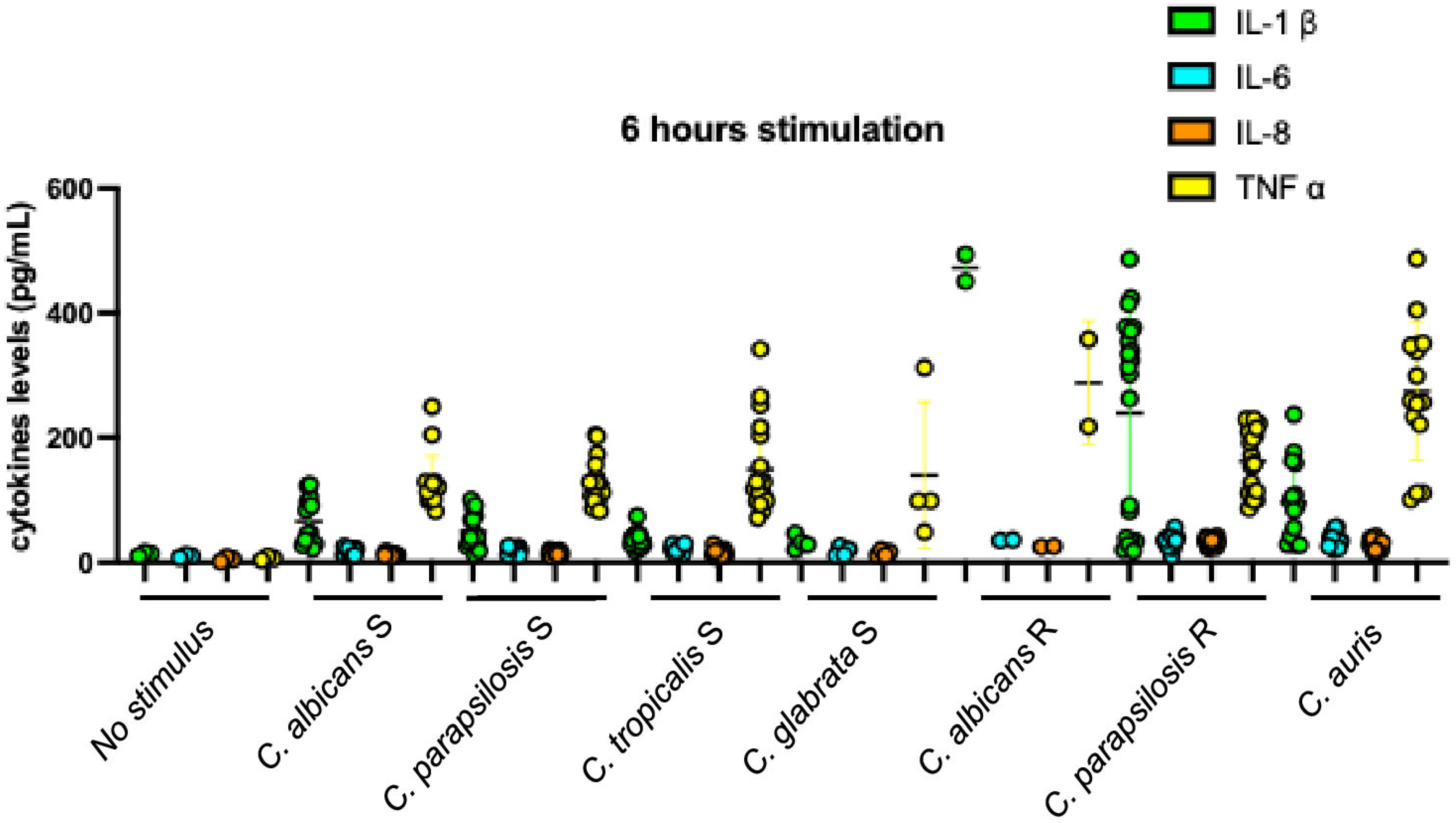
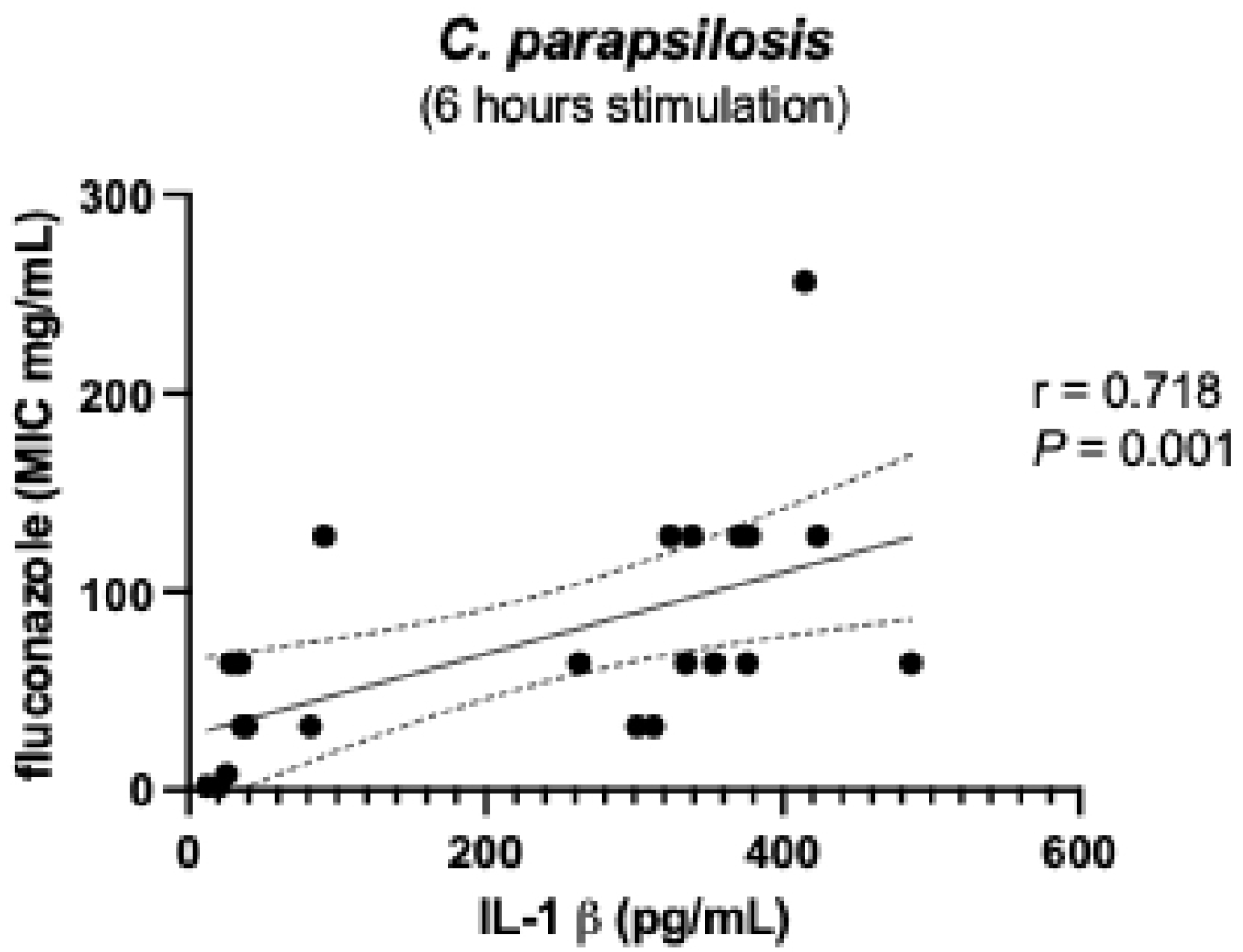
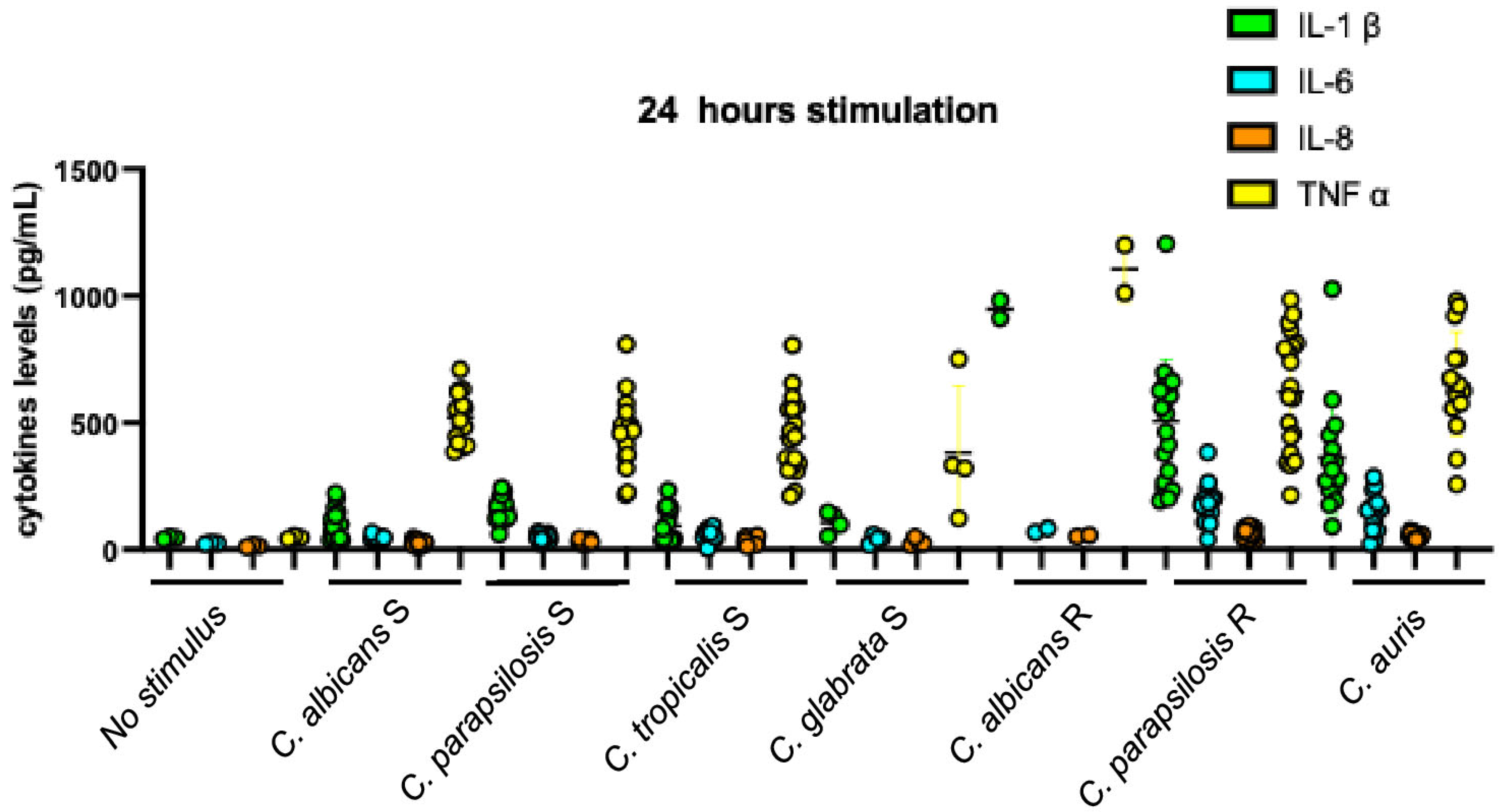
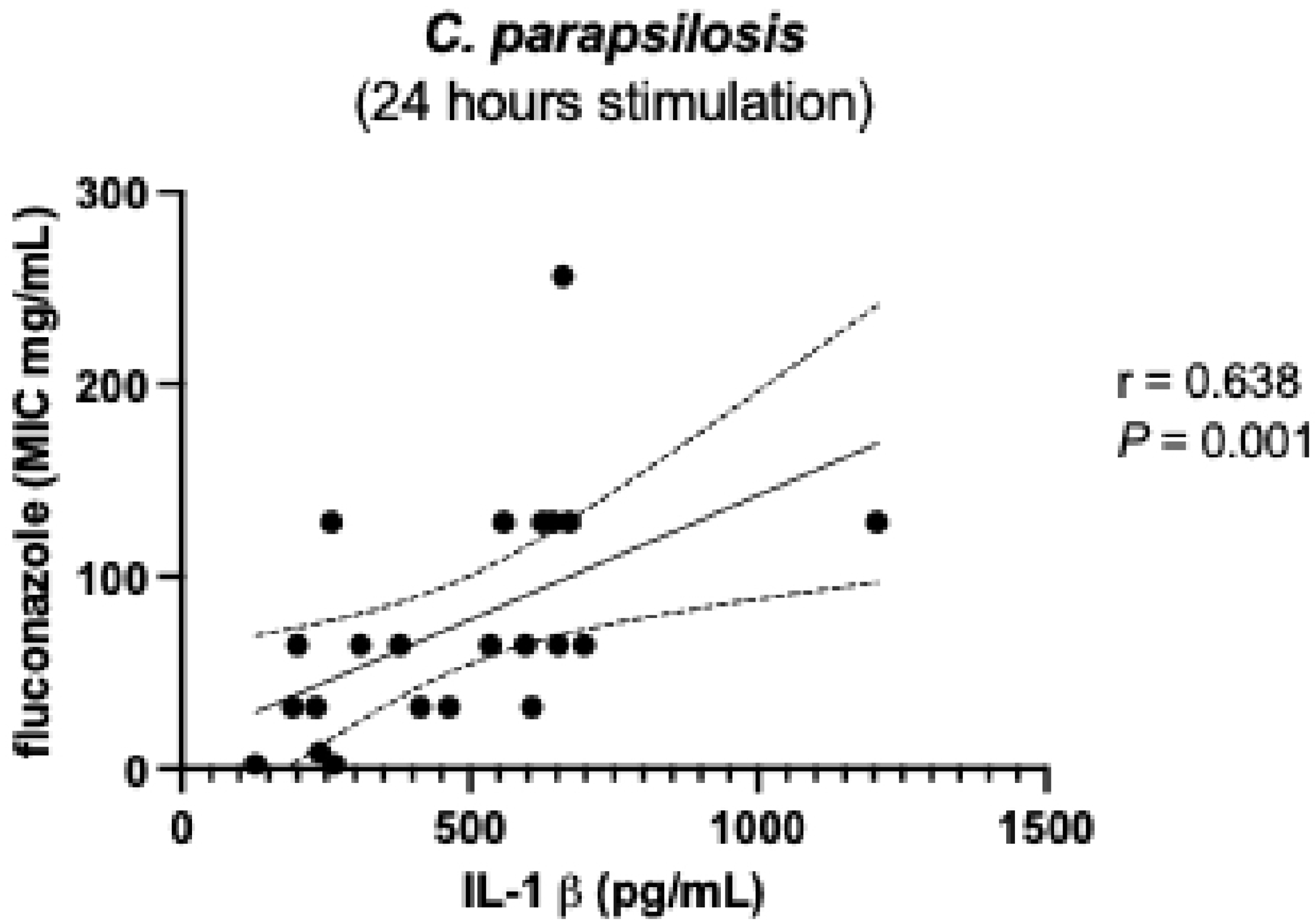
To further elucidate the biological significance of the dual effect observed in IL-1β production induced by resistant strains after 6 h of stimulation (Figure 2), we analyzed the responses elicited by individual Candida species among the resistant strains. Analysis of the data presented in Figure 4 shows that C. parapsilosis strains accounted for the observed variability in IL-1β induction. Further examination of this phenotype revealed a positive correlation between IL-1β levels at 6 h and the fluconazole MICs of these strains (Figure 5); strains with higher drug resistance induced higher IL-1β production (r = 0.674, p-value = 0.001). This IL-1β induction phenotype appeared to diminish after 24 h of stimulation (compare Figure 3 with Figure 6). Nonetheless, the correlation between IL-1β levels and fluconazole MICs remained statistically significant (Figure 7). No additional correlations between antifungal resistance and cytokine induction were observed.
Susceptible strains and C. auris induced lower amounts of IL-1β than TNF-α, with negative IL-1β/TNF-α ratios (0.52 from C. albicans and C. parapsilosis, 0.25 from C. tropicalis, 0.23 from C. glabrata, and 0.35 from C. auris stimulation). Resistant strains induced higher amounts of IL-1β than TNF-α, with positive IL-1β/TNF-α ratios (1.64 from C. albicans and 1.45 from C. parapsilosis stimulation).
After 24 hours of stimulation, inductions by susceptible strains and C. auris were still characterized by IL-1β levels lower than TNF-α, despite increasing or decreasing IL-1β/TNF-α ratios themselves (0.19 from C. albicans, 0.35 from C. parapsilosis, 0.21 from C. tropicalis, 0.28 from C. glabrata, and 0.56 from C. auris stimulation) (Figure 6). IL-1β/TNF-α ratios from resistant strain inductions changed to negative, similar to those obtained with the susceptible and C. auris strains, although the ratios were higher (0.90 from C. albicans and 0.80 from C. parapsilosis stimulation). It is apparent that the change was due to the remarkable increase in induction of TNF-α by resistant strains, not to the reduction in IL-1β quantity. Indeed, following prolonged stimulation, the quantified levels of IL-1β derived from C. auris strains remained significantly lower than those observed in the globally resistant strains (p-value < 0.05; specifically, p-value = 0.033). Conversely, the measured concentrations of TNF-α exhibited no statistically significant difference between the two groups under the same experimental conditions (p-value > 0.05; specifically, p-value = 0.615). These findings are visually represented in Figure 2.

4. Discussion

Candida colonizes the surfaces of healthy individuals as commensal but can cause life-threatening disseminated infections of endogenous origin in compromised and hospitalized hosts [4,5,27]. C. albicans can invade epithelial cell barriers, mostly of the gastrointestinal tract, through active endocytosis and a paracellular route involving the proteolytic digestion of tight junctions [3,4,28]. The early stages of Candida–host interactions correspond to the first defense response by the mucosal epithelial cells. These cells are equipped with PRRs and produce pro-inflammatory cytokines as effector weapons against the MAMP stimulus [7,29]. The fungal countermeasures towards host immune surveillance may be a critical point of transition from colonization to infection and are now considered part of the fitness attributes of yeasts [14]. Indeed, fungal fitness refers to the overall ability of a fungal organism to survive, grow, reproduce, and successfully interact with its environment, including the host, during infection. It encompasses a range of biological traits that contribute to the organism’s adaptability and competitiveness under various conditions and can be altered by the mechanisms underlying the antifungal resistance [21].
Our experiments, stimulating the human intestinal cell line DLD-1 with Candida strains differing in species and susceptibility phenotype to fluconazole, revealed preliminary but interesting results that we consider as possible starting points for further in-depth investigations.
The levels of cytokines induced by the fluconazole susceptible Candida were significantly different from and lower than those induced by C. auris strains, and the relation between the stimulating activity of these two groups was the same for all the tested cytokines, at the shorter and longer stimulation times. Differently, the relation between the stimulating activity of resistant strains (other than C. auris) and that of C. auris strains, and the relation between the stimulation activity of resistant and susceptible strains were different depending on the cytokine. IL-6 and IL-8 levels induced by the fluconazole susceptible Candida were significantly different from and lower than those induced by the resistant strains and by C. auris; IL-6 and IL-8 levels induced by the resistant strains and by C. auris were not different. The level of IL-1β obtained with the resistant strains was significantly different and higher than that obtained with C. auris at two stimulation times. The level of TNF-α obtained with the resistant strains was significantly lower than that obtained with C. auris, which was no longer evident after prolonged stimulation, giving the idea of delayed stimulation activity. The level of TNF-α obtained with the resistant strains was not different from that obtained with the sensitive strains at two stimulation times. These aspects were also represented by the profiles of induction and the relative ratios between diverse cytokines for each group of stimulating strains. The profiles obtained with susceptible strains and C. auris groups (see the most evident negative IL-1β/TNF-α ratio) were like each other over time, and different from those obtained with the group of resistant strains. In fact, resistant Candida strains gave profiles showing positive IL-1β/TNF-α ratios, being the level of the first cytokine higher than the second one.
The differences in immunostimulant ability that emerged within individual Candida species and among different susceptibility phenotypes suggest that the early host–parasite interaction can be considered a part of fungal fitness, which includes many aspects of their biology [29,30,31]. Vincent et al. first considered the fungus–host interaction as part of yeast fitness, having shown that resistance to amphotericin B in clinical and evolved-laboratory strains of C. albicans came at a high cost, i.e., reduced opposition to the host’s innate immune defense and less infectious damage than its susceptible counterpart [32]. In addition, to our best knowledge, this is the first study demonstrating this kind of relation concerning the very early host–pathogen immune response mediated by epithelial mucosal cells.
It is anyway hard to attribute the different behaviors of susceptible and resistant strains to advantages or disadvantages for the latter, given the fungal genetic plasticity that can translate mutational changes into gains of function [33,34,35]. In our study, the high level of cytokines induced by resistant strains could be beneficial to them by leading to high inflammation and permeability of mucosa lamina propria, in turn promoting microbial invasion into the favorable bloodstream [36,37,38]. Conversely, the ability of susceptible strains to modulate and inhibit mucosal defense signals could be impaired by mutations, responsible for resistance, which may come at a cost, as often observed among bacteria [39,40]. In our view, the different level and timing of IL-1β and TNF-α in comparison with IL-6 and IL-8 production by resistant strains, particularly by C. parapsilosis, might suggest that their stimulatory behavior is the result of a possible mutational suffering.
Another noteworthy point emerging from our data concerns C. auris. Assuming that fluconazole-susceptible Candida strains represent the wild-type phenotype, the similar stimulating profile induced by C. auris may also reflect a wild-type origin; its high fluconazole MICs, elevated cytokine induction levels, and genomic features reported in the literature may represent inherited traits of C. auris. Several studies have reported that, despite the presence of independently arising mutations, the distinction between intrinsic and acquired resistance among C. auris isolates is less clear than in other Candida species [13,41,42,43]. In this context, it is noteworthy that multidrug-resistant C. auris has emerged simultaneously on three continents [44].
Finally, our data permitted not to exclude a role for ergosterol in fungi–host interaction in animals, which has already been demonstrated in plants [9,10,11]. The cited work of Vincent et al. reinforces this possibility. They demonstrated that Candida strains resistant to amphotericin B, characterized by a reduced ergosterol content in the cell membrane, showed performance in parasite–host interactions and phagocytosis [12]. It can be envisaged that similar behaviors observed in this study could be due to other external fungal structures. A possible actor could be the wall glucan, already known to be a MAMP, whose folding might be improper, given the improper structure of the underlying fungal membrane rich in ergosterol. Structural differences between the glucans of fluconazole-susceptible and -resistant Candida strains may in turn result from independent nontarget effects induced by the stress response [45].
This study has limitations inherent to a preliminary investigation, but we hope it will serve as a foundation for more in-depth future research. Specifically, the absence of isogenic strains, resistance-related genomic data, and clade identification for C. auris [41] limits the accurate interpretation of the functional impact of resistance on fungal fitness at the strain level. Equally important and intended for future evaluation is the lack of phenotypic assessment of biochemical structures of the fungal membrane and cell wall. As previously discussed, mutations in genes involved in ergosterol biosynthesis can lead to drug resistance and overexpression due to gain-of-function, a possibility that must be evaluated [17].
Although this study requires further investigation, it offers valuable insights that enhance our understanding of fungal drug resistance. The diversity of fitness during the early immune response between fluconazole-sensitive and fluconazole-resistant Candida strains within the same species is evident. Another finding relates to the intriguing behavior of C. auris toward mammalian host cells in vitro; notably, this species appears to lie somewhere between fluconazole-sensitive and fluconazole-resistant Candida strains. Finally, our findings echo observations in plant cells on the role of ergosterol in interacting with host innate immunity.

Author Contributions

G.P. planned the study, supervised the experimental project, wrote the first draft, and supervised the final draft of the manuscript. A.V. contributed to strain isolations and performed cellular stimulation experiments. S.P. contributed to cellular stimulation. A.M.S. supervised the final draft of the manuscript. D.S. planned this study, performed cellular stimulation, measurement of cytokines, and statistical analysis, and supervised the experimental plan definition. All authors have read and agreed to the published version of the manuscript.

Funding

This research received no external funding.

Institutional Review Board Statement

This study was approved by the Ethical Committee of the University of Genoa (CERA 2024/26, 8 March 2024), and all participants provided written informed consent.

Informed Consent Statement

Informed consent was obtained from all subjects involved in the study. The authors assert that all procedures contributing to this work comply with the ethical standards of the relevant national and institutional committees on human experimentation and with the Helsinki Declaration of 1975, as revised in 2000.

Data Availability Statement

The original contributions presented in this study are included in the article. Further inquiries can be directed to the corresponding authors.

Conflicts of Interest

The authors declare no conflicts of interest.

References

  1. Brown, G.D.; Denning, D.W.; Gow, N.A.; Levtz, S.M.; Netea, M.G.; White, T.C. Hidden killers: Human fungal infections. Sci. Transl. Med. 2012, 4, 165rv113. [Google Scholar] [CrossRef]
  2. Hohmann, F.B.; Chaves, R.C.F.; Olivato, G.B.; Souza, G.M.; Galindo, V.B.; Silva, M., Jr.; Martino, M.D.V.; Menezes, F.G.; Corrêa, T.D. Characteristics, risk factors, and outcomes of bloodstream Candida infections in the intensive care unit: A retrospective cohort study. J. Int. Med. Res. 2023, 51, 3000605221131122. [Google Scholar] [CrossRef]
  3. Basmaciyan, L.; Bon, F.; Paradis, T.; Lapaquette, P.; Dalle, F. Candida albicans interactions with the host: Crossing the intestinal epithelial barrier. Tissue Barriers. 2019, 7, 1612661. [Google Scholar] [CrossRef]
  4. Sheppard, D.C.; Filler, S.G. Host cell invasion by medically important fungi. Cold Spring Harb. Perspect. Med. 2014, 5, a019687. [Google Scholar] [CrossRef] [PubMed]
  5. Qin, Y.; Zhang, L.; Xu, Z.; Zhang, J.; Jiang, Y.Y.; Cao, Y.; Yan, T. Innate immune cell response upon Candida albicans infection. Virulence 2016, 7, 512–526. [Google Scholar] [CrossRef]
  6. Jensen, O.; Trujillo, E.; Hanson, L.; Ost, K.S. Controlling Candida: Immune regulation of commensal fungi in the gut. Infect. Immun. 2024, 92, e0051623. [Google Scholar] [CrossRef]
  7. Plato, A.; Hardison, S.E.; Brown, G.D. Pattern recognition receptors in antifungal immunity. Semin. Immunopathol. 2015, 37, 97–106. [Google Scholar] [CrossRef]
  8. Netea, M.G.; Brown, G.D.; Kullberg, B.J.; Gow, N.A.R. An integrated model of the recognition of Candida albicans by the innate immune system. Nat. Rev. Microbiol. 2008, 6, 67–78. [Google Scholar] [CrossRef]
  9. Klemptner, R.L.; Sherwood, J.S.; Tugizimana, F.; Dubery, I.A.; Piater, L.A. Ergosterol, an orphan fungal microbe-associated molecular pattern (MAMP). Mol. Plant Pathol. 2014, 15, 747–761. [Google Scholar] [CrossRef] [PubMed]
  10. Rossard, S.; Roblin, G.; Atanassova, R. Ergosterol triggers characteristic elicitation steps in Beta vulgaris leaf tissues. J. Exper. Botany. 2010, 61, 1807–1816. [Google Scholar] [CrossRef] [PubMed]
  11. Kazan, K.; Gardiner, D.M. Targeting pathogen sterols: Defence and counterdefence? PLoS Pathog. 2017, 13, e1006297. [Google Scholar] [CrossRef]
  12. Perlin, D.S.; Rautema-Richardson, R.; Alastruey-Izquierdo, A. The global problem of antifungal resistence: Prevalence, mechanisms, and management. Lancet Infect. Dis. 2017, 12, e383–e392. [Google Scholar] [CrossRef] [PubMed]
  13. Revie, N.M.; Iyer, K.R.; Robbins, N.; Cowen, L.E. Antifungal drug resistance: Evolution, mechanisms and impact. Curr. Opin. Microbiol. 2018, 45, 70–76. [Google Scholar] [CrossRef]
  14. Clancy, C.J.; Nguyen, M.H. At what cost echinocandin resistance? J. Infect. Dis. 2011, 204, 499–501. [Google Scholar] [CrossRef]
  15. Robbins, N.; Caplan, T.; Cowen, L.E. Molecular evolution of antifungal drug resistance. Microbiol. Spectr. 2017, 71, 753–775. [Google Scholar] [CrossRef] [PubMed]
  16. Vale-Silva, L.A.; Coste, A.T.; Ischer, F.; Parker, J.E.; Kelly, S.L.; Pinto, E.; Sanglard, D. Azole resistance by loss of function of the sterol Δ5,6-desaturase gene (ERG3) in Candida albicans does not necessarily decrease virulence. Antimicrob. Agents Chemother. 2012, 56, 1960–1968. [Google Scholar] [CrossRef]
  17. Flowers, S.A.; Barker, K.S.; Berkow, E.l.; Toner, G.; Chadwick, S.D.; Gygax, S.E.; Morschhäuser, J.; Rogers, P.D. Gain-of-function mutations in UPC2 are a frequent cause of ERG11 upregulation in azole-resistant clinical isolates of Candida albicans. Eukaryot. Cell 2012, 11, 1289–1299. [Google Scholar] [CrossRef] [PubMed]
  18. Dykes, C.; Demeter, L.M. Clinical significance of human immunodeficiency virus type 1 replication fitness. Clin. Microbiol. Rev. 2007, 20, 550–578. [Google Scholar] [CrossRef]
  19. Goig, G.A.; Windels, E.M.; Loiseau, C.; Stritt, C.; Biru, L.; Borrell, S.; Brites, D.; Gagneux, S. Ecology, global diversity and evolutionary mechanisms in the Mycobacterium tuberculosis complex. Nat. Rev. Microbiol. 2025. epub ahead of print. [Google Scholar] [CrossRef]
  20. Borrell, S.; Gagneux, S. Infectiousness, reproductive fitness and evolution of drug-resistant Mycobacterium tuberculosis. Int. J. Tuberc. Lung. Dis. 2009, 13, 1456–1466. [Google Scholar]
  21. Cowen, L.E.; Kohn, L.M.; Anderson, J.B. Divergence in fitness and evolution of drug resistance in experimental populations of Candida albicans. J. Bacteriol. 2001, 183, 2971–2978. [Google Scholar] [CrossRef]
  22. Anderson, J.B. Evolution of antifungal-drug resistance: Mechanisms and pathogen fitness. Nat. Rev. Microbiol. 2005, 3, 547–556. [Google Scholar] [CrossRef]
  23. Ben-Ami, R.; Garcia-Effron, G.; Lewis, R.E.; Gamarra, S.; Leventakos, K.; Perlin, D.S.; Kontoyiannis, D.P. Fitness and virulence costs of Candida albicans FKS1 Hot Spot Mutations associated with echinocandin resistance. J. Infect. Dis. 2011, 204, 626–635. [Google Scholar] [CrossRef]
  24. CLSI. Performance Standards for Antifungal Susceptibility Testing of Yeasts, 3rd ed.; CLSI supplement M27M44S; Clinical and Laboratory Standards Institute: Wayne, PA, USA, 2022. [Google Scholar]
  25. NCCLS. Reference Method for Broth Dilution Antifungal Susceptibility Testing of Yeasts, 2nd ed.; Approved standard, NCCLS document M27-A2; NCCLS: Wayne, PA, USA, 2002. [Google Scholar]
  26. Navarro-Arias, M.J.; Hernández-Chávez, M.J.; García-Carnero, L.C.; Amezcua-Hernández, D.G.; Lozoya-Pérez, N.E.; Estrada-Mata, E.; Martínez-Duncker, I.; Franco, B.; Mora-Montes, H.M. Differential recognition of Candida tropicalis, Candida guilliermondii, Candida krusei, and Candida auris by human innate immune cells. Infect. Drug Resist. 2019, 12, 783–794. [Google Scholar] [CrossRef] [PubMed]
  27. Estrada-Mata, E.; Navarro-Arias, M.J.; Pérez-García, L.A.; Mellado-Mojica, E.; López, M.G.; Csonka, K.; Gacser, A.; Mora-Montes, H.M. Members of the Candida parapsilosis complex and Candida albicans are differentially recognized by human peripheral blood mononuclear cells. Front. Microbiol. 2016, 6, 1527. [Google Scholar] [CrossRef]
  28. Wachtler, B.; Citiulo, F.; Jablonowski, N.; Förster, S.; Dalle, F.; Schaller, M.; Wilson, D.; Hube, B. Candida albicans-epithelial interactions: Dissecting the roles of active penetration, induced endocytosis and host factors on the infection process. PLoS ONE 2012, 7, e36952. [Google Scholar] [CrossRef] [PubMed]
  29. Prieto, D.; Carpena, N.; Maneu, V.; Gil, M.L.; Pla, J.; Gozalbo, D. TLR2 modulates gut colonization and dissemination of Candida albicans in a murine model. Microbes Infect. 2016, 18, 656–660. [Google Scholar] [CrossRef] [PubMed]
  30. Singh-Babak, S.D.; Babak, T.; Diezmann, S.; Hill, J.A.; Xie, J.L.; Chen, Y.L.; Poutanen, S.M.; Rennie, R.P.; Heitman, J.; Cowen, L.E. Global analysis of the evolution and mechanism of echinocandin resistance in Candida glabrata. PLoS Pathog. 2012, 8, e1002718. [Google Scholar] [CrossRef]
  31. Cowen, L.E.; Sanglard, D.; Calabrese, D.; Sirjusingh, C.; Anderson, J.B.; Kohn, L.M. Evolution of drug resistance in experimental population of Candida albicans. J. Bacteriol. 2000, 182, 1515–1522. [Google Scholar] [CrossRef]
  32. Vincent, B.M.; Lancaster, A.K.; Scherz-Shouval, R.; Whitesell, L.; Lindquist, S. Fitness trade-offs restrict the evolution of resistance to amphotericin B. PLoS Biol. 2013, 11, e1001692. [Google Scholar] [CrossRef]
  33. Andersson, D.I. The biological cost of mutational antibiotoc resistance: Any practical conclusion? Curr. Opin. Microbiol. 2006, 9, 461–465. [Google Scholar] [CrossRef]
  34. Selmecki, A.M.; Dulmage, K.; Cowen, L.E.; Anderson, J.B.; Berman, J. Acquisition of aneuploidy provides increased fitness during the evolution of antifungal resistance. PLoS Genet. 2009, 5, e1000705. [Google Scholar] [CrossRef]
  35. Borghi, E.; Andreoni, S.; Cirasola, D.; Ricucci, V.; Sciota, R.; Morace, G. Antifungal resistance does not necessarily affect Candida glabrata fitness. J. Chemother. 2014, 26, 32–36. [Google Scholar] [CrossRef] [PubMed]
  36. Gow, N.A.R.; van de Veerdonk, F.L.; Brown, A.J.P.; Netea, M.G. Candida albicans morphogenesis and host defence: Discriminating invasion from colonization. Nat. Rev. Microbiol. 2013, 10, 112–122. [Google Scholar] [CrossRef]
  37. Tong, Y.; Tang, J. Candida albicans infection and intestinal immunity. Microbiol. Res. 2017, 198, 27–35. [Google Scholar] [CrossRef]
  38. Imbert, S.; Castain, L.; Pons, A.; Jacob, S.; Meyer, I.; Palous, M.; Vezinet, C.; Langeron, O.; Hennequin, C.; Monse, A.; et al. Discontinuation of echinocandin and azole treatments led to the disappearance of an FKS alteration but not azole resistance during clonal Candida glabrata persistent candidaemia. Clin. Microbiol. Infect. 2016, 22, 891.e5–891.e8. [Google Scholar] [CrossRef] [PubMed]
  39. Zang, Q.; Sahin, O.; McDermott, P.F.; Payot, S. Fitness of antimicrobial-resistant Campylobacter and Salmonella. Microbes Infect. 2006, 8, 1972–1978. [Google Scholar] [CrossRef]
  40. Alame Emane, A.K.; Guo, X.; Takiff, H.E.; Liu, S. Drug resistance, fitness and compensatory mutations in Mycobacterium tuberculosis. Tuberculosis 2021, 129, 102091. [Google Scholar] [CrossRef]
  41. Du, H.; Bing, J.; Hu, T.; Ennis, C.L.; Nobile, C.J.; Huang, G. Candida auris: Epidemiology, biology, antifungal resistance, and virulence. PLoS Pathog. 2020, 16, e1008921. [Google Scholar] [CrossRef] [PubMed]
  42. Sharma, C.; Kumar, N.; Pandey, R.; Meis, J.F.; Chowdhary, A. Whole genome sequencing of emerging multidrug resistant Candida auris isolates in India demonstrates low genetic variation. New Microbes New Infect. 2016, 13, 77–82. [Google Scholar] [CrossRef]
  43. Chatterjee, S.; Alampalli, S.V.; Nageshan, R.K.; Chettiar, S.T.; Joshi, S.; Tatu, U.S. Draft genome of a commonly misdiagnosed multidrug resistant pathogen Candida auris. BMC Genom. 2015, 16, 686. [Google Scholar] [CrossRef]
  44. Lockhart, E.R.; Etienne, K.A.; Vallabhaneni, S.; Farooqi, J.; Chowdhary, A.; Govender, N.P.; Colombo, A.L.; Calvo, B.; Cuomo, C.A.; Desjardins, C.A.; et al. Simultaneous emergence of multidrug-resistant Candida auris on 3 continents confirmed by whole-genome sequencing and epidemiological analyses. Clin. Infect. Dis. 2017, 64, 134–140. [Google Scholar] [CrossRef] [PubMed]
  45. Cowen, L.E.; Sanglard, D.; Howard, S.J.; Roger, P.D.; Perlin, D.S. Mechanisms of antifungal drug resistance. Cold Spring Harb. Perspect. Med. 2015, 5, a019752. [Google Scholar] [CrossRef] [PubMed]
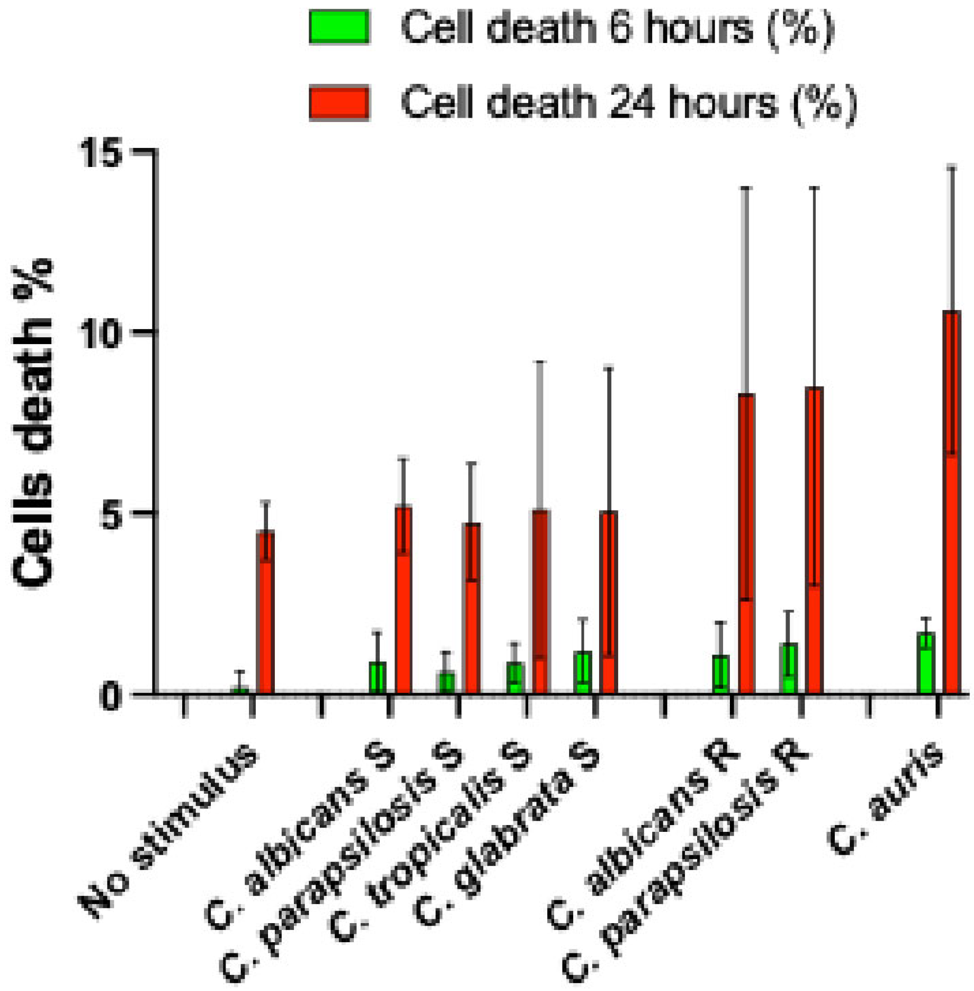
Figure 1. No significant differences in cell death were observed across experimental conditions. Sensitive (S) and resistant (R) fluconazole Candida strains. Pp-value (significant when <0.05); for all stimulatory condition, S 6 and 24 h, R 6 and 24 h, p-value > 0.05.
Microorganisms 13 01777 g001
Figure 2. Cytokines produced after 6 h of stimulation. IL-1β—interleukin-1β; IL-6—interleukin-6; IL-8—interleukin-8; TNF-α—tumor necrosis factor α. Sensitive—fluconazole sensitive Candida strains; Resistant—fluconazole resistant Candida strains. Pp-value (significant when <0.05).
Microorganisms 13 01777 g002
Figure 3. Cytokines produced after 24 h stimulation. IL-1β—interleukin-1β; IL-6—interleukin-6; IL-8—interleukin-8; TNF-α—tumor necrosis factor α; Sensitive— fluconazole sensitive Candida strains; Resistant—fluconazole resistant Candida strains. Pp-value (significant when <0.05).
Microorganisms 13 01777 g003
Figure 4. Cytokines produced after 6 h of stimulation according to species and fluconazole susceptibility phenotype of Candida stimulating strains. C. albicans S—C. albicans susceptible to fluconazole; C. parapsilosis S—C. parapsilosis susceptible to fluconazole; C. tropicalis S—C. tropicalis susceptible to fluconazole; C. glabrata S—C. glabrata susceptible to fluconazole; C. albicans R—C. albicans resistant to fluconazole; C. parapsilosis R—C. parapsilosis resistant to fluconazole IL-1β—interleukin-1β; IL-6—interleukin-6; IL-8—interleukin-8; TNF-α—tumor necrosis factor α.
Microorganisms 13 01777 g004
Figure 5. Positive correlation between IL-1 β levels induced after 6 h stimulation by C. parapsilosis resistant strains and their MIC to fluconazole. IL-1β—interleukin-1β. p < 0.05 significant. r—Spearman correlation; Pp-value (significant when <0.05).
Microorganisms 13 01777 g005
Figure 6. Cytokines produced after 24 h of stimulation according to species and fluconazole susceptibility phenotype of Candida stimulating strains. C. albicans S—C. albicans susceptible to fluconazole; C. parapsilosis S—C. parapsilosis susceptible to fluconazole; C. tropicalis S—C. tropicalis susceptible to fluconazole; C. glabrata S—C. glabrata susceptible to fluconazole; C. albicans R—C. albicans resistant to fluconazole; C. parapsilosis R—C. parapsilosis resistant to fluconazole; IL-1β—interleukin-1β; IL-6—interleukin-6; IL-8—interleukin-8; TNF-α—tumor necrosis factor α.
Microorganisms 13 01777 g006
Figure 7. Positive correlation between IL-1 β levels induced after 24 h stimulation by C. parapsilosis resistant strains and their MIC to fluconazole. IL-1β—interleukin-1β. p < 0.05 significant. r—Spearman correlation; Pp-value (significant when <0.05).
Microorganisms 13 01777 g007
Table 1. Susceptibility phenotypes of Candida strains.
C. albicansC. parapsilosisC. tropicalisC. glabrataC. auris
N strainMIC mg/LN strainMIC mg/LN strainMIC mg/LN strainMIC mg/LN StrainMIC mg/LN strainMIC mg/L
FZAB FZAB FZAB FZAB FZAB FZABVORCAS
A111P181P21321T121G120.25Au1>256220.12
A20.51P20.250.5P22322T241G221Au2>256220.12
A320.5P30.121P23322T311G30.50.25Au3>256120.25
A40.250.5P421P2424T411G420.5Au4>256240.12
A50.251P50.51P25640.5T50.51 Au5>256220.12
A60.251P60.50.25P261281T60.51 Au6>256220.12
A70.50.5P70.51P27640.5T711 Au7>256220.12
A80.251P80.51P28642T810.5 Au8>256440.06
A90.251P911P291282T911 Au9>25622>4
A100.50.5P100.50.5P301281T1011 Au10>256240.12
A110.50.5P110.50.5P31320.5T110.50.5 Au11>256240.03
A120.51P120.50.25P32320.5T1211 Au12>256240.25
A1310.5P130.50.5P33641T1321
A14<0.120.5P140.50.5P341281T1412
A150.251P150.250.5P352560.5T1541
A160.251P160.50.5P361280.5T1621
A17>2561P1711P37641T1711
A18>2561P1811P381281T1811
P19640.5P39641T1941
P20160.5
FZ—fluconazole; AB—amphotericin B; VOR—voriconazole; CAS—caspofungin.
Table 2. Comparison between cellular cytokine responses to stimulation with different Candida species.
Stimulation By
Cytokine and incubation time (hours)Sensitive
C. albicans
vs.
Sensitive
C. parapsilosis
vs.
Sensitive
C. tropicalis
vs.
Sensitive
C. albicans
vs.
Sensitive
C. parapsilosis
vs.
Resistant
C. albicans
Sensitive
C. parapsilosis
Sensitive
C. tropicalis
Sensitive
C. glabrata
Sensitive
C. tropicalis
Sensitive
C. glabrata
Sensitive
C. glabrata
Resistant
C. albicans
Resistant
C. parapsilosis
Resistant
C. parapsilosis
PPPPPPPPP
IL-1β (6)0.0580.0570.0970.1860.25304560.0010.0130.012 *
IL-1β (24)0.018 *0.5540.3070.0860.1660.4930.015<0.0010.032 *
IL-6 (6)0.4260.017 *0.8710.048 *0.9500.2590.003<0.0010.707
IL-6 (24)0.3570.008 *0.8670.0960.3000.1440.015<0.0010.032 *
IL-8 (6)0.046 *0.041 *0.1330.8000.8530.7090.001<0.0010.216
IL-8 (24)0.0750.1300.9040.8890.1870.3180.003<0.0010.557
TNF-α (6)0.9790.6010.2530.5730.5250.3990.0290.0230.431
TNF-α (24)0.9290.0540.1520.0900.1950.4030.0410.0430.008 *
IL-1 β (6)—interleukin-1β after 6 h stimulation; IL-1β (24)—interleukin-1β after 24 h stimulation; IL-6 (6)—interleukin-6 after 6 h stimulation; IL-6 (24)—interleukin-6 after 24 h stimulation; IL-8 (6)—interleukin-8 after 6 h stimulation; IL-8 (24)—interleukin-8 after 24 h stimulation; TNF-α (6)—tumor necrosis factor after 6 h stimulation; TNF-α (24)—tumor necrosis factor α after 24 h stimulation. Sensitive—fluconazole sensitive Candida strains; Resistant—fluconazole resistant Candida strains. Pp-value (significant when <0.05); * p-value < 0.05.
Table 3. Comparison between cellular cytokine responses to stimulation with C. auris strains and different Candida species.
Stimulation By
Cytokine and incubation time (hours)C. auris versus
Sensitive
C. albicans
Sensitive
C. parapsilosis
Sensitive
C. tropicalis
Sensitive
C. glabrata
Resistant
C. albicans
Resistant
C. parapsilosis
PPPPPP
IL-1β (6)0.262 *0.151 *0.0070.062 *0.0570.066
IL-1β (24)<0.001<0.001<0.0010.0040.0590.063
IL-6 (6)<0.001<0.0010.0040.0020.9410.663
IL-6 (24)0.002<0.0010.0050.0140.2350.088
IL-8 (6)<0.001<0.001<0.0010.0400.5290.230
IL-8 (24)<0.001<0.001<0.0010.0490.9410.235
TNF-α (6)0.0050.0010.0010.0360.9260.003 *
TNF-α (24)0.0300.0020.0010.0770.015 *0.981
IL-1β (6)—interleukin-1β after 6 h stimulation; IL-1β (24)—interleukin-1β after 24 h stimulation; IL-6 (6)—interleukin-6 after 6 h stimulation; IL-6 (24)—interleukin-6 after 24 h stimulation; IL-8 (6)—interleukin-8 after 6 h stimulation; IL-8 (24)—interleukin-8 after 24 h stimulation; TNF-α (6)—tumor necrosis factor after 6 h stimulation; TNF-α (24)—tumor necrosis factor α after 24 h stimulation. Sensitive—Candida strains sensitive to fluconazole; Resistant—Candida strains resistant to fluconazole. P—p-value (significant when <0.05); * p-value highlighted in the result section.
Disclaimer/Publisher’s Note: The statements, opinions and data contained in all publications are solely those of the individual author(s) and contributor(s) and not of MDPI and/or the editor(s). MDPI and/or the editor(s) disclaim responsibility for any injury to people or property resulting from any ideas, methods, instructions or products referred to in the content.