Article

Isolation of ESBL-Producing Enterobacteriaceae in Food of Animal and Plant Origin: Genomic Analysis and Implications for Food Safety

1
Istituto Zooprofilattico Sperimentale della Puglia e della Basilicata, via Manfredonia 20, 71121 Foggia, Italy
2
Department of Veterinary Medicine, University Aldo Moro of Bari, Strada per Casamassima Km 3, Valenzano, 70010 Bari, Italy
*
Author to whom correspondence should be addressed.
Microorganisms 2025, 13(8), 1770; https://doi.org/10.3390/microorganisms13081770
Submission received: 23 May 2025 / Revised: 23 July 2025 / Accepted: 25 July 2025 / Published: 29 July 2025
(This article belongs to the Special Issue Food Microorganisms and Genomics, 2nd Edition)

Abstract

Background: The spread of ESBL-producing Enterobacteriaceae (ESBL-PE) strains in food poses a potential risk to human health. The aim of the study was to determine the occurrence of ESBL-PE and to investigate their distribution on foods. Methods: A total of 1000 food samples, including both raw and ready-to-eat products, was analyzed for the presence of ESBL-producing Enterobacteriaceae using chromogenic selective agar. Antibiotic resistance in the isolated strains was assessed using conventional methods, while whole-genome sequencing was employed to predict antimicrobial resistance and virulence genes. Results: The overall occurrence of ESBL-PE strains was 2.8%, with the highest contamination in raw meat samples (10%). A total of 31 multidrug-resistant (MDR) strains was isolated, mainly Escherichia coli, followed by Klebsiella pneumoniae, Salmonella enterica, and Enterobacter hormaechei. All strains exhibited high levels of resistance to at least four different β-lactam antibiotics, as well as to other antimicrobial classes including sulfonamides, tetracyclines, aminoglycosides, and quinolones. Whole-genome sequencing identified 63 antimicrobial resistance genes, with blaCTX-M being the most prevalent ESBL gene. Twenty-eight (90%) isolates carried Inc plasmids, known vectors of multiple antimicrobial resistance genes, including those associated with ESBLs. Furthermore, several virulence genes were identified. Conclusions: The contamination of food with ESBL-PE represents a potential public health risk, underscoring the importance of the implementation of genomic surveillance to monitor and control the spread of antimicrobial resistance.

1. Introduction

The production of enzymes capable of hydrolyzing and inactivating β-lactam antibiotics is the most important mechanism of resistance in the Enterobacteriaceae family. Extended-spectrum β-lactamases (ESBLs) are enzymes that confer resistance to penicillins, first- to third-generation cephalosporins and monobactams (e.g., aztreonam), but not to cephamycin (e.g., cefoxitin) or carbapenems (e.g., meropenem, imipenem) [1]. However, these enzymes are inhibited by β-lactamase inhibitors such as clavulanic acid [2,3]. The first ESBLs were identified in the 1980s as variants of the TEM-1 and SHV-1 enzymes, resulting from specific amino acid substitutions in K. pneumoniae and E. coli strains, respectively [4,5,6]. In recent years, novel ESBLs have emerged, with the cefotaximase-M (CTX-M) family becoming the most prevalent group in many European countries. Among these, CTX-M-15 is the most frequently detected variant. Over 140 TEM and 60 SHV derivatives capable of hydrolyzing third-generation cephalosporins and aztreonam have been identified [5,7]. CTX-M-type ESBLs are now the most widely distributed globally, particularly in K. pneumoniae and E. coli, which are major pathogens in both healthcare-associated and community-acquired infections [8]. These enzymes are not exclusively chromosomally encoded but are frequently carried on mobile genetic elements, especially plasmids. Notably, blaCTX-M-like genes are often co-located with other antimicrobial resistance determinants, such as aac (6’)-Ib-cr, blaOXA, catB, tet, aadA, dfrA17, sul genes [9]. ESBL-producing Enterobacteriaceae are often multidrug-resistant, with additional resistance to non-β-lactam antibiotics such as aminoglycosides (e.g., amikacin, gentamicin, streptomycin), fluoroquinolones (e.g., ciprofloxacin), trimethoprim, tetracyclines, sulfonamides (e.g., sulfisoxazole), and chloramphenicol [10,11]. The spread of ESBL-producing Enterobacteriaceae in food represents a potential threat to human health, as the resistance genes are frequently associated with mobile genetic elements capable of transferring to both commensal and pathogenic bacteria, even across different genera and species [12].
This study aims to enhance our understanding of the occurrence of ESBL-producing Enterobacteriaceae across various food categories by screening samples collected from the southern Italian regions of Apulia and Basilicata, and to assess differences in contamination levels between raw and ready-to-eat foods. Antibiotic resistance in the isolated strains will be assessed using conventional phenotypic methods, while whole-genome sequencing (WGS) will be employed to predict the presence of resistance genes. Comparing results from both approaches will aid in the classification of isolates based on their resistance profiles and contribute to a deeper understanding of antimicrobial resistance in the food chain.

2. Materials and Methods

2.1. Sampling

Samples were collected using a non-probabilistic convenience sampling method. All samples were transported in temperature-controlled containers maintained at 4 ± 2 °C and promptly delivered to the laboratory. A total of 1000 food samples was analyzed, consisting of 500 raw and 500 ready-to-eat (RTE) products. All samples were sourced from the retail market across the southern Italian regions of Apulia and Basilicata between 2018 and 2023.

2.2. Bacterial Strain Isolation

The food samples were analyzed with the aim of detecting ESBL-producing Enterobacteriaceae, as described in a previous study [13] with the exception that CHROMagar™ ESBL (CHROMagar, Paris, France) was used as the chromogenic isolation medium.

2.3. ESBL-Producing Screening

All presumptive ESBL-producing Enterobacteriaceae strains, previously isolated, were evaluated for ESBL production by determining the Minimum Inhibitory Concentration (MIC), in accordance with the Clinical and Laboratory Standards Institute (CLSI) guidelines [14]. The Sensititre™ EUVSEC® panel (Trek Diagnostic Systems, Westlake, OH, USA), which includes the ESBL-producing screening combination test method, was employed. A reduction of two or more dilution steps in the MIC of cefotaxime and/or ceftazidime in the presence of clavulanic acid, compared to the MIC of cephalosporin alone, was interpreted as indicative of ESBL production.

2.4. Whole-Genome Sequencing

For each sample, DNA was purified using the QIAmp DNA mini kit (Qiagen, Hilden, Germany), according to the manufacturer’s instructions. DNA quality was assessed by calculating the absorbance ratio at 260/280 nm, while DNA quantification was performed by measuring the optical density (OD) at 260 nm using a BioPhotometer (Eppendorf, Milan, Italy). DNA samples purified with an A260/A280 ratio between 1.8 and 2.0 were considered to be of high quality and stored at −20 °C until analysis. Libraries were prepared using the Illumina DNA Prep kit (Illumina, San Diego, CA, USA) and sequenced using a MiSeq platform (Illumina, San Diego, CA, USA) with a 2 × 250 bp paired end approach [15]. Bioinformatic analyses were conducted through the European Galaxy server (https://usegalaxy.eu, accessed on 18 March 2025). Raw reads were assembled using SPAdes version 3.15 [16] and assembly quality was assessed with Quast version 5.0.2 [17]. Genome assemblies were subsequently analyzed to screen for (i) ribosomal multilocus sequence typing (rMLST) by PubMLST species identification database (https://pubmlst.org/species-id, accessed on 21 March 2024), (ii) multilocus sequence typing (MLST) [18], and (iii) detection of antimicrobial resistance (AMR) genes and point mutations, virulence genes (VGs) and plasmid sequences using both ABRIcate (Galaxy Version 1.0.1) and StarAMR (Galaxy Version 0.11.0) tools. Default parameters were applied for all tools.
The nucleotide sequences of strains were deposited in GenBank under the BioProject accession PRJNA1076372 (Supplementary Table S1).

2.5. Antimicrobial Susceptibility Test

The ESBL-producing strains were analyzed for their resistance profiles against the antibiotics listed in Table 1, by the minimum inhibitory concentration (MIC) method, following the CLSI guidelines for Enterobacterales as reported in a previous study [13]. E. coli ATCC 25,922 was used as the control strain. MIC values were interpreted according to the guidelines provided by the CLSI [14].

2.6. Statistical Analysis

Differences in the percentages of isolates were compared using the Chi-square test, performed with Epi Info software version 3.3.2. A p-value < 0.05 was considered statistically significant.

3. Results

3.1. Contamination Rate of Food Samples with ESBL-Producing Enterobacteriaceae

A total of 1000 food samples was cultured to isolate ESBL-producing Enterobacteriaceae (ESBL-PE). Strains were successfully recovered from 5% (25/500) of the raw food samples and from 0.6% (3/500) of the ready-to-eat (RTE) food samples (Table 2 and Table 3). A statistically significant difference was observed between these two groups (χ2 = 17.8; p < 0.05). Although all samples were cultured for the detection of ESBL-producing Enterobacteriaceae, no strains were isolated from raw vegetables, RTE dried or cooked sausages, RTE ready meals or RTE bakery and pastry products. The highest contamination rate was observed in raw meat samples (10%), in which the occurrence of ESBL-PE was significantly higher compared to other food categories (p < 0.05). A total of 31 ESBL-PE strains were isolated from 28 (2.8%; 28/1000) food samples. Specifically, 27 strains were isolated from 25 raw food samples, with two samples yielding two strains each. Four strains were isolated from 3 RTE food samples, including one ice cream sample, from which two strains were isolated (Table 2 and Table 3).
Draft assemblies of the isolated strains, combined with rMLST analysis of the draft genomes, enabled accurate taxonomic assignment (Table 2 and Table 3). Escherichia coli (20/31; 65%) was the most commonly isolated species, followed by six Klebsiella pneumoniae (6/31; 19%), two Enterobacter hormaechei (2/31; 6%), two Salmonella enterica (2/31; 6%) and one Klebsiella quasipneumoniae (1/31; 3%). MLST analysis identified 16 sequence types (STs) among E. coli strains, five STs in K. pneumoniae and one ST each in E. hormaechei and S. enterica strains. For one K. quasipneumoniae strain, the sequence type could not be determined (Table 2 and Table 3).

3.2. Antimicrobial Resistance Profiles of the ESBL-Producing Enterobacteriaceae Isolates

Antimicrobial susceptibility testing (including 31 antibiotics grouped in 11 classes) was conducted on 31 strains and the results are collected in Figure 1. All (100%) strains showed a multidrug resistance (MDR).
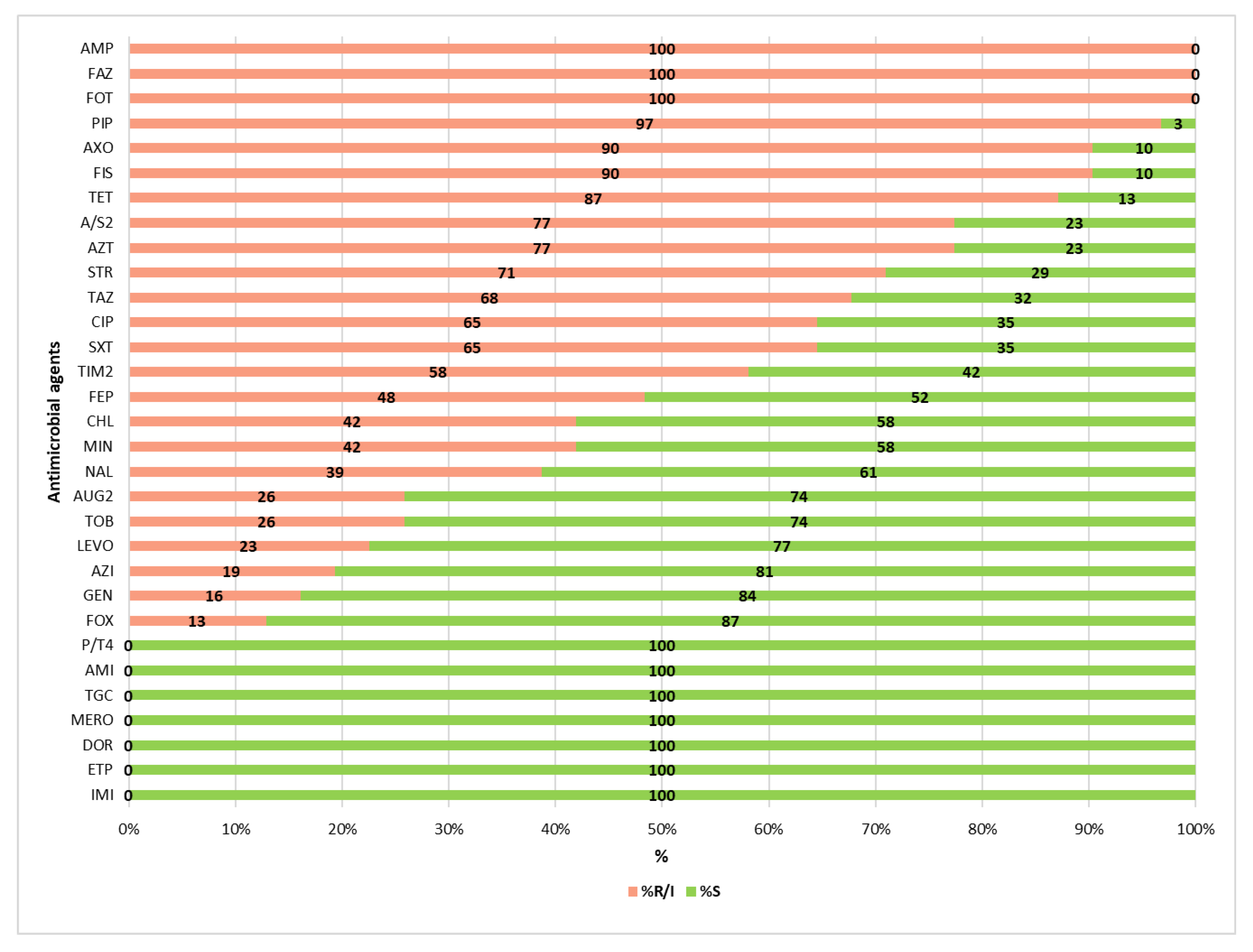
The two E. hormaechei isolates exhibited intrinsic resistance to ampicillin (AMP), amoxicillin/clavulanic acid (AUG2), ampicillin/sulbactam (A/S2), cefazolin (FAZ), and cefoxitin (FOX). Seven K. pneumoniae isolates showed intrinsic resistance to ampicillin. The highest resistance rates were observed for ampicillin, cefazolin and cefotaxime, with all 31 isolates (100%) resistant. This was followed by resistance to piperacillin (n = 30; 97%), ceftriaxone and sulfisoxazole (n = 28; 90%), tetracycline (n = 27; 87%) and both ampicillin/sulbactam and aztreonam (n = 24; 77%). For fifteen additional antimicrobials, resistance rates ranged from 71% to 13% (Figure 2). No resistance was observed to piperacillin/tazobactam, carbapenems, amikacin or tigecycline.

3.3. Antimicrobial Resistance Determinants

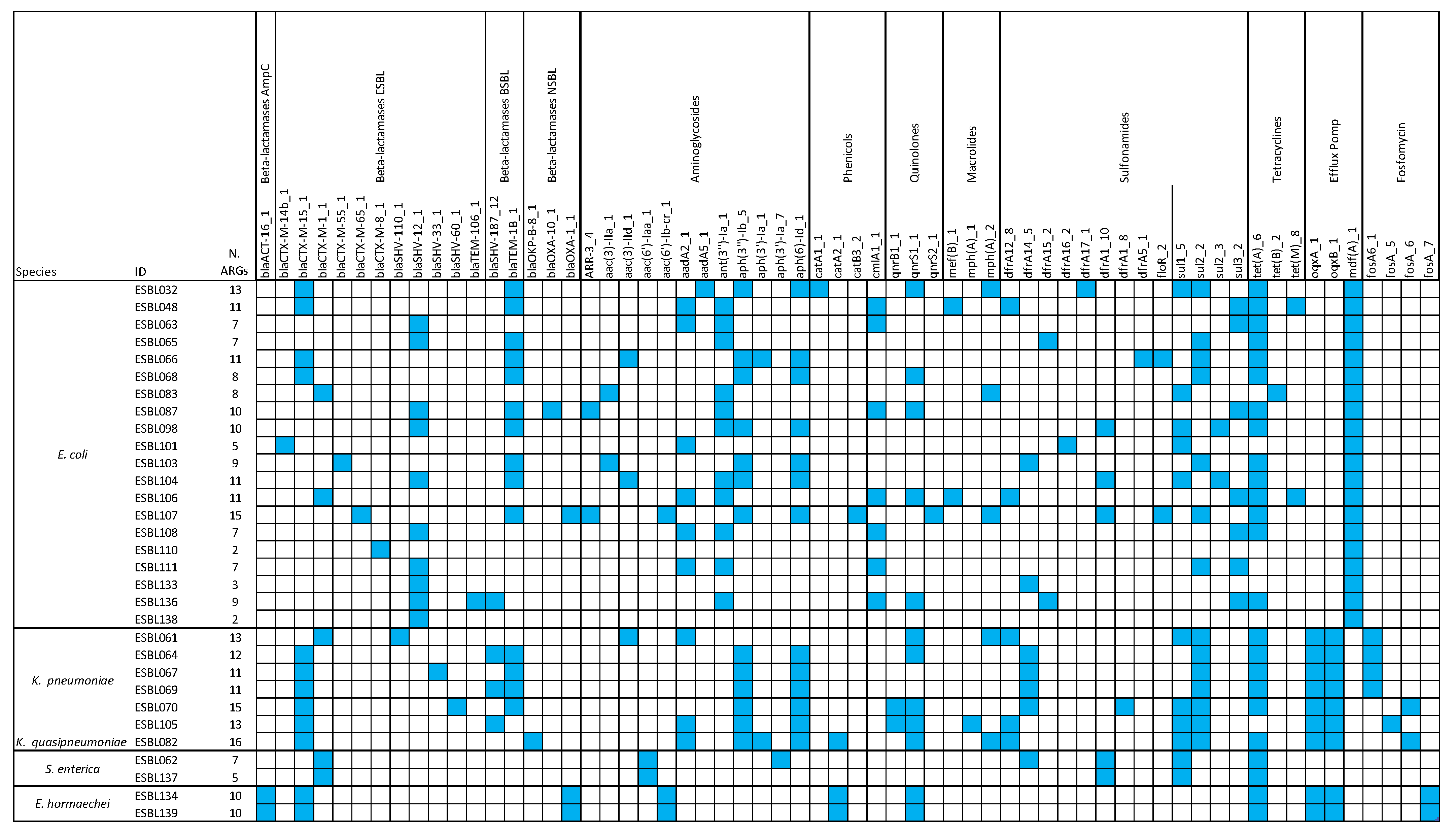
This investigation identified 63 antimicrobial resistance genes (ARGs) using the AMR ResFinder database in combination with the ABRIcate tool. These ARGs were further grouped into 14 classes based on their antibiotic resistance profiles (Figure 3). All isolates carried at least one gene encoding ESBL β-lactamases (blaCTX-M, blaSHV, blaTEM). Additionally, ARGs conferring resistance to aminoglycosides, phenicols, macrolides, quinolones, sulfonamides, and tetracyclines were detected in all isolates (Figure 3). A strong correlation was observed between genotypic predictions and phenotypic resistance profiles. Specifically, there was 100% concordance for β-lactamase genes, while 27 out of 28 sulfonamide-resistant isolates and 25 out of 27 tetracycline-resistant isolates harbored genes associated with phenotypical resistance. Additionally, mutations in gyrA (S83L; D87N), parC (A56T; S80I; S80R) and parE (L416F; S458A) were detected in E. coli strains (Table 4). Among these, the non-synonymous mutation S83L in gyrA was identified in eight E. coli strains, with three of these also carrying the D87N mutation. The S80I substitution in parC was the most prevalent. The two substitutions in parE were identified in two strains of E. coli.

3.4. Correlation Between the Presence of ARGs and Phenotypic Resistance

All ESBL-producing isolates carried extended-spectrum β-lactamase (ESBL) genes (blaCTX-M; blaSHV; blaTEM). Among the 31 isolates, 21 (66%) harbored the blaCTX-M gene. The most prevalent were those encoding enzymes from the CTX-M-1 group, particularly CTX-M-15 and CTX-M-1, found in 17 isolates (55%), with CTX-M-15 alone detected in 12 isolates (39%). Genes from the CTX-M-9 group (CTX-M-14, CTX-M-55, CTX-M-65) and the CTX-M-8 group were less frequent, with frequencies of 10% (3/31) and 3% (1/31), respectively. Thirteen isolates (42%) carried ESBL blaSHV genes, while the ESBL variant blaTEM-106 gene was detected in only one isolate (3%). Additionally, genes encoding the non-ESBL enzyme TEM-1 were present in 14 isolates (45%). Two E. hormaechei strains displayed phenotypic resistance to first-generation cephalosporins and cefoxitin, consistent with the presence of the blaACT gene encoding AmpC β-lactamases.
Further correlations between genotype and phenotype were observed for other antibiotic classes. Aminoglycoside resistance genes (aac, aph, aad, ant) were identified in 28 isolates (90%), of which 22 exhibited resistance to streptomycin, tobramycin, or gentamicin. Chloramphenicol resistance genes (catA, catB, clmA) were found in 12 isolates (39%), with 11 also exhibiting phenotypic resistance. However, two E. coli strains were phenotypically resistant despite lacking any known chloramphenicol resistance genes. Sulfonamide resistance genes (dfrA, floR, sul) were detected in 27 isolates (87%), all of which were phenotypically resistant to sulfisoxazole and/or trimethoprim/sulfamethoxazole. One E. coli isolate, however, showed phenotypic resistance to sulfisoxazole without any corresponding ARGs. Tetracycline resistance genes (tet) were identified in 25 strains (81%), according with phenotypic resistance. Notably, two E. coli strains exhibited resistance to tetracycline in the absence of identifiable tet genes. Quinolone resistance genes (qnr) were present in 13 isolates (42%), of which 11 demonstrated phenotypic resistance to ciprofloxacin, levofloxacin, or nalidixic acid. Conversely, eight E. coli and two S. enterica isolates were phenotypically resistant despite lacking the qnr gene, while two isolates carried the qnr gene but remained phenotypically susceptible. Additionally, non-synonymous mutations were identified in 4% of E. coli strains, suggesting a role in resistance to fluoroquinolones. Macrolide resistance genes (mef, mph) were detected in eight isolates (26%), with phenotypic resistance observed in four. Two strains showed phenotypic resistance to azithromycin despite the absence of corresponding resistance genes. The fosA gene, conferring resistance to fosfomycin, was identified in nine isolates (29%). However, phenotypic resistance to fosfomycin was not assessed in this study. Finally, 29 isolates (94%), excluding the two S. enterica strains, harbored efflux pump genes (oqxA, oqxB, mdfA), which are known to contribute to multidrug resistance.

3.5. Detection of Plasmid Genes

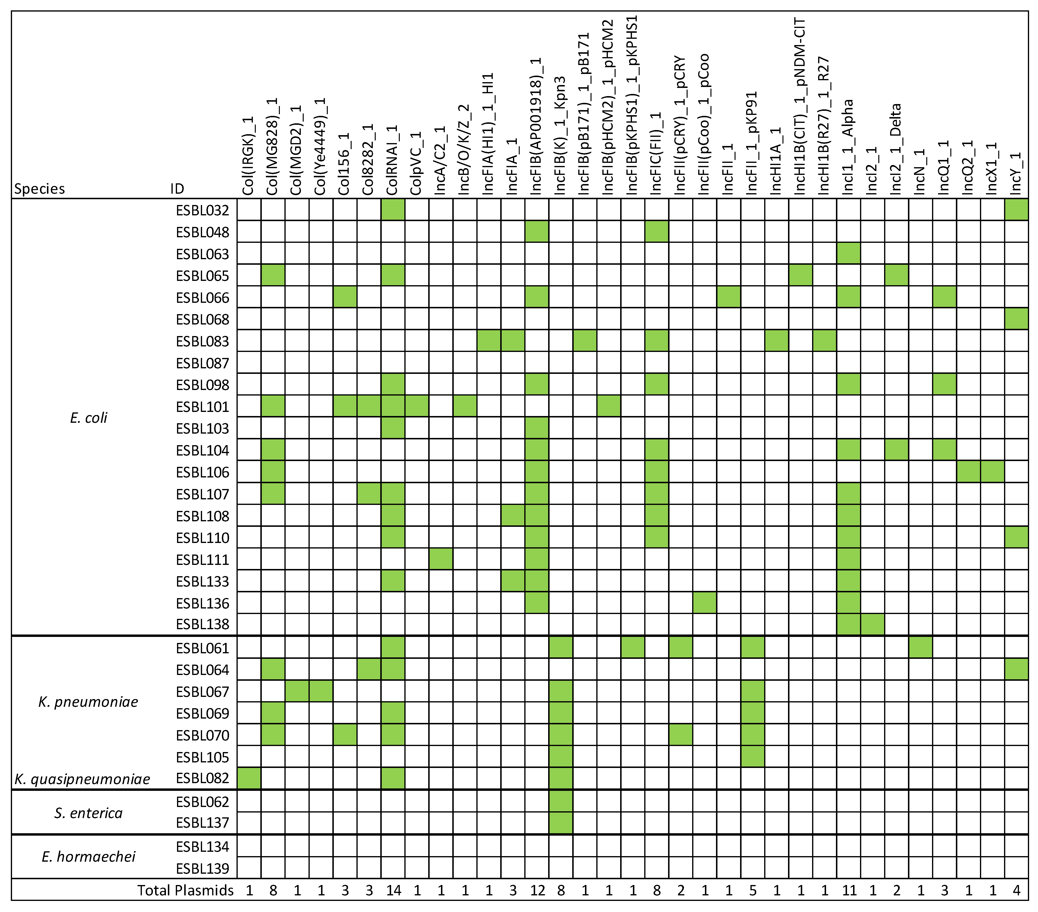
Plasmid gene prediction was performed using the PlasmidFinder database. It is important to note that only partial plasmid nucleotide sequences were detected in the analyzed strains, rather than complete plasmid assemblies. A total of 105 plasmid replicons or replicon fragments were identified across the isolates (Figure 4). The most frequently detected replicon was ColRNAI_1, present in 14 strains, followed by IncFIB (AP001918)_1, IncI1_1_Alpha and IncFII, found in 12, 11 and 7 strains, respectively. Notably, strain ESBL101 harbored the highest number (n = 7) of distinct replicon types. In contrast, no replicon sequences were detected in two E. hormaechei strains (ESBL134 and ESBL139) and one E. coli strain (ESBL087) (Figure 4).

3.6. Detection of Virulence Genes

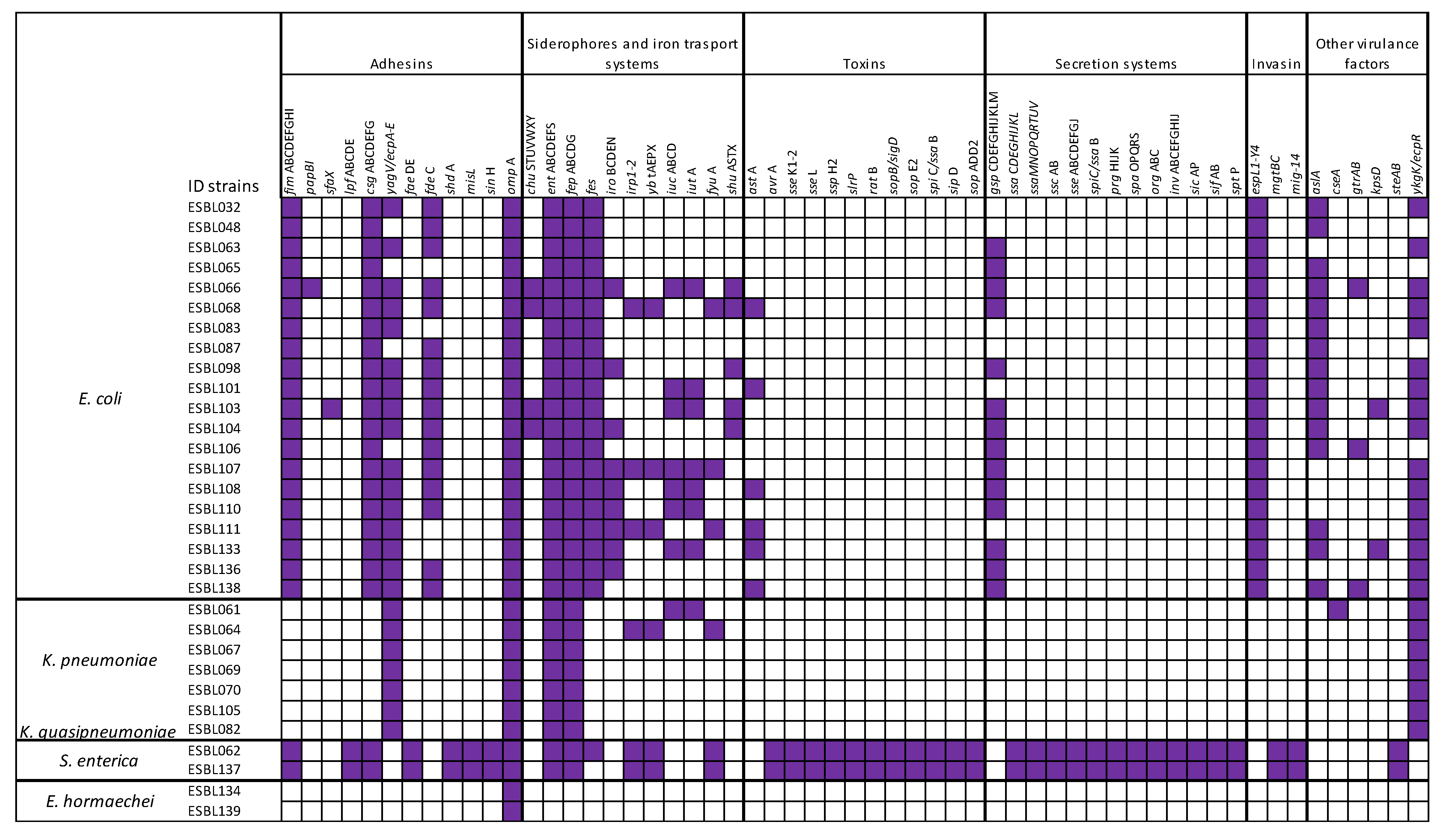
The virulence genes (VGs) prediction was carried out using the Virulence Factors Database (VFDB). A total of 191 VGs, encoding adhesins, siderophores/iron transport systems, toxins, secretion systems, invasins or others, was identified in the isolates (Supplementary Table S2 and Figure 5). Regardless of taxonomic species, the outer membrane protein gene ompA proved the most prevalent, detected in 100% of the isolates, followed by 29 (94%) isolates that carried ent and fep genes necessary for the biosynthesis and transport of the siderophore enterobactin; 22 (71%) strains carried csg genes and 23 (74%) isolates showed yag/ecp genes, both essential for surface adhesion, biofilm formation and interaction with host cells. The distribution of the number of VGs per strain is reported in Figure 5. Notably, the two S. enterica strains exhibited the highest number of virulence genes (117), indicating a potentially enhanced pathogenicity. These strains were distinguished by the presence of genes encoding adhesins (e.g., fim, ipf, csg, fae), siderophores and iron transport systems (e.g., ent, fep, irp, ybt, fyu), invasins (mgt, mig), as well as a variety of toxins and secretion systems.
Among the E. coli isolates, the number of virulence genes ranged from 78 to 37. Most E. coli strains harbored genes encoding adhesins (e.g., fim, cgs, yag/ecp, fde), siderophores/iron transport systems (e.g., ent, fep, fes), secretion systems (e.g., gsp), and invasins systems (e.g., esp). K. pneumoniae strains exhibited a lower level of virulence genes, ranging from 21 to 10 genes. All the K. pneumoniae strains carried yagV/ecpA-E for adhesins genes, ent and fep for siderophores/iron transport systems and ykgK/ecpR for other virulence genes. The two E. hormaechei strains carried only a single virulence gene: the adhesins gene ompA.

4. Discussion

This study investigated the occurrence of extended-spectrum β-lactamase-producing Enterobacteriaceae (ESBL-PE) in 1000 food samples purchased in the Southern Italian regions of Apulia and Basilicata between 2018 and 2023. Half of the samples (n = 500) were ready-to-eat (RTE) foods, with a low positivity rate (0.3%), while the remaining 500 were raw foods, among which 5% tested positive. The overall prevalence of ESBL-PE contamination was 2.8% (28/1000). Notably, raw meat samples showed a significantly higher positivity rate (10%) compared to other food categories (range: 0.0–4.0%; p < 0.05). Among the 207 raw meat samples, poultry meat (n = 50) showed the highest contamination rate, with 32% (16/50) testing positive. This result is consistent with international reports, which have documented poultry meat contamination rates of 36% in Ghana, 21% in Brazil and the United States, and 25.9% in Switzerland [19,20]. Even higher prevalence rates have been reported in parts of Europe, reaching 93% in Spain and 80% in the Netherlands [21,22].
A statistically significant decreasing trend in the prevalence of ESBL-producing bacteria in food-producing animals has been reported by the European Food Safety Authority and the European Centre for Disease Prevention and Control (EFSA-ECDC) with a notable decline in the prevalence of ESBL-producing E. coli in Italy, from 82.3% during 2017–2018 to 43.4% in the 2021–2022 period [23].
In our study, among RTE foods, ESBL-PE were detected in one cheese sample, one salad sample and one ice cream sample. No positive samples were identified in other RTE foods investigated. Among raw foods, 4% of milk samples tested positive, while no ESBL-PE were detected in raw vegetables. A recent study reported contamination rates of about 18.0% in raw milk, 1.3% in cheese and 0.25% in vegetables [24]. Notably, the raw milk contamination was relatively low in Europe (2%), Malaysia (3.18%), and India (6.6%) [25,26,27].
Among the food samples that tested positive for ESBL-PE, a total of 31 bacterial isolates were obtained. Whole-genome sequencing revealed four species: E. coli, predominantly isolated from raw meat, with the highest frequency in poultry meat; K. pneumoniae and K. quasipneumoniae, primarily detected in turkey and pork meat; S. enterica, exclusively isolated from poultry meat; and E. hormaechei, isolated from ice cream and raw seafood.
In a similar study, E. coli accounted for 61% of isolates, while K. pneumoniae represented 9.1% [20]. A study conducted in the United Kingdom reported a prevalence of ESBL-producing E. coli in 27.5% of analyzed meat samples [28]. In Germany, the highest prevalence of ESBL-producing E. coli was observed in chicken meat (74.9%), followed by turkey meat (40.1%) [24]. The prevalence of ESBL-producing K. pneumoniae in animal-derived food products shows substantial variation, with reported rates ranging from 3% in retail chicken meat samples in the Netherlands and 3.7% in chicken liver samples in Algeria, to 23.4% in raw milk samples in Lebanon and 25% in food fish samples in India [29,30,31,32]. The ability of Salmonella spp. to produce ESBLs, similar to other members of the Enterobacteriaceae family, has led the World Health Organization (WHO) to classify them as high-priority pathogens [33]. Two studies published in 2022, one from Poland and one from Italy, reported relatively high prevalence rates (14.3% and 11.7%, respectively) of ESBL-producing Salmonella spp. in poultry meat [34,35]. In contrast, lower prevalence rates have been reported in studies from Canada, China, Egypt and Southeast Asia, with contamination levels in foods of animal origin ranging from 0.05% to 3.5% [1,36,37,38].
In this study, Enterobacter hormaechei, a member of the Enterobacter cloacae complex (ECC), was the least frequently isolated species. According to the literature, ECC strains are known to produce extended-spectrum β-lactamases (ESBLs) and represent the third most prevalent group of drug-resistant Enterobacterales, following Escherichia coli and Klebsiella pneumoniae [39].
In this study, MLST analysis of 31 isolates identified distinct sequence types (STs), reflecting a high degree of genetic diversity. However, in four isolates the STs could not be determined due to unmatched allelic profiles in the Achtman MLST database. The most frequently identified STs among E. coli were ST10 (n = 3), ST69 (n = 2), and ST1011 (n = 2), while the remaining STs were represented by single isolates. Among the STs identified in E. coli, ST10 represents a globally disseminated lineage commonly associated with both commensal and extraintestinal pathogenic strains, often harboring extended-spectrum β-lactamase (ESBL) genes [40,41]. Notably, an E. coli ST10 isolate was recovered from an RTE cheese sample. ST69 is a well-known uropathogenic clone, frequently associated with antimicrobial resistance [42], while ST155 has been implicated in zoonotic transmission and multidrug resistance [43,44]. In K. pneumoniae, the detection of ST29 is of particular concern, as this lineage has been implicated in nosocomial outbreaks and is known to carry carbapenemase-producing plasmids [45]. Both S. enterica isolates belonged to ST32, corresponding to serovar Infantis, a major foodborne pathogen known for its association with multidrug resistance [46]. The presence of these high-risk clones underscores the importance of continuous genomic surveillance to monitor the spread of clinically significant lineages.
All strains isolated in this study were tested for antimicrobial susceptibility against 31 antibiotics. As expected for ESBL-producing isolates, the highest resistance levels were observed among β-lactam antibiotics. High resistance rates were also observed among non-β-lactam antibiotics, including sulfonamides, tetracycline, aminoglycosides and fluoroquinolones. The production of extended-spectrum β-lactamases (ESBLs) is frequently associated with co-resistance to other antibiotic classes, such as aminoglycosides (e.g., amikacin, gentamicin, streptomycin), fluoroquinolones (e.g., ciprofloxacin), trimethoprim, tetracyclines (e.g., tetracycline), sulfonamides (e.g., sulfisoxazole), and chloramphenicol [11]. All isolates were classified as multidrug-resistant (MDR), since they exhibited resistance to at least one antibiotic in three or more distinct antimicrobial classes [47].
Additionally, genomic analysis identified 63 ARGs potentially associated with phenotypic resistance. The most frequently detected genes were those conferring resistance to aminoglycosides (aac, aph, aad, ant) and β-lactams (blaACT; blaCTX-M; blaSHV; blaTEM), followed by genes associated with resistance to sulfonamides (dfrA, floR, sul), efflux pumps (oqxA, oqxB, mdfA), tetracyclines (tet), phenicols (catA, catB, clmA), fosfomycin (fosA), macrolides (mef, mph) and quinolones (qnr). Among this class of antibiotics, non-synonymous mutations in gyrA, parC and parE were predicted, but only in some E. coli strains. The co-occurrence of the S83L and D87N substitutions in gyrA was detected in three E. coli isolates, while the S83L mutation alone was identified in five additional isolates. This substitution has also been reported as the most common in other studies [48,49]. Both variants are associated with fluoroquinolone resistance and evidence suggests that the mutation at amino-acid position 83 confers a higher level of resistance than the one at position 87 [50,51]. Nevertheless, the co-occurrence of mutations in gyrA or in combination with other genes may contribute to higher levels of resistance. In six E. coli isolates, substitutions were also identified in parC and parE, two genes mapped within the quinolone resistance-determining regions (QRDRs), suggesting their involvement in fluoroquinolone resistance [52].
When comparing phenotypic antimicrobial resistance with genomic sequencing-based predictions, a notable degree of concordance was observed. All isolated strains harbored at least one ESBL gene (blaCTX-M; blaSHV; blaTEM). The CTX-M gene was detected in 66% of the isolates, with CTX-M-15 being the most common variant, identified in 39% of the strains. Similar findings have been reported in previous studies, where CTX-M-type ESBLs were predominant in E. coli, K. pneumoniae and Salmonella enterica serovar Typhimurium [53,54]. In European countries, ESBL production in clinical isolates of human origin is most frequently mediated by CTX-M-15 enzymes, whereas their presence remains relatively low in isolates from livestock [55,56]. A study conducted in the UK reported the absence of CTX-M-15-producing E. coli in food samples derived from both animal and non-animal sources [28]. In another study, the blaCTX-M-15 gene was detected in 5.2% (n = 21) of all strains isolated from foods [9]. SHV-type ESBLs remain among the most commonly detected β-lactamases in Enterobacterales, particularly in K. pneumoniae and E. coli, while TEM-type ESBLs have largely declined in detection rates [5].
A strong correlation between the presence of ARGs and phenotypic resistance was particularly evident for β-lactamases, sulfonamides, aminoglycosides and tetracyclines. However, the presence of acquired ARGs in bacterial genomes does not always correlate directly with phenotypic resistance. The detection of amino acid mutations associated with fluoroquinolone resistance may explain the phenotypic resistance observed in strains where multiple mutations co-occurred. Conversely, phenotypic resistance may be observed in the absence of detectable ARGs. This discrepancy may be attributed to other resistance mechanisms not assessed in this study [57,58].
Genomic sequencing revealed a total of 191 virulence genes among the isolated strains, associated with various mechanisms of virulence that contribute to their ability to cause infections. These genes encode proteins involved in key virulence functions, such as adhesion to surfaces, evasion of the host immune response, cellular invasion and other processes that ultimately contribute to disease development [59].
Consistent with previous studies, all isolates harbored the ompA gene, which encodes an outer membrane protein involved in key processes such as biofilm formation, adhesion and invasion of eukaryotic cells, modulation of host immune responses and contribution to antibiotic resistance [60]. All S. enterica and E. coli strains tested were positive for the main virulence genes involved in the synthesis of adhesin proteins, such as intimin, fimbriae and curli which are critical for attachment to host cells during colonization. Additionally, these strains possessed genes encoding iron acquisition systems, particularly enterobactin, that facilitate iron uptake from the host, a mechanism frequently employed by Enterobacterales [61,62].
The two S. enterica strains also carried invasion-associated genes encoding proteins involved in cell invasion, allowing them to enter and survive within host cells. Furthermore, they contained multiple genes associated with secretion systems and toxins, which are known to contribute to severe diarrheal diseases [63].
Among the E. coli strains, the only detected toxin gene was astA, which encodes the enteroaggregative heat-stable toxin 1 (EAST1), a toxin implicated in diarrheal illness [64]. Notably, one E. coli strain carrying this gene was isolated from an ice cream sample. All seven K. pneumoniae/K quasipneumoniae isolates were positive for adhesin and iron acquisition system genes but tested negative for toxin and secretion system genes. In this study, a total of 33 different plasmid replicons or replicon fragments were identified among the analyzed strains. Notably, 90% of the isolates harbored Inc plasmids, which are known to carry multiple antimicrobial resistance (AMR) genes, including those encoding extended-spectrum β-lactamases (ESBLs). IncFII plasmids, which frequently carry the blaCTX-M-15 gene and are known for their high transferability, were detected in two E. coli and five K. pneumoniae strains isolated from raw meat samples [65,66].

5. Conclusions

The findings of this study highlight the presence, albeit relatively low overall, of extended-spectrum β-lactamase-producing Enterobacteriaceae (ESBL-PE) in food products intended for human consumption, with a particularly concerning occurrence in raw poultry meat (32%). This result, consistent with other reports, indicates that such products may represent a relevant source of human exposure to multidrug-resistant (MDR) bacteria. The widespread detection of MDR strains exhibiting high levels of resistance to β-lactams and other commonly used antimicrobial classes raises serious concerns regarding food safety and the future effectiveness of antimicrobial therapies.
The associated risk extends beyond foodborne infections to include silent colonization of the human gastrointestinal tract, facilitating horizontal gene transfer of antimicrobial resistance genes (ARGs) to both commensal and pathogenic bacteria, contributing to the broader spread and persistence of resistance within the human population. Of particular concern is the identification of the blaCTX-M-15 gene, frequently associated with highly transferable IncFII plasmids and widely reported in clinical isolates across Europe; its detection in E. coli and K. pneumoniae strains from raw meat reinforces the hypothesis of a direct connection between the food chain and public health.
Moreover, the co-occurrence of multiple virulence genes (e.g., ompA, siderophores, adhesins), although the gene expression has not been evaluated, suggests that these isolates are not only resistance carriers but also possess considerable pathogenic potential, particularly threatening to immunocompromised individuals. These findings suggest that food products may act as vehicles for the transmission of MDR Enterobacteriaceae capable of causing difficult-to-treat infections. The identification of MDR phenotypes, mobile resistance determinants such as IncFII plasmids and clinically significant ESBL genes (e.g., blaCTX-M-15, blaSHV, blaTEM), jointly with virulence factors, underscores the urgent need for a comprehensive One Health approach that integrates human, animal and environmental health.
The global spread of ESBL-producing Escherichia coli has been recognized as a critical public health threat by the World Health Organization (WHO) due to its rapid dissemination and major clinical implications [33,67]. In this context, our data could add valuable genomic information to the global effort to monitor the emergence and transmission of ESBL-producing Enterobacteriaceae in Southern Italy and may serve as a reference for comparative analyses of this One Health-associated clone. In this regard, the application of next-generation sequencing and in silico genome analysis using global sequence typing and source-tracking databases is crucial for elucidating the origins, evolution and spread of clinically relevant high-risk clones and their resistance mechanisms.

Supplementary Materials

The following supporting information can be downloaded at https://www.mdpi.com/article/10.3390/microorganisms13081770/s1: Table S1: ID genomes deposited in GenBank; Table S2: Complete list of the AMR genes, virulence factors and plasmids predicted by ABRIcate and StarAMR.

Author Contributions

Conceptualization, R.F., A.B., M.C., M.T. and A.P.; Data curation, R.F., A.B., L.M.D., S.C. and A.P.; Formal analysis, R.F., A.B., L.M.D., L.C., L.D.S. and A.D.; Funding acquisition, R.F. and A.P.; Investigation, R.F., A.B., L.M.D., L.C. and L.D.S.; Methodology, R.F., A.B., L.M.D., M.C. and A.P.; Project administration, R.F. and M.C.; Resources, R.F.; Software, S.C., D.P. and A.P.; Supervision, M.T. and A.P.; Validation, R.F., A.B., D.P. and M.C.; Visualization, M.T. and A.P.; Writing—original draft, R.F.; Writing—review and editing, R.F., A.B. and A.P. All authors have read and agreed to the published version of the manuscript.

Funding

This research was funded by the Ministry of Health, Italy (Rome) (Project Code: IZS PB RC 05/2020).

Institutional Review Board Statement

Not applicable.

Informed Consent Statement

Not applicable.

Data Availability Statement

The data that support the findings of this study are available from the corresponding author upon reasonable request.

Conflicts of Interest

The authors declare no conflicts of interest.

Abbreviations

The following abbreviations are used in this manuscript:
AMRantimicrobial resistance
ARGsantimicrobial resistance genes
CLSIClinical and Laboratory Standards Institute
ESBLExtended Spectrum β-Lactamase
ESBL-PEExtended Spectrum β-Lactamase producing Enterobacteriaceae
MDRMultidrug resistance
MICMinimum inhibitory concentration
RTEReady-to-eat
VGVirulence gene
WGSWhole-Genome Sequencing

References

  1. Lay, K.K.; Jeamsripong, S.; Sunn, K.P.; Angkititrakul, S.; Prathan, R.; Srisanga, S.; Chuanchuen, R. Colistin Resistance and ESBL Production in Salmonella and Escherichia coli from Pigs and Pork in the Thailand, Cambodia, Lao PDR, and Myanmar Border Area. Antibiotics 2021, 10, 657. [Google Scholar] [CrossRef] [PubMed]
  2. Bush, K.; Jacoby, G.A. Updated Functional Classification of β-Lactamases. Antimicrob. Agents Chemother. 2010, 54, 969–976. [Google Scholar] [CrossRef]
  3. Shaikh, S.; Fatima, J.; Shakil, S.; Danish Rizvi, S.M.; Kamal, M.A. Antibiotic resistance and extended spectrum beta-lactamases: Types, epidemiology and treatment. Saudi J. Biol. Sci. 2015, 22, 90–101. [Google Scholar] [CrossRef]
  4. Drawz, S.M.; Bonomo, R.A. Three Decades of β-Lactamase Inhibitors. Clin. Microbiol. Rev. 2010, 23, 160–201. [Google Scholar] [CrossRef]
  5. Livermore, D.M.; Canton, R.; Gniadkowski, M.; Nordmann, P.; Rossolini, G.M.; Arlet, G.; Ayala, J.; Coque, T.M.; Kern-Zdanowicz, I.; Luzzaro, F.; et al. CTX-M: Changing the Face of ESBLs in Europe. J. Antimicrob. Chemother. 2006, 59, 165–174. [Google Scholar] [CrossRef] [PubMed]
  6. Paterson, D.L.; Bonomo, R.A. Extended-Spectrum β-Lactamases: A Clinical Update. Clin. Microbiol. Rev. 2005, 18, 657–686. [Google Scholar] [CrossRef] [PubMed]
  7. Amelia, A.; Nugroho, A.; Harijanto, P.N. Diagnosis and Management of Infections Caused by Enterobacteriaceae Producing Extended-Spectrum b-Lactamase. Acta Medica Indones. 2016, 48, 156–166. [Google Scholar]
  8. Kiratisin, P.; Chattammanat, S.; Sa-Nguansai, S.; Dansubutra, B.; Nangpatharapornthawee, P.; Patthamalai, P.; Tirachaimongkol, N.; Nunthanasup, T. A 2-Year Trend of Extended-Spectrum β-Lactamase-Producing Escherichia coli and Klebsiella pneumoniae in Thailand: An Alert for Infection Control. Trans. R. Soc. Trop. Med. Hyg. 2008, 102, 460–464. [Google Scholar] [CrossRef]
  9. Irrgang, A.; Hammerl, J.A.; Falgenhauer, L.; Guiral, E.; Schmoger, S.; Imirzalioglu, C.; Fischer, J.; Guerra, B.; Chakraborty, T.; Käsbohrer, A. Diversity of CTX-M-1-Producing E. coli from German Food Samples and Genetic Diversity of the Bla CTX-M-1 Region on IncI1 ST3 Plasmids. Vet. Microbiol. 2018, 221, 98–104. [Google Scholar] [CrossRef]
  10. Karisik, E.; Ellington, M.J.; Pike, R.; Warren, R.E.; Livermore, D.M.; Woodford, N. Molecular Characterization of Plasmids Encoding CTX-M-15-Lactamases from Escherichia coli Strains in the United Kingdom. J. Antimicrob. Chemother. 2006, 58, 665–668. [Google Scholar] [CrossRef]
  11. Moawad, A.A.; Hotzel, H.; Neubauer, H.; Ehricht, R.; Monecke, S.; Tomaso, H.; Hafez, H.M.; Roesler, U.; El-Adawy, H. Antimicrobial Resistance in Enterobacteriaceae from Healthy Broilers in Egypt: Emergence of Colistin-Resistant and Extended-Spectrum β-Lactamase-Producing Escherichia coli. Gut Pathog. 2018, 10, 39. [Google Scholar] [CrossRef] [PubMed]
  12. Partridge, S.R.; Kwong, S.M.; Firth, N.; Jensen, S.O. Mobile Genetic Elements Associated with Antimicrobial Resistance. Clin. Microbiol. Rev. 2018, 31, e00088-17. [Google Scholar] [CrossRef]
  13. Fraccalvieri, R.; Bianco, A.; Difato, L.M.; Capozzi, L.; Del Sambro, L.; Castellana, S.; Donatiello, A.; Serrecchia, L.; Pace, L.; Farina, D.; et al. Isolation and Characterization of Colistin-Resistant Enterobacteriaceae from Foods in Two Italian Regions in the South of Italy. Microorganisms 2025, 13, 163. [Google Scholar] [CrossRef]
  14. Weinstein, M.P. (Ed.) Performance Standards for Antimicrobial Susceptibility Testing, 31st ed.; Clinical and Laboratory Standards Institute: Wayne, PA, USA, 2021; ISBN 978-1-68440-104-8. [Google Scholar]
  15. Bianco, A.; Capozzi, L.; Monno, M.R.; Del Sambro, L.; Manzulli, V.; Pesole, G.; Loconsole, D.; Parisi, A. Characterization of Bacillus cereus Group Isolates From Human Bacteremia by Whole-Genome Sequencing. Front. Microbiol. 2021, 11, 599524. [Google Scholar] [CrossRef]
  16. Bankevich, A.; Nurk, S.; Antipov, D.; Gurevich, A.A.; Dvorkin, M.; Kulikov, A.S.; Lesin, V.M.; Nikolenko, S.I.; Pham, S.; Prjibelski, A.D.; et al. SPAdes: A New Genome Assembly Algorithm and Its Applications to Single-Cell Sequencing. J. Comput. Biol. 2012, 19, 455–477. [Google Scholar] [CrossRef]
  17. Gurevich, A.; Saveliev, V.; Vyahhi, N.; Tesler, G. QUAST: Quality Assessment Tool for Genome Assemblies. Bioinformatics 2013, 29, 1072–1075. [Google Scholar] [CrossRef] [PubMed]
  18. Laukkanen-Ninios, R.; Didelot, X.; Jolley, K.A.; Morelli, G.; Sangal, V.; Kristo, P.; Brehony, C.; Imori, P.F.M.; Fukushima, H.; Siitonen, A.; et al. Population Structure of the Yersinia Pseudotuberculosis Complex According to Multilocus Sequence Typing. Environ. Microbiol. 2011, 13, 3114–3127. [Google Scholar] [CrossRef]
  19. Eibach, D.; Dekker, D.; Gyau Boahen, K.; Wiafe Akenten, C.; Sarpong, N.; Belmar Campos, C.; Berneking, L.; Aepfelbacher, M.; Krumkamp, R.; Owusu-Dabo, E.; et al. Extended-Spectrum Beta-Lactamase-Producing Escherichia coli and Klebsiella pneumoniae in Local and Imported Poultry Meat in Ghana. Vet. Microbiol. 2018, 217, 7–12. [Google Scholar] [CrossRef]
  20. Gómez-Sanz, E.; Bagutti, C.; García-Martín, A.B.; Roth, J.A.; Alt Hug, M.; Maurer Pekerman, L.; Schindler, R.; Furger, R.; Eichenberger, L.; Steffen, I.; et al. Extended-Spectrum β-Lactamase-Producing Enterobacterales in Diverse Foodstuffs: A Prospective, Longitudinal Study in the City of Basel, Switzerland. Front. Microbiol. 2023, 14, 1295037. [Google Scholar] [CrossRef]
  21. Overdevest, I.; Willemsen, I.; Rijnsburger, M.; Eustace, A.; Xu, L.; Hawkey, P.; Heck, M.; Savelkoul, P.; Vandenbroucke-Grauls, C.; van der Zwaluw, K.; et al. Extended-Spectrum B-Lactamase Genes of Escherichia coli in Chicken Meat and Humans, the Netherlands. Emerg. Infect. Dis. 2011, 17, 1216–1222. [Google Scholar] [CrossRef] [PubMed]
  22. Egea, P.; López-Cerero, L.; Torres, E.; Gómez-Sánchez, M.D.C.; Serrano, L.; Navarro Sánchez-Ortiz, M.D.; Rodriguez-Baño, J.; Pascual, A. Increased Raw Poultry Meat Colonization by Extended Spectrum Beta-Lactamase-Producing Escherichia coli in the South of Spain. Int. J. Food Microbiol. 2012, 159, 69–73. [Google Scholar] [CrossRef]
  23. Plain Language Summary of the European Union Summary Report on Antimicrobial Resistance in Zoonotic and Indicator Bacteria from Humans, Animals and Food in 2021–2022. EFSA J. 2024, 22, 220202. [CrossRef]
  24. Kaesbohrer, A.; Bakran-Lebl, K.; Irrgang, A.; Fischer, J.; Kämpf, P.; Schiffmann, A.; Werckenthin, C.; Busch, M.; Kreienbrock, L.; Hille, K. Diversity in Prevalence and Characteristics of ESBL/pAmpC Producing E. coli in Food in Germany. Vet. Microbiol. 2019, 233, 52–60. [Google Scholar] [CrossRef] [PubMed]
  25. Damianos, A.; Tsitsos, A.; Economou, V.; Gioula, G.; Haidich, A.-B. Systematic Review and Meta-Analysis of the Occurrence of ESBL-Producing Escherichia coli and Salmonella spp. in Foods of Animal Origin in Europe. Food Control 2025, 171, 111127. [Google Scholar] [CrossRef]
  26. Kamaruzzaman, E.A.; Abdul Aziz, S.; Bitrus, A.A.; Zakaria, Z.; Hassan, L. Occurrence and Characteristics of Extended-Spectrum β-Lactamase-Producing Escherichia coli from Dairy Cattle, Milk, and Farm Environments in Peninsular Malaysia. Pathogens 2020, 9, 1007. [Google Scholar] [CrossRef]
  27. Batabyal, K.; Banerjee, A.; Pal, S.; Dey, S.; Joardar, S.N.; Samanta, I.; Isore, D.P.; Singh, A.D. Detection, Characterization, and Antibiogram of Extended-Spectrum Beta-Lactamase Escherichia coli Isolated from Bovine Milk Samples in West Bengal, India. Vet. World 2018, 11, 1423–1427. [Google Scholar] [CrossRef]
  28. Randall, L.P.; Lodge, M.P.; Elviss, N.C.; Lemma, F.L.; Hopkins, K.L.; Teale, C.J.; Woodford, N. Evaluation of Meat, Fruit and Vegetables from Retail Stores in Five United Kingdom Regions as Sources of Extended-Spectrum Beta-Lactamase (ESBL)-Producing and Carbapenem-Resistant Escherichia coli. Int. J. Food Microbiol. 2017, 241, 283–290. [Google Scholar] [CrossRef]
  29. Chenouf, N.S.; Carvalho, I.; Messaï, C.R.; Ruiz-Ripa, L.; Mama, O.M.; Titouche, Y.; Zitouni, A.; Hakem, A.; Torres, C. Extended Spectrum β-Lactamase-Producing Escherichia coli and Klebsiella pneumoniae from Broiler Liver in the Center of Algeria, with Detection of CTX-M-55 and B2/ST131-CTX-M-15 in Escherichia coli. Microb. Drug Resist. 2021, 27, 268–276. [Google Scholar] [CrossRef]
  30. Huizinga, P.; Kluytmans-van Den Bergh, M.; Rossen, J.W.; Willemsen, I.; Verhulst, C.; Savelkoul, P.H.M.; Friedrich, A.W.; García-Cobos, S.; Kluytmans, J. Decreasing Prevalence of Contamination with Extended-Spectrum Beta-Lactamase-Producing Enterobacteriaceae (ESBL-E) in Retail Chicken Meat in the Netherlands. PLoS ONE 2019, 14, e0226828. [Google Scholar] [CrossRef] [PubMed]
  31. Diab, M.; Hamze, M.; Bonnet, R.; Saras, E.; Madec, J.-Y.; Haenni, M. OXA-48 and CTX-M-15 Extended-Spectrum Beta-Lactamases in Raw Milk in Lebanon: Epidemic Spread of Dominant Klebsiella pneumoniae Clones. J. Med. Microbiol. 2017, 66, 1688–1691. [Google Scholar] [CrossRef]
  32. Sivaraman, G.K.; Sudha, S.; Muneeb, K.H.; Shome, B.; Holmes, M.; Cole, J. Molecular Assessment of Antimicrobial Resistance and Virulence in Multi Drug Resistant ESBL-Producing Escherichia coli and Klebsiella pneumoniae from Food Fishes, Assam, India. Microb. Pathog. 2020, 149, 104581. [Google Scholar] [CrossRef]
  33. Tacconelli, E.; Carrara, E.; Savoldi, A.; Harbarth, S.; Mendelson, M.; Monnet, D.L.; Pulcini, C.; Kahlmeter, G.; Kluytmans, J.; Carmeli, Y.; et al. Discovery, Research, and Development of New Antibiotics: The WHO Priority List of Antibiotic-Resistant Bacteria and Tuberculosis. Lancet Infect. Dis. 2018, 18, 318–327. [Google Scholar] [CrossRef]
  34. Pławińska-Czarnak, J.; Wódz, K.; Kizerwetter-Świda, M.; Bogdan, J.; Kwieciński, P.; Nowak, T.; Strzałkowska, Z.; Anusz, K. Multi-Drug Resistance to Salmonella spp. When Isolated from Raw Meat Products. Antibiotics 2022, 11, 876. [Google Scholar] [CrossRef]
  35. Gambino, D.; Gargano, V.; Butera, G.; Sciortino, S.; Pizzo, M.; Oliveri, G.; Cardamone, C.; Piraino, C.; Cassata, G.; Vicari, D.; et al. Food Is Reservoir of MDR Salmonella: Prevalence of ESBLs Profiles and Resistance Genes in Strains Isolated from Food. Microorganisms 2022, 10, 780. [Google Scholar] [CrossRef]
  36. Primeau, C.A.; Bharat, A.; Janecko, N.; Carson, C.A.; Mulvey, M.; Reid-Smith, R.; McEwen, S.; McWhirter, J.E.; Parmley, E.J. Integrated Surveillance of Extended-Spectrum Beta-Lactamase (ESBL)-Producing Salmonella and Escherichia coli from Humans and Animal Species Raised for Human Consumption in Canada from 2012 to 2017. Epidemiol. Infect. 2023, 151, e14. [Google Scholar] [CrossRef] [PubMed]
  37. Ye, Q.; Wu, Q.; Zhang, S.; Zhang, J.; Yang, G.; Wang, J.; Xue, L.; Chen, M. Characterization of Extended-Spectrum β-Lactamase-Producing Enterobacteriaceae From Retail Food in China. Front. Microbiol. 2018, 9, 1709. [Google Scholar] [CrossRef]
  38. Adel, W.A.; Ahmed, A.M.; Hegazy, Y.; Torky, H.A.; Shimamoto, T. High Prevalence of ESBL and Plasmid-Mediated Quinolone Resistance Genes in Salmonella enterica Isolated from Retail Meats and Slaughterhouses in Egypt. Antibiotics 2021, 10, 881. [Google Scholar] [CrossRef] [PubMed]
  39. Yeh, T.-K.; Lin, H.-J.; Liu, P.-Y.; Wang, J.-H.; Hsueh, P.-R. Antibiotic Resistance in Enterobacter hormaechei. Int. J. Antimicrob. Agents 2022, 60, 106650. [Google Scholar] [CrossRef] [PubMed]
  40. Manges, A.R.; Geum, H.M.; Guo, A.; Edens, T.J.; Fibke, C.D.; Pitout, J.D.D. Global Extraintestinal Pathogenic Escherichia coli (ExPEC) Lineages. Clin. Microbiol. Rev. 2019, 32, e00135-18. [Google Scholar] [CrossRef] [PubMed]
  41. Oteo, J.; Diestra, K.; Juan, C.; Bautista, V.; Novais, Â.; Pérez-Vázquez, M.; Moyá, B.; Miró, E.; Coque, T.M.; Oliver, A.; et al. Extended-Spectrum β-Lactamase-Producing Escherichia coli in Spain Belong to a Large Variety of Multilocus Sequence Typing Types, Including ST10 Complex/A, ST23 Complex/A and ST131/B2. Int. J. Antimicrob. Agents 2009, 34, 173–176. [Google Scholar] [CrossRef]
  42. Doumith, M.; Day, M.; Ciesielczuk, H.; Hope, R.; Underwood, A.; Reynolds, R.; Wain, J.; Livermore, D.M.; Woodford, N. Rapid Identification of Major Escherichia coli Sequence Types Causing Urinary Tract and Bloodstream Infections. J. Clin. Microbiol. 2015, 53, 160–166. [Google Scholar] [CrossRef]
  43. Aworh, M.K.; Kwaga, J.K.P.; Hendriksen, R.S.; Okolocha, E.C.; Thakur, S. Genetic Relatedness of Multidrug Resistant Escherichia coli Isolated from Humans, Chickens and Poultry Environments. Antimicrob. Resist. Infect. Control 2021, 10, 58. [Google Scholar] [CrossRef]
  44. Maluta, R.P.; Logue, C.M.; Casas, M.R.T.; Meng, T.; Guastalli, E.A.L.; Rojas, T.C.G.; Montelli, A.C.; Sadatsune, T.; De Carvalho Ramos, M.; Nolan, L.K.; et al. Overlapped Sequence Types (STs) and Serogroups of Avian Pathogenic (APEC) and Human Extra-Intestinal Pathogenic (ExPEC) Escherichia coli Isolated in Brazil. PLoS ONE 2014, 9, e105016. [Google Scholar] [CrossRef] [PubMed]
  45. Chaudhry, T.H.; Aslam, B.; Arshad, M.I.; Alvi, R.F.; Muzammil, S.; Yasmeen, N.; Aslam, M.A.; Khurshid, M.; Rasool, M.H.; Baloch, Z. Emergence of BlaNDM-1 Harboring Klebsiella pneumoniae ST29 and ST11 in Veterinary Settings and Waste of Pakistan. Infect. Drug Resist. 2020, 13, 3033–3043. [Google Scholar] [CrossRef] [PubMed]
  46. Kang, M.-S.; Oh, J.-Y.; Kwon, Y.-K.; Lee, D.-Y.; Jeong, O.-M.; Choi, B.-K.; Youn, S.-Y.; Jeon, B.-W.; Lee, H.-J.; Lee, H.-S. Public Health Significance of Major Genotypes of Salmonella enterica Serovar Enteritidis Present in Both Human and Chicken Isolates in Korea. Res. Vet. Sci. 2017, 112, 125–131. [Google Scholar] [CrossRef]
  47. Magiorakos, A.-P.; Srinivasan, A.; Carey, R.B.; Carmeli, Y.; Falagas, M.E.; Giske, C.G.; Harbarth, S.; Hindler, J.F.; Kahlmeter, G.; Olsson-Liljequist, B.; et al. Multidrug-Resistant, Extensively Drug-Resistant and Pandrug-Resistant Bacteria: An International Expert Proposal for Interim Standard Definitions for Acquired Resistance. Clin. Microbiol. Infect. 2012, 18, 268–281. [Google Scholar] [CrossRef]
  48. Hopkins, K.L.; Davies, R.H.; Threlfall, E.J. Mechanisms of quinolone resistance in Escherichia coli and Salmonella: Recent developments. Int. J. Antimicrob. Agents 2005, 25, 358–373. [Google Scholar] [CrossRef]
  49. Johnning, A.; Kristiansson, E.; Fick, J.; Weijdegård Band Larsson, D.G.J. Resistance Mutations in gyrA andpar Care Common in Escherichia Communities of both Fluoroquinolone-Polluted and Uncontaminated Aquatic Environments. Front. Microbiol. 2015, 6, 1355. [Google Scholar] [CrossRef] [PubMed]
  50. Marcusson, L.L.; Frimodt-Møller, N.; Hughes, D. Interplay in the selection of fluoroquinolone resistance and bacterial fitness. PLoS Pathog. 2009, 5, e1000541. [Google Scholar] [CrossRef] [PubMed]
  51. Machuca, J.; Briales, A.; Labrador, G.; Díaz-De-Alba, P.; López-Rojas, R.; Docobo-Pérez, F.; Martínez-Martínez, L.; Rodríguez-Baño, J.; Pachón, M.E.; Pascual, Á.; et al. Interplay between plasmid-mediated and chromosomal-mediated fluoroquinolone resistance and bacterial fitness in Escherichia coli. J. Antimicrob. Chemother. 2014, 69, 3203–3215. [Google Scholar] [CrossRef]
  52. Redgrave, L.S.; Sutton, S.B.; Webber, M.A.; Piddock, L.J. Fluoroquinolone resistance: Mechanisms, impact on bacteria, and role in evolutionary success. Trends Microbiol. 2014, 22, 438–445. [Google Scholar] [CrossRef]
  53. Bialvaei, A.Z.; Kafil, H.S.; Asgharzadeh, M.; Aghazadeh, M.; Yousefi, M. CTX-M Extended-Spectrum β-Lactamase-Producing Klebsiella spp, Salmonella spp, Shigella spp and Escherichia coli Isolates in Iranian Hospitals. Braz. J. Microbiol. 2016, 47, 706–711. [Google Scholar] [CrossRef]
  54. Paulitsch-Fuchs, A.H.; Melchior, N.; Haitzmann, T.; Fingerhut, T.; Feierl, G.; Baumert, R.; Kittinger, C.; Zarfel, G. Analysis of Extended Spectrum Beta Lactamase (ESBL) Genes of Non-Invasive ESBL Enterobacterales in Southeast Austria in 2017. Antibiotics 2022, 12, 1. [Google Scholar] [CrossRef]
  55. Brolund, A. Overview of ESBL-Producing Enterobacteriaceae from a Nordic Perspective. Infect. Ecol. Epidemiol. 2014, 4, 24555. [Google Scholar] [CrossRef] [PubMed]
  56. Valentin, L.; Sharp, H.; Hille, K.; Seibt, U.; Fischer, J.; Pfeifer, Y.; Michael, G.B.; Nickel, S.; Schmiedel, J.; Falgenhauer, L.; et al. Subgrouping of ESBL-Producing Escherichia coli from Animal and Human Sources: An Approach to Quantify the Distribution of ESBL Types between Different Reservoirs. Int. J. Med. Microbiol. 2014, 304, 805–816. [Google Scholar] [CrossRef] [PubMed]
  57. Boolchandani, M.; D’Souza, A.W.; Dantas, G. Sequencing-Based Methods and Resources to Study Antimicrobial Resistance. Nat. Rev. Genet. 2019, 20, 356–370. [Google Scholar] [CrossRef] [PubMed]
  58. Tang, B.; Chang, J.; Chen, Y.; Lin, J.; Xiao, X.; Xia, X.; Lin, J.; Yang, H.; Zhao, G.; Van Tyne, D. Escherichia fergusonii, an Underrated Repository for Antimicrobial Resistance in Food Animals. Microbiol. Spectr. 2022, 10, e01617-21. [Google Scholar] [CrossRef]
  59. Bujňáková, D.; Puvača, N.; Ćirković, I. Virulence Factors and Antibiotic Resistance of Enterobacterales. Microorganisms 2022, 10, 1588. [Google Scholar] [CrossRef]
  60. Scheller, D.; Twittenhoff, C.; Becker, F.; Holler, M.; Narberhaus, F. OmpA, a Common Virulence Factor, Is Under RNA Thermometer Control in Yersinia pseudotuberculosis. Front. Microbiol. 2021, 12, 687260. [Google Scholar] [CrossRef]
  61. Ahmad, O.M.; Rukh, S.; Dos Santos Pereira, S.; Saran, A.; Chandran, V.I.; Muneeb, A.; Banderas Echeverry, W.M.; Shoyoye, M.; Akintunde, D.M.; Hassan, D.; et al. A Comprehensive Review of the Role of Virulence Factors in Enteropathogenic Escherichia coli-Induced Intestinal Injury. Cureus 2025, 17, e83475. [Google Scholar] [CrossRef]
  62. Assouma, F.F.; Sina, H.; Adjobimey, T.; Noumavo, A.D.P.; Socohou, A.; Boya, B.; Dossou, A.D.; Akpovo, L.; Konmy, B.B.S.; Mavoungou, J.F.; et al. Susceptibility and Virulence of Enterobacteriaceae Isolated from Urinary Tract Infections in Benin. Microorganisms 2023, 11, 213. [Google Scholar] [CrossRef] [PubMed]
  63. Pakbin, B.; Brück, W.M.; Rossen, J.W.A. Virulence Factors of Enteric Pathogenic Escherichia coli: A Review. Int. J. Mol. Sci. 2021, 22, 9922. [Google Scholar] [CrossRef] [PubMed]
  64. Paiva De Sousa, C.; Dubreuil, J.D. Distribution and Expression of the astA Gene (EAST1 Toxin) in Escherichia coli and Salmonella. Int. J. Med. Microbiol. 2001, 291, 15–20. [Google Scholar] [CrossRef]
  65. Rozwandowicz, M.; Brouwer, M.S.M.; Fischer, J.; Wagenaar, J.A.; Gonzalez-Zorn, B.; Guerra, B.; Mevius, D.J.; Hordijk, J. Plasmids Carrying Antimicrobial Resistance Genes in Enterobacteriaceae. J. Antimicrob. Chemother. 2018, 73, 1121–1137. [Google Scholar] [CrossRef]
  66. Carattoli, A. Resistance Plasmid Families in Enterobacteriaceae. Antimicrob. Agents Chemother. 2009, 53, 2227–2238. [Google Scholar] [CrossRef]
  67. Fuga, B.; Sellera, F.P.; Cerdeira, L.; Esposito, F.; Cardoso, B.; Fontana, H.; Moura, Q.; Cardenas-Arias, A.; Sano, E.; Ribas, R.M.; et al. WHO Critical Priority Escherichia coli as One Health Challenge for a Post-Pandemic Scenario: Genomic Surveillance and Analysis of Current Trends in Brazil. Microbiol. Spectr. 2022, 10, e01256-21. [Google Scholar] [CrossRef] [PubMed]
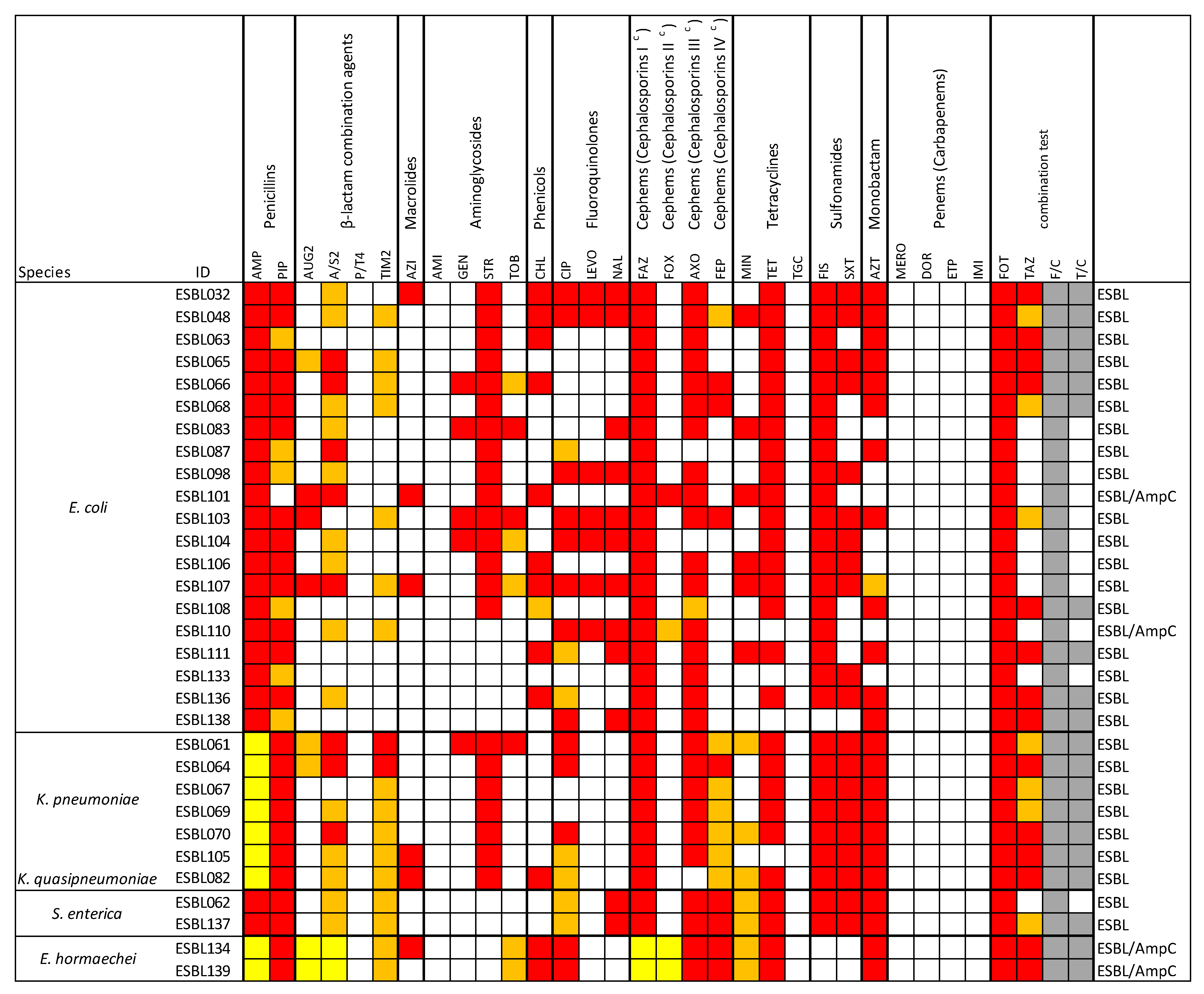
Figure 1. Antimicrobial resistance revealed on 31 strains of Enterobacteriaceae. The species are indicated in the first column. Antimicrobial agents, grouped by drug class, are listed in the remaining columns. c = subclass of generation I, II, III, or IV. Red: Resistance; yellow: intrinsic resistance; white: susceptibility; orange: intermediate resistance; gray: positive result in the combination test for ESBL production. AMP: Ampicillin; PIP: Piperacillin; AUG2: Amoxicillin/Clavulanic acid; A/S2: Ampicillin/Sulbactam; P/T4: Piperacillin/Tazobactam; TIM2: Ticarcillin/Clavulanic acid; AZI: Azithromycin; AMI: Amikacin; GEN: Gentamicin; STR: Streptomycin; TOB: Tobramycin; CHL: Chloramphenicol; CIP: Ciprofloxacin; LEVO: Levofloxacin; NAL: Nalidixic Acid; FAZ: Cefazolin; FOX: Cefoxitin; AXO: Ceftriaxone; FEP: Cefepime; MIN: Minocycline; TET: Tetracycline; TGC: Tigecycline; FIS: Sulfisoxazole; SXT: Trimethoprim/Sulfamethoxazole; AZT: Aztreonam; MERO: Meropenem; DOR: Doripenem; ETP: Ertapenem; IMI: Imipenem; FOT: Cefotaxime; TAZ: Ceftazidime; F/C: Cefotaxime/Clavulanic acid; F/C: Ceftazidime/Clavulanic acid.
Microorganisms 13 01770 g001
Figure 2. Antimicrobial resistance distribution. R = resistant; I = intermediate-resistant; S = susceptible. Tested antibiotics: AMP: Ampicillin; FAZ: Cefazolin; FOT: Cefotaxime; PIP: Piperacillin; AXO: Ceftriaxone; FIS: Sulfisoxazole; TET: Tetracycline; A/S2: Ampicillin/Sulbactam; AZT: Aztreonam; STR: Streptomycin; TAZ: Ceftazidime; CIP: Ciprofloxacin; SXT: Trimethoprim/Sulfamethoxazole; TIM2: Ticarcillin/Clavulanic acid; FEP: Cefepime; CHL: Chloramphenicol; MIN: Minocycline; NAL: Nalidixic Acid; AUG2: Amoxicillin/Clavulanic acid; TOB: Tobramycin; LEVO: Levofloxacin; AZI: Azithromycin; GEN: Gentamicin; FOX: Cefoxitin; P/T4: Piperacillin/Tazobactam; AMI: Amikacin; TGC: Tigecycline; MERO: Meropenem; DOR: Doripenem; ETP: Ertapenem; IMI: Imipenem.
Microorganisms 13 01770 g002
Figure 3. Representation of resistome profile related to the 31 strains (ESBL#ID). The first column lists the species predicted by WGS. Additionally, the table reports the AMR genes identified by WGS. Strains that harbored AMR genes, identified by WGS, are highlighted in blue.
Microorganisms 13 01770 g003
Figure 4. Schematic view of replicons related to the 31 strains (ESBL#ID). For each strain, the presence of a replicon is shown in green.
Microorganisms 13 01770 g004
Figure 5. Schematic view of virulence genes related to the 31 strains (ESBL#ID). For each strain, the presence of VGs is shown in purple.
Microorganisms 13 01770 g005
Table 1. Antimicrobials tested.
Antimicrobial Class (Subclass)AgentRange µg/mL
PenicillinsAMP—Ampicillin1–32
PIP—Piperacillin16–64
β-lactam combination agentsAUG2—Amoxicillin/Clavulanic acid1/0.5–32/16
A/S2—Ampicillin/Sulbactam4/2–16/8
P/T4—Piperacillin/Tazobactam8/4–128/4
TIM2—Ticarcillin/Clavulanic acid8/2–64/2
MacrolidesAZI—Azithromycin0.25–32
AminoglycosidesAMI—Amikacin2–8
GEN—Gentamicin0.25–16
STR—Streptomycin2–64
TOB—Tobramycin2–8
PhenicolsCHL—Chloramphenicol2–32
Quinolones (Fluoroquinolones)CIP—Ciprofloxacin0.015–4
LEVO—Levofloxacin1–8
QuinolonesNAL—Nalidixic Acid0.5–32
Cephems (Cephalosporins I c)FAZ—Cefazolin1–16
Cephems (Cephalosporins II c)FOX—Cefoxitin0.5–64
Cephems (Cephalosporins III c)AXO—Ceftriaxone 0.25–64
Cephems (Cephalosporins IV c)FEP—Cefepim0.06–32
TetracyclinesMIN—Minocycline1–8
TET—Tetracycline4–32
Tetracycline (Glycylcycline)TGC—Tigecyline1–8
SulfonamidesFIS—Sulfisoxazole16–256
SXT—Trimethoprim/Sulfamethoxazole0.12/2.38–4/76
MonobactamAZT—Aztreonam1–16
Penems (Carbapenems)MERO—Meropenem0.03–16
DOR—Doripenem4–8
ETP—Ertapenem0.25–8
IMI—Imipenem0.12–16
ESBL-producing screening combination test methodFOT—Cefotaxime0.25–64
TAZ—Ceftazidime0.25–128
F/C—Cefotaxime/Clavulanic acid0.06/4–64/4
T/C—Ceftazidime/Clavulanic acid0.12/4–128/4
Table 2. ESBL-producing Enterobacteriaceae isolated from raw food samples. N. = number. ID = Identification. * One E. coli and one Salmonella enterica were revealed in the same poultry meat sample; ** one E. coli and one K. pneumoniae were isolated from the same minced turkey sample.
Raw Food SamplesIsolates
SourceN. Analyzed SamplesN. ESBL Positive (Type of Food)ESBL Positive (%)N. ESBL PositiverMLST SpeciesSequence Type (ST)ID Strain
Raw Milk522 (raw milk from vending machine)42E. coliST744ESBL032
E. coliST10ESBL048
Raw Meat20710 (poultry meat) 9E. coliST223ESBL063
E. coliST1011ESBL098
E. coliST457ESBL103
E. coliST1011ESBL104
E. coliST155ESBL108
E. coliST88ESBL110
E. coliST8132ESBL111
E. coliST93ESBL133
E. coli  *ST4162ESBL136
2S. enterica  *ST32ESBL137
S. entericaST32ESBL062
2 (turkey meat) 2K. pneumoniaeST3069ESBL070
K. pneumoniaeST187ESBL105
1 (minced turkey meat) 2E. coli  **ST2705ESBL065
K. pneumoniae  **ST29ESBL064
1 (minced horse meat) 1E. coliST69ESBL068
2 (minced beef and pork) 2E. coliST69ESBL066
K. pneumoniaeST35ESBL067
1 (pork meat) 1K. pneumoniaeST29ESBL069
2 (fresh pork sausage) 2K. pneumoniaeST25ESBL061
E. coliST2179ESBL107
1 (hamburger) 1E. coliST218ESBL106
Subtotal 201022
Seafood products1331 (mussel) 2E. coliST10ESBL101
1 (frozen squid rings)E. hormaecheiST109ESBL134
Subtotal 21.52
Bakery and pastry products, fresh pasta281 (fresh egg pasta)3.61E. coliST515ESBL087
Vegetables80000/ /
TOT50025527
Table 3. ESBL-producing Enterobacteriaceae isolated from RTE food samples. N. = number. ID = Identification. ND = Not Detected. One E. coli and one E. hormaechei were isolated from the same packaged ice cream.
Ready-To-Eat Food SamplesIsolates
SourceN. Analyzed SamplesN. ESBL Positive (Type of Food)ESBL Positive (%)N. ESBL Positive rMLST SpeciesSequence Type (ST)ID Strain
Milk and Cheese2401 (canestrello cheese)0.51E. coliST10ESBL083
Dried or cooked Sausages35000/ /
Ready meals100000/ /
Bakery and pastry products, fresh pasta27000/ /
Ice cream661 (packaged ice cream)1.52E. coliST2223ESBL138
E. hormaecheiST109ESBL139
Vegetables321 (mixed salad)3.01K. quasipneumoniaeNDESBL082
TOT50030.64
Table 4. Mutations detected in the isolates. The presence of each mutation is marked in red. Green color indicates the absence of mutations.
gyr AparCparE
Isolate IDS83LD87NA56TS80IS80RL416FS458A
ESBL032
ESBL048
ESBL061
ESBL062
ESBL063
ESBL064
ESBL065
ESBL066
ESBL067
ESBL068
ESBL069
ESBL070
ESBL083
ESBL087
ESBL098
ESBL101
ESBL103
ESBL104
ESBL105
ESBL106
ESBL107
ESBL108
ESBL110
ESBL111
ESBL133
ESBL134
ESBL136
ESBL137
ESBL138
ESBL139
ESBL82
Disclaimer/Publisher’s Note: The statements, opinions and data contained in all publications are solely those of the individual author(s) and contributor(s) and not of MDPI and/or the editor(s). MDPI and/or the editor(s) disclaim responsibility for any injury to people or property resulting from any ideas, methods, instructions or products referred to in the content.