Article

Comparative Genomics and Phylogenomics of Novel Radiation-Resistant Bacterium Paracoccus qomolangmaensis sp. nov. S3-43T, Showing Pyrethroid Degradation

1
Key Laboratory of Cryospheric Science and Frozen Soil Engineering, Northwest Institute of Eco-Environment and Resources, Chinese Academy of Sciences, Lanzhou 730000, China
2
Key Laboratory of Extreme Environmental Microbial Resources and Engineering, Lanzhou 730000, China
3
Key Laboratory of Ecological Safety and Sustainable Development in Arid Lands, Northwest Institute of Eco-Environment and Resources, Chinese Academy of Sciences, Lanzhou 730000, China
4
Key Laboratory of Desert and Desertification, Northwest Institute of Eco-Environment and Resources, Chinese Academy of Sciences, Lanzhou 730000, China
5
Gansu Academy of Aco-Environmental Sciences, Lanzhou 730020, China
*
Author to whom correspondence should be addressed.
Microorganisms 2025, 13(11), 2441; https://doi.org/10.3390/microorganisms13112441 (registering DOI)
Submission received: 8 September 2025 / Revised: 15 October 2025 / Accepted: 22 October 2025 / Published: 24 October 2025
(This article belongs to the Section Environmental Microbiology)

Abstract

This study focused on the multifunctional characteristics and bioremediation potential of Paracoccus spp. A novel Gram-stain-negative, aerobic, non-motile, ellipsoidal bacterium, named Paracoccus qomolangmaensis S3-43T, was isolated from moraine samples collected from the north slope of Mount Everest at an altitude of 6109 m above sea level (a.s.l.). To clarify the phylogenetic relationship of this strain within the Paracoccus genus and systematically characterize its features, analyses were conducted using polyphasic taxonomy and comparative genomics. Results revealed two distinct functional characteristics of strain S3-43T: First, strain S3-43T exhibits exceptional radiation resistance, particularly tolerance to ionizing radiation. Genome annotation indicates abundant DNA repair and antioxidant-related genes (e.g., vsr, mutL, mutS, ruvC, radA, addA, recA, recN, recO). Second, strain S3-43T contained several pyrethroids degradation related genes, including cytochrome P450, monooxygenase, and aminopeptidase. The results of the genomic comparison of strain S3-43T with related type strains also revealed differences and distribution of key genes related to stress response, environmental variables, and bioactive metabolites. Based on the results of the polyphasic taxonomic analysis, strain S3-43T (=KCTC 8297T = GDMCC 1.3460T) should be classified as a novel species of the genus Paracoccus, designated as Paracoccus qomolangmaensis sp. nov.

1. Introduction

Mount Everest, the core of the Earth’s Third Pole, presents an extreme “multiple stressor” environment, characterized by high altitude, strong radiation (10–20X cosmic rays sea level annual dose, intensified UV inducing DNA breaks, and protein/lipid oxidation, demanding robust DNA repair and antioxidant systems), oligotrophic (the moraine composed of glacial rock debris, sand, and fines exhibits oligotrophy, total organic carbon <0.1%), and low temperatures (annual averages of −15 °C to −10 °C, extremes to −40 °C, with >30 °C diurnal fluctuations), making it difficult for most organisms to survive there in the long term [1,2,3]. Especially at higher altitudes, the cosmic rays received there further hindered the evolution and development of life in such extremely glacial environments [2]. However, the extreme pressures of glacial environments also drive microorganisms to evolve unique adaptive strategies. Proteobacteria, as the dominant group in glacial environments [4] (including moraine, ice, and glacial meltwater [5]), exhibits significant advantages under glacial conditions. Moreover, species of Pseudomonadota, which are oligotrophic bacteria inhabiting nutrient-poor environments like deep-sea sediments, glacial ice, deep subsoil, and so on, possess well-known capabilities of antioxidation, radiation-resistance, degradation of pollutants, and production of abundant secondary metabolites with important applications [6,7,8,9,10]. Therefore, there is an urgent need to discover more stress-resistant microbial resources in glacial environments to explore their mechanisms of stress resistance and uncover their potential applications.
The genus Paracoccus, belonging to the phylum Pseudomonadota, was first proposed by Davis et al. [11], with Paracoccus denitrificans designated as the type species [12]. All species of the genus Paracoccus are Gram-negative. Members of this genus have been isolated from various environments, such as soil, air, plant inter-roots, horse blood, etc. To date, the genus Paracoccus includes 91 validly published novel species (https://lpsn.dsmz.de/genus/paracoccus, accessed on 15 May 2025); marine isolates from different habitats have the highest abundance (~34%), followed by soil isolates (~28%) [13]. Strains affiliated to Paracoccus isolated from oligotrophic environments are still rare. For instance, only four type strains were isolated from extremely glacial habitats [14,15,16,17,18]. Furthermore, from the perspective of molecular phylogenetics, single-gene-based phylogenies are not suitable for Paracoccus spp., as many species of Paracoccus spp. sequences with higher than 99% 16S rRNA gene similarity, but were identified as novel species based on DNA-DNA hybridization (DDH) values [13,19]; genomic metrics such as DDH, Average Nucleotide Identity (ANI), and Average Amino Acid Identity (AAI) were used to overcome the limitations based on single gene [20].
In addition, with regard to the potential applications of Paracoccus species, most Paracoccus spp. have been reported to their bioremediation potential to degrade various environmental pollutants, such as deltamethrin, lindane, carbofuran, polycyclic aromatic hydrocarbons (PAH), etc. For example, Paracoccus sp. YM3 can metabolize carbofuran (50 mg/L) to carbofuran-7-phenol, using the pesticide as the only source of carbon. Paracoccus sp. NITDBR1 can use lindane as the sole source of carbon and could degrade up to 90%, and there are also strains that can act as methylotrophs utilizing C1-carbon compounds as a substrate and accumulating polyhydroxyalkanoates (PHA) [21,22,23,24]. Due to its environmental restoration capabilities and resilience to adverse conditions, previous studies have reported that only Paracoccus everestensis S8-55T isolated from the north slope of Mount Everest has been reported to exhibit antioxidant capacity [14]. Paracoccus yibinensis WLY502T contains genes such as malK, ugpE, ugpA, crtY, and ispA, which are involved in astaxanthin production to repair oxidant damage [25].
Although the above findings indicate that Paracoccus spp. are potential genera for bioremediation of the environment, systematic studies of Paracoccus species derived from strong-irradiated glacial environments remain scarce, especially radiation-resistant species. Specifically, existing relevant studies have primarily focused on the isolation of subglobular bacteria in cold environments (non-radiation-dominated): Paracoccus gahaiensis CUG 00006T, Paracoccus tibetensis S9a3T, Paracoccus nototheniae I-41R45T, and Paracoccus liaowanqingii 2251T were isolated from Gahai Lake and permafrost of Qinghai-Tibetan Plateau, black rock cod fish from the Chilean Antarctic, and Tibetan antelope [15,16,17,18]. Although these strains are adapted to low temperatures or oligotrophic conditions, the radiation intensity in their habitats has not been explicitly correlated. Only Cui Et Al. isolated Paracoccus everestensis S8-55T with antioxidant capability from the north slope of Mount Everest [15]. However, this study did not delve into its radiation resistance mechanism nor address its pollutant degradation function. As mentioned, low-temperature glacial environments typically receive intense solar radiation, especially high-altitude mountain glaciers [1,2]. Microorganisms surviving under such environmental stress may evolve multifunctional adaptive mechanisms that combine cold tolerance, radiation resistance, and efficient nutrient utilization. Therefore, radiation-resistant strain resources and potential applications of Paracoccus species derived from glacier environments require further exploration and investigation. The purpose of this study is to describe a novel species in the genus Paracoccus and to show some of the species’ capabilities via polyphasic taxonomic methods, and by combining genome sequencing with comparative genomic analysis, elucidate the molecular mechanisms of radiation resistance and pyrethroids degradation capacity of strain S3-43T. The findings not only expand our understanding of Paracoccus species diversity and adaptive strategies in extreme cryogenic and highly irradiated environments, but also provide valuable genomic resources for studying radiation resistance mechanisms and developing novel pollutant degradation processes.

2. Materials and Methods

2.1. Isolation, Identification, and Growth Conditions

Moraine samples were collected near the East Rongbuk Glacier on the northern slope of Mount Everest in the Tibet Autonomous Region of China (28.02° N, 86.56° E). Samples were collected in May 2022 at a depth of 0~5 cm, stored at −20 °C in Labplas sterile bags (EFR-5590E, TWIRL’EM®, Labplas Inc., Montreal, QC, Canada). Strain was isolated from moraine on Reasoner’s 2A (R2A) agar [26]. Take 5 g of moraine sample with 20 mL of sterile saline (0.85%, m/v) into a solution 30 °C, 200 rpm shaking for 40 min, stand for 2 min, then dilute the supernatant to a concentration of one part per thousand, and then 200 μL of the supernatant was pipetted and spread on R2A agar medium. After 15 days of incubation at 30 °C, strain S3-43T was isolated and purified on R2A agar medium. Strain was deposited in the Guangdong Microbial Culture Collection Center (GDMCC) and Japan Collection of Microorganisms (JCM). Using universal bacterial primers 27F and 1492R, the complete 16S rRNA gene was amplified by Polymerase Chain Reaction (PCR) [27]. Upon receiving the purified PCR products, Tsingke Company (Xi’an, China) performed bidirectional sequencing using the dideoxy chain termination method with ABI 3730XL analyzer (Applied Biosystems v1.3.1, Waltham, MA, USA). Sequences were compiled using SeqMan software (Lasergen v18.0.3, Houston, TX, USA). The 16S rRNA gene sequence database in EzBioCloud performs 16S rRNA sequence alignment using the BLAST method, matching and comparing with the most closely related type strain [28]. The 16S rRNA sequence of strain S3-43T was deposited in the GenBank database with DDBJ/EMBL/GenBank accession number ON527558. Phylogenetic trees for the 16S rRNA sequences, including the 41 closest type strains of the genus Paracoccus, with Pikeienue piscinae RR4-56T used as an outgroup, were reconstructed using the neighbor-joining [29], minimum-evolution [30] and maximum-likelihood [31] method in Mega V11.0 [32], and topologies of the resultant trees were calculated by bootstrap analysis according to 1000 resamplings [31].

2.2. Morphological, Physiological, Biochemical, and Chemotaxonomic Analysis

Multiple identifications were performed on the strains. The cell morphology of strain S3-43T was observed [33], and cells were subjected to Gram staining using the Solarbio Gram-Stain Kit (Solarbio, Beijing, China). Experimental measurements were also conducted to determine the tolerance of strain S3-43T to temperature (10, 20, 30, 35, 40, and 45 °C), pH (pH 4.0–12.0, interval 1), and NaCl (0–10%, w/v, interval 1%). The methods of carbohydrate and nitrogen utilization tests were performed according to Shirling et al. and Williams et al. [34,35]. Using the method of Kurup and Schmitt, we determined the production of catalase and oxidase and evaluated the hydrolysis of Tween 20, Tween 80, starch, and gelatin [36]. Additional physiological and biochemical characteristics were analyzed for cells cultured at 30 °C. The assays were performed using API ZYM and API 20NE test strips (bioMérieux, Craponne, France) in accordance with the manufacturer’s protocols. The whole-cell sugar composition [37] and polar lipid pattern [38] of strain S3-43T were detected by TLC. Cellular fatty acids were extracted, methylated, and identified using Sherlock MIDI (Newark, DE, USA, Microbial Identification System 6.2b) and a gas chromatograph (6890N; Agilent, Santa Clara, CA, USA), followed by peak identification via the TSBA 6 (version 6.21) database [39]. The analysis method of menaquinones was described by Collins et al. [40].

2.3. Whole Genome Sequencing, Assembly, and Annotation

We extracted genomic DNA from 72h aerobic cultures grown in R2A liquid medium at 30 °C and 160 rpm, using the Bacterial Genomic DNA Extraction Kit (Omega, Norcross, GA, USA), following the manufacturer’s recommendations. The genomic DNA extracted from strain S3-43T was paired-end sequenced on an Illumina HiSeq2000 (Illumina, Inc., San Diego, CA, USA). The genome was assembled de novo by the Velvet 1.2.10 program [41,42]. Sequencing depth was 100× to use high-quality datasets for analysis. Genome assembly was conducted in two stages: the draft genome was generated with SOAPdenovo2 [41], and the complete genome was finalized using Unicycler v0.4.8 [42]. Subsequently, protein-coding genes were predicted from the complete chromosomal assemblies with Glimmer 3.02 (http://ccb.jhu.edu/software/glimmer/index.shtml accessed on 10 May 2025). The NCBI prokaryotic genome annotation pipeline (PGAP) [43] was used to predict tRNA genes, rRNA genes, and non-coding rRNA genes. Genome annotation was carried out employing the RAST system (Rapid Annotation using Subsystem Technology) [44]. The UBCG phylogenomic tree was reconstructed based on the genome using the UBCG pipeline (Up-to-Date Bacterial Core Gene set and pipeline) for 92 core genes [45]. A total of 68 genomes from the Paracoccus type strains were retrieved from the NCBI database on 22 July 2024.

2.4. Radiation Resistance Analysis

The radiation resistance of strain S3-43T was assessed. We used the reference type strains Deinococcus radiodurans R1T as the positive control and Escherichia coli BL 21T as the negative control. Strains S3-43T, R1T, and BL21T were evaluated for resistance to UV-C irradiation, γ-ray irradiation, heavy-ion irradiation, and antioxidant. Strains S3-43T, R1T, and BL21T were inoculated into R2A liquid medium and cultured to the logarithmic growth phase (OD600 = 1). The bacterial suspensions were then diluted to a concentration of 10−4 relative to the original concentration. Take 100 μL of strain dilutions and spread it on R2A agar medium and irradiate it with UV-C irradiation at 0, 50, 100 J/m2, incubated for 7–14 days, and counted. Take 20 mL of the original solution and irradiate it with γ-rays using a Co-60 source γ-ray meter (Qingdao, China, Zhongke Tongxing Nuclear Technology Co., Ltd.) at 0, 50, and 100 kGy. Take 1 mL of diluted solution in a 3 cm diameter Petri dish and subject it to 12C6+ ion beam irradiation at 0, 250, and 500 Gy. (The 12C6+ ion beam irradiation parameters are as follows: after passing through a 50 μm stainless steel window, a 20 μm Mylar film, and a 1.3 m of air gap, the energy of the 12C6+ ions from 80 MeV/u decays to 76.37 MeV/u; the range in the water was expected to be 16 mm, and the peak position was 15.5 mm.) After γ-rays and ion beam irradiation, 100 μL of irradiated bacterial suspension was coated on R2A agar medium, incubated for 7–14 days, and counted. The colonies of the irradiated plates were counted as N; the colonies of the unirradiated plates were counted as N0, and the survival rate of the irradiated strains was calculated as SR (Survival Rate) = N/N0 × 100% to determine the strength of the strain’s resistance to radiation [7]. All tests had three replicates.

2.5. Antioxidant Analysis

The antioxidant capacity of the strains was evaluated by assessing their viability in the presence of H2O2, a weak oxidant that permeates cell membranes and induces intracellular damage [46]. We evaluated the antioxidant capacity based on the survival rate of the strain after exposure to hydrogen peroxide. Use the reference type strains Deinococcus radiodurans R1T as the positive control and Escherichia coli BL 21T as the negative control. Strains were cultured in R2A liquid medium, and the bacterial suspension (OD600 = 1) was mixed with H2O2 solution to make the final mixed solution of hydrogen peroxide concentration, which was 0–100 mM at intervals of 10 mM. Subsequently, the centrifuge tubes were incubated for four hours at 30 °C under agitation (160 rpm). Diluted the mixed solution into 10,000 dilutions with 0.9% normal saline, inoculated 100 μL diluent on R2A agar medium, cultured it in a constant temperature incubator at 30 °C for 7 days, and counted. The colonies of the treated plates were counted as N; the colonies of the untreated plates were counted as N0, and the survival rate of the strains under oxidative stress was calculated as SR (Survival Rate) = N/N0 × 100% to determine the strength of the strain’s resistance. All tests had three replicates.

2.6. Cyhalothrin Degradation Analysis

The cyhalothrin degradation experiment was carried out in 500 mL flasks containing 150 mL of sterile R2A liquid medium containing 50 mg/L cyhalothrin. The strain was inoculated into the medium and incubated in a shaker at 30 °C, 180 rpm for 5 days, and the concentration of residual cyhalothrin was measured by high performance liquid chromatography (HPLC) every 12 h. The growth of the strain was determined by OD600. All tests had three replicates.

2.7. Comparative Genomic Analysis

Genomic data of species related to strains S3-43T were obtained from GenBank to evaluate their similarity. We computed the Average Nucleotide Identity (ANI) with the OrthoANIu (OrthoANI), BLAST (ANIb), and MUMmer (ANIm) algorithms [28,47,48,49]. We computed the digital DNA–DNA hybridization (dDDH) values using the GGDC 3.0 tool [50]. Specifically, the values were derived from the recommended formula 2, chosen for its independence from genome length and robustness when handling incomplete draft genomes. We computed the Average Amino acid Identity (AAI) with the online tool developed by the Konstantinidis group (http://enve-omics.ce.gatech.edu/aai/ accessed on 10 May 2025) [51]. KEGG (Kyoto Encyclopedia of Genes and Genomes) [52], COG (Clusters of Orthologous Groups of proteins) [53], NR (NCBI Non-Redundant Protein) [54], Pfam (Protein Families) [55], Swiss-Prot [56], and CAZy (Carbohydrate Active Enzymes) [57] databases were selected for retrieval to improve the general functional annotation. The biosynthetic gene cluster of secondary metabolites was predicted by silicon calculation using AntiSMASH 7.0 [58]. Pan-genomes were analyzed using PGAweb [59].

2.8. Data Analysis and Statistics

All experiments were performed in triplicate. The phylogenetic tree was constructed by MEGA 11.0. Statistical analysis was carried out using SPSS 16.0 and R 4.1.0 software. Data analysis and graphical representation were conducted with OriginPro 2022. Adobe illustrator 2020 was used to draw figures.

3. Results and Discussion

3.1. Phenotypic, Physiological, Phylogenetic, and Phylogenomic Characteristics

The colonies of strain S3-43T cultured in R2A medium after 72h were milky white and spherical. The cells of S3-43T were aerobic, Gram-negative, and ellipsoid (0.6–0.9 μm × 0.7–1.1 μm) (Figure 1a), without spore formation and motility. The strain grew with the optimal temperature of 30 °C, the optimal pH = 8.0, and without NaCl. Strain S3-43T was positive for oxidase activity, reduction in nitrate, and alkaline phosphatase, but negative for esterase lipase (C8) and N-acetyl-β-glucosaminidase. Strain S3-43T could not use L-Arabinose or D-xylose as the sole carbon source. The different physiological characteristics of S3-43T and its reference strains are shown in Table S1. The physiological characteristics distinguish strain S3-43T from its closest type species in the genus Paracoccus. The chemotaxonomic characteristics such as major fatty acids, predominant respiratory menaquinone, and polar lipids of strain S3-43T correspond to those of other members of the genus Paracoccus (Table S2, Figure 1b and Figure S4).
Pairwise 16S rRNA gene sequence comparisons showed that strain S3-43T has the highest pairwise similarity of 98.76% with P. haematequi M1-83T. The 16S rRNA-based neighbor-joining, minimum-evolution, maximum-likelihood phylogenetic trees (Figures S1–S3) and the core gene-based UBCG phylogenomic tree collectively determined the placement of strain S3-43T within the genus Paracoccus cluster (Figure 1c). The assembled genome sequence of strain S3-43T (DDBJ/EMBL/GenBank accession number CP119082) encodes 3746 CDS, 46 tRNAs, and two copies of the 16S rRNA gene. Genomic characterization of strain S3-43T is similar to other species of the genus Paraucoccus. Detailed genomic information on the genome sequences of the reference strains is provided in Table S3. ANIb, ANIm, and OrthoANI evaluations indicate that the overall genome relatedness between the strain S3-43T (Figure S5) and its closest relatives is below the 95% cut-off value for grouping genomes of the same species [60]. The genome sequences of strain S3-43T and related species had the highest dDDH value of 49.10% (P. haematequi M1-83T). The results of the dDDH analysis of the strain S3-43T and its closest neighbors were below the threshold of 70% established for delineating prokaryotic species (Figure S5) [61]. The highest AAI value between S3-43T and related species was 91.91% (Figure S5), which is also lower than 95%, the threshold defined by prokaryotic species [62]. These suggest that strain S3-43T represents a novel species of the genus Paracoccus.
Based on the presented results for phylogenomic, genome, biochemical, and physiological analyses, strain S3-43T appears to be a novel species of the genus Paracoccus, for which the name Paracoccus qomolangaensis sp. nov. is proposed.

3.2. The Radiation Resistance and Antioxidant of S3-43T

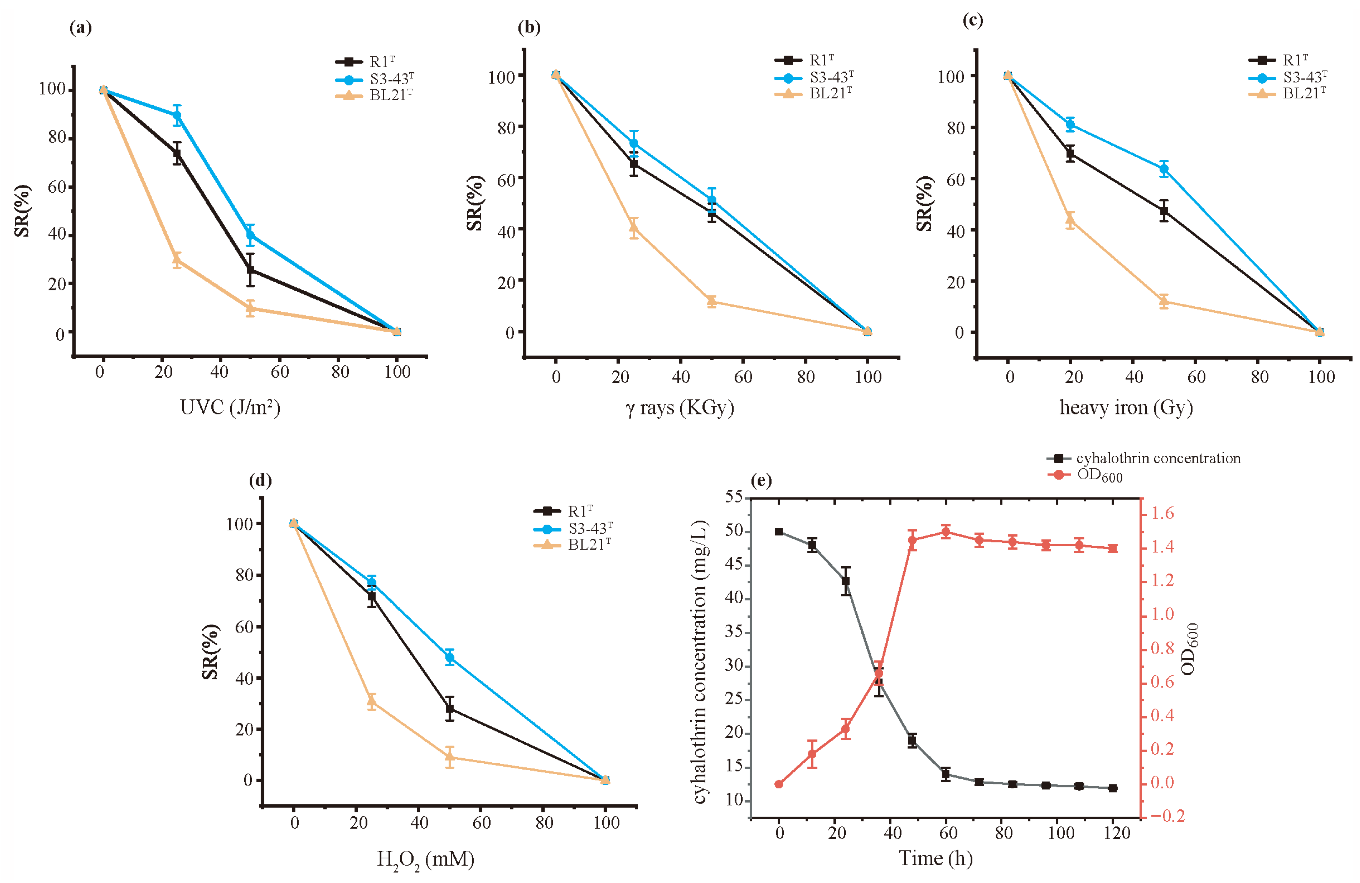
Figure 2 shows the radiation resistance and antioxidant activity of strain S3-43T compared to the control strains D. radiodurans R1T and E. coli BL21T. The radiation resistance of strain S3-43T was evaluated through non-ionizing radiation (UV-C 254nm) and ionizing radiation (γ-rays and heavy-ion beam). The results show that the radiation resistance of strain S3-43T is generally higher than that of the positive control D. radiodurans R1T, performing particularly well when exposed to UV-C irradiation. At the radiation dose of 25 J/m2, the survival rate of strain S3-43T reached 89.67%, while the survival rate of D. radiodurans R1T only reached 74.00%. Under γ-ray irradiation, the advantage of strain S3-43T did not exist. However, under heavy-ion beam irradiation at a dose of 100 Gy, the survival rate of strain S3-43T reached 81.00%, which is higher than that of strain D. radiodurans R1T. In addition, the antioxidant capacity of strain S3-43T was assessed, showing great antioxidant ability with a slower decline in survival rate (Figure 2d). When the UV-C dose was 100 J/m2, the γ-ray dose was 100 KGy, the heavy-ion dose was 500 Gy, and the hydrogen peroxide concentration was 100 mM, no strain survived. This indicated that even if there were anti-radiation and antioxidant genes in the strains that expressed gene repair function, excessive UV-C radiation and hydrogen peroxide concentration would also cause cell damage.

3.3. Cyhalothrin Degradation Capability

Figure 2e shows the growth of strain S3-43T under 50 mg/L cyhalothrin conditions and the degradation process of cyhalothrin. In the first 0–24 h, bacterial cell growth was slow, and the cyhalothrin concentration remained virtually unchanged. In the logarithmic growth phase (1–2 days), both bacterial cell numbers and rates of cyhalothrin biodegradation showed a fast upward trend, reaching a biodegradation rate of 62% by 48 h. After 60 h of incubation, bacterial cell numbers reached a peak and then gradually decreased. After 5 days, the degradation rates of cyhalothrin reached 76.2%. In conclusion, strain S3-43T can effectively degrade cyhalothrin.

3.4. Genomic Insights into a Novel Species Related to Biological Functions

A series of comparative genomic analyses was performed to further characterize Paracoccus qomolangaensis S3-43T and its closely related type strains at the genomic level.

3.4.1. Pan-Genome Analysis

The pan-genome represents the complete genetic makeup of a species, serving as the gene pool of all strains within that species. It comprises core genes (homologous gene clusters/genes present in all samples), dispensable genes (homologous gene clusters/genes coexisting in two or more samples), and unique genes (homologous gene clusters/genes present in only a single sample). Bacterial pangenomes can be categorized into open pan-genome and closed pan-genome. It was shown that the genus Paracoccus supports an open pan-genome [20]. The 12 strains of genus Paracoccus in this study showed an open pan-genome based on a power-law regression function (b = 0.646, which is in the range of 0 < b > 1) (Figure S5), in which each Paracoccus sp. contains several unique genes, and as the genomes increase, the pan-genome pool increases further. The core size accumulation curve reached saturation after two genomes, indicating the existence of a stable minimum core. The 3365 CDS present in the genome of strain S3-43T were divided into 1958 homologous gene clusters (Figure 3b), most of these gene clusters were present in the core genes (48.96%), compared to the dispensable genes (1208 gene clusters, 38.16%) and unique genes (408 gene clusters, 12.89%) genome (Figure 3a). We compared the homologous genes between strain S3-43T and its closely related strains and showed that 1550 core gene clusters were shared among the genomes; the percentage range of core genes for each Paracoccus is between 37.71% and 53.54%. Of the 18,876 genes identified in the 1550 homologous gene clusters shared by the genomes, 17,718 genes have COG annotations, including repair genes such as superoxide dismutase, recombinant DNA repair gene recR, recO, recA, and radA, and DNA mismatch repair gene mutS and mutL (Table 1). This suggests that Paracoccus spp. have the potential for DNA repair. The percentage of the dispensable gene ranged from 25.00 to 48.68% among 12 genomes of Paracoccus. The unique genes had a wide percentage range from 6.87 to 43.28% (Figure 3a). These results underscore the high adaptability of Paracoccus genomes and the non-conservative nature of their gene counts. Strain S3-43T has 408 unique gene clusters out of a total of 1958 clusters, and of the 423 genes identified in these unique clusters, 243 genes have COG annotations, including antioxidant genes and DNA repair genes, such as the organic hydrogen peroxide reductase OsmC/OhrA, site-specific recombinant DNA repair gene xerD, etc. This suggests that strain S3-43T exhibited a superior capacity for DNA repair in the genus Paracoccus.
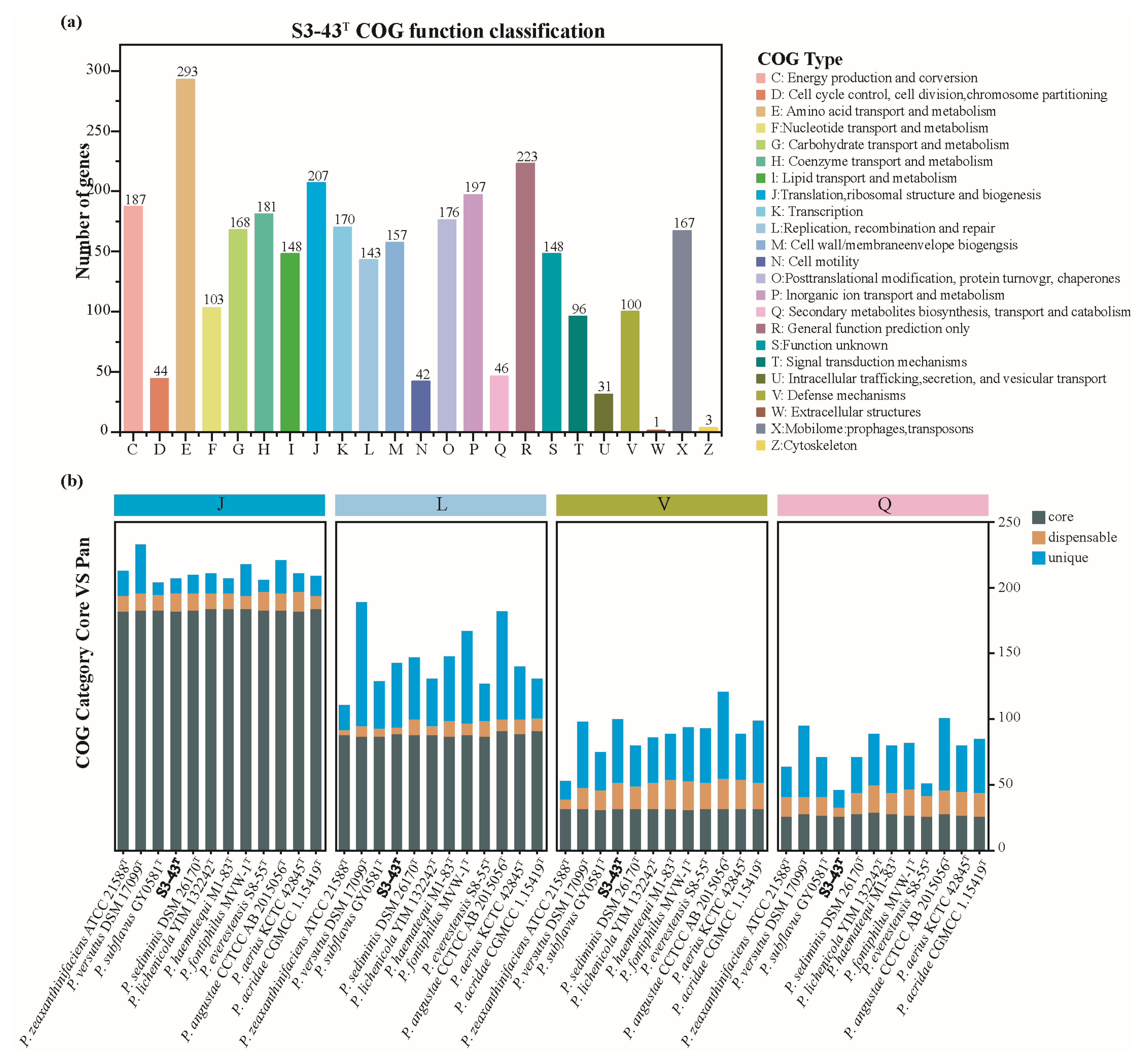
The Clusters of Orthologous Genes (COG) database was used to study conserved protein functions among different species, reveal their evolutionary relationships, and compare species by COG functional categories [63]. The proteins that make up each homologous gene cluster are assumed to come from an ancestral protein and have the same function [64]. A total of 2761 CDS of strain S3-43T (82.05% of all) were distributed in 23 COG functional categories (Figure 4a). The major functional category includes genes containing amino acid transport and metabolism (COG-E, 293 genes), general function prediction only (COG-R, 223 genes), translation, ribosomal structure and biogenesis (COG-J, 207 genes), inorganic ion transport and metabolism (COG-P, 197 genes), energy production and conversion (COG-C, 187 genes), coenzyme transport and metabolism (COG-H, 181 genes), posttranslational modification, protein turnover, chaperones (COG-O, 176 genes), transcription (COG-K, 170 genes), carbohydrate transport and metabolism (COG-G, 168 genes), mobilome: prophages, transposons (COG-X, 167 genes), cell wall/membrane/envelope biogenesis (COG-M, 157 genes), lipid transport and metabolism (COG-I, 148 genes), function unknown (COG-S, 148 genes), replication, recombination and repair (COG-L, 143 genes), nucleotide transport and metabolism (COG-F, 103 genes), and defense mechanisms (COG-V, 100 genes). Analysis of the pan-genomes of 12 strains of genus Paracoccus showed broadly similar functional categories related to radiation resistance and antioxidant (Figure 4b).

3.4.2. Genes Potentially Associated with Radiation Resistance and Antioxidant Capabilities

As mentioned, it is speculated that strain S3-43T has the potential for radiation resistance and antioxidant as isolated from the northern slope of Mount Everest were with strong radiation. Radiation damage to bacteria is mainly divided into direct and indirect damage; high doses of ionizing radiation can directly damage DNA, causing single-strand breaks (SSBs) and double-strand breaks (DSBs), with SSBs occurring 40 times more than DSBs [65]. Usually, direct damage accounts for only 20% of the cellular damage caused by radiation and 80% of the cellular damage comes from indirect damage due to oxidative stress [66,67]. Research has shown that exposure to ionizing radiation causes severe oxidative stress to all macromolecules within cells [65]. Oxidative stress refers to cellular damage caused by elevated levels of intracellular oxygen radicals and accumulation of reactive oxygen species (ROS) [68]. The accumulation of ROS arises from the radiolytic decomposition of water, including hydroxyl radicals (HO), superoxide (O2), and hydrogen peroxide (H2O2), leading to cellular oxidative stress and damage to proteins, lipids, nucleic acids, and carbohydrates [67,69]. Bacteria protect themselves from oxidative damage by turning on the antioxidant system and DNA repair system. Oxidative stress causes DNA damage, including oxidative damage to bases and sugar-phosphates, and single or double-strand breaks (SSB or DSB) in DNA [70]. ROS react with nitrogenous bases and deoxyribose to cause oxidative reactions leading to DNA base alterations or double helix breaks [71]. ROS also causes lipid peroxidation, where the free radicals oxidize unsaturated lipid strands, leading to the formation of hydroperoxyl lipids and alkyl radicals [72].
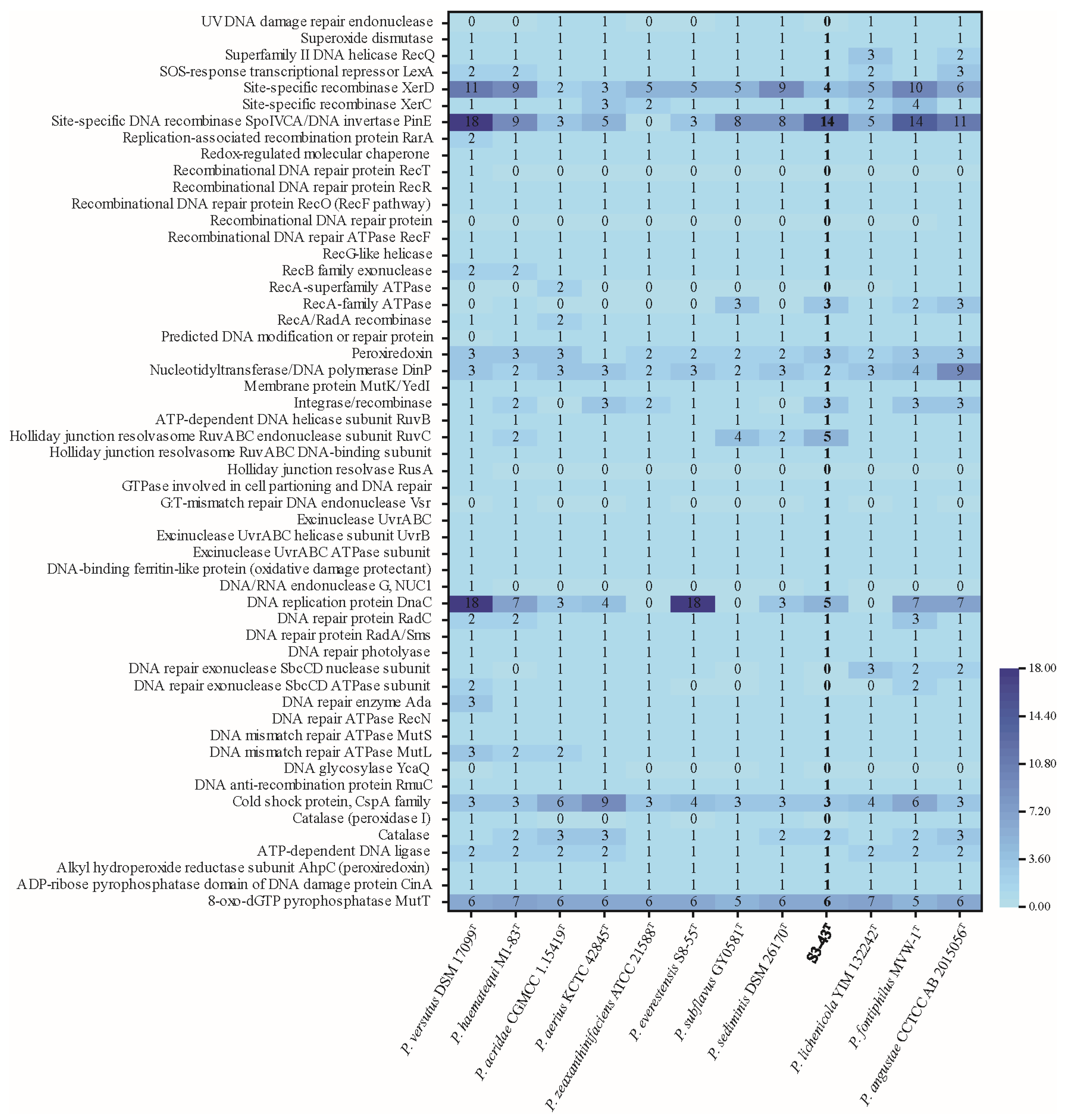
The strains in this study have a series of DNA repair genes to respond to oxidative stress-induced DNA damage (Figure 5). For example, the most common base damage caused by oxidative stress is 8-oxoguanine [70], which is mainly repaired by the base excision repair pathway (BER) initiated by the DNA N-glycosidases Fpg and OGG1, respectively, and is ameliorated by the involvement of MutT in the “GO repair system” [73,74]. In bacteria, UvrA and UvrB proteins encoded by genes uvrA and uvrB recognize and cleave damaged DNA in a multistep reaction to complete the nucleotide excision repair (NER) process [75] in response to DNA damage induced by radiation stress and oxidative stress. The results show that genes involved in antioxidant and DNA restoration described in previous studies were located in the strain S3-43T genome. The genomes of 12 Paracoccus spp. strains were annotated to ahpC (peroxiredoxin), catalase, peroxiredoxin, and SOD (superoxide dismutase), constituting the enzymatic antioxidant system (Figure 5). Gene ahpC can encode a key antioxidant enzyme belonging to the thiol peroxidases (peroxiredoxins) family that scavenges micromolar concentrations of H2O2. Peroxidase as AhpC can be activated when millimolar concentrations of H2O2 are saturated [76,77]. SOD scavenges superoxide radicals, which generate less reactive hydrogen peroxide and oxygen with two superoxide radicals and two protons. Hydrogen peroxide is, in turn, converted to water and oxygen by catalase [70].
The possession of these antioxidant enzymes by strain S3-43T is comparable to that of the other 11 strains, and it is hypothesized that Paracoccus spp. have a stable enzymatic antioxidant system. Meanwhile, KEGG metabolic pathway analysis showed that strain S3-43T had 30 genes involved in glutathione metabolism, including pepN, gshA, gshB, and gst, suggesting that strain S3-43T may metabolize glutathione as a non-enzymatic antioxidant to scavenge ROS [78]. The gene crtB of strain S3-43T is involved in the carotenoid biosynthesis pathway, and carotenoids, as antioxidants, can greatly inhibit the effects of oxidative stress on redox status [79]. The other 11 related strains have more genes involved in the carotenoid biosynthesis pathway (gene numbers: 1–7); thus, it is hypothesized that strain S3-43T does not mainly rely on carotenoid antioxidants in response to oxidative stress. A complete antioxidant system gives the strain S3-43T excellent resistance to ionizing radiation.

3.4.3. Gene Potentially Associated with Cyhalothrin Degradation Capability

Although pyrethroids are considered safer than other insecticides, their persistence in the environment poses high risks to non-target organisms and humans. Humans can be exposed to pyrethroid insecticides through direct skin contact and inhalation [80]. Many studies have confirmed that microorganisms can degrade pyrethroids directly by using them as carbon sources or through co-metabolism [81]. The cleavage of ester bonds is generally considered the main step in the degradation pathway of pyrethroids [82]. In the process of microbial degradation of pyrethroids, various enzymes responsible for ester bond hydrolysis (carboxylesterase, monooxygenase, and aminopeptidase) have been purified and characterized [83,84,85,86,87]. Previous research has shown that the metabolic resistance of pyrethroids is mainly related to cytochrome P450 monooxygenases (P450s), followed by carboxylesterases (CarEs) [88,89]. The biodegradation of cyhalothrin in Cunninghamella elegans is catalyzed by the monooxygenase cytochrome P450 [85]. The resistance to λ—cypermethrin in the Helicoverpa armigera SYR of cotton bollworm in Shenyang, China, is due to metabolic enhancement caused by overexpression of multiple P450 enzymes [90]. Heterologously expressed P450 enzymes can also metabolize pyrethroid insecticides in cotton bollworms [91]. The results indicate that the genes involved in cyhalothrin degradation described in previous studies are located in the genome of strain S3-43T. It is hypothesized that cytochrome P450 may be involved in the metabolism of cyhalothrin by strain S3-43T, catalyzing the hydrolysis of ester bonds. Strain S3-43T possessed cytochrome P450 comparable to the other 11 strains (Table 2). In addition, the genome of strain S3-43T was annotated with various monooxygenases and aminopeptidases, which are hypothesized to play a role in cyhalothrin degradation. Among these twelve genomes, aminopeptidase FrvX was annotated only in P. haematequi, P. acridae, P. aerius, and S3-43T, suggesting a greater potential for pyrethroids degradation by strain S3-43T. Strain S3-43T was isolated from the northern slope of Mount Everest and has good adaptability to cold environments. Therefore, its enzymes can maintain good activity in cold environments, which is helpful for the treatment of vegetables and fruits contaminated with pyrethroids in subsequent applications. Therefore, further functional expression and metabolic analyses are needed to confirm the role of cytochrome P450, monooxygenase, and aminopeptidase in the metabolism of cyhalothrin resistance in strain S3-43T.

3.4.4. Horizontal Gene Transfer Analysis

Genomic islands (GIs) are a prevalent form of horizontally transferred elements, typically comprising independent DNA fragments within prokaryotic chromosomes (Table S4). Based on the functions encoded by their genes, genomic islands can be categorized into virulence, resistance, metabolic, and symbiotic islands. Beyond several fundamental and homologous proteins that are functionally and structurally analogous to those found in other known strains, the genomic islands of S3-43T encompass some non-homologous proteins capable of expressing additional functions. The existence of these non-homologous proteins may enable the strain to exhibit unique biological characteristics or adaptations, conferring it with enhanced viability or a competitive edge in a specific environment.
Most of the genes identified within the genomic islands of these twelve model strains were classified as putative genes or features with other movable genetic elements, such as insertion sequences and transposases. However, each strain carries genes related to enhanced adaptability, including genes encoding the secretion system (TSS), toxin antitoxin system, antibiotic resistance, and heavy metal transporters. Through genetic testing, we identified 22 GIs in S3-43T containing 529 genes ranging from 5628 to 86,802 bp in length. Based on the functional analysis of GI genes, most of these known functional genes are involved in cellular metabolism and membrane transport functions. Annotation of the predicted gene functions of S3-43T GIs revealed that some of these gene sequences could be expressed as DNA repair systems, such as the ruvC, dnaE, vsr, and xerD. Genes ruvA, ruvB, and ruvC can affect the effective survival rate of cells after radiation. The production of RuvA, RuvB, and RuvC proteins is required for the recombinational repair of UV-induced damage to DNA [92,93]. Genes uvrA, uvrB, uvrC, and uvrD are involved in the NER (nucleotide excision repair mechanism) repair pathway capable of dealing with a variety of potentially lethal DNA damage induced by radiation [94,95,96]. Gene vsr encodes a DNA mismatch endonuclease that can initiate VSP (Very Short Patch) mismatch repair [97,98], and is presumed t to be involved in radiation-induced DNA mismatches. Gene osmC plays an important role in peroxide metabolism and protects strains from oxidative stress [76,99].

4. Conclusions

This is the first study to describe the bacterial strain Paracoccus qomolangaensis S3-43T, isolated from the north slope of Mount Everest, and to investigate its mechanisms of radiation resistance and genomic function under extreme environmental stress. A variety of radiation-resistant antioxidant genes were identified, including recR, recO, recA, radA, and ahpC, among others. Therefore, we analyze that this is the reason why this strain was able to survive at high altitude in a cold-resistant, radiation-resistant, and oxidation-resistant environment. In addition, strain S3-43T contained a number of cyhalothrin degradation-related genes, including cytochrome P450, monooxygenase, and aminopeptidase.
In conclusion, experimental and genomic analyses showed that strain S3-43T possesses radiation and antioxidant resistance. A good antioxidant system gives it good resistance to ionizing radiation, while also exhibiting potential for pesticide degradation. Our results provide a foundation for investigating microbial radiation resistance and antioxidant mechanisms.

Description of Paracoccus qomolangaensis sp. nov.

Paracoccus qomolangaensis (qomolangma. en’sis. N.L. masc. adj. qomolangmaensis about Mount Qomolangma, Tibet, China, where the type strain was isolated.)
Strain S3-43T exhibits Gram-negative cells, with non-flagellated, ellipsoid (0.6–0.9 μm × 0.7–1.1 μm). Colonies on R2A are circular, convex, smooth, opaque, milky white, and approximately 2.0–3.0 mm in diameter after 72 h at 30 °C. It can grow at 10–35 °C (optimum 30 °C) and pH 6.0–11.0 (optimum pH 8.0), but it is intolerant of NaCl. Cells are positive for oxidase activity and reduction in nitrate, but urea, gelatin, starch, cellulose, Tweens 20, and 80 were not hydrolyzed. The strain is negative for production of H2S. In the API ZYM strip, it is positive for alkaline phosphatase, esterase (C4), leucine arylamidasem, naphthol-AS-BI-phosphohydrolase, and α-Glucosidase and negative for esterase lipase (C8), lipase (C14), acid phosphatase, N-acetyl-β-glucosaminidase, α-mannosidase, and β-fucosidase. Strain S3-43T could not use L-Arabinose, D-Fructose, D-Glucose, D-Lactose, D-Galactose, D-Mannitol, D-Raffinose, D-Rhamnose, sucrose, or D-xylose as the sole carbon source. Meanwhile, strain S3-43T could utilize L-Tyrosine as a unique nitrogen source but could not utilize L-Alanine, L-Aspartate, L-Histidine, and L-Glycine. Ubiquinone-10 (Q-10) is the sole respiratory quinone. The major polar lipids are diphosphatidylglycerol (DPG), four phospholipids (PL), an unidentified lipid (L), an unidentified aminolipid (AL), and an unidentified glycolipid (GL). The major cellular fatty acids (>10.0%) are summed feature 8 (comprising C18:1 ω7c and/or C18:1 ω6c).
The type strain S3-43T (=KCTC 8297T = GDMCC 1.3460T) was isolated from moraine samples being collected from the north slope of Mount Everest (28.02° N, 86.56° E), PR China. The DNA G+C content of the type strain is 67.2%. The GenBank/EMBL/DDBJ accession numbers for the genome and 16S rRNA gene sequences of type strain S3-43T are CP119082 and ON527558, respectively.

Supplementary Materials

The following supporting information can be downloaded at: https://www.mdpi.com/article/10.3390/microorganisms13112441/s1, Figure S1: Neighbor-joining phylogenetic tree based on 16S rRNA gene sequences of the strain S3-43T and the type strains of other closely related species in the genus Paracoccus and Pikeienuella, Figure S2: Minimum-evolution phylogenetic tree based on 16S rRNA gene sequences of the strain S3-43T and the type strains of other closely related species in the genus Paracoccus and Pikeienuella, Figure S3: Maximum-likelihood phylogenetic tree based on 16S rRNA gene sequences of the strain S3-43T and the type strains of other closely related species in the genus Paracoccus and Pikeienuella, Figure S4: Polar lipids profile of strain S3-43T, Figure S5: Genome comparisons of strain S3-43T and their related reference strains, and Figure S6: Characteristic curves of the pan-genome and core genome of S3-43T; Table S1: Phenotypic characteristics of strain S3-43T and its closely related type strains in the genus Paracoccus, Table S2: Whole cellular fatty acids composition of S3-43T and the closely related type strains of the genus Paracoccus, Table S3: General genomic characteristics comparison of strain S3-43T and its closely related species, and Table S4: Features of the GIs found in the genome of S3-43T. References [100,101,102,103,104] are cited in the supplementary materials.

Author Contributions

Conceptualization, Y.L. and G.Z.; Data Curation, X.C.; Formal Analysis, Y.L. and L.Z.; Funding Acquisition, W.Z.; Investigation, T.C. (Tuo Chen); Methodology, Y.L.; Project Administration, W.Z.; Resources, T.C. (Tuo Chen), G.L. and W.Z.; Software, X.C. and T.C. (Tian Cheng); Supervision, G.Z.; Validation, Y.L. and Y.Z.; Visualization, Y.L.; Writing—Original Draft, Y.L.; Writing—Review and Editing, Y.L. and G.Z. All authors have read and agreed to the published version of the manuscript.

Funding

This research was funded by the Scientific Project of Gansu Province, grant number 25ZDWA005, and the National Science Foundation of Gansu Province, grant number 25JRRA503.

Institutional Review Board Statement

Not applicable.

Informed Consent Statement

Not applicable.

Data Availability Statement

The GenBank/EMBL/DDBJ accession numbers for the genome and 16S rRNA gene sequences of type strain S3-43T are CP119082 and ON527558, respectively. The data can be accessed at https://www.ncbi.nlm.nih.gov/.

Acknowledgments

We thank our colleagues at HIRFL of the Institute of Modern Physics, Chinese Academy of Sciences, for providing high-quality carbon ion beam irradiation.

Conflicts of Interest

The authors declare no conflicts of interest.

References

  1. Sood, U.; Dhingra, G.G.; Anand, S.; Hira, P.; Kumar, R.; Kaur, J.; Verma, M.; Singhvi, N.; Lal, S.; Rawat, C.D.; et al. Microbial Journey: Mount Everest to Mars. Indian J. Microbiol. 2022, 62, 323–337. [Google Scholar] [CrossRef]
  2. Moore, G.W.; Semple, J.L. The impact of global warming on Mount Everest. High Alt. Med. Biol. 2009, 10, 383–385. [Google Scholar] [CrossRef]
  3. Lieberman, P.; Morey, A.; Hochstadt, J.; Larson, M.; Mather, S. Mount Everest: A space analogue for speech monitoring of cognitive deficits and stress. Aviat. Space Environ. Med. 2005, 76, B198–B207. [Google Scholar]
  4. Oren, A.; Garrity, G.M. Valid publication of the names of forty-two phyla of prokaryotes. Int. J. Syst. Evol. Microbiol. 2021, 71, 5056. [Google Scholar] [CrossRef] [PubMed]
  5. Kim, K.M.; Hwang, K.; Lee, H.; Cho, A.; Davis, C.L.; Christner, B.C.; Priscu, J.C.; Kim, O.S. Genetic isolation and metabolic complexity of an Antarctic subglacial microbiome. Nat. Commun. 2025, 16, 7501. [Google Scholar] [CrossRef]
  6. Asaf, S.; Numan, M.; Khan, A.L.; Al-Harrasi, A. Sphingomonas: From diversity and genomics to functional role in environmental remediation and plant growth. Crit. Rev. Biotechnol. 2020, 40, 138–152. [Google Scholar] [CrossRef] [PubMed]
  7. Liu, Y.; Chen, T.; Cui, X.; Xu, Y.; Hu, S.; Zhao, Y.; Zhang, W.; Liu, G.; Zhang, G. Sphingomonas radiodurans sp. nov., a novel radiation-resistant bacterium isolated from the north slope of Mount Everest. Int. J. Syst. Evol. Microbiol. 2022, 72, 5312. [Google Scholar] [CrossRef] [PubMed]
  8. Wilkes, R.A.; Aristilde, L. Degradation and metabolism of synthetic plastics and associated products by Pseudomonas sp.: Capabilities and challenges. J. Appl. Microbiol. 2017, 123, 582–593. [Google Scholar] [CrossRef]
  9. Kang, M.; Choi, T.R.; Ahn, S.; Heo, H.Y.; Kim, H.; Lee, H.S.; Lee, Y.K.; Joo, H.S.; Yune, P.S.; Kim, W.; et al. Arctic Psychrotolerant Pseudomonas sp. B14-6 Exhibits Temperature-Dependent Susceptibility to Aminoglycosides. Antibiotics 2022, 11, 1019. [Google Scholar] [CrossRef]
  10. Desriac, F.; Jégou, C.; Balnois, E.; Brillet, B.; Le Chevalier, P.; Fleury, Y. Antimicrobial peptides from marine proteobacteria. Mar. Drugs 2013, 11, 3632–3660. [Google Scholar] [CrossRef]
  11. Davis, D.H.; Doudoroff, M.; Stanier, R.Y.; Mandel, M. Proposal to reject the genus Hydrogenomonas: Taxonomic implications. Int. J. Syst. Bacteriol. 1969, 19, 375–390. [Google Scholar] [CrossRef]
  12. Steinrücke, P.; Ludwig, B. Genetics of Paracoccus denitrificans. FEMS Microbiol. Rev. 1993, 10, 83–117. [Google Scholar] [CrossRef]
  13. Puri, A.; Bajaj, A.; Singh, Y.; Lal, R. Harnessing taxonomically diverse and metabolically versatile genus Paracoccus for bioplastic synthesis and xenobiotic biodegradation. J. Appl. Microbiol. 2022, 132, 4208–4224. [Google Scholar] [CrossRef]
  14. Cui, X.; Liu, Y.; Xu, Y.; Chen, T.; Zhang, S.; Wang, J.; Yang, R.; Liu, G.; Zhang, W.; Zhang, G. Paracoccus everestensis sp. nov., a novel bacterium with great antioxidant capacity isolated from the north slope of Mount Everest. Int. J. Syst. Evol. Microbiol. 2022, 72, 5562. [Google Scholar] [CrossRef]
  15. Zhang, G.; Xian, W.; Yang, J.; Liu, W.; Jiang, H.; Li, W. Paracoccus gahaiensis sp. nov. isolated from sediment of Gahai Lake, Qinghai-Tibetan Plateau, China. Arch. Microbiol. 2016, 198, 227–232. [Google Scholar] [CrossRef]
  16. Li, J.; Lu, S.; Jin, D.; Yang, J.; Lai, X.H.; Huang, Y.; Tian, Z.; Dong, K.; Zhang, S.; Lei, W.; et al. Paracoccus liaowanqingii sp. nov., isolated from Tibetan antelope (Pantholops hodgsonii). Int. J. Syst. Evol. Microbiol. 2020, 70, 744–750. [Google Scholar] [CrossRef]
  17. Kampfer, P.; Irgang, R.; Poblete-Morales, M.; Fernandez-Negrete, G.; Glaeser, S.P.; Fuentes-Messina, D.; Avendano-Herrera, R. Paracoccus nototheniae sp. nov., isolated from a black rock cod fish (Notothenia coriiceps) from the Chilean Antarctic. Int. J. Syst. Evol. Microbiol. 2019, 69, 2794–2800. [Google Scholar] [CrossRef] [PubMed]
  18. Zhu, S.; Zhao, Q.; Zhang, G.; Jiang, Z.; Sheng, H.; Feng, H.; An, L. Paracoccus tibetensis sp. nov., isolated from Qinghai-Tibet Plateau permafrost. Int. J. Syst. Evol. Microbiol. 2013, 63, 1902–1905. [Google Scholar] [CrossRef]
  19. Lee, J.H.; Kim, Y.S.; Choi, T.J.; Lee, W.J.; Kim, Y.T. Paracoccus haeundaensis sp. nov., a Gram-negative, halophilic, astaxanthin-producing bacterium. Int. J. Syst. Evol. Microbiol. 2004, 54, 1699–1702. [Google Scholar] [CrossRef]
  20. Puri, A.; Bajaj, A.; Lal, S.; Singh, Y.; Lal, R. Phylogenomic Framework for Taxonomic Delineation of Paracoccus spp. and Exploration of Core-Pan Genome. Indian J. Microbiol. 2021, 61, 180–194. [Google Scholar] [CrossRef] [PubMed]
  21. Ning, M.; Hao, W.; Cao, C.; Xie, X.; Fan, W.; Huang, H.; Yue, Y.; Tang, M.; Wang, W.; Gu, W.; et al. Toxicity of deltamethrin to Eriocheir sinensis and the isolation of a deltamethrin-degrading bacterium, Paracoccus sp. P-2. Chemosphere 2020, 257, 127162. [Google Scholar] [CrossRef] [PubMed]
  22. Sahoo, B.; Ningthoujam, R.; Chaudhuri, S.J.I.M. Isolation and characterization of a lindane degrading bacteria Paracoccus sp. NITDBR1 and evaluation of its plant growth promoting traits. Int. Microbiol. 2018, 22, 155–167. [Google Scholar] [CrossRef]
  23. Peng, X.; Zhang, J.S.; Li, Y.Y.; Li, W.; Xu, G.M.; Yan, Y.C. Biodegradation of insecticide carbofuran by Paracoccus sp. YM3. J. Environ. Sci. health. Part. B—Pestic. Food Contam. Agric. Wastes 2008, 43, 588–594. [Google Scholar] [CrossRef] [PubMed]
  24. Liu, X.; Ge, W.; Zhang, X.; Chai, C.; Wu, J.; Xiang, D.; Chen, X. Biodegradation of aged polycyclic aromatic hydrocarbons in agricultural soil by Paracoccus sp. LXC combined with humic acid and spent mushroom substrate. J. Hazard. Mater. 2019, 379, 120820. [Google Scholar] [CrossRef] [PubMed]
  25. Luo, Q.; Lu, Y.; Li, X.; Su, J.; Zhao, P.; Zhang, Z.; Gu, Y.; Zhao, D.; Zheng, J. Paracoccus yibinensis sp. nov., a novel bacterium with astaxanthin producing isolated from the environment of Chinese distilled Baijiu. Antonie Leeuwenhoek 2025, 118, 74. [Google Scholar] [CrossRef]
  26. Katayama, Y.; Hiraishi, A.; Kuraishi, H. Paracoccus thiocyanatus sp. nov., a new species of thiocyanate-utilizing facultative chemolithotroph, and transfer of Thiobacillus versutus to the genus Paracoccus as Paracoccus versutus comb. nov. with emendation of the genus. Microbiology 1995, 141, 1469–1477. [Google Scholar] [CrossRef]
  27. Weisburg, W.G.; Barns, S.M.; Pelletier, D.A.; Lane, D.J. 16S ribosomal DNA amplification for phylogenetic study. J. Bacteriol. 1991, 173, 697–703. [Google Scholar] [CrossRef]
  28. Yoon, S.H.; Ha, S.M.; Kwon, S.; Lim, J.; Kim, Y.; Seo, H.; Chun, J. Introducing EzBioCloud: A taxonomically united database of 16S rRNA gene sequences and whole-genome assemblies. Int. J. Syst. Evol. Microbiol. 2017, 67, 1613–1617. [Google Scholar] [CrossRef]
  29. Saitou, N.; Nei, M. The neighbor-joining method: A new method for reconstructing phylogenetic trees. Mol. Biol. Evol. 1987, 4, 406–425. [Google Scholar] [CrossRef]
  30. Tamura, K.; Peterson, D.; Peterson, N.; Stecher, G.; Nei, M.; Kumar, S. MEGA5: Molecular evolutionary genetics analysis using maximum likelihood, evolutionary distance, and maximum parsimony methods. Mol. Biol. Evol. 2011, 28, 2731–2739. [Google Scholar] [CrossRef]
  31. Felsenstein, J. Evolutionary trees from DNA sequences: A maximum likelihood approach. J. Mol. Evol. 1981, 17, 368–376. [Google Scholar] [CrossRef] [PubMed]
  32. Tamura, K.; Stecher, G.; Kumar, S. MEGA11: Molecular Evolutionary Genetics Analysis Version 11. Mol. Biol. Evol. 2021, 38, 3022–3027. [Google Scholar] [CrossRef]
  33. Logan, N.A.; Berge, O.; Bishop, A.H.; Busse, H.J.; De Vos, P.; Fritze, D.; Heyndrickx, M.; Kämpfer, P.; Rabinovitch, L.; Salkinoja-Salonen, M.S.; et al. Proposed minimal standards for describing new taxa of aerobic, endospore-forming bacteria. Int. J. Syst. Evol. Microbiol. 2009, 59, 2114–2121. [Google Scholar] [CrossRef]
  34. Shirling, E.B.; Gottlieb, D. Methods for characterization of Streptomyces species. Int. J. Syst. Evol. Microbiol. 1966, 16, 313–340. [Google Scholar] [CrossRef]
  35. Williams, S.T.; Goodfellow, M.; Alderson, G.; Wellington, E.M.; Sneath, P.H.; Sackin, M.J. Numerical classification of Streptomyces and related genera. J. Gen. Microbiol. 1983, 129, 1743–1813. [Google Scholar] [CrossRef] [PubMed]
  36. Kurup, P.V.; Schmitt, J.A. Numerical taxonomy of Nocardia. Can. J. Microbiol. 1973, 19, 1035–1048. [Google Scholar] [CrossRef] [PubMed]
  37. Staneck, J.L.; Roberts, G.D. Simplified approach to identification of aerobic actinomycetes by thin-layer chromatography. Appl. Microbiol. 1974, 28, 226–231. [Google Scholar] [CrossRef]
  38. Minnikin, D.E.; O’Donnell, A.G.; Goodfellow, M.; Alderson, G.; Athalye, M.; Schaal, A.; Parlett, J.H. An integrated procedure for the extraction of bacterial isoprenoid quinones and polar lipids. J. Microbiol. Methods 1984, 2, 233–241. [Google Scholar] [CrossRef]
  39. Sasser, M. Identification of Bacteria by Gas Chromatography of Cellular Fatty Acids; MIDI Technical Note 101; MIDI: Newark, NJ, USA, 1990. [Google Scholar]
  40. Collins, M.D.; Pirouz, T.; Goodfellow, M.; Minnikin, D.E. Distribution of menaquinones in actinomycetes and corynebacteria. J. Gen. Microbiol. 1977, 100, 221–230. [Google Scholar] [CrossRef]
  41. Luo, R.; Liu, B.; Xie, Y.; Li, Z.; Huang, W.; Yuan, J.; He, G.; Chen, Y.; Pan, Q.; Liu, Y.; et al. SOAPdenovo2: An empirically improved memory-efficient short-read de novo assembler. Gigascience 2012, 1, 18. [Google Scholar] [CrossRef]
  42. Wick, R.R.; Judd, L.M.; Gorrie, C.L.; Holt, K.E. Unicycler: Resolving bacterial genome assemblies from short and long sequencing reads. PLoS Comput. Biol. 2017, 13, e1005595. [Google Scholar] [CrossRef]
  43. Tatusova, T.; DiCuccio, M.; Badretdin, A.; Chetvernin, V.; Nawrocki, E.P.; Zaslavsky, L.; Lomsadze, A.; Pruitt, K.D.; Borodovsky, M.; Ostell, J. NCBI prokaryotic genome annotation pipeline. Nucleic Acids Res. 2016, 44, 6614–6624. [Google Scholar] [CrossRef]
  44. Overbeek, R.; Olson, R.; Pusch, G.D.; Olsen, G.J.; Davis, J.J.; Disz, T.; Edwards, R.A.; Gerdes, S.; Parrello, B.; Shukla, M.; et al. The SEED and the Rapid Annotation of microbial genomes using Subsystems Technology (RAST). Nucleic Acids Res. 2014, 42, D206–D214. [Google Scholar] [CrossRef]
  45. Na, S.I.; Kim, Y.O.; Yoon, S.H.; Ha, S.M.; Baek, I.; Chun, J. UBCG: Up-to-date bacterial core gene set and pipeline for phylogenomic tree reconstruction. J. Microbiol. 2018, 56, 280–285. [Google Scholar] [CrossRef] [PubMed]
  46. Feng, T.; Wang, J. Oxidative stress tolerance and antioxidant capacity of lactic acid bacteria as probiotic: A systematic review. Gut Microbes 2020, 12, 1801944. [Google Scholar] [CrossRef] [PubMed]
  47. Lee, I.; Ouk Kim, Y.; Park, S.C.; Chun, J. OrthoANI: An improved algorithm and software for calculating average nucleotide identity. Int. J. Syst. Evol. Microbiol. 2016, 66, 1100–1103. [Google Scholar] [CrossRef]
  48. Richter, M.; Rosselló-Móra, R.; Oliver Glöckner, F.; Peplies, J. JSpeciesWS: A web server for prokaryotic species circumscription based on pairwise genome comparison. Bioinformatics 2016, 32, 929–931. [Google Scholar] [CrossRef]
  49. Chun, J.; Oren, A.; Ventosa, A.; Christensen, H.; Arahal, D.R.; da Costa, M.S.; Rooney, A.P.; Yi, H.; Xu, X.W.; De Meyer, S.; et al. Proposed minimal standards for the use of genome data for the taxonomy of prokaryotes. Int. J. Syst. Evol. Microbiol. 2018, 68, 461–466. [Google Scholar] [CrossRef]
  50. Meier-Kolthoff, J.P.; Carbasse, J.S.; Peinado-Olarte, R.L.; Göker, M. TYGS and LPSN: A database tandem for fast and reliable genome-based classification and nomenclature of prokaryotes. Nucleic Acids Res. 2022, 50, D801–D807. [Google Scholar] [CrossRef] [PubMed]
  51. Rodríguez-R, L.; Konstantinidis, K. Bypassing Cultivation To Identify Bacterial Species: Culture-independent genomic approaches identify credibly distinct clusters, avoid cultivation bias, and provide true insights into microbial species. Microbe Mag. 2014, 9, 111–118. [Google Scholar] [CrossRef]
  52. Kanehisa, M.; Goto, S.; Kawashima, S.; Okuno, Y.; Hattori, M. The KEGG resource for deciphering the genome. Nucleic Acids Res. 2004, 32, D277–D280. [Google Scholar] [CrossRef] [PubMed]
  53. Cantalapiedra, C.P.; Hernández-Plaza, A.; Letunic, I.; Bork, P.; Huerta-Cepas, J. eggNOG-mapper v2: Functional Annotation, Orthology Assignments, and Domain Prediction at the Metagenomic Scale. Mol. Biol. Evol. 2021, 38, 5825–5829. [Google Scholar] [CrossRef]
  54. O’Leary, N.A.; Wright, M.W.; Brister, J.R.; Ciufo, S.; Haddad, D.; McVeigh, R.; Rajput, B.; Robbertse, B.; Smith-White, B.; Ako-Adjei, D.; et al. Reference sequence (RefSeq) database at NCBI: Current status, taxonomic expansion, and functional annotation. Nucleic Acids Res. 2016, 44, D733–D745. [Google Scholar] [CrossRef]
  55. Finn, R.D.; Coggill, P.; Eberhardt, R.Y.; Eddy, S.R.; Mistry, J.; Mitchell, A.L.; Potter, S.C.; Punta, M.; Qureshi, M.; Sangrador-Vegas, A.; et al. The Pfam protein families database: Towards a more sustainable future. Nucleic Acids Res. 2016, 44, D279–D285. [Google Scholar] [CrossRef]
  56. Bairoch, A.; Apweiler, R. The SWISS-PROT protein sequence database and its supplement TrEMBL in 2000. Nucleic Acids Res. 2000, 28, 45–48. [Google Scholar] [CrossRef]
  57. Cantarel, B.L.; Coutinho, P.M.; Rancurel, C.; Bernard, T.; Lombard, V.; Henrissat, B. The Carbohydrate-Active EnZymes database (CAZy): An expert resource for Glycogenomics. Nucleic Acids Res. 2009, 37, D233–D238. [Google Scholar] [CrossRef]
  58. Blin, K.; Shaw, S.; Augustijn, H.E.; Reitz, Z.L.; Biermann, F.; Alanjary, M.; Fetter, A.; Terlouw, B.R.; Metcalf, W.W.; Helfrich, E.J.N.; et al. antiSMASH 7.0: New and improved predictions for detection, regulation, chemical structures and visualisation. Nucleic Acids Res. 2023, 51, W46–W50. [Google Scholar] [CrossRef] [PubMed]
  59. Chen, X.; Zhang, Y.; Zhang, Z.; Zhao, Y.; Sun, C.; Yang, M.; Wang, J.; Liu, Q.; Zhang, B.; Chen, M.; et al. PGAweb: A Web Server for Bacterial Pan-Genome Analysis. Front. Microbiol. 2018, 9, 1910. [Google Scholar] [CrossRef]
  60. Kim, M.; Oh, H.S.; Park, S.C.; Chun, J. Towards a taxonomic coherence between average nucleotide identity and 16S rRNA gene sequence similarity for species demarcation of prokaryotes. Int. J. Syst. Evol. Microbiol. 2014, 64, 346–351. [Google Scholar] [CrossRef]
  61. Wayne, L.G. International Committee on Systematic Bacteriology announcement of the report of the ad hoc Committee on Reconciliation of Approaches to Bacterial Systematics. J. Appl. Bacteriol. 1988, 64, 283–284. [Google Scholar] [CrossRef] [PubMed]
  62. Konstantinidis, K.T.; Tiedje, J.M. Towards a genome-based taxonomy for prokaryotes. J. Bacteriol. 2005, 187, 6258–6264. [Google Scholar] [CrossRef]
  63. Galperin, M.Y.; Kristensen, D.M.; Makarova, K.S.; Wolf, Y.I.; Koonin, E.V. Microbial genome analysis: The COG approach. Brief. Bioinform. 2019, 20, 1063–1070. [Google Scholar] [CrossRef]
  64. Tatusov, R.L.; Natale, D.A.; Garkavtsev, I.V.; Tatusova, T.A.; Shankavaram, U.T.; Rao, B.S.; Kiryutin, B.; Galperin, M.Y.; Fedorova, N.D.; Koonin, E.V. The COG database: New developments in phylogenetic classification of proteins from complete genomes. Nucleic Acids Res. 2001, 29, 22–28. [Google Scholar] [CrossRef]
  65. Ranawat, P.; Rawat, S. Radiation resistance in thermophiles: Mechanisms and applications. World J. Microbiol. Biotechnol. 2017, 33, 112. [Google Scholar] [CrossRef]
  66. Halliwell, B.; Gutteridge, J.M.C. Free Radicals in Biology and Medicine; Oxford University Press: Oxford, UK, 2015. [Google Scholar]
  67. Jung, K.W.; Lim, S.; Bahn, Y.S. Microbial radiation-resistance mechanisms. J. Microbiol. 2017, 55, 499–507. [Google Scholar] [CrossRef]
  68. Wang, Y.; Wu, Y.; Wang, Y.; Xu, H.; Mei, X.; Yu, D.; Wang, Y.; Li, W. Antioxidant Properties of Probiotic Bacteria. Nutrients 2017, 9, 521. [Google Scholar] [CrossRef]
  69. Hutchinson, F. Chemical changes induced in DNA by ionizing radiation. Prog. Nucleic Acid Res. Mol. Biol. 1985, 32, 115–154. [Google Scholar] [CrossRef]
  70. Slupphaug, G.; Kavli, B.; Krokan, H.E. The interacting pathways for prevention and repair of oxidative DNA damage. Mutat. Res. 2003, 531, 231–251. [Google Scholar] [CrossRef]
  71. Juan, C.A.; Pérez de la Lastra, J.M.; Plou, F.J.; Pérez-Lebeña, E. The Chemistry of Reactive Oxygen Species (ROS) Revisited: Outlining Their Role in Biological Macromolecules (DNA, Lipids and Proteins) and Induced Pathologies. Int. J. Mol. Sci. 2021, 22, 4642. [Google Scholar] [CrossRef]
  72. Yadav, D.K.; Kumar, S.; Choi, E.H.; Chaudhary, S.; Kim, M.H. Molecular dynamic simulations of oxidized skin lipid bilayer and permeability of reactive oxygen species. Sci. Rep. 2019, 9, 4496. [Google Scholar] [CrossRef]
  73. Boiteux, S.; Coste, F.; Castaing, B. Repair of 8-oxo-7,8-dihydroguanine in prokaryotic and eukaryotic cells: Properties and biological roles of the Fpg and OGG1 DNA N-glycosylases. Free Radic. Biol. Med. 2017, 107, 179–201. [Google Scholar] [CrossRef]
  74. Tahara, Y.K.; Kietrys, A.M.; Hebenbrock, M.; Lee, Y.; Wilson, D.L.; Kool, E.T. Dual Inhibitors of 8-Oxoguanine Surveillance by OGG1 and NUDT1. ACS Chem. Biol. 2019, 14, 2606–2615. [Google Scholar] [CrossRef]
  75. Wagner, K.; Moolenaar, G.F.; Goosen, N. Role of the insertion domain and the zinc-finger motif of Escherichia coli UvrA in damage recognition and ATP hydrolysis. DNA Repair 2011, 10, 483–496. [Google Scholar] [CrossRef]
  76. Dubbs, J.M.; Mongkolsuk, S. Peroxiredoxins in bacterial antioxidant defense. In Peroxiredoxin Systems; Subcellular Biochemistry; Springer: Dordrecht, The Netherlands, 2007; Volume 44, pp. 143–193. [Google Scholar] [CrossRef]
  77. Alharbi, A.; Rabadi, S.M.; Alqahtani, M.; Marghani, D.; Worden, M.; Ma, Z.; Malik, M.; Bakshi, C.S. Role of peroxiredoxin of the AhpC/TSA family in antioxidant defense mechanisms of Francisella tularensis. PLoS ONE 2019, 14, e0213699. [Google Scholar] [CrossRef]
  78. Wang, D.; Zhu, F.; Wang, Q.; Rensing, C.; Yu, P.; Gong, J.; Wang, G. Disrupting ROS-protection mechanism allows hydrogen peroxide to accumulate and oxidize Sb(III) to Sb(V) in Pseudomonas stutzeri TS44. BMC Microbiol. 2016, 16, 279. [Google Scholar] [CrossRef]
  79. Tomášek, O.; Gabrielová, B.; Kačer, P.; Maršík, P.; Svobodová, J.; Syslová, K.; Vinkler, M.; Albrecht, T. Opposing effects of oxidative challenge and carotenoids on antioxidant status and condition-dependent sexual signalling. Sci. Rep. 2016, 6, 23546. [Google Scholar] [CrossRef]
  80. Saillenfait, A.M.; Ndiaye, D.; Sabaté, J.P. Pyrethroids: Exposure and health effects—An update. Int. J. Hyg. Environ. Health 2015, 218, 281–292. [Google Scholar] [CrossRef]
  81. Zhan, H.; Huang, Y.; Lin, Z.; Bhatt, P.; Chen, S. New insights into the microbial degradation and catalytic mechanism of synthetic pyrethroids. Environ. Res. 2020, 182, 109138. [Google Scholar] [CrossRef]
  82. Zhan, H.; Wang, H.; Liao, L.; Feng, Y.; Fan, X.; Zhang, L.; Chen, S. Kinetics and Novel Degradation Pathway of Permethrin in Acinetobacter baumannii ZH-14. Front. Microbiol. 2018, 9, 98. [Google Scholar] [CrossRef]
  83. Cai, X.; Wang, W.; Lin, L.; He, D.; Huang, G.; Shen, Y.; Wei, W.; Wei, D. Autotransporter domain-dependent enzymatic analysis of a novel extremely thermostable carboxylesterase with high biodegradability towards pyrethroid pesticides. Sci. Rep. 2017, 7, 3461. [Google Scholar] [CrossRef]
  84. Chen, S.; Lin, Q.; Xiao, Y.; Deng, Y.; Chang, C.; Zhong, G.; Hu, M.; Zhang, L.H. Monooxygenase, a novel beta-cypermethrin degrading enzyme from Streptomyces sp. PLoS ONE 2013, 8, e75450. PLoS ONE 2013, 8, e75450. [Google Scholar] [CrossRef]
  85. Palmer-Brown, W.; de Melo Souza, P.L.; Murphy, C.D. Cyhalothrin biodegradation in Cunninghamella elegans. Environ. Sci. Pollut. Res. Int. 2019, 26, 1414–1421. [Google Scholar] [CrossRef]
  86. Tang, A.; Wang, B.; Liu, Y.; Li, Q.; Tong, Z.; Wei, Y. Biodegradation and extracellular enzymatic activities of Pseudomonas aeruginosa strain GF31 on β-cypermethrin. Environ. Sci. Pollut. Res. Int. 2015, 22, 13049–13057. [Google Scholar] [CrossRef]
  87. Tang, A.X.; Liu, H.; Liu, Y.Y.; Li, Q.Y.; Qing, Y.M. Purification and Characterization of a Novel β-Cypermethrin-Degrading Aminopeptidase from Pseudomonas aeruginosa GF31. J. Agric. Food Chem. 2017, 65, 9412–9418. [Google Scholar] [CrossRef]
  88. Yang, X.Q.; Wang, W.; Tan, X.L.; Wang, X.Q.; Dong, H. Comparative Analysis of Recombinant Cytochrome P450 CYP9A61 from Cydia pomonella Expressed in Escherichia coli and Pichia pastoris. J. Agric. Food Chem. 2017, 65, 2337–2344. [Google Scholar] [CrossRef]
  89. Wheelock, C.E.; Shan, G.; Ottea, J. Overview of Carboxylesterases and Their Role in the Metabolism of Insecticides. J. Pestic. Sci. 2005, 30, 75–83. [Google Scholar] [CrossRef]
  90. Wang, Z.G.; Jiang, S.S.; Mota-Sanchez, D.; Wang, W.; Li, X.R.; Gao, Y.L.; Lu, X.P.; Yang, X.Q. Cytochrome P450-Mediated λ-Cyhalothrin-Resistance in a Field Strain of Helicoverpa armigera from Northeast China. J. Agric. Food Chem. 2019, 67, 3546–3553. [Google Scholar] [CrossRef]
  91. Yang, Y.; Yue, L.; Chen, S.; Wu, Y. Functional expression of Helicoverpa armigera CYP9A12 and CYP9A14 in Saccharomyces cerevisiae. Pestic. Biochem. Physiol. 2008, 92, 101–105. [Google Scholar] [CrossRef]
  92. Sharples, G.J.; Benson, F.E.; Illing, G.T.; Lloyd, R.G. Molecular and functional analysis of the ruv region of Escherichia coli K-12 reveals three genes involved in DNA repair and recombination. Mol. Gen. Genet.—MGG 1990, 221, 219–226. [Google Scholar] [CrossRef]
  93. West, S.C.; Connolly, B. Biological roles of the Escherichia coli RuvA, RuvB and RuvC proteins revealed. Mol. Microbiol. 1992, 6, 2755–2759. [Google Scholar] [CrossRef]
  94. Newton, K.N.; Courcelle, C.T.; Courcelle, J. UvrD Participation in Nucleotide Excision Repair Is Required for the Recovery of DNA Synthesis following UV-Induced Damage in Escherichia coli. J. Nucleic Acids 2012, 2012, 271453. [Google Scholar] [CrossRef]
  95. Lage, C.; Gonçalves, S.R.F.; Souza, L.L.; Pádula, M.d.; Leitão, A.C. Differential survival of Escherichia coli uvrA, uvrB, and uvrC mutants to psoralen plus UV-A (PUVA): Evidence for uncoupled action of nucleotide excision repair to process DNA adducts. J. Photochem. Photobiol. B—Biol. 2010, 98, 40–47. [Google Scholar] [CrossRef]
  96. Thakur, M.; Agarwal, A.; Muniyappa, K. The intrinsic ATPase activity of Mycobacterium tuberculosis UvrC is crucial for its damage-specific DNA incision function. FEBS J. 2021, 288, 1179–1200. [Google Scholar] [CrossRef]
  97. Hennecke, F.; Kolmar, H.; Bründl, K.; Fritz, H.J. The vsr gene product of E. coli K-12 is a strand- and sequence-specific DNA mismatch endonuclease. Nature 1991, 353, 776–778. [Google Scholar] [CrossRef]
  98. Dar, M.E.; Bhagwat, A.S. Mechanism of expression of DNA repair gene vsr, an Escherichia coli gene that overlaps the DNA cytosine methylase gene, dcm. Mol. Microbiol. 1993, 9, 823–833. [Google Scholar] [CrossRef]
  99. Saikolappan, S.; Das, K.; Sasindran, S.J.; Jagannath, C.; Dhandayuthapani, S. OsmC proteins of Mycobacterium tuberculosis and Mycobacterium smegmatis protect against organic hydroperoxide stress. Tuberculosis 2011, 91 (Suppl. S1), S119–S127. [Google Scholar] [CrossRef]
  100. Sheu, S.Y.; Hsieh, T.Y.; Young, C.C.; Chen, W.M. Paracoccus fontiphilus sp. nov., isolated from a freshwater spring. Int. J. Syst. Evol. Microbiol. 2018, 68, 2054–2060. [Google Scholar] [CrossRef]
  101. Berry, A.; Janssens, D.; Humbelin, M.; Jore, J.P.M.; Hoste, B.; Cleenwerck, I.; Vancanneyt, M.; Bretzel, W.; Mayer, A.F.; Lopez-Ulibarri, R.; et al. Paracoccus zeaxanthinifaciens sp. nov., a zeaxanthin-producing bacterium. Int. J. Syst. Evol. Microbiol. 2003, 53, 231–238. [Google Scholar] [CrossRef]
  102. Lang, L.; An, D.F.; Jiang, L.Q.; Li, G.D.; Wang, L.S.; Wang, X.Y.; Li, Q.Y.; Jiang, C.L.; Jiang, Y. Paracoccus lichenicola sp. nov., Isolated from Lichen. Curr. Microbiol. 2021, 78, 816–821. [Google Scholar] [CrossRef]
  103. Pan, J.; Sun, C.; Zhang, X.Q.; Huo, Y.Y.; Zhu, X.F.; Wu, M. Paracoccus sediminis sp. nov., isolated from Pacific Ocean marine sediment. Int. J. Syst. Evol. Microbiol. 2014, 64, 2512–2516. [Google Scholar] [CrossRef]
  104. Zhang, G.; Jiao, K.; Xie, F.; Pei, S.; Jiang, L. Paracoccus subflavus sp. nov., isolated from Pacific Ocean sediment. Int. J. Syst. Evol. Microbiol. 2019, 69, 1472–1476. [Google Scholar] [CrossRef] [PubMed]
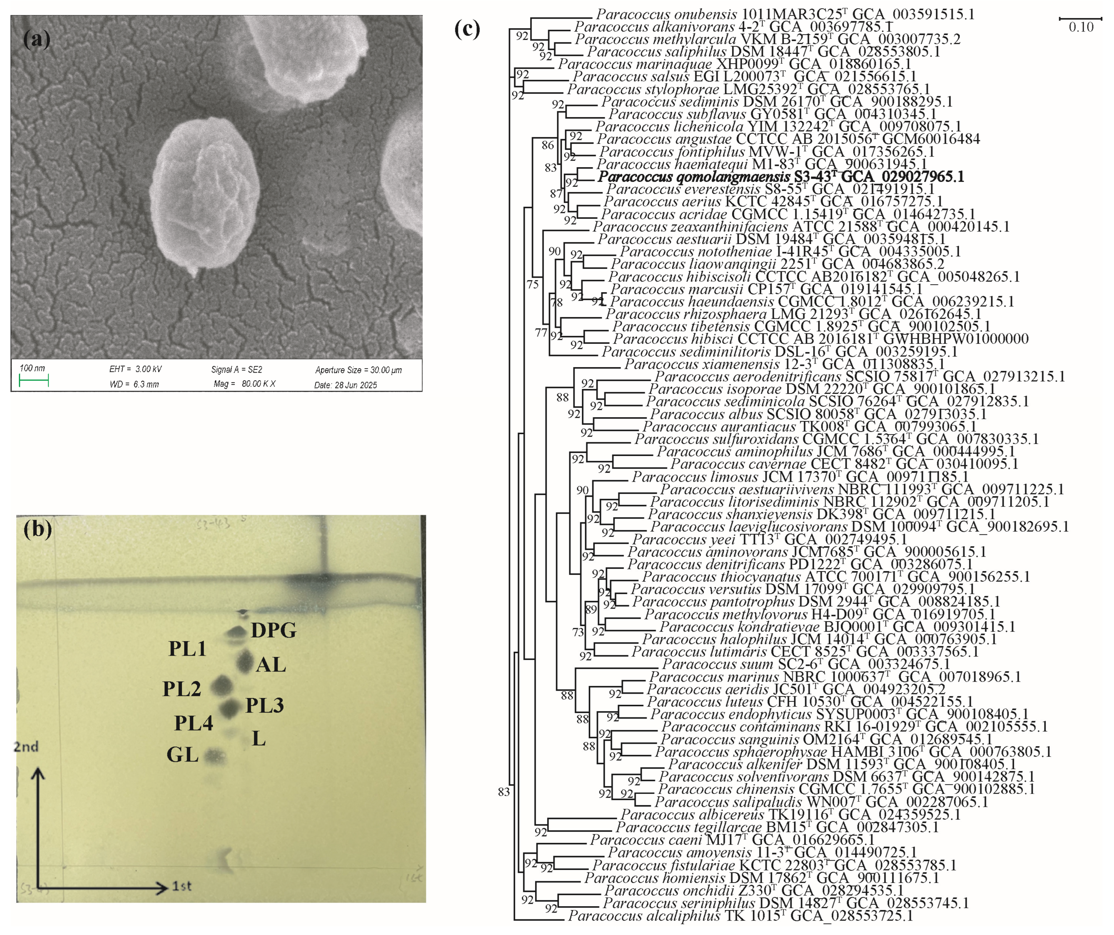
Figure 1. Chart of basic properties of strain S3-43T. (a) Scanning electron microscope photo of strain S3-43T, (b) Polar lipids profile of strain S3-43T, and (c) UBCG phylogenetic tree based on the Up-to-Date Core Gene set and pipeline of strains S3-43T, and strains of other 69 species in the genus Paracoccus. Phylogenetic tree generated with UBCG using the amino acid sequences. The number at the nodes indicates the gene support index. The numbers at the nodes indicate the gene support index. Bar, 0.10 substitutions per nucleotide position.
Microorganisms 13 02441 g001
Figure 2. The radiation resistance, antioxidant, and cyhalothrin degradation of S3-43T. (a) Survival rates of strains after exposure to different doses of UV- C radiation, (b) Survival rates of strains after exposure to different doses of γ-rays, (c) Survival rates of strains after exposure to different doses of heavy-ion beam, (d) Survival rates of strains after treated with H2O2, and (e) Cyhalothrin degradation rate of strain S3-43T.
Microorganisms 13 02441 g002
Figure 3. Comparative analysis of orthologous protein groups in S3-43T and related 11 Paracoccus genomes. (a) Percentage of core, dispensable, and unique gene clusters in each of the 12 genomes. (b) Venn diagram displaying the number of core and unique gene clusters for each of S3-43T and related type strains.
Microorganisms 13 02441 g003
Figure 4. COG functional categories of strains S3-43T (a) and COG category core vs. pan (b). (a) The horizontal axis represents different COG types, while the vertical axis represents the number of genes. For detailed functional descriptions of each COG type, please refer to the legend on the right. (b) The horizontal axis represents different strains and functional names at various taxonomic levels. The vertical axis indicates gene counts. Different colors within the bars denote distinct pangenome classifications.
Microorganisms 13 02441 g004
Figure 5. Gene lists potentially associated with radiation resistance and antioxidant activity of Paracoccus spp. The horizontal axis represents different strains, while the vertical axis represents the number of functional genes.
Microorganisms 13 02441 g005
Table 1. List of genes encoding antioxidant DNA repair response proteins in the genomes of type strain S3-43T.
GenBank IDSymbolProtein NameGenBank IDSymbolProtein Name
RS08485-cold-shock proteinRS02950yghUglutathione-dependent disulfide-bond oxidoreductase
RS14830-cold-shock proteinRS13140ruvBHolliday junction branch migration DNA helicase RuvB
RS13155ruvCcrossover junction endodeoxyribonuclease RuvCRS13150ruvAHolliday junction branch migration protein RuvA
RS06430-DNA alkylation repair proteinRS15020ruvXHolliday junction resolvase RuvX
RS01405recQDNA helicase RecQRS00030-ligase-associated DNA damage response DEXH box helicase
RS10405vsrDNA mismatch endonuclease VsrRS00025pdeMligase-associated DNA damage response endonuclease PdeM
RS03235mutLDNA mismatch repair endonuclease MutLRS00040-ligase-associated DNA damage response exonuclease
RS01015mutSDNA mismatch repair protein MutSRS15440msrBpeptide-methionine (R)-S-oxide reductase MsrB
RS10955-DNA mismatch repair protein MutTRS14820msrApeptide-methionine (S)-S-oxide reductase MsrA
RS07735rmuCDNA recombination protein RmuCRS15445msrApeptide-methionine (S)-S-oxide reductase MsrA
RS12135radADNA repair protein RadARS12420recArecombinase RecA
RS01075radCDNA repair protein RadCRS08935recRrecombination mediator RecR
RS04590recNDNA repair protein RecNRS15945recJsingle-stranded-DNA-specific exonuclease RecJ
RS13770recODNA repair protein RecORS01565-UvrD-helicase domain-containing protein
RS00015recFDNA replication/repair protein RecFRS08415-UvrD-helicase domain-containing protein
RS00950addAdouble-strand break repair helicase AddARS13235-UvrD-helicase domain-containing protein
RS00955addBdouble-strand break repair protein AddBRS16030-NAD(P)H-quinone oxidoreductase
RS08490-DsbA family oxidoreductaseRS00945trxAthioredoxin
RS05580uvrAexcinuclease ABC subunit UvrARS16155trxAthioredoxin
RS05645uvrBexcinuclease ABC subunit UvrBRS12075trxBthioredoxin-disulfide reductase
RS15085uvrCexcinuclease ABC subunit UvrC
Table 2. List of genes encoding pyrethroids degradation in the genomes of type strain S3-43T and its related strains.
Gene Description123456789101112
Cytochrome P450434343222434
Heme-degrading monooxygenase HmoA and related ABM domain proteins212220222222
Quinol monooxygenase YgiN100002011202
Aminopeptidase N, contains DUF3458 domain111111111111
D-aminopeptidase000110000000
L-aminopeptidase/D-esterase111221111111
Leucyl aminopeptidase222222222322
Methionine aminopeptidase211121111121
Putative aminopeptidase FrvX101110000100
Xaa-Pro aminopeptidase244322213233
Strains: 1. Paracoccus qomolangmaensis S3-43T, 2. Paracoccus versutus IAM 12814T, 3. Paracoccus haematequi LMG 30633T, 4. Paracoccus acridae KCTC 42932T, 5. Paracoccus aerius KCTC 42845T, 6. Paracoccus zeaxanthinifaciens ATCC 21588T, 7. Paracoccus everestensis S8-55T, 8. Paracoccus subflavus GY0581T, 9. Paracoccus sediminis CMB17T, 10. Paracoccus lichenicola YIM 132242T, 11. Paracoccus fontiphilus MVW-1T, and 12. Paracoccus angustae CCTCC AB 2015056T.
Disclaimer/Publisher’s Note: The statements, opinions and data contained in all publications are solely those of the individual author(s) and contributor(s) and not of MDPI and/or the editor(s). MDPI and/or the editor(s) disclaim responsibility for any injury to people or property resulting from any ideas, methods, instructions or products referred to in the content.