Article

The Cell Wall Deacetylases Spy1094 and Spy1370 Contribute to Streptococcus pyogenes Virulence

by
Tiger Aspell
1,
Adrina Hema J. Khemlani
1,
Catherine Jia-Yun Tsai
1,2,
Jacelyn Mei San Loh
1,2 and
Thomas Proft
1,2,*
1
Department of Molecular Medicine & Pathology, School of Medical Sciences, The University of Auckland, Private Bag 92019, Auckland 1142, New Zealand
2
Maurice Wilkins Centre for Molecular Biodiscovery, The University of Auckland, Private Bag 92019, Auckland 1142, New Zealand
*
Author to whom correspondence should be addressed.
Microorganisms 2023, 11(2), 305; https://doi.org/10.3390/microorganisms11020305
Submission received: 24 November 2022 / Revised: 21 January 2023 / Accepted: 22 January 2023 / Published: 24 January 2023
(This article belongs to the Special Issue Pathogenic Streptococci: Virulence, Host Response and Therapy)

Abstract

:
Streptococcus pyogenes, or Group A Streptococcus (GAS), is a strictly human pathogen that causes a wide range of diseases, including skin and soft tissue infections, toxic shock syndrome and acute rheumatic fever. We have recently reported that Spy1094 and Spy1370 of S. pyogenes serotype M1 are N-acetylglucosamine (GlcNAc) deacetylases. We have generated spy1094 and spy1370 gene deletion mutants in S. pyogenes and gain-of-function mutants in Lactococcus lactis. Similar to other cell wall deacetylases, our results show that Spy1094 and Spy1370 confer lysozyme-resistance. Furthermore, deletion of the genes decreased S. pyogenes virulence in a human whole blood killing assay and a Galleria mellonella (Greater wax moth) larvae infection model. Expression of the two genes in L. lactis resulted in increased lysozyme resistance and survival in whole human blood, and reduced survival of infected G. mellonella larvae. Deletion of the spy1370, but not the spy1094 gene, decreased resistance to the cationic antimicrobial peptide cecropin B, whereas both enzymes increased biofilm formation, probably resulting from the increase in positive charges due to deacetylation of the cell wall. In conclusion, Spy1094 and Spy1370 are important S. pyogenes virulence factors and might represent attractive targets for the development of antibacterial agents.

1. Introduction

Streptococcus pyogenes, or Group A Streptococcus (GAS), is an exclusively human pathogen that can cause a wide range of diseases. These include common skin and soft tissue infections such as pharyngitis, tonsillitis, impetigo, erysipelas and cellulitis [1,2]. Severe invasive GAS diseases such as necrotising fasciitis (‘flesh-eating disease’) and streptococcal toxic shock syndrome are less common but have high mortality rates (30–70%) [3,4,5,6]. Untreated cases of pharyngitis and skin infections can result in post-streptococcal autoimmune diseases such as acute rheumatic fever, rheumatic heart disease and acute glomerulonephritis [7,8,9]. It is estimated that S. pyogenes is responsible for approximately 500,000 deaths, globally, each year [10].
The success of S. pyogenes as a major human pathogen can be attributed to the production of a large arsenal of virulence factors, which include adhesins [11,12], pili [13], superantigens [14], cytolysins [15], fibrinolysin [16] and complement evasion factors [17]. Another virulence strategy of some bacterial pathogens involves the deacetylation of peptidoglycan in the cell wall which is made up of alternating β-1,4-linked N-acetylglucosamine (GlcNAc) and N-acetylmuramic acid (MurNAc) residues cross-linked by a short peptide [18]. Deacetylation of the peptidoglycan cell wall results in decreased susceptibility to lysozyme, a muramidase that cleaves the amide bond between GlcNAc and MurNAc [19]. Lysozyme is a naturally occurring antimicrobial agent found in bodily secretions such as tears, saliva and milk. Lysosomal enzymes are also found in human phagocytes and assist in bacterial killing [20]. Streptococcus pneumoniae (pneumococcus) produces a peptidoglycan deacetylase (SpPgdA) and a pgdA gene deletion mutant showed increased susceptibility to lysozyme and reduced virulence in an intraperitoneal mouse model [21]. PGDAs have also been reported in other bacterial pathogens (reviewed in [22]). Another deacetylase has been reported in Streptococcus iniae and named polysaccharide deacetylase of S. iniae (SiPdi) [23]. The substrate for this enzyme is unknown, but a role in adherence and host invasion has been demonstrated.
The Staphylococcus epidermidis deacetylase (IcaB) alters bacterial biofilms by modifying poly-β-1,6-N-acetyl-d-glucosamine (PNAG), also referred to as polysaccharide intercellular adhesin (PIA), an exopolysaccharide that forms the extracellular matrix of some bacterial biofilms [24]. Deletion of the icaB gene resulted in impaired biofilm formation and colonisation of the bacteria [25].
Recently, we reported the biochemical characterisation of two novel S. pyogenes deacetylases, Spy1370 and Spy1094 [26]. Recombinant forms of the two enzymes deacetylated the pseudosubstrate GlcNAc3, suggesting a role in the modification of the bacterial peptidoglycan cell wall. In this study, we investigated the involvement of Spy1094 and Spy1370 on S. pyogenes virulence by generating S. pyogenes gene deletion mutants and Lactococcus lactis gain-of-function mutants and analysed them for lysozyme sensitivity, antimicrobial resistance, biofilm formation, survival in human whole blood and in-vivo virulence using a Galleria mellonella (greater wax moth) larvae infection model.

2. Materials and Methods

2.1. Bacterial Strains and Growth Conditions

S. pyogenes SF370 (M1 serotype, ATCC 700294) was grown in Brain Heart Infusion (BHI) (BD Biosciences, San Jose, CA, USA) at 37 °C under static conditions. Lactococcus lactis MG1363 was cultured in M17 + 0.5% glucose (GM17) medium (BD Biosciences) at 28 °C under static conditions. Escherichia coli DH5α was grown in Luria Bertani (LB, BD Biosciences) broth at 37 °C with aeration. Solid BHI, GM17 or LB plates were made by adding 1.5% Bacto agar (BD Biosciences) to the liquid medium. When appropriate, antibiotics were added: spectinomycin at 50 μg/mL for S. pyogenes and 100 μg/mL for E. coli; kanamycin at 200 μg/mL for S. pyogenes and L. lactis, and 50 μg/mL for E. coli.

2.2. Generation of S. pyogenes and L. lactis Mutants

S. pyogenes Δspy1094 and S. pyogenes Δspy1370 mutants were generated by allelic replacement using the pFW11 vector [27] (a gift from Andreas Podbielski, University of Rostock) as previously described [28,29]. In brief, ~1000 bp sequences flanking the upstream (FR1) and downstream (FR2) regions of the spy1370 and spy1094 genes, respectively, were amplified from genomic S. pyogenes DNA by 30 cycles of PCR with iProof polymerase (Bio Rad, Hercules, CA, USA) and specific primers (Sigma-Aldrich, St. Louis, MO, USA) (see Table 1). Each flanking region was cloned into the pFW11 MCS-I and MCS-II regions, respectively, which flank the spectinomycin-resistance gene aad9, using restriction enzymes (NEB, Table 1) and T4 DNA-ligase (Biolab, Tokyo, Japan). The recombinant constructs were electroporated into S. pyogenes SF370 using a Gene Pulser Xcell—(Bio-Rad), and the transformants were selected on BHI agar plates containing spectinomycin. Replacement of the spy1094 or spy1370 genes with aad9 was confirmed by PCR using flanking region (FR) primers and aad9.fw primers (Table 1). In addition, the deletion mutants were validated at the protein level by Western blotting (see below). The S. pyogenes gene deletion mutants were complemented with the complete spy1094 gene and spy1370 gene, respectively, cloned into the pLZ12-Km2-P23R plasmid [27] using gene-specific primers (Table 1).
L. lactis gain-of-function strains were generated by electroporating the recombinant pLZ12-km2-P23R plasmids into L. lactis. The strains were validated by PCR and Western blotting.

2.3. Western Blots

Whole bacterial cell lysates were separated on a 12.5% polyacrylamide gel and transferred onto a nitrocellulose membrane (Bio Rad) in a transfer buffer (25 mM Tris-HCl, 192 mM glycine, 20% (v/v) methanol, pH 8.3) using a TE77 semi-transfer unit (Hoefer, Holliston, MA, USA) at 200 V, 50 mA/gel for 1 h. The membrane was incubated with TBS-T (20 mM Tris-HCl pH 7.6, 150 mM NaCl, 0.1% (v/v) Tween-20) plus 5% (w/v) skim milk powder at RT for 1 h and then washed once with TBS-T. Antibodies against recombinant forms of Spy1094 and Spy1370 were generated in rabbits as previously described [26]. Rabbit-sera were diluted 1/1000 with TBS-T plus 2.5% (w/v) milk powder and incubated with the membranes for 1 h at RT. After three washes with TBS-T, the membranes were incubated with HRP-cojugated goat anti-rabbit IgG (Abcam, Cambridge, UK), diluted 1/1000 in TBST for 1 h at RT. The membranes were then washed three times with TBS and developed using ECL detection reagent (Amersham Biosciences, Slough, UK) and analysed with a ChemiDocTM imaging system (Bio Rad).

2.4. Effects of Lysozyme on the Survival of S. pyogenes and L. lactis Strains

The protocol for lysozyme killing assays were adapted from Milani et al. [23]. Overnight cultures of S. pyogenes wildtype and mutant strains were grown in BHI with appropriate antibiotics to the mid exponential phase (OD600 = 0.6–0.8). Bacteria were then washed with PBS and resuspended in BHI to approximately 2 × 10⁵ CFU. An amount of 20 μL chicken egg white lysozyme (Sigma-Aldrich) was added to 180 μL of diluted bacteria to a final concentration of 80 μg/mL. The bacteria were then incubated at 37 °C for 30 min, serially diluted in sterile PBS and enumerated by spot-plating in triplicates onto BHI agar plates containing the appropriate antibiotics. Cultures without lysozyme treatment were grown as controls. Percentage survival was calculated using the following equation: % survival = ([CFU with treatment]/[CFU without treatment]) × 100. Four independent experiments were carried out.

2.5. Effects of Cecropin B on the Survival of GAS Mutants

This method was adopted from Wang et al. [27]. Overnight cultures of each S. pyogenes strain were grown to the mid exponential phase (OD600 = 0.6–0.8) at 37 °C in BHI with the appropriate antibiotics. Cecropin B (Sigma-Aldrich) was serially diluted (0–25 μM) and added to bacterial cultures in a final volume of 100 μL in a 96-well plate. The cultures were incubated further at 37 °C until the stationary phase was reached and then serially diluted for enumeration by spot-plating in triplicates. Cultures without cecropin B treatment were grown as controls. Percentage survival was calculated using the following equation: % survival = ([CFU with treatment]/[CFU without treatment]) × 100. Three independent experiments were carried out.

2.6. Effects of H2O2 on the Survival of GAS Mutants

This method was adopted from Pericone et al. [28]. Overnight cultures of each GAS strain were grown to the mid-exponential phase (OD600 = 0.6–0.8) at 37 °C, supplemented with the appropriate antibiotics). Then, 100 μL aliquots were added to 100 μL of fresh BHI media containing 0–5% H2O2. The samples were then incubated at 37 °C for 1 h before spot-plating in triplicates for bacterial enumeration. Control cultures without H2O2 treatment were grown in parallel. Percentage survival was calculated using the following equation: % survival = ([CFU with treatment]/[CFU without treatment]) × 100. Four independent experiments were carried out.

2.7. Biofilm Formation in GAS Mutants

S. pyogenes wildtype and mutant strains were grown at 37 °C overnight and then diluted 1:10 in fresh BHI media containing the appropriate antibiotics. The cultures were then seeded into uncoated 96-well plates (Sigma-Aldrich) in triplicates and further incubated at 37 °C overnight without aeration. Unbound, planktonic cells were removed by washing the wells with sterile PBS before the addition of crystal violet dye as described previously [29]. Biofilm formation was quantified by measuring absorbance at 595 nm using an EnSightTM Multimode plate reader (Perkin Elmer, Waltham, MA, USA). Four independent experiments were carried out.

2.8. Whole Blood Killing Assay

This assay was carried out as described recently [30]. S. pyogenes wildtype and mutant strains were grown in BHI with appropriate antibiotics to the late exponential phase. Approximately 1 × 10⁵ CFU of bacteria in a volume of 50 μL were added to 1 mL of fresh heparinised human whole blood from a consented donor and incubated for 2.5 h at 37 °C with constant rotation. An amount of 100 μL samples were taken every 30 min, serially diluted and plated onto BHI agar plates in triplicates. The percentage of survival was calculated as [CFU (at a given time point)/CFU (at the start)] × 100.
L. lactis was grown in a GM17 medium with appropriate antibiotics to the late exponential phase, pelleted at 5000× g and resuspended in Hank’s Balanced Salt Solution (HBSS). Approximately 1 × 10³ or 1 × 10⁵ bacteria were added to 1 ml of heparinised human blood and incubated for 2.5 h at 37 °C. Serially diluted samples were plated on GM17 agar plates in triplicates. Bacterial samples were also cultured on agar plates and enumerated to confirm the injected doses. Two independent experiments with two different blood donors were carried out.
Collection of human blood was approved by the Auckland Health Research Ethics Committee (AH24859) for a project entitled “Investigation of host-pathogen interactions to identify new targets and develop new detection/diagnostic tools for anti-microbial therapy”. Volunteers without a previous episode of pharyngitis/tonsillitis were informed that their blood would be used for research only, assigned a code and de-identified by the phlebotomist. No blood samples were stored after the assays.

2.9. Galleria Mellonella Larvae Infection Model

G. mellonella (greater wax moth) larvae experiments were carried out as previously described [31,32]. In brief, a group of 10 antibiotic-free, healthy (with no melanisation) and medium-sized (~1.5 cm) G. mellonella larvae (Biosuppliers-New Zealand) were used for each experiment. Twenty microlitres of bacteria (1.5 × 10⁶ CFU for S. pyogenes wildtype and mutant strains and 1 × 10⁵ CFU for L. lactis wildtype and mutant strains) were injected into the lower left proleg using an insulin syringe (BD Biosciences). A control group was injected with sterile PBS only. Bacterial samples were also cultured on agar plates and enumerated to confirm the injected doses. The larvae were incubated in Falcon®12-well plates (Biocompare) at 37 °C without food. Survival of the larvae was monitored over a 5-day period post-injection. In addition, a health index score was calculated as described previously [31,32].

2.10. Statistical Analysis

All statistical analyses were conducted using GraphPad Prism software (version V7.03). Statistical significance was calculated using one-way ANOVA with Tukey’s multiple comparisons to compare two or more data sets. p values of <0.05 were considered statistically significant. Survival curves were estimated using the Kaplan–Meier estimator in GraphPad Prism.

3. Results

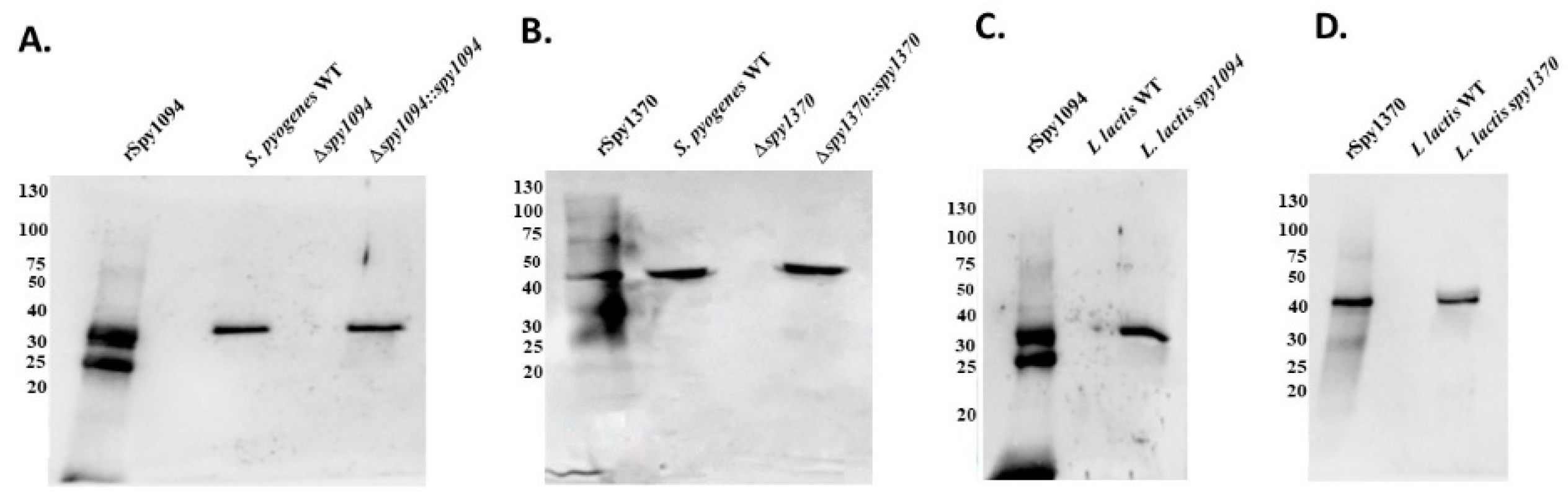
3.1. Generation of S. pyogenes Gene Deletion Strain and L. lactis Gain-of-Function Strains

We have previously shown that recombinant forms of Spy1094 and Spy1370 possess deacetylase activity for the pseudosubstrate GlcNAc3 which is commonly used to demonstrate the ability to deacetylase peptidoglycan cell walls [26]. To demonstrate that the two enzymes play a crucial role in S. pyogenes virulence, we first generated isogenic gene deletion mutants. Allelic replacement was used to exchange the target gene for the spectinomycin-resistance gene aad9. The deletion mutants were then complemented with the respective complete genes driven by the constitutive lactococcal P23 promotor [33]. The successful gene deletion was confirmed by Western blot analysis with specific antisera raised against recombinant forms of the enzymes (Figure 1A). Bands were detected in the wildtype strain at ~32 kDa (Spy1094) and ~44 kDa (Spy1370) which correspond to the correct sizes of the enzymes as determined previously [26]. Those bands were not detected in the deletion mutants but were visible in the complementation mutants (Figure 1A,B).
The complementation plasmids were also used to generate gain-of-function mutants in L. lactis. As shown in Figure 1C,D, the specific antisera did not react with any protein in the wildtype L. lactis whole cell lysate, but recognised proteins at the expected sizes in the extracts of the gain-of-function mutants, confirming expression of Spy1094 and Spy1370.
Growth kinetics were conducted with wildtype and mutant strains. The S. pyogenes Δspy1094 mutant showed a slightly decreased growth compared to the parent strain, whereas S. pyogenes Δspy1370 grew considerably slower and failed to reach the same cell density as the wildtype strain after 24 h (Figure 2A). Complementation of the mutant strains resulted in growth kinetics similar to the S. pyogenes wildtype, suggesting that the growth defects could be solely attributed to the deleted genes. No difference in growth was observed between the L. lactis mutants compared to the parent strain (Figure 2B).

3.2. Deletion of spy1094 and spy1370 Decreases Lysozyme Resistance

There is a direct relationship between increasing levels of deacetylation of the peptidoglycan cell wall and increasing resistance to lysozyme [34]. We previously showed that recombinant forms of Spy1094 and Spy1370 both deacetylate the pseudosubstrate GlcNAc3, suggesting a biological role in peptidoglycan modification [26]. To further analyse this, we treated wildtype and mutant strains with 80 μg/mL lysozyme for 30 min and enumerated the surviving bacteria. As shown in Figure 3A, deletion of the spy1094 gene led to an ~85% reduction of survival (p < 0.001), whereas the S. pyogenes Δspy1370 mutant was completely killed by the lysozyme treatment. Complementation of the genes restored lysozyme resistance to approximately 75% (spy1094) and ~80% (spy1370) of wildtype S. pyogenes. Heterologous expression of Spy1094 and Spy1370 in L. lactis led to a ~35% increase in lysozyme resistance (p < 0.05) (Figure 3B).

3.3. Deletion of spy1370 Confers Resistance to Cationic AMP, but Not to Oxidative Killing

Deacetylation of the peptidoglycan cell wall leads to an increase in the net positive charge which might affect the activity of cationic antimicrobial peptides (CAMPs) by preventing translocation to the cell membrane. It has been demonstrated that deletion of the Staphylococcus epidermidis icaB gene, which encodes an exopolysaccharide deacetylase, results in decreased resistance against CAMPs and neutrophil phagocytosis [35]. We, therefore, analysed our wildtype and mutant strains for resistance to Cecropin B, a CAMP that was first isolated from the hemolymph of the silk moth Hyalophora cecropia [36] and to H2O2, a reactive oxygen species produced inside phagosomes during the oxidative burst.
Cecropin B showed a dose-dependent effect on S. pyogenes and L. lactis with complete bacterial killing observed at concentrations of 25 μM (Figure 4A–C). Deletion of spy1370 resulted in substantially decreased survival at the concentrations tested (p < 0.001). This was partially restored in the S. pyogenes Δspy1370::spy1370 complementation mutant (Figure 4B). No differences were observed between the spy1094 deletion mutant and wildtype S. pyogenes (Figure 4A) or between wildtype L. lactis and the gain-of-function mutants (Figure 4C). The effect of H2O2 was measured at various concentrations up to 5%, which resulted in the complete killing of the bacteria (Figure 4D–F). No significant differences were observed between wildtype bacteria and mutant strains, suggesting that Spy1094 and Spy1370 have no protective effect against exogenous H2O2.

3.4. Deletion of spy1094 and spy1370 Decreases Biofilm Formation

Biofilms are responsible for a large medical burden on a global scale. S. pyogenes is able to form biofilms which have been associated with diseases such as tonsilitis and cellulitis [37]. Recently, it was shown that S. pyogenes serotype M1 strains consistently formed biofilms in patients with necrotising fasciitis (‘flesh-eating disease’) [38].
As the S. epidermitis exopolysaccharide deacetylase IcaB plays an important role in biofilm formation, we analysed the ability of Spy1094 and Spy1370 to contribute to biofilm formation in-vitro. Biofilm formation was observed with the wildtype S. pyogenes SF370 strain (serotype M1) but was significantly reduced to <5% in the spy1370 deletion mutant (p < 0.001) (Figure 5A). The S. pyogenes Δspy1370::spy1370 complementation mutant restored biofilm formation to approximately 75%. Deletion of spy1094 also resulted in significantly decreased biofilm formation (p < 0.05) to ~70% of the wildtype level and was also partly restored in the complementation mutant to 80% (Figure 5A). These findings could be confirmed with the L. lactis gain-of-function mutants where the L. lactis spy1094 and L. lactis spy1370 mutants showed increased biofilm formation (p < 0.001) compared to L. lactis wildtype at levels of 140% and 150%, respectively (Figure 5B).

3.5. Spy1094 and Spy1370 Promote Survival in Human Blood

To test if the two deacetylases contribute to the overall virulence of S. pyogenes, we used a human whole blood killing assay [30]. Freshly collected human blood was inoculated with 1 × 10⁵ CFU S. pyogenes wildtype and mutant strains, and survival was analysed by enumerating the surviving bacteria (Figure 6A). The S. pyogenes wildtype showed about a 10-fold increase in CFU during the 2.5 h incubation period. In contrast, survival of S. pyogenes Δspy1094 and S. pyogenes Δspy1370 both significantly decreased during incubation and were almost completely killed within the first 1.5 h (p < 0.001; Figure 6A). A difference between the mutant strains was observed after 1 h when approximately 50% of the S. pyogenes Δspy1094 still survived, whereas S. pyogenes Δspy1370 was almost completely killed. Survival in blood was mostly restored in the complemented mutants. We also analysed L. lactis wildtype and mutant strains (Figure 6B) by inoculating human blood with 1 × 10⁵ CFU bacteria and found that both mutants survived in whole human blood with a ~2.2-fold increase in CFU for L. lactis spy1094 and ~3-fold increase in CFU for L. lactis spy1370 at the 2.5 h endpoint. In contrast, <50% of the L. lactis wildtype survived after the 2.5 h incubation showing that the two deacetylases significantly increase survival of L. lactis in human blood (p < 0.001). Similar results were achieved when human blood was inoculated with 1 × 10³ CFU of the L. lactis wildtype or mutant strains (Supplementary Figure S1).

3.6. Deletion of spy1094 and spy1370 Reduces Virulence of S. pyogenes in a Galleria Mellonella Infection Model

G. mellonella (wax moth) larvae have been introduced as an alternative model to study microbial infections and we have previously shown that this model is useful to study virulence in S. pyogenes [31,32,39]. Although insects lack an adaptive immune response, their innate immune response shows remarkable similarities with the immune response in vertebrates, including the presence of lysozyme and AMPs [39].
Compared to infection with wildtype S. pyogenes, deletion of spy1094 or spy1370 substantially increased survival of infected G. mellonella larvae (Figure 7A). Infection with wildtype S. pyogenes resulted in the complete killing of the larvae after three days. In contrast, only 10% killing was observed after three days with the S. pyogenes Δspy1094 mutant and 60% of the larvae were still alive after five days. The complementation mutant partially restored the wildtype phenotype with larval survival reduced to 40% after five days. An even larger effect was observed with the S. pyogenes Δspy1370 mutant which did not kill any larvae three days post-infection and 80% of the larvae were still alive after five days. Only 30% larval survival five days post-infection was observed with the complementation mutant. We have previously developed a health score index which allows assessment of the G. mellonella larvae not only based on survival, but also certain health indicators such as melanisation, cocoon formation and mobility [31,39]. On a scale from 1 to 10 with 10 being the most healthy larvae, larvae infected with the S. pyogenes spy1094 and spy1370 deletion mutants both showed substantially increased health scores. Larvae inoculated with either S. pyogenes Δspy1094 or S. pyogenes Δspy1370 both scored about 9 after the first three days post-infection, dropping to 7 after five days. In contrast, larvae infected with wildtype S. pyogenes scored only 0.5 three days post-infection (Figure 7B). Infection of G. mellonella larvae with the L. lactis gain-of-function mutants further confirmed the role of Spy1094 and Spy1370 in S. pyogenes virulence. L. lactis expressing Spy1094 killed 60% of the larvae after five days compared to the 100% survival of larvae infected with wildtype L. lactis. The L. lactis spy1370 mutant killed 50% of larvae after five days (Figure 7C). For the health score index, larvae inoculated with the L. lactis mutants expressing Spy1094 or Spy1370 scored 5 two days post-infection. After five days, the scores were 3 and 5, respectively. In contrast, the health score in larvae inoculation with wildtype L. lactis dropped only slightly to 9 after five days (Figure 7D).

4. Discussion

We have recently reported the biochemical analysis of the S. pyogenes deacetylases Spy1094 and Spy1370. Recombinant forms of both enzymes were able to deacetylate the pseudosubstrate chitotriose (GlcNAc3) with high substrate affinity but low substrate turnover. Here, we describe the further characterisation of the enzymes using gene deletions introduced by allelic replacement with the spectinomycin-resistance gene aad9. In addition, L. lactis gain-of-function mutants were generated. Our results suggest that both enzymes deacetylate the peptidoglycan cell wall, contributing to lysozyme resistance and increased virulence during infection.
Peptidoglycan GlcNAc deacetylases (PgdA enzymes) have been described in several pathological bacterial species, mainly in Gram-positive bacteria. The first description of a PgdA enzyme was reported in 2000 when it was shown that deletion of the pgdA gene in Streptococcus pneumoniae resulted in increased susceptibility to lysozyme [40]. Lysozyme is an enzyme that is abundantly found in mucosal secretions, including tears and saliva. Lysozyme hydrolyses the β-1,4-glycosidic bonds between MurNAc and GlcNAc and plays an important role in innate immunity [41]. PgdA enzymes have also been reported in the swine pathogen Streptococcus suis and in Streptococcus mutans but play no role in lysozyme resistance despite the fact that SmPgdA is able to deacetylate chitohexaose (GlnNAc6) [42,43]. The fish pathogen Streptococcus iniae expresses a paralogue deacetylase (Pdi) with unknown substrate specificity. However, a pdi gene deletion revealed a role for Pdi in lysozyme resistance [23]. The S. pyogenes serotype M1 Spy1370 is 35% identical to PgdA from S. pneumoniae, whereas Spy1094 shares 72% identity with Pdi from S. iniae, suggesting orthologue genes [26]. In line with the results reported for S. pneumoniae PgdA, spy1370 gene deletion resulted in significantly decreased lysozyme resistance (p < 0.001). More recently, a Spy1094 variant was characterised in the S. pyogenes NZ131 strain (serotype M49) and named polysaccharide-peptidoglycan linkage deacetylase (PplD). The authors found that PplD deacetylates the GlcNAc that is linked with the rhamnopolysaccharide of the Group A antigen [44]. Notably, the study shows no decrease in lysozyme resistance after deletion of the pplD gene, which is in contrast to Pdi [23] and our results, which show a significant decrease in lysozyme resistance in the spy1094 deletion mutant (p < 0.001). A possible reason might be the growth phase of the bacteria used in the lysozyme assays. It was shown that S. iniae Δpdi mutants were substantially more susceptible to lysozyme when static bacteria were exposed compared to growing cells [23]. In our assay, the bacteria were also used at static conditions. We also provide further evidence for the role of Spy1094 as a PgdA-like enzyme by generating L. lactis gain-of-function mutants. L. lactis is a non-pathogenic bacterium that expresses a PgdA enzyme that provides resistance to autolysis. It was also shown that overexpression of PdgA results in a higher percentage of deacetylated peptidoglycan [45]. Our results indicate that expression of either Spy1094 or Spy1370 in L. lactis leads to a significant increase in lysozyme resistance (p < 0.05). This suggests that either PplD/Spy1094 deacetylates other GlcNAc residues in the cell wall and, therefore, has at least a partly overlapping function with PgdA or that deacetylation of the cell wall linkage unit is sufficient to confer some resistance to lysozyme.
Notably, lysozyme can kill bacteria due to a non-enzymatic mechanism similar to cationic antimicrobial peptides (CAMPs) that requires a negatively charged cell wall which facilitates the transport across the cell wall [41]. Deacetylation of GlcNAc increases the cell wall net positive charge which might lead to repulsion of CAMPs. Cecropin B is a CAMP that was shown to be highly effective against E. coli and the Gram-positive Staphylococcus aureus [27]. Our results show that Spy1370, but not Spy1094, confers resistance to cecropin B (p < 0.001) which is in line with the results reported for S. iniae where neither moronecidine nor polymyxin B showed increased killing in the pdi gene deletion mutant [23]. In contrast, S. pyogenes NZ131 strains with either pgdA or pplD gene deletions showed reduced resistance to killing by the cationic enzyme human group IIA secreted phospholipase A2 (hGIIA), which might be due to the very high positive charge of hGIIA [44].
Deacetylation of peptidoglycan might also reduce recognition by pattern-recognition receptors (PRRs) such as Toll-like receptor 2 (TLR-2) [46]. Furthermore, TLR-signalling results in the production of AMPs [41]. We provided further evidence for a role of GlcNAc deacetylases in immune modulation using a human whole blood killing assay. The decreased survival of the S. pyogenes Δspy1094 and S. pyogenes Δspy1370 mutants, as well as the increased survival of L. lactis bacteria expressing Spy1094 or Spy1370 suggests an immune evasion strategy conferred by the deacetylases. This might be mediated by the increased positive cell surface charge due to cell wall deacetylation and consequently, the repulsion of CAMPs as discussed earlier. However, additional other mechanisms can’t be excluded. Our results are further supported by a transcriptome analysis of S. pyogenes grown in human whole blood compared to bacteria grown in a growth medium. After 30 min, the spy1370 transcript was upregulated a massive 1541-fold, whereas spy1094 mRNA was increased 146-fold, suggesting major roles in virulence [47]. Milani et al. reported an approximately 50% reduced survival for the S. iniae pdi deletion mutant after 1 h in whole fish blood and suggested that this might be due to higher susceptibility to oxidative killing, but this could not be confirmed experimentally [23]. This is in line with our observations as deletion of either spy1094 or spy1370 failed to increase killing of S. pyogenes by hydrogen peroxide. A longitudinal transcriptome analysis of S. pyogenes in an experimental rhesus macaque pharyngitis model provided some evidence for the involvement of Spy1370 in inflammation. The regulation of spy1370 mRNA transcription strongly correlated with C-reactive protein levels which was used as a phenotypic marker for inflammation. No differential regulation for spy1094 was reported [48].
The attenuated virulence of S. pyogenes Δspy1094 and S. pyogenes Δspy1370 was further evidenced in a G. mellonella larvae infection model. Our results show that both S. pyogenes deletion mutants had a significantly lower larvae killing effect compared to wildtype S. pyogenes (p < 0.001). In case of the spy1370 deletion mutant, this might partially be due to a lower growth kinetic that we have observed in-vitro. However, both L. lactis gain-of-function mutants showed a significantly increased ability to kill G. mellonella larvae (p < 0.001), and showed a comparable in-vitro growth kinetic with wildtype L. lactis. This is in line with a study showing decreased larval killing with the S. iniae pdi deletion mutant [23]. The reason for the reduced growth of S. pyogenes Δspy1370 is unclear. The complete restoration of the wildtype phenotype in the complementation mutant indicates that the enzymatic activity of Spy1370 is responsible. Spy1370 might be a promiscuous enzyme that also recognises a substrate that is involved in cell growth regulation. An alternative role for PdgA has been suggested for the swine pathogen S. suis, which contains peptidoglycan with low levels of deacetylated components and is lysozyme sensitive. However, deletion of the pdgA gene resulted in severely impaired virulence in animal infection models [43]. Alternatively, peptidoglycan deacetylation might have an effect on cell wall morphology causing reduced growth. Notably, this reduced growth has not been reported for PdgA enzymes from any other species, but this might simply have been unrecognised. Interestingly, an S. iniae mutant lacking the pdi gene was unable to form chains and showed increased buoyancy in a liquid culture [23]. A similar defect was reported in the L. lactis pdgA deletion mutant [45]. However, we did not observe any differences in buoyancy or chain formation in S. pyogenes Δspy1370 (data not shown).
We demonstrate that both Spy1094 and Spy1370 play a role in biofilm formation which was significantly decreased in the gene deletion mutants (p > 0.001) and increased in L. lactis gain-of-function mutants (p < 0.05). Involvement of PgdA deacetylases or other cell wall deacetylases in biofilm formation has not been reported in the literature thus far. However, Freiberg et al. reported a 3.7-fold upregulation of spy1094 mRNA when S. pyogenes was grown in biofilm compared to planktonic growth at the stationary phase [49]. Staphylococcus epidermitis produces a deacetylase, IcaB, which targets polysaccharide intercellular adhesin (PIA), an important component of many bacterial biofilms. IcaB also deacetylates GlnNAc components. However, in PIA, these are β-1,6-linked and the enzyme lacks activity against β-1,4-linked GlnNAcs found in the peptidoglycan of the cell wall [35]. It, therefore, seems more likely that the deacetylation of the peptidoglycan GlcNAc and the resulting increase of the positive net charge might contribute to biofilm formation, e.g., by increasing bacterial cell aggregation. Notably, the S. iniae pdi deletion mutant had an approximately two-fold reduced ability to adhere to fish epithelial cells, and deletion of the pdgA gene in S. mutans resulted in a different colony texture and increased cell surface hydrophobicity [42].

5. Conclusions

We have generated spy1094 and spy1370 gene deletions in S. pyogenes SF370 (serotype M1) and gain-of-function mutants in L. lactis. Using these strains, we show that Spy1094/PplD and Spy1370/PgdA play crucial roles in S. pyogenes virulence in a human blood killing assay and a G. mellonella infection model. Both proteins are cell wall deacetylases that target GlcNAc components and contribute to lysozyme and biofilm formation. Furthermore, Spy1370/PgdA, but not Spy1094/PlpD, confers resistance to the cationic antimicrobial peptide cecropin B. Our results might provide a basis for the future development of specific treatment options against S. pyogenes.

Supplementary Materials

The following supporting information can be downloaded at: https://www.mdpi.com/article/10.3390/microorganisms11020305/s1, Figure S1: Survival of wildtype and mutant L. lactis strains in whole human blood after incubation with 1 × 10³ CFU.

Author Contributions

Conceptualisation, T.P.; methodology, T.A. and A.H.J.K.; formal analysis, T.A.; investigation, T.A., J.M.S.L. and C.J.-Y.T.; data curation, T.A. and T.P.; writing—original draft preparation, T.A.; writing—review and editing, T.P., J.M.S.L. and C.J.-Y.T.; visualisation, T.A. and T.P.; supervision, T.P., J.M.S.L. and C.J.-Y.T.; project administration, T.P.; funding acquisition, T.P. All authors have read and agreed to the published version of the manuscript.

Funding

This work was financially supported by a Graduate Student Fund from the University of Auckland. J.M.S.L. is a New Zealand National Heart Foundation Senior Research Fellow. C.J.-Y.T. is an Auckland Medical Research Foundation Postdoctoral Research Fellow.

Institutional Review Board Statement

This study was conducted in accordance with University of Auckland Principles for Conducting Research involving Human Participants and approved by the Auckland Health Research Ethics Committee (AH24859) in October 2022.

Informed Consent Statement

Informed consent was obtained from all subjects involved in the study.

Data Availability Statement

Data is contained within the article and in Supplementary Figure S1.

Acknowledgments

We would like to thank Reuben McGregor (University of Auckland) for assistance with the statistical analysis.

Conflicts of Interest

The authors declare no conflict of interest.

References

  1. Anjos, L.M.; Marcondes, M.B.; Lima, M.F.; Mondelli, A.L.; Okoshi, M.P. Streptococcal acute pharyngitis. Rev. Soc. Bras. Med. Trop. 2014, 47, 409–413. [Google Scholar] [CrossRef]
  2. Stevens, D.L.; Bryant, A.E. Impetigo, Erysipelas and Cellulitis. In Streptococcus pyogenes: Basic Biology to Clinical Manifestations; Ferretti, J.J., Stevens, D.L., Fischetti, V.A., Eds.; University of Oklahoma Health Sciences Center: Oklahoma City, OK, USA, 2016. [Google Scholar]
  3. Botteaux, A.; Budnik, I.; Smeesters, P.R. Group A Streptococcus infections in children: From virulence to clinical management. Curr. Opin. Infect. Dis. 2018, 31, 224–230. [Google Scholar] [CrossRef]
  4. Cunningham, M.W. Pathogenesis of group A streptococcal infections. Clin. Microbiol. Rev. 2000, 13, 470–511. [Google Scholar] [CrossRef]
  5. Stevens, D.L. Streptococcal toxic-shock syndrome: Spectrum of disease, pathogenesis, and new concepts in treatment. Emerg. Infect. Dis. 1995, 1, 69–78. [Google Scholar] [CrossRef]
  6. Stevens, D.L.; Bryant, A.E. Severe Group A Streptococcal Infections. In Streptococcus pyogenes: Basic Biology to Clinical Manifestations; Ferretti, J.J., Stevens, D.L., Fischetti, V.A., Eds.; The University of Oklahoma Health Sciences Center: Oklahoma City, OK, USA, 2016. [Google Scholar]
  7. Cunningham, M.W. Streptococcus and rheumatic fever. Curr. Opin. Rheumatol. 2012, 24, 408–416. [Google Scholar] [CrossRef]
  8. Martin, W.J.; Steer, A.C.; Smeesters, P.R.; Keeble, J.; Inouye, M.; Carapetis, J.; Wicks, I.P. Post-infectious group A streptococcal autoimmune syndromes and the heart. Autoimmun. Rev. 2015, 14, 710–725. [Google Scholar] [CrossRef]
  9. Mosquera, J.; Pedreanez, A. Acute post-streptococcal glomerulonephritis: Analysis of the pathogenesis. Int. Rev. Immunol. 2020, 1–20. [Google Scholar] [CrossRef]
  10. Carapetis, J.R.; Steer, A.C.; Mulholland, E.K.; Weber, M. The global burden of group A streptococcal diseases. Lancet Infect. Dis. 2005, 5, 685–694. [Google Scholar] [CrossRef]
  11. Brouwer, S.; Barnett, T.C.; Rivera-Hernandez, T.; Rohde, M.; Walker, M.J. Streptococcus pyogenes adhesion and colonization. FEBS Lett. 2016, 590, 3739–3757. [Google Scholar] [CrossRef]
  12. Raynes, J.M.; Young, P.G.; Proft, T.; Williamson, D.A.; Baker, E.N.; Moreland, N.J. Protein adhesins as vaccine antigens for Group A Streptococcus. Pathog. Dis. 2018, 76, fty016. [Google Scholar] [CrossRef] [Green Version]
  13. Nakata, M.; Kreikemeyer, B. Genetics, Structure, and Function of Group A Streptococcal Pili. Front. Microbiol. 2021, 12, 616508. [Google Scholar] [CrossRef]
  14. Proft, T.; Fraser, J.D. Streptococcal superantigens. Chem. Immunol. Allergy 2007, 93, 1–23. [Google Scholar] [CrossRef]
  15. Tabata, A.; Nagamune, H. Diversity of beta-hemolysins produced by the human opportunistic streptococci. Microbiol. Immunol. 2021, 65, 512–529. [Google Scholar] [CrossRef]
  16. Verhamme, I.M.; Panizzi, P.R.; Bock, P.E. Pathogen activators of plasminogen. J. Thromb. Haemost. 2015, 13 (Suppl. 1), S106–S114. [Google Scholar] [CrossRef] [Green Version]
  17. Laabei, M.; Ermert, D. Catch Me if You Can: Streptococcus pyogenes Complement Evasion Strategies. J. Innate Immun. 2019, 11, 3–12. [Google Scholar] [CrossRef]
  18. Moynihan, P.J.; Sychantha, D.; Clarke, A.J. Chemical biology of peptidoglycan acetylation and deacetylation. Bioorg. Chem. 2014, 54, 44–50. [Google Scholar] [CrossRef]
  19. Glick, A.D.; Ranhand, J.M.; Cole, R.M. Degradation of group A streptococcal cell walls by egg-white lysozyme and human lysosomal enzymes. Infect. Immun. 1972, 6, 403–413. [Google Scholar] [CrossRef] [Green Version]
  20. di Luzio, N.R. Lysozyme activity: An index of macrophage functional status. Front. Biol. 1979, 48, 447–462. [Google Scholar]
  21. Vollmer, W.; Tomasz, A. Peptidoglycan N-acetylglucosamine deacetylase, a putative virulence factor in Streptococcus pneumoniae. Infect. Immun. 2002, 70, 7176–7178. [Google Scholar] [CrossRef] [Green Version]
  22. Planas, A. Peptidoglycan Deacetylases in Bacterial Cell Wall Remodeling and Pathogenesis. Curr. Med. Chem. 2022, 29, 1293–1312. [Google Scholar] [CrossRef]
  23. Milani, C.J.E.; Aziz, R.K.; Locke, J.B.; Dahesh, S.; Nizet, V.; Buchanan, J.T. The novel polysaccharide deacetylase homologue Pdi contributes to virulence of the aquatic pathogen Streptococcus iniae. Microbiology (Reading) 2010, 156, 543–554. [Google Scholar] [CrossRef] [PubMed] [Green Version]
  24. Branda, S.S.; Vik, S.; Friedman, L.; Kolter, R. Biofilms: The matrix revisited. Trends Microbiol. 2005, 13, 20–26. [Google Scholar] [CrossRef] [PubMed]
  25. Vuong, C.; Voyich, J.M.; Fischer, E.R.; Braughton, K.R.; Whitney, A.R.; DeLeo, F.R.; Otto, M. Polysaccharide intercellular adhesin (PIA) protects Staphylococcus epidermidis against major components of the human innate immune system. Cell. Microbiol. 2004, 6, 269–275. [Google Scholar] [CrossRef] [PubMed]
  26. Aspell, T.; Khemlani, A.H.J.; Loh, J.M.S.; Tsai, C.J. Functional characterisation of two novel deacetylases from Streptococcus pyogenes. Microbiol. Res. 2022, 13, 323–331. [Google Scholar] [CrossRef]
  27. Wang, J.; Ma, K.; Ruan, M.; Wang, Y.; Li, Y.; Fu, Y.V.; Song, Y.; Sun, H.; Wang, J. A novel cecropin B-derived peptide with antibacterial and potential anti-inflammatory properties. PeerJ 2018, 6, e5369. [Google Scholar] [CrossRef] [Green Version]
  28. Pericone, C.D.; Park, S.; Imlay, J.A.; Weiser, J.N. Factors contributing to hydrogen peroxide resistance in Streptococcus pneumoniae include pyruvate oxidase (SpxB) and avoidance of the toxic effects of the fenton reaction. J. Bacteriol. 2003, 185, 6815–6825. [Google Scholar] [CrossRef] [Green Version]
  29. Kimura, K.R.; Nakata, M.; Sumitomo, T.; Kreikemeyer, B.; Podbielski, A.; Terao, Y.; Kawabata, S. Involvement of T6 pili in biofilm formation by serotype M6 Streptococcus pyogenes. J. Bacteriol. 2012, 194, 804–812. [Google Scholar] [CrossRef] [Green Version]
  30. Aghababa, H.; Ting, Y.T.; Pilapitiya, D.; Loh, J.M.S.; Young, P.G.; Proft, T. Complement evasion factor (CEF), a novel immune evasion factor of Streptococcus pyogenes. Virulence 2022, 13, 225–240. [Google Scholar] [CrossRef]
  31. Loh, J.M.; Adenwalla, N.; Wiles, S.; Proft, T. Galleria mellonella larvae as an infection model for group A streptococcus. Virulence 2013, 4, 419–428. [Google Scholar] [CrossRef] [Green Version]
  32. Tsai, C.J.; Loh, J.M.S.; Proft, T. The Use of Galleria mellonella (Wax Moth) as an Infection Model for Group A Streptococcus. Methods Mol. Biol 2020, 2136, 279–286. [Google Scholar] [CrossRef]
  33. Que, Y.A.; Haefliger, J.A.; Francioli, P.; Moreillon, P. Expression of Staphylococcus aureus clumping factor A in Lactococcus lactis subsp. cremoris using a new shuttle vector. Infect. Immun. 2000, 68, 3516–3522. [Google Scholar] [CrossRef] [Green Version]
  34. Clarke, A.J.; Dupont, C. O-acetylated peptidoglycan: Its occurrence, pathobiological significance, and biosynthesis. Can. J. Microbiol. 1992, 38, 85–91. [Google Scholar] [CrossRef]
  35. Pokrovskaya, V.; Poloczek, J.; Little, D.J.; Griffiths, H.; Howell, P.L.; Nitz, M. Functional characterization of Staphylococcus epidermidis IcaB, a de-N-acetylase important for biofilm formation. Biochemistry 2013, 52, 5463–5471. [Google Scholar] [CrossRef]
  36. Xanthopoulos, K.G.; Lee, J.Y.; Gan, R.; Kockum, K.; Faye, I.; Boman, H.G. The structure of the gene for cecropin B, an antibacterial immune protein from Hyalophora cecropia. Eur. J. Biochem. 1988, 172, 371–376. [Google Scholar] [CrossRef]
  37. Young, C.; Holder, R.C.; Dubois, L.; Reid, S.D. Streptococcus pyogenes Biofilm. In Streptococcus pyogenes: Basic Biology to Clinical Manifestations; Ferretti, J.J., Stevens, D.L., Fischetti, V.A., Eds.; University of Oklahoma Health Sciences Center: Oklahoma City, OK, USA, 2016. [Google Scholar]
  38. Skutlaberg, D.H.; Wiker, H.G.; Mylvaganam, H.; Group, I.S.; Norrby-Teglund, A.; Skrede, S. Consistent Biofilm Formation by Streptococcus pyogenes emm 1 Isolated from Patients with Necrotizing Soft Tissue Infections. Front. Microbiol. 2022, 13, 822243. [Google Scholar] [CrossRef]
  39. Tsai, C.J.; Loh, J.M.; Proft, T. Galleria mellonella infection models for the study of bacterial diseases and for antimicrobial drug testing. Virulence 2016, 7, 214–229. [Google Scholar] [CrossRef] [Green Version]
  40. Vollmer, W.; Tomasz, A. The pgdA gene encodes for a peptidoglycan N-acetylglucosamine deacetylase in Streptococcus pneumoniae. J. Biol. Chem. 2000, 275, 20496–20501. [Google Scholar] [CrossRef] [Green Version]
  41. Ragland, S.A.; Criss, A.K. From bacterial killing to immune modulation: Recent insights into the functions of lysozyme. PLoS Pathog. 2017, 13, e1006512. [Google Scholar] [CrossRef] [Green Version]
  42. Deng, D.M.; Urch, J.E.; ten Cate, J.M.; Rao, V.A.; van Aalten, D.M.; Crielaard, W. Streptococcus mutans SMU.623c codes for a functional, metal-dependent polysaccharide deacetylase that modulates interactions with salivary agglutinin. J. Bacteriol. 2009, 191, 394–402. [Google Scholar] [CrossRef] [Green Version]
  43. Fittipaldi, N.; Sekizaki, T.; Takamatsu, D.; de la Cruz Dominguez-Punaro, M.; Harel, J.; Bui, N.K.; Vollmer, W.; Gottschalk, M. Significant contribution of the pgdA gene to the virulence of Streptococcus suis. Mol. Microbiol. 2008, 70, 1120–1135. [Google Scholar] [CrossRef]
  44. Rush, J.S.; Parajuli, P.; Ruda, A.; Li, J.; Pohane, A.A.; Zamakhaeva, S.; Rahman, M.M.; Chang, J.C.; Gogos, A.; Kenner, C.W.; et al. PplD is a de-N-acetylase of the cell wall linkage unit of streptococcal rhamnopolysaccharides. Nat. Commun. 2022, 13, 590. [Google Scholar] [CrossRef] [PubMed]
  45. Meyrand, M.; Boughammoura, A.; Courtin, P.; Mezange, C.; Guillot, A.; Chapot-Chartier, M.P. Peptidoglycan N-acetylglucosamine deacetylation decreases autolysis in Lactococcus lactis. Microbiology (Reading) 2007, 153, 3275–3285. [Google Scholar] [CrossRef] [PubMed] [Green Version]
  46. Wolf, A.J.; Underhill, D.M. Peptidoglycan recognition by the innate immune system. Nat. Rev. Immunol. 2018, 18, 243–254. [Google Scholar] [CrossRef] [PubMed]
  47. Graham, M.R.; Virtaneva, K.; Porcella, S.F.; Barry, W.T.; Gowen, B.B.; Johnson, C.R.; Wright, F.A.; Musser, J.M. Group A Streptococcus transcriptome dynamics during growth in human blood reveals bacterial adaptive and survival strategies. Am. J. Pathol. 2005, 166, 455–465. [Google Scholar] [CrossRef] [Green Version]
  48. Virtaneva, K.; Porcella, S.F.; Graham, M.R.; Ireland, R.M.; Johnson, C.A.; Ricklefs, S.M.; Babar, I.; Parkins, L.D.; Romero, R.A.; Corn, G.J.; et al. Longitudinal analysis of the group A Streptococcus transcriptome in experimental pharyngitis in cynomolgus macaques. Proc. Natl. Acad. Sci. USA 2005, 102, 9014–9019. [Google Scholar] [CrossRef] [Green Version]
  49. Freiberg, J.A.; Le Breton, Y.; Tran, B.Q.; Scott, A.J.; Harro, J.M.; Ernst, R.K.; Goo, Y.A.; Mongodin, E.F.; Goodlett, D.R.; McIver, K.S.; et al. Global Analysis and Comparison of the Transcriptomes and Proteomes of Group A Streptococcus Biofilms. mSystems 2016, 1, e00149-16. [Google Scholar] [CrossRef] [Green Version]
Figure 1. Expression of Spy1094 and Spy1370 in wild-type and mutant strains. Bacterial whole cell lysates were separated on a 12.5% PAGE gel, transferred onto a nitrocellulose membrane and probed with rabbit antiserum against rSpy1094 or rSpy1370. (A) Expression of Spy1094 in wildtype and mutant S. pyogenes. (B) Expression of Spy1370 in wildtype and mutant S. pyogenes. (C) Expression of Spy1094 in wildtype and mutant L. lactis. (D) Expression of Spy1370 in wildtype and mutant L. lactis. Recombinant Spy1094 or rSpy1370 were used as positive control.
Microorganisms 11 00305 g001
Figure 2. In-vitro growth kinetics of wildtype and mutant strains. Overnight cultures of wildtype and mutant strains were adjusted to an OD600 of 0.04 and grown for 24 h at 37 °C. (A) S. pyogenes strains. (B) L. lactis strains.
Microorganisms 11 00305 g002
Figure 3. Spy1094 and Spy1370 contribute to lysozyme resistance. Wildtype and mutant strains were treated with 80 μg/mL lysozyme and incubated for 30 min at 37 °C. Surviving bacteria were enumerated by spot plating in triplicates on agar plates. (A) S. pyogenes strains. (B) L. lactis strains. Data are mean values from four independent experiments in triplicates +/− SD. p values were calculated using the one-way ANOVA tests. ** p < 0.001; * p < 0.05.
Microorganisms 11 00305 g003
Figure 4. Survival of wild-type and mutant strains after treatment with the CAMP cecropin B or with H2O2. Wildtype and mutant strains were grown to mid-exponential phase (OD600 = 0.6–0.8), treated with cecropin B at various concentrations (0–25 μM) and incubated for another 3 h at 37 °C. (A,B) S. pyogenes wildtype and mutants. (C) L. lactis wild-type and mutants. To measure susceptibility to oxidative killing, bacteria were treated with H2O2 at concentrations of 0–5% and incubated for another 30 min at 37 °C. (D,E) S. pyogenes wildtype and mutants. (F) L. lactis wild-type and mutants. Surviving bacteria were enumerated by spot plating in triplicates on agarose. Data are mean values from two independent experiments (AC) or four independent experiments (DF) in triplicates +/− SD. Notably, the S. pyogenes Δspy1370 mutant showed decreased growth in culture medium which could be restored in a complementation mutant (Figure 2A).
Microorganisms 11 00305 g004
Figure 5. Spy1094 and Spy1370 contribute to biofilm formation. Wildtype and mutant strains were grown on uncoated 96-well plates and incubated at 37 °C overnight. Biofilms were measured in triplicates after addition of crystal violet dye by measuring absorbance at 595 nm. (A) S. pyogenes wildtype and mutants. (B) L. lactis wild-type and mutants. Data are mean values from four independent experiments in triplicates +/− SD. p values were calculated using the one-way ANOVA tests. ** p < 0.001; * p < 0.05.
Microorganisms 11 00305 g005
Figure 6. Survival of wildtype and mutant bacteria in whole human blood. Freshly collected human blood was inoculated with 1 × 10⁵ CFU per ml with (A) S. pyogenes and mutant strains or (B) with L. lactis and mutant strains. Surviving bacteria were enumerated by spot-plating in triplicates on BHI and GM17 agar plates, respectively. Data are mean values from two independent experiments with two different blood donors in triplicates +/− SD for S. pyogenes (A). For L. lactis, one representative experiment in triplicates is shown and a second experiment with a different blood donor and a lower inoculation (1 × 10³ CFU) is shown in supplementary Figure S1. p values were calculated using the one-way ANOVA tests. ** p < 0.001.
Microorganisms 11 00305 g006
Figure 7. Infection of G. mellonella larvae shows a role for Spy1094 and Spy1370 in S. pyogenes virulence. Twenty microlitres of bacteria (1.5 × 10⁶ CFU for S. pyogenes wildtype and mutant strains and 1 × 10⁵ CFU for L. lactis wildtype and mutant strains) were injected into the lower left proleg of G. mellonella larvae (n = 10). A control group was injected with sterile PBS only. Survival of the larvae was monitored over a 5-day period post-injection. (A) S. pyogenes wildtype and mutants. (C) L. lactis wild-type and mutants. In addition, a health index score was calculated which includes mobility, cocoon formation and melanisation of the infected larvae. (B) S. pyogenes wildtype and mutants. (D) L. lactis wild-type and mutants. p values were calculated using the one-way ANOVA tests. ** p < 0.001.
Microorganisms 11 00305 g007
Table 1. Primers used in this study. The restriction enzyme sites are underlined.
A: Primers Used to Generate and Confirm S. pyogenesΔspy1094 and S. pyogenesΔspy1370
PrimerSequence (5′-3′)Restriction enzyme
spy1094FR1.fwtgcctcgagtcgagctgacgggttttcXhoI
spy1094FR1.revaggtaagcttgaaaatgagggtcaaaccaaHindIII
spy1094FR2.fw agctgcagccaaatcatactcactgtaaacPstI
spy1094FR2.revagcccgggttcagttcaagacctgttgacXmaI
spy1370FR1.fwcggtcgaccagttggtttagttcttgccSalI
spy1370FR1.revcgggatccaataaacacaatagctaaacBamHI
spy1370FR2.fwagctgcaggtaatatcgttatgtttcPstI
spy1370FR2.revatacccgggcttagcttatgtctttcctaXmaI
aad9.fwccttattggtacttacatgtttgnone
aad9.revccattcaatattctctccaagnone
B: Primers used to generate L. lactisspy1094 and L. lactisspy1370
PrimerSequence (5′-3′)Restriction enzyme
spy1094RBS.fwtcaggatccgattggagcaaataaatatgaacaatagacataaacggcBamHI
spy1094.revgaattcttatggttccattgtttgEcoRI
spy1370RBS.fwtcaggatccgattggagcaaataaatatgaaaaaattaaatgttattcttgttgBamHI
spy1370.revccgctcgagttactgatgcgcatagagXhoI
Disclaimer/Publisher’s Note: The statements, opinions and data contained in all publications are solely those of the individual author(s) and contributor(s) and not of MDPI and/or the editor(s). MDPI and/or the editor(s) disclaim responsibility for any injury to people or property resulting from any ideas, methods, instructions or products referred to in the content.

