Article

Effects of Nutritional Mode on the Physiological and Biochemical Characteristics of the Mixotrophic Flagellate Poterioochromonas malhamensis and the Potential Ecological Implications

1
Center for Microalgal Biotechnology and Biofuels, Institute of Hydrobiology, Chinese Academy of Sciences, Wuhan 430072, China
2
Key Laboratory for Algal Biology, Institute of Hydrobiology, Chinese Academy of Sciences, Wuhan 430072, China
3
Institute for Advanced Study, Shenzhen University, Shenzhen 518060, China
4
Hydrobiological Data Analysis Center, Institute of Hydrobiology, Chinese Academy of Sciences, Wuhan 430072, China
*
Authors to whom correspondence should be addressed.
Microorganisms 2022, 10(5), 852; https://doi.org/10.3390/microorganisms10050852
Submission received: 8 April 2022 / Revised: 18 April 2022 / Accepted: 19 April 2022 / Published: 20 April 2022
(This article belongs to the Section Environmental Microbiology)

Abstract

:
Mixotrophic flagellates play an important role in connecting the classical food chain and microbial food loop. The feeding characteristics of the mixotrophic flagellate Poterioochromonas malhamensis have been well studied, but its role as a food source for other large zooplankton is less studied. This study focuses on the physiological and biochemical changes in P. malhamensis when using autotrophy, chemoheterotrophy, and phagotrophy, and the effect of these changes on the feeding ability of one of its predators, the ciliate Paramecium caudatum. The results showed that chemoheterotrophic P. malhamensis had a higher growth rate and larger cell size than autotrophic and phagotrophic P. malhamensis. The biochemical composition of P. malhamensis also varied greatly between the three nutritional modes. The protein, total absolute amino acid, and fucoxanthin contents were highest for autotrophic P. malhamensis, while chemoheterotrophic P. malhamensis had the highest contents of total sugar and total absolute fatty acid. The contents of most biochemical components in phagotrophic P. malhamensis fell between those in autotrophic and chemoheterotrophic P. malhamensis. A feeding experiment showed that the grazing ability of P. caudatum on chemoheterotrophic P. malhamensis was significantly higher than that on phagotrophic P. malhamensis and autotrophic P. malhamensis. This study showed that the transformation of nutritional modes can alter the biochemical composition of the mixotrophic flagellate P. malhamensis and, as a result, affect the grazing ability of its predator P. caudatum.

1. Introduction

Mixotrophy—the combination of autotrophy and heterotrophy, the latter meaning utilization of dissolved organic substrates (i.e., osmotrophy) or grazing on particulate organic matter (i.e., phagotrophy)—is a common nutritional mode in algal protists [1,2]. Among the different types of mixotrophs, some mixotrophic flagellates (mainly the members of the Chrysophyceae, Dinophyceae, Cryptophyceae, and Dictyochophyceae) are capable of living by photosynthesis, uptake of dissolved organic substrates, and grazing on particulate food simultaneously [3,4]. These mixotrophic flagellates are widely distributed in marine or freshwater ecosystems and play an important role in the cycling of matter and the flow of energy in natural ecosystems [5,6,7]. They are an important component of the primary producers, and in some acidic lakes they can dominate the plankton [8]. Furthermore, they can graze on a large number of bacteria or phototrophic pico-phytoplankton and then, in turn, be consumed as food by zooplankton or fish. These mixotrophic flagellates are, therefore, an important link connecting the classical food chain and the microbial loop [9]. However, most studies only focus on their importance as bacterivores or algivores [10,11], while their role as a food source for other large zooplankton is less studied.
The chrysophyte Poterioochromonas, which has two unequal flagella and one golden-brown plastid, has been widely studied as a model mixotrophic microorganism. Poterioochromonas has a wide distribution globally and has been found in many aquatic environments [12]. Through previous studies, the role of Poterioochromonas as a predator and its feeding characteristics are well known. For example, Poterioochromonas malhamensis has proved to be an omnivorous grazer that can graze on a range of bacteria and microalgae [13], with its feeding ability largely influenced by the size and shape of prey, environmental temperature, and pH value [12,14,15]. On the other hand, P. malhamensis can also be consumed by large zooplankton, such as Daphnia magna and the rotifer Brachionus angularis [16,17]. However, the factors affecting P. malhamensis as a food source for large zooplankton remain less studied.
The morphological characteristics and biochemical composition of prey are generally considered as the two main factors that influence the feeding behavior of large zooplankton [18,19]. It is well known that the biochemical composition of predators is closely correlated with the biochemical composition of their prey. In addition, the physiological and biochemical composition of mixotrophic microorganisms is largely dependent on the nutritional conditions in their environment. For instance, Chlorella cells can accumulate a large amount of lipid during the transition from heterotrophic to autotrophic nutrition [20], and autotrophic Chlamydomonas cells can have a lower content of protein than that found in heterotrophic cells [21]. Changes in the physiological characteristics and biochemical composition of mixotrophic microorganisms when they change to a different trophic mode are therefore expected to further influence the feeding behavior of their predators.
Paramecium caudatum, a free-living transparent unicellular microorganism, is widely distributed in freshwater ecosystems. It is omnivorous, and its diet consists of bacteria, microalgae, and yeast [22]. It is, however, also the high-quality prey of other zooplankton and fish larvae. Therefore, P. caudatum is an important model microorganism for research into the food-chains of aquatic ecosystems [23]. In this study, we hypothesize that the transformation of nutritional modes can alter the physiological and/or biochemical characteristic of the mixotrophic flagellate P. malhamensis and further affect the grazing ability of its predator P. caudatum. To test this hypothesis, we first establish the axenic culture of P. malhamensis isolated from Chlorella culture and then explore the effect of nutritional mode on the physiological and biochemical characteristics of P. malhamensis. Furthermore, the feeding abilities of P. caudatum on P. malhamensis in its different trophic modes are determined. Based on these results, we discuss the relationship between the feeding ability of P. caudatum on P. malhamensis and the physiological and biochemical characteristics of P. malhamensis.

2. Materials and Methods

2.1. Isolation and Cultivation of P. malhamensis

The mixotrophic flagellate P. malhamensis was isolated from a collapsed culture of Chlorella sorokiniana GT-1 [12] and was preserved in the China General Microbiological Culture Collection Center (CGMCC, No. 11620). The isolated flagellate was cultivated in autotrophic AF-6 medium [24] with a culture temperature of 22–25 °C and a light intensity of 30–50 μmol photons m−2 s−1 maintained on a 24:0 L:D cycle. The AF-6 medium consisted of: (1) NaNO3 (140 mg L−1), NH4NO3 (22 mg L−1), MgSO4·7H2O (30 mg L−1), KH2PO4 (10 mg L−1), K2HPO4 (5 mg L−1), CaCl2·2H2O (10 mg L−1), CaCO3 (10 mg L−1), citric acid (2 mg L−1), and Fe-citrate (2 mg L−1); (2) biotin (0.002 mg L−1), vitamin B1 (0.01 mg L−1), vitamin B6 (0.001 mg L−1), and vitamin B12 (0.001 mg L−1); and (3) Na2EDTA·2H2O (3.75 mg L−1), FeCl3·6H2O (0.485 mg L−1), MnCl2·4H2O (0.205 mg L−1), ZnCl2·7H2O (0.025 mg L−1), CoCl2·6H2O (0.01 mg L−1), and Na2MoO4·2H2O (0.02 mg L−1).

2.2. Purification of P. malhamensis Culture

The original culture of P. malhamensis was found to contain bacterial contaminants, and so the seed culture of P. malhamensis was purified using a modification of the method of [25,26], in which P. malhamensis culture contaminated by bacteria was repeatedly treated with mixed antibiotics, followed by the wide-spectrum antibiotic ciprofloxacin. The mixed antibiotics consisted of streptomycin, chloramphenicol, and gentamicin, with a weight ratio of 1:1:0.5. The final concentrations of the mixed antibiotics and the wide-spectrum antibiotic were all 100 mg L−1. To obtain a high cell concentration, the flagellate P. malhamensis was cultivated in BG-11 medium [27] containing killed cells of the prey bacterium Bacillus sabtilis at a concentration of 107 cells mL−1. The B. sabtilis was isolated from a contaminated Scenedesmus acuminatus culture and preserved in our laboratory. The killed bacteria were prepared by subjecting live bacteria to a temperature of 121 °C for 15 min to achieve cell death.
The purification process began by transferring aliquots of the original P. malhamensis culture into 24-well plates with 1 mL per well. The P. malhamensis was first treated with the mixed antibiotics at 10–20 μmol photons m−2 s−1 for 12–14 h. The supernatant from the treated P. malhamensis cultures was then transferred into BG-11 medium containing the killed bacteria. After substantial proliferation, the high-concentration P. malhamensis cultures were then treated with the wide-spectrum antibiotic. The purification process using the wide-spectrum antibiotic was basically the same as that using mixed antibiotics except for the antibiotic used. The purified P. malhamensis cultures were examined under light microscopy (BX53, Olympus, Tokyo, Japan) to check for bacteria. The treatment of mixed antibiotics and a wide-spectrum antibiotic was repeated 2–3 times until no bacteria were observed, indicating that an axenic culture of P. malhamensis had been obtained. To confirm its axenic status, this culture was then cultivated with an enriched medium consisting of tryptone (1 g L−1), yeast extract (1 g L−1), beef liver extract powder (1 g L−1), and glucose (10 g L−1).

2.3. Cultivation of P. malhamensis with Different Nutritional Modes

Axenic P. malhamensis was cultivated under three sets of nutritional conditions, each promoting one mode of nutrition, i.e., autotrophy, chemoheterotrophy, and phagotrophy. For autotrophic growth, the culture medium was AF-6 medium. For chemoheterotrophic growth, P. malhamensis was cultivated in the enriched medium. For phagotrophic growth, P. malhamensis was cultivated in BG-11 medium containing prey Chlorella sorokiniana cells at a concentration of 1.6 × 10⁷ cells mL−1. According to microscopic observation, few C. sorokiniana cells were left after feeding by P. malhamensis for 10 days. It should be noted that the seeds for the different nutritional modes were all from chemoheterotrophic P. malhamensis. For each of the three nutritional modes, the culture system was 250 mL flasks, set up in quadruplicate. In each case, the initial cell concentration of P. malhamensis was 3 × 10⁵ cells mL−1. The temperature of the culture room was 21–23 °C. The cultures were all kept at an illumination of 40 μmol photons m−2 s−1, which was because P. malhamensis in continuous darkness cannot grow well for longer than 5 days and would die after 10 days [14,15]. The prevailing nutritional mode of the mixotrophic flagellate P. malhamensis in the coexistence of light and organic matter is heterotrophy [28,29]. Therefore, in this study, chemoheterotrophy and phagotrophy actually represented ‘predominantly chemoheterotrophic mixotrophy’ and ‘predominantly phagotrophic mixotrophy’, respectively.

2.4. Growth and Cell Morphology of P. malhamensis with Different Nutritional Modes

The cell concentration of P. malhamensis for each of the three nutritional modes was measured daily over a 10-day period using a hemocytometer (Improved Neubauer, Marienfeld, Thuringia, Germany) under light microscopy (CX31, Olympus, Tokyo, Japan) at 400× magnification. Lugol’s iodine with a final concentration of 1% [30] was used to stain the P. malhamensis cells before counting. Cell sizes of P. malhamensis were determined for each nutritional mode every other day using a light microscope equipped with a measurement function (BX53, Olympus, Tokyo, Japan). For each sample, at least 100 P. malhamensis cells were measured to calculate the mean value. In addition, the BX53 microscope was also used to observe and record the cell morphologies of P. malhamensis for each nutritional mode every other day.

2.5. Analysis of Biochemical Composition of P. malhamensis with Different Nutritional Modes

The biochemical composition of P. malhamensis, including total lipid, total protein, total sugar, fucoxanthin, amino acid, and fatty acid, was analyzed. The analysis was performed using P. malhamensis cells harvested by centrifugation after cultivation for 10 days. Prior to analysis, the harvested cells were freeze-dried using a lyophilizer (Labconco, Freezone®, Kansas City, MO, USA) to create a powder. Each sample was determined in triplicate.
The analytical methods for the total protein content and total sugar content were as previously described [31]. The total lipid content was measured according to [32] with a slight modification. The lipid was extracted from dried sample (50–100 mg) using chloroform:methanol (2:1), with violent shaking at room temperature for 1 h, and then separated into aqueous, powder residue and organic layers by the addition of 1.5 mL 0.7% KCl. The lowest organic layer was carefully transferred into a new vial after the extract was centrifuged at 1000× g for 10 min. The organic layer was then evaporated using nitrogen gas. The dried lipid was resuspended with 1 mL n-hexane and transferred into a new pre-weighed centrifuge tube. After drying with nitrogen gas, the increased weight of the centrifuge tube was the weight of extracted lipid. The ratio of net lipid weight to net P. malhamensis powder weight was the total lipid content.
The fucoxanthin content of P. malhamensis was extracted using ethanol and analyzed with high-performance liquid chromatography (HPLC) according to the method of [33]. In brief, a freeze-dried P. malhamensis sample (10 mg) was dissolved with 2 mL absolute ethanol in a brown bottle. The sample solution was then incubated at 45 °C for 4 h with a shaking every 30 min. After filtration with a membrane filter (pore size 0.22 μm), the sample was analyzed with a Waters Alliance e2695 HPLC (Waters Corporation, Milford, MA, USA).
The content of hydrolyzed amino acid was determined with a method modified from [34]. A freeze-dried P. malhamensis sample (30–50 mg) was weighed and transferred into an acid hydrolysis tube. Then, 10 mL hydrochloric acid (6 mol L−1) was added to hydrolyze the sample, which was held at 110 °C for 24 h under the protection of nitrogen gas. The hydrolyzed sample was then evaporated using nitrogen gas. The dried hydrolysate was resuspended with double-distilled water. The resuspended sample was filtered with a membrane filter (pore size 0.22 μm) and measured using an amino acid analyzer (A300, MembraPure, Berlin, Germany).
The fatty acid composition of P. malhamensis was determined using the method of in situ transesterification as previously described [35] with a slight modification. A freeze-dried P. malhamensis sample (5 mg) was solubilized with 200 μL of chloroform:methyl alcohol (2:1, v/v) and transesterified with 300 μL HCl:MeOH (5%, v/v). Then, 25 μL of tridecanoic acid (0.2 mg mL−1) was added as the standard substance. The reaction was carried out in a water bath at 85 °C for 1 h. The reactant was then cooled at room temperature for 30 min. The fatty acid methyl esters (FAMEs) thus formed were extracted using 1 mL n-hexane at room temperature for 1–4 h until two phases had been formed. The upper phase (200 μL) containing FAMEs was transferred into a new vial. Pentadecane (5 μL, 0.2 mg mL−1) was added to the vial as the internal standard. The analysis was carried out using a gas chromatograph with a flame ionization detector (7890B-5977A, Agilent, Santa Clara, CA, USA) and an HP-88 capillary column. The initial column temperature was set at 50 °C for 2 min and gradually increased to 175 °C for 5 min at rate of 25 °C min−1, followed by a rise to 210 °C for 2 min at rate of 7 °C min−1, and finally increased to 230 °C for 1 min at rate of 2 °C min−1. The injector temperature was set at 250 °C.

2.6. Grazing Abilities of Paramecium caudatum on P. malhamensis Cells with Different Trophic Modes

Poterioochromonas malhamensis, cultivated with the methods described in Section 2.3 to promote three different nutritional modes (i.e., autotrophy, chemoheterotrophy, and phagotrophy), was used as the prey of Paramecium caudatum. The P. caudatum originated from the China Zebrafish Resource Center (CZRC, http://en.zfish.cn/ (accessed on 18 April 2022)) and was cultivated using boiled rice straw (10–30 g rice straw per 1 L distilled water). The feeding experiment was performed in 50 mL flasks, with triplicate flasks for each nutritional mode of P. malhamensis. An aliquot of supernatant (20 mL) of boiled rice straw containing P. caudatum (110 cells mL−1) was added to each flask. Poterioochromonas malhamensis cells for each nutritional mode were harvested at 1500× g for 5 min and then added to these flasks with a final cell concentration of 106 cells mL−1. After co-culture for 24 h, the remaining P. malhamensis cells were counted using a hemocytometer under light microscopy. The clearance rate (η, %) of prey P. malhamensis, calculated according to the method of [31], was used to evaluate the grazing ability of P. caudatum on P. malhamensis. The clearance rate was calculated using the equation: η = (1 − Nt/N0) × 100%, where Nt and N0 are the cell concentrations of P. malhamensis after and before the grazing experiment, respectively. The experiment was carried out in a culture room with a temperature of 22–25 °C and continuous illumination at a light intensity of 20–30 μmol photons m−2 s−1.

2.7. Statistical Analyses

Differences in biochemical composition between the three nutritional modes of P. malhamensis were tested with one-way analysis of variance (ANOVA) followed by the Tukey honestly significant difference test. The correlation between the grazing ability of Paramecium caudatum on P. malhamensis cells and the physiological/biochemical parameters of P. malhamensis was analyzed using Pearson’s correlation coefficient for normally distributed data and Spearman’s rank correlation coefficient for non-normally distributed data. The normality of data was analyzed by the Shapiro–Wilk test. The physiological/biochemical parameters analyzed included cell size, total protein, total lipid, total sugar, total amino acid, total absolute/relative essential amino acids, total absolute/relative non-essential amino acids, total fatty acid, total absolute/relative saturated fatty acids, total absolute/relative monounsaturated fatty acid, and total absolute/relative polyunsaturated fatty acid of P. malhamensis cells. Among these parameters, cell size, absolute/relative monounsaturated fatty acid, and relative polyunsaturated fatty acid were non-normally distributed data. Data were analyzed using the SPSS 18.0 software platform.

3. Results

3.1. Purification of P. malhamensis Culture

When untreated non-axenic P. malhamensis was cultivated using the enriched medium, the contaminant bacteria multiplied greatly and resulted in the cessation of P. malhamensis growth (Figure 1A). After repeated treatments using multifarious antibiotics, the P. malhamensis culture grew well in the enriched medium, and no bacteria were observed under the microscope (Figure 1B). This indicated that an axenic P. malhamensis culture had been obtained.

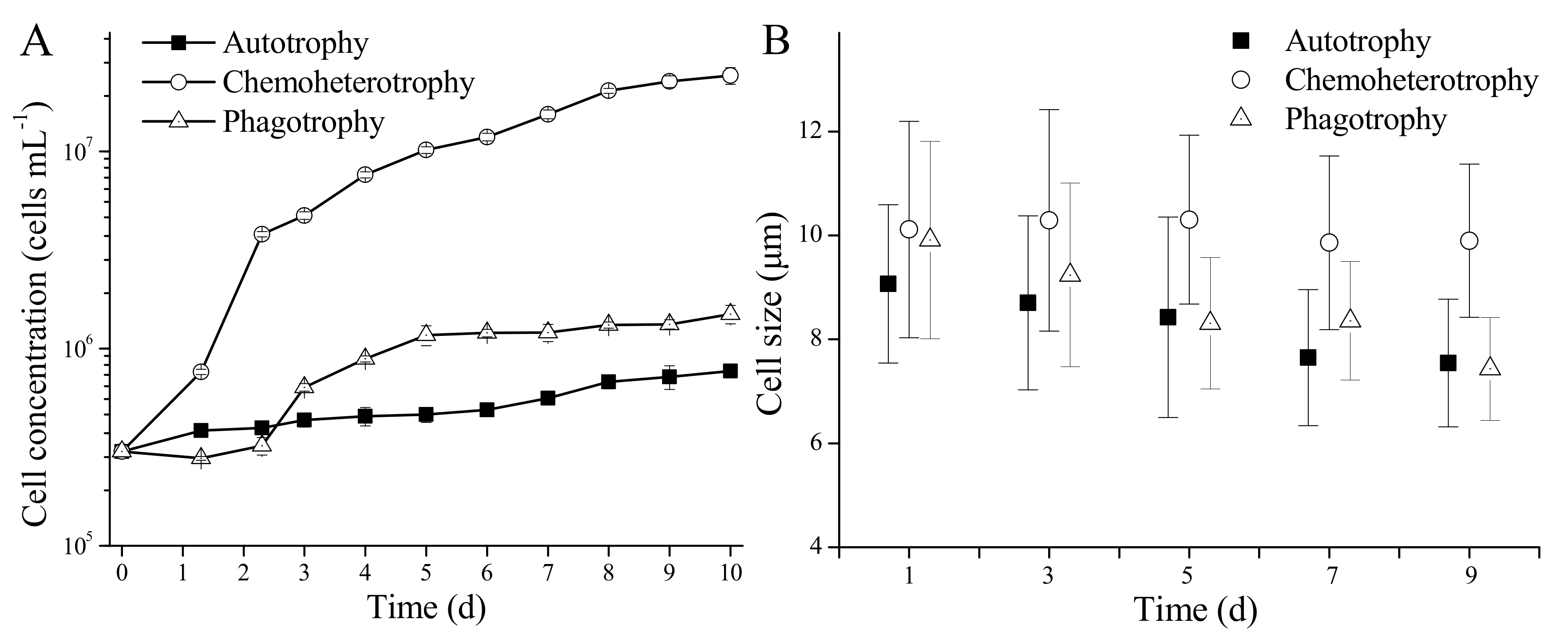
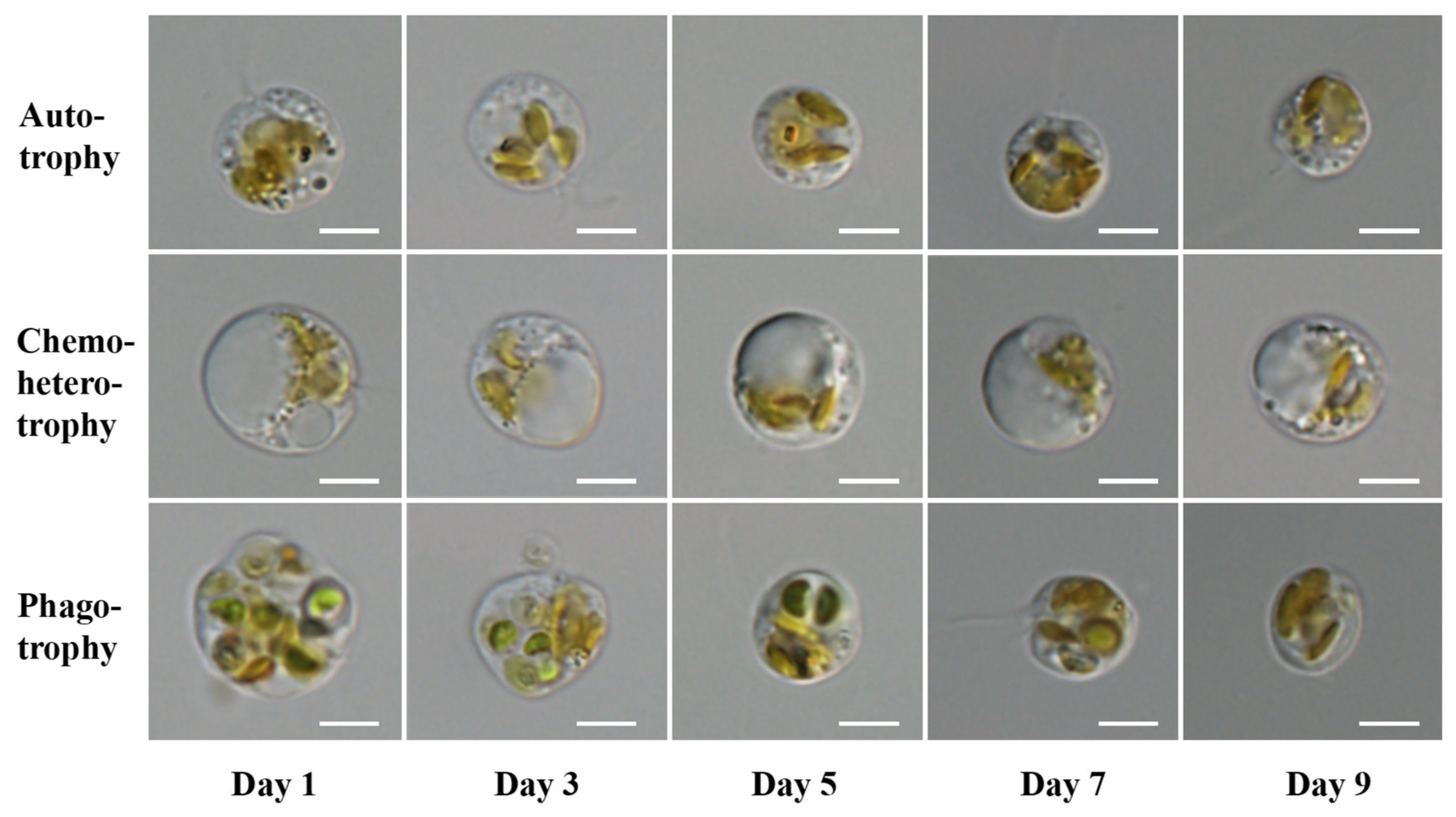
3.2. Growth and Cell Morphology of P. malhamensis with Different Nutritional Modes

The growth of axenic P. malhamensis differed depending on its nutritional mode (Figure 2A). With autotrophy, the cell concentration of P. malhamensis increased to 7 × 10⁵ cells mL−1 from the initial cell concentration of 3 × 10⁵ cells mL−1 after a 10-day cultivation period. However, the cell concentration of P. malhamensis reached up to 1 × 10⁶ cells mL−1 when prey Chlorella was present (i.e., phagotrophy). Furthermore, the cell concentration of P. malhamensis under chemoheterotrophic conditions increased to 1 × 10⁷ cells mL−1 after cultivation for 4–5 days. This showed that the growth rate of P. malhamensis was better with chemoheterotrophy than with autotrophy or phagotrophy. Changes in P. malhamensis cell size varied between the different nutritional modes. The cell size of chemoheterotrophic P. malhamensis basically remained around the initial value of 10 μm during the whole cultivation period, while the cell sizes of autotrophic and phagotrophic P. malhamensis both decreased gradually to 7.5 μm after cultivation for 9 days (Figure 2B).
However, compared to autotrophic P. malhamensis, the cell size of phagotrophic P. malhamensis decreased slowly in the first 3 days because of its ability to graze on prey Chlorella cells (Figure 3). The cell morphology of P. malhamensis also varied greatly between different nutritional modes (Figure 3). The chloroplasts of autotrophic and phagotrophic P. malhamensis in the later period of culture (after 7 days) were intact and distinct, while the chloroplasts of chemoheterotrophic P. malhamensis over the whole cultivation period were rather amorphous. However, one big vacuole, which occupied more than two-thirds of the total cell volume, was always observed in chemoheterotrophic P. malhamensis cells.

3.3. Biochemical Composition of P. malhamensis with Different Nutritional Modes

The total lipid content of P. malhamensis was similar for all three nutritional modes, all being ca. 28% (Table 1). However, the total protein, total sugar, and fucoxanthin contents of P. malhamensis varied greatly between different nutritional modes. The total protein content of autotrophic P. malhamensis was higher than that of chemoheterotrophic P. malhamensis and phagotrophic P. malhamensis (ANOVA, p < 0.01). The same trend occurred for fucoxanthin content, with autotrophic P. malhamensis exhibiting higher percentages of fucoxanthin than chemoheterotrophic P. malhamensis and phagotrophic P. malhamensis (ANOVA, p < 0.01). However, the total sugar content of P. malhamensis was lower with autotrophy than with chemoheterotrophy and phagotrophy (ANOVA, p < 0.01).
With respect to the absolute amino acid content (Table 2), the total amino acid content of autotrophic P. malhamensis was higher than that of chemoheterotrophic P. malhamensis and phagotrophic P. malhamensis (ANOVA, p < 0.05), which corresponded to the higher total protein content. The contents of total essential amino acids (EAAs) and total non-essential amino acids (NEAAs) in autotrophic P. malhamensis cells were also both higher than those in chemoheterotrophic P. malhamensis cells and phagotrophic P. malhamensis cells. Interestingly, the relative amino acid content for total EAAs and total NEAAs was the same for all three trophic modes, all being around 50%. Furthermore, the relative contents of most amino acids were similar for the three modes, except for arginine in phagotrophic P. malhamensis, where the relative content was nearly two times higher than that in chemoheterotrophic P. malhamensis and autotrophic P. malhamensis (ANOVA, p < 0.01). With all three nutritional modes, the amino acid with the richest content in P. malhamensis cells was always glutamic acid.
The fatty acid contents of different nutritional P. malhamensis cells were different from the results of total lipid content (Table 1 and Table 3). The absolute content of total fatty acid in autotrophic P. malhamensis was much lower than that in chemoheterotrophic P. malhamensis and phagotrophic P. malhamensis (ANOVA, p < 0.01). The difference in total fatty acid was mainly caused by the diverse contents of saturated fatty acids (SFAs), with the absolute content of SFAs in autotrophic P. malhamensis being much lower than that in chemoheterotrophic P. malhamensis and phagotrophic P. malhamensis (ANOVA, p < 0.01) (Table 3). To be more specific, chemoheterotrophic P. malhamensis was rich in the SFAs C14:0 (6.29%) and C16:0 (4.51%), while phagotrophic P. malhamensis mainly contained the SFA C16:0 with a content of 5.17%. The content of monounsaturated fatty acids (MUFAs) in autotrophic P. malhamensis (0.48%) was also lower than that in chemoheterotrophic P. malhamensis and phagotrophic P. malhamensis (both 2.75%). The absolute contents of total polyunsaturated fatty acids (PUFAs) in different trophic P. malhamensis cells were all around 10%, while the individual PUFA content in different trophic P. malhamensis cells varied greatly. Autotrophic P. malhamensis had the lowest absolute content of the PUFA C18:2. The absolute content of the PUFA C18:3 in chemoheterotrophic P. malhamensis cells was much lower than that in autotrophic P. malhamensis cells and phagotrophic P. malhamensis cells (ANOVA, p < 0.01). Phagotrophic P. malhamensis cells had a lower absolute content of the PUFA C22:5, compared to chemoheterotrophic P. malhamensis cells and autotrophic P. malhamensis cells. However, with respect to the relative fatty acid content, the relative content of PUFAs in autotrophic P. malhamensis cells was much higher than that in chemoheterotrophic P. malhamensis and phagotrophic P. malhamensis (ANOVA, p < 0.01).

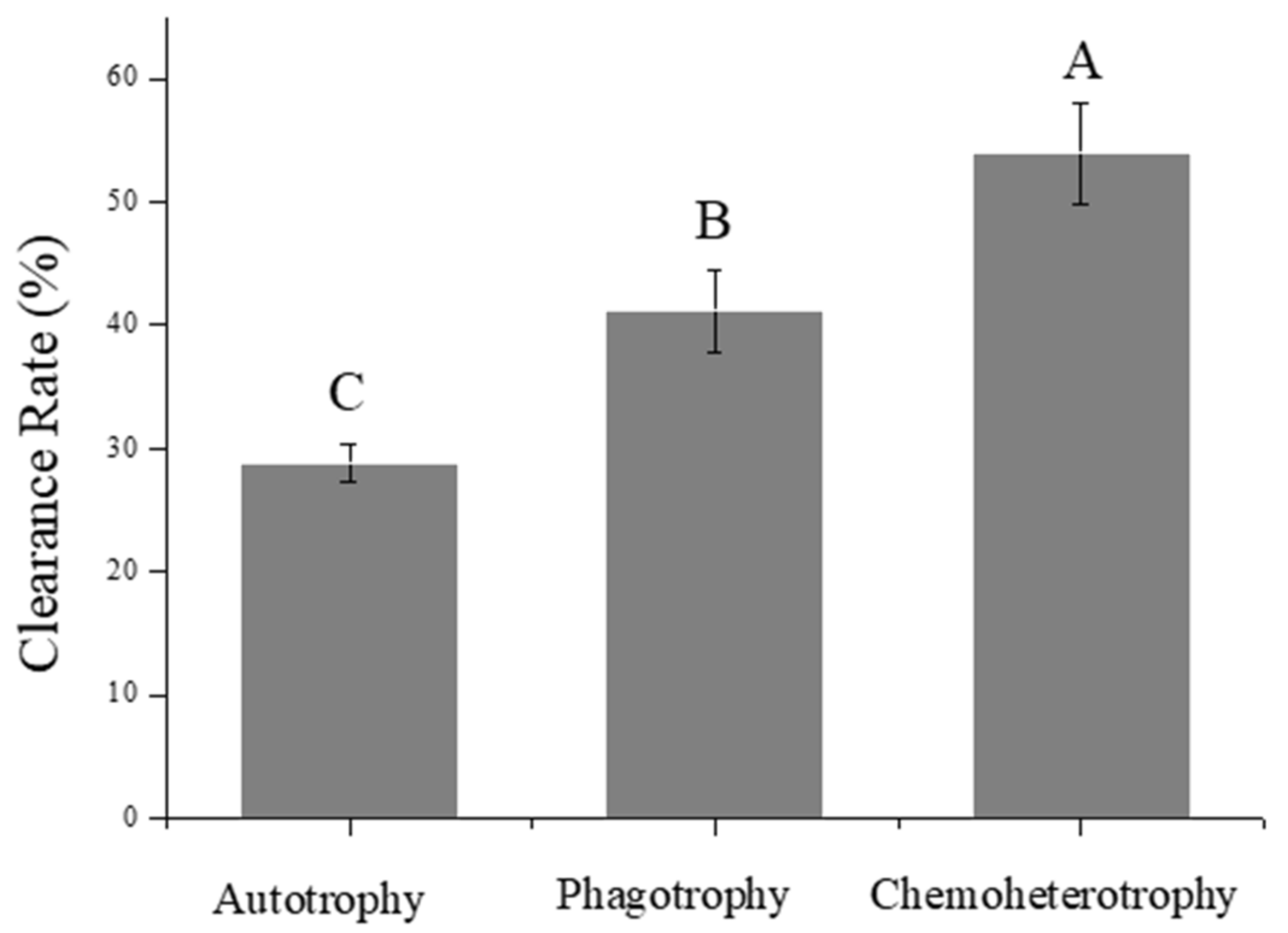
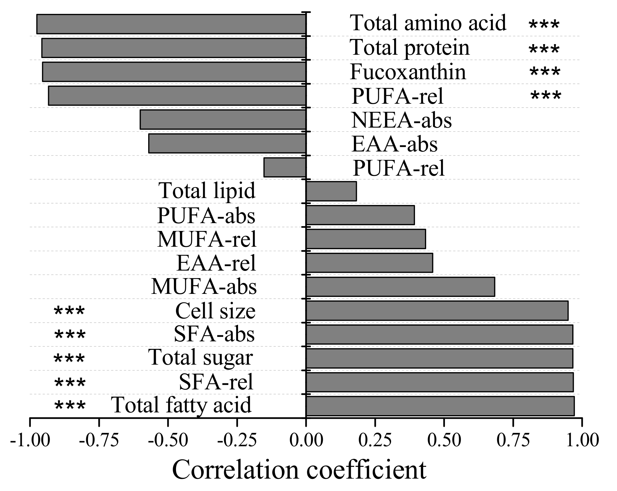
3.4. Grazing Abilities of Paramecium caudatum on P. malhamensis with Different Trophic Modes

The clearance rate of P. caudatum on chemoheterotrophic P. malhamensis cells (53.88%) was significantly higher than that on phagotrophic P. malhamensis cells (41.17%) and autotrophic P. malhamensis (28.86%) (ANOVA, p < 0.01) (Figure 4). This showed that the grazing ability of P. caudatum on P. malhamensis cells differs depending on the nutritional mode of the P. malhamensis.
Correlation analysis showed that the grazing ability of P. caudatum on P. malhamensis cells had significant negative correlations (r < −0.95, p < 0.001) with the total amino acid, total protein content, total relative PUFA content, and fucoxanthin content of P. malhamensis cells (Figure 5). On the other hand, the grazing ability of P. caudatum on P. malhamensis cells had significant positive correlations (r > 0.95, p < 0.001) with the total fatty acid content, total relative SFA content, total sugar content, total absolute SFA content, and cell size of P. malhamensis cells.

4. Discussion

4.1. Purification and Cultivation of P. malhamensis

Obtaining an axenic culture of P. malhamensis was a critical step in studying the nutritional modes of this mixotrophic flagellate. However, as most mixotrophic flagellates lack a cell wall and cannot grow well on solid media, axenic seed cultures of mixotrophic flagellates are difficult to obtain. Therefore, the mixotrophic flagellate cultures involved in some studies are actually not axenic [36]. In our study, an axenic P. malhamensis seed culture was obtained through repeated treatments using multifarious antibiotics. The axenic P. malhamensis culture allowed a more precise study of the physiological and biochemical characteristics of P. malhamensis. Looking beyond the present study, having an axenic seed culture of P. malhamensis also makes it possible to exploit the flagellate’s commercial application value. Poterioochromonas malhamensis could be used to produce antimicrobial malhamensilipin A [37], active polysaccharides β-1,3-glucan [38], or even to control Microcystis blooms [39]. However, its application values have been restricted greatly by the undeveloped mass cultivation methods commonly used for P. malhamensis. In this study, the maximum cell concentration of P. malhamensis was in the order of magnitude of 107 cells mL−1, but we have established a chemoheterotrophic fermentation technology for cultivation of P. malhamensis using the axenic seed culture, which yields a maximum cell concentration and dry weight of P. malhamensis under optimal fermentation conditions of 3 × 10⁸ cells mL−1 [40] and 32.8 g L−1 [38], respectively.
Our study showed that P. malhamensis exhibited an extremely low growth rate when relying upon autotrophy (Figure 2A), which is a similar finding to that of a previous study [28], which indicates that the photosynthetic ability of P. malhamensis is low. The low photosynthetic ability of P. malhamensis is speculated to result from the lack of a CO2 concentrating mechanism as in other species of chrysophyte [41]. The nutritional mode of P. malhamensis was once defined as being predominately phagotrophic mixotrophy, which means that it can achieve its maximum specific growth rate (μmax) only when particulate prey is provided [4]. However, a higher μmax can be observed when P. malhamensis is cultivated with dissolved organic matter [42], and this is consistent with our results. It is notable that the total organic carbon concentrations under phagotrophic conditions and chemoheterotrophic conditions in different studies were different. Therefore, it is more appropriate to describe the nutrition mode of P. malhamensis as being ‘predominately heterotrophic mixotrophy’.

4.2. Effects of Nutritional Mode on the Morphological and Biochemical Characteristics of P. malhamensis

Over the whole cultivation period, the cell size of P. malhamensis under chemoheterotrophic conditions was always larger than that under autotrophic conditions, this resulting from the formation of a bulky chrysolaminarin vesicle (Figure 3) containing a large amount of β-1,3-glucan as the storage substance [38]. The same phenomenon has also been observed in another chrysophyte, Ochromonas [43]. The mixotrophic microalga Chlorella sorokiniana has also been found to increase its cell volume when using heterotrophy by accumulating an excess of storage starch [44]. On the other hand, the protein and total amino acid contents of autotrophic P. malhamensis were both significantly higher than those of chemoheterotrophic P. malhamensis (ANOVA, p < 0.05). This indicates that the storage substance chrysolaminarin might be utilized to synthesize other functional biomolecules (e.g., protein and polypeptides) when P. malhamensis transforms from chemoheterotrophy to autotrophy.
With respect to pigment content, it has been widely proved that chlorophyll concentrations of the mixotrophic flagellates Poterioochromonas or Ochromonas decline after the addition of organics to the culture medium [28,42,45]. Correspondingly, in the present study, P. malhamensis cells under chemoheterotrophic conditions exhibited a pale chloroplast color and a low content of fucoxanthin (Figure 3 and Table 1), which indicated that the photosynthetic system of heterotrophic P. malhamensis cells might be closed when exogenous organic matter is available.
To date, the biochemical composition of P. malhamensis, especially in terms of amino acids and fatty acids, has rarely been studied. Essential amino acids are those that animals or humans cannot synthesize de novo and so can only obtain from a food source. However, most plant cells can synthesize all kinds of amino acids de novo [46]. In the present study (Table 2), all the amino acids were present at higher ratios in autotrophic P. malhamensis than in chemoheterotrophic P. malhamensis, which showed that autotrophic P. malhamensis can synthesize all amino acids de novo. Therefore, the mixotrophic P. malhamensis cells are more like plant cells from the perspective of biochemical characteristics. With respect to fatty acids, PUFAs were considered in a previous study [36] to be the most reliable biochemical variable at indicating the nutritional mode of Ochromonas sp., since the total absolute PUFA contents of Ochromonas sp. varied greatly between different nutritional modes. In contrast, the total absolute PUFA contents in P. malhamensis cells with different trophic modes were the same (Table 3), which indicated that Poterioochromonas and Ochromonas might have different PUFA metabolism pathways. In contrast, the total absolute SAF content of autotrophic P. malhamensis was lower than that of heterotrophic P. malhamensis, which was consistent with the variations observed in Ochromonas sp. [36]. Therefore, it is likely that the SAFs are another form of storage in P. malhamensis and could be used as one of the biochemical variables reflecting the nutritional mode of P. malhamensis.

4.3. Effects of Variation in the Biochemical Composition of P. malhamensis, Resulting from Different Nutritional Modes, on Its Predators’ Feeding Behavior and the Implications for Aquatic Food Webs

Recently, the important roles of mixotrophic flagellates in connecting the classical food chain and microbial loop have attracted more and more attention from ecologists [47,48]. The present study has shown that the transformation of nutritional modes can alter the biochemical composition of the mixotrophic flagellate P. malhamensis and, as a result, affect the grazing ability of its predator Paramecium caudatum. Moreover, a recent study has shown that the grazing ability of P. malhamensis on its prey Microcystis aeruginosa also varies depending on the nutritional mode of the P. malhamensis [40]. These studies imply that changes in environmental nutrient resources would greatly affect the flow directions within food chains centered on mixotrophic flagellates. However, it is difficult to be sure about the precise causal relationships between apparent grazing abilities of predators and nutritional modes of prey. In the present study, Pearson’s correlation analysis showed that the grazing ability of P. caudatum on P. malhamensis cells is positively correlated with the content of fatty acids (especially the SFAs) in P. malhamensis. As certain fatty acids (e.g., SFA C18:0) have generally been considered as indispensable nutrients in artificially synthesized media for Paramecium [49], we have been convinced to some extent that the stronger grazing ability of the ciliate P. caudatum on chemoheterotrophic P. malhamensis cells may result from the higher content of SFAs in chemoheterotrophic P. malhamensis cells. In addition, our analytical results also showed that the ciliate P. caudatum was seemingly better at grazing on chemoheterotrophic P. malhamensis with a high content of chrysolaminarin (i.e., β-1, 3-glucan). To date, the ability of P. caudatum to utilize chrysolaminarin remains uncertain, while a previous study [50] revealed that the addition of the storage substance glycogen could also improve the growth of Paramecium. However, it is notable that most biochemical variables are correlated with each other. For instance, increasing contents of total sugar and lipid are generally accompanied by a decreased content of protein. Therefore, the correlations between grazing ability and any one biochemical variable will be affected by the internal interactions between different biochemical variables. So, although the correlation results in the present study showed that the grazing ability of P. caudatum on P. malhamensis had significant negative correlations with the protein (or amino acid) content and relative PUFA content of P. malhamensis cells, this does not necessarily mean that there is a direct causal link between grazing ability and protein or PUFA content. Indeed, amino acids and PUFA C18:2 have always been considered as important nutrients supporting the growth of Paramecium [49]. Therefore, it is still difficult for us to judge which biochemical variable of P. malhamensis is the main or the real factor affecting P. caudatum grazing on P. malhamensis with different nutritional modes.
The present study has provided a case report illustrating that changes in environmental resources (presented by different nutritional modes) can alter the biochemical composition of a mixotrophic flagellate and, as a consequence, affect the grazing ability of its predator. However, in natural ecosystems, the mixotrophic flagellate P. malhamensis could also be grazed by many other zooplankton species, such as Daphnia magna and Brachionus angularis [16,51], and the altered biochemical composition of P. malhamensis may bring about more complicated influences on diverse higher trophic predators that have different nutritional requirements. Conceivably, some consumers (e.g., P. caudatum in this study) will prefer to graze on chemoheterotrophic P. malhamensis cells rich in sugar and fatty acid, while other consumers may show no interest in feeding on chemoheterotrophic P. malhamensis cells because of their low amino acid content. This speculation is supported by a previous study, which revealed that the feeding preferences of various rotifers (including Brachionus sericus, Cephalodella sp., and Elosa worallii) for autotrophic, heterotrophic, and mixotrophic Chlamydomonas acidophila cells differed between the rotifer species [11]. Given these complex influences, we therefore suppose that the direction of carbon transfer and energy flow in aquatic food webs centered on mixotrophic flagellates may be altered as a result of changes to the nutritional mode used by these flagellates and the concomitant changes in their biochemical composition.

5. Conclusions

The physiological and biochemical composition of axenic P. malhamensis cells, which was obtained by use of multifarious antibiotics, was closely related to the nutritional mode being used by the P. malhamensis cells. Under chemoheterotrophic conditions, P. malhamensis cells exhibited the highest growth rate and largest cell size and simultaneously had the highest content of total sugar as well as total absolute fatty acid. Autotrophic P. malhamensis cells exhibited the lowest growth rate and smallest cell size but had the highest content of protein, total absolute amino acid, and fucoxanthin. The contents of most biochemical components in phagotrophic P. malhamensis cells fell between those in autotrophic and chemoheterotrophic P. malhamensis cells. A feeding experiment showed that P. caudatum was better at grazing on chemoheterotrophic P. malhamensis cells, followed by phagotrophic and then autotrophic P. malhamensis. These results indicate that changes in nutritional mode can alter the biochemical composition of the mixotrophic flagellate P. malhamensis, and this, in turn, can affect the grazing ability of its predator Paramecium caudatum. Further studies should focus on collecting more field evidence to verify the effect of environmental changes on the direction of carbon transfer and energy flow in aquatic food webs centered on mixotrophic flagellates.

Author Contributions

M.M. performed the experiments, analyzed the data, and wrote the paper. C.W. and M.C. participated in analyzing the data. H.W. helped in preparing the P. malhamensis culture. Q.H. and Y.G. contributed to the design of the experiments, the drafting of the paper, and revising it critically. All authors gave approval for publication. All authors have read and agreed to the published version of the manuscript.

Funding

This work was funded by the National Natural Science Foundation of China (No. 32002413, No. 31872201, and No. 31772419) and the China Postdoctoral Science Foundation (No. 2019M662749).

Institutional Review Board Statement

Not applicable.

Informed Consent Statement

Not applicable.

Data Availability Statement

Not applicable.

Acknowledgments

The authors thank Wanwan Ke for providing the prey bacteria. They thank Yu Peng in the Institute of Hydrobiology, Chinese Academy of Sciences, for providing Paramecium caudatum.

Conflicts of Interest

The authors declare no conflict of interest.

References

  1. Burkholder, J.M.; Glibert, P.M.; Skelton, H.M. Mixotrophy, a major mode of nutrition for harmful algal species in eutrophic waters. Harmful Algae 2008, 8, 77–93. [Google Scholar] [CrossRef]
  2. Jones, R.I. Mixotrophy in planktonic protists: An overview. Freshw. Biol. 2000, 45, 219–226. [Google Scholar] [CrossRef]
  3. Stoecker, D.K.; Hansen, P.J.; Caron, D.A.; Mitra, A. Mixotrophy in the marine plankton. Annu. Rev. Mar. Sci. 2017, 9, 311–335. [Google Scholar] [CrossRef] [PubMed] [Green Version]
  4. Holen, D.A.; Boraas, M.E. Mixotrophy in chrysophytes. In Chrysophyte Algae: Ecology, Phylogeny and Development; Cambridge University Press: Cambridge, UK, 1995; pp. 119–140. [Google Scholar]
  5. Moorthi, S.; Caron, D.A.; Gast, R.J.; Sanders, R.W. Mixotrophy: A widespread and important ecological strategy for planktonic and sea-ice nanoflagellates in the Ross Sea, Antarctica. Aquat. Microb. Ecol. 2009, 54, 269–277. [Google Scholar] [CrossRef]
  6. Sanders, R.W. Alternative nutritional strategies in protists: Symposium introduction and a review of freshwater protists that combine photosynthesis and heterotrophy. J. Eukaryot. Microbiol. 2011, 58, 181–184. [Google Scholar] [CrossRef] [PubMed]
  7. Sanders, R.W.; Gast, R.J. Bacterivory by phototrophic picoplankton and nanoplankton in Arctic waters. FEMS Microbiol. Ecol. 2012, 82, 242–253. [Google Scholar] [CrossRef] [PubMed] [Green Version]
  8. Sanders, R.W.; Berninger, U.G.; Lim, E.L.; Kemp, P.F.; Caron, D.A. Heterotrophic and mixotrophic nanoplankton predation on picoplankton in the Sargasso Sea and on Georges Bank. Mar. Ecol. Prog. Ser. 2000, 192, 103–118. [Google Scholar] [CrossRef]
  9. Azam, F.; Fenchel, T.; Field, J.; Grey, J.; Meyer, R.L.; Thingstad, F. The ecological role of water-column microbes. Mar. Ecol. Prog. Ser. 1983, 10, 257–263. [Google Scholar] [CrossRef]
  10. Unrein, F.; Gasol, J.M.; Not, F.; Forn, I.; Massana, R. Mixotrophic haptophytes are key bacterial grazers in oligotrophic coastal waters. ISME J. 2014, 8, 164. [Google Scholar] [CrossRef] [Green Version]
  11. Weithoff, G.; Wacker, A. The mode of nutrition of mixotrophic flagellates determines the food quality for their consumers. Funct. Ecol. 2007, 21, 1092–1098. [Google Scholar] [CrossRef]
  12. Ma, M.; Gong, Y.; Hu, Q. Identification and feeding characteristics of the mixotrophic flagellate Poterioochromonas malhamensis, a microalgal predator isolated from outdoor massive Chlorella culture. Algal Res. 2018, 29, 142–153. [Google Scholar] [CrossRef]
  13. Zhang, X.; Watanabe, M.M. Light and electron microscopy of grazing by Poterioochromonas malhamensis (Chrysophyceae) on a range of phytoplankton taxa. J. Phycol. 1996, 32, 37–46. [Google Scholar] [CrossRef]
  14. Zhang, X.; Hu, H.; Men, Y.; Yang, J.; Christoffersen, K. Feeding characteristics of a golden alga (Poterioochromonas sp.) grazing on toxic cyanobacterium Microcystis aeruginosa. Water Res. 2009, 43, 2953–2960. [Google Scholar] [CrossRef]
  15. Zhang, X.; Watanabe, M.M. Grazing and growth of the mixotrophic chrysomonad Poterioochromonas malhamensis feeding on algae. J. Phycol. 2001, 37, 738–743. [Google Scholar] [CrossRef]
  16. Zhang, X.; Hu, H.; Warming, T.P.; Christoffersen, K.S. Life history response of Daphnia magna to a mixotrophic golden alga, Poterioochromonas sp., at different food levels. Bull. Environ. Contam. Toxicol. 2011, 87, 117–123. [Google Scholar] [CrossRef]
  17. Boxhorn, J.E.; Holen, D.A.; Boraas, M.E. Toxicity of the chrysophyte flagellate Poterioochromonas malhamensis to the rotifer Brachionus angularis. Hydrobiologia 1998, 387, 283–287. [Google Scholar] [CrossRef]
  18. Sterner, R.W.; Hagemeier, D.D.; Smith, W.L.; Smith, R.F. Phytoplankton nutrient limitation and food quality for Daphnia. Limnol. Oceanogr. 1993, 38, 857–871. [Google Scholar] [CrossRef] [Green Version]
  19. Stoecker, D.K.; Capuzzo, J.M. Predation on protozoa: Its importance to zooplankton. J. Plankton Res. 1990, 12, 891–908. [Google Scholar] [CrossRef] [Green Version]
  20. Fan, J.; Cui, Y.; Zhou, Y.; Wan, M.; Wang, W.; Xie, J.; Li, Y. The effect of nutrition pattern alteration on Chlorella pyrenoidosa growth, lipid biosynthesis-related gene transcription. Bioresour. Technol. 2014, 164, 214–220. [Google Scholar] [CrossRef]
  21. Lalibertè, G.; de la Noüie, J. Auto-, hetero-, and mixotrophic growth of Chlamydomonas humicola (chlorophyceae) on acetate. J. Phycol. 1993, 29, 612–620. [Google Scholar] [CrossRef]
  22. Kryuchkova, M.; Danilushkina, A.; Lvov, Y.; Fakhrullin, R. Evaluation of toxicity of nanoclays and graphene oxide in vivo: A Paramecium caudatum study. Environ. Sci-Nano. 2016, 3, 442–452. [Google Scholar] [CrossRef] [Green Version]
  23. Gupta, G.S.; Kumar, A.; Senapati, V.A.; Pandey, A.K.; Shanker, R.; Dhawan, A. Laboratory scale microbial food chain to study bioaccumulation, biomagnification, and ecotoxicity of cadmium telluride quantum dots. Environ. Sci. Technol. 2017, 51, 1695–1706. [Google Scholar] [CrossRef]
  24. Kato, S. Laboratory culture and morphology of Colacium vesiculosum Ehrb.(Euglenophyceae). Jpn. J. Phycol. 1982, 30, 63–67. [Google Scholar]
  25. Corno, G.; Jurgens, K. Direct and indirect effects of protist predation on population size structure of a bacterial strain with high phenotypic plasticity. Appl. Environ. Microb. 2006, 72, 78–86. [Google Scholar] [CrossRef] [Green Version]
  26. Destain, J.; Haubruge, É.; Thonart, P.; Portetelle, D.; Francis, F.; Bauwens, J.; Tarayre, C.; Brasseur, C.; Mattéotti, C.; Vandenbol, M. Isolation of an amylolytic chrysophyte, Poterioochromonas sp., from the digestive tract of the termite Reticulitermes santonensis. Biotechnol. Agron. Soc. Environ. 2014, 18, 19–31. [Google Scholar]
  27. Rippka, R.; Deruelles, J.; Waterbury, J.B.; Herdman, M.; Stanier, R.Y. Generic assignments, strain histories and properties of pure cultures of cyanobacteria. J. Gen. Microbiol. 1979, 111, 1–61. [Google Scholar] [CrossRef] [Green Version]
  28. Sanders, R.W.; Porter, K.G.; Caron, D.A. Relationship between phototrophy and phagotrophy in the mixotrophic chrysophyte Poterioochromonas malhamensis. Microb. Ecol. 1990, 19, 97–109. [Google Scholar] [CrossRef]
  29. Terrado, R.; Pasulka, A.L.; Lie, A.A.; Orphan, V.J.; Heidelberg, K.B.; Caron, D.A. Autotrophic and heterotrophic acquisition of carbon and nitrogen by a mixotrophic chrysophyte established through stable isotope analysis. ISME J. 2017, 11, 2022–2034. [Google Scholar] [CrossRef] [Green Version]
  30. Leakey, R.; Burkill, P.; Sleigh, M. A comparison of fixatives for the estimation of abundance and biovolume of marine planktonic ciliate populations. J. Plankton Res. 1994, 16, 375–389. [Google Scholar] [CrossRef]
  31. Ma, M.; Wei, C.; Wang, H.; Sha, C.; Chen, M.; Gong, Y.; Hu, Q. Isolation and evaluation of a novel strain of Chlorella sorokiniana that resists grazing by the predator Poterioochromonas malhamensis. Algal Res. 2019, 38, 101429. [Google Scholar] [CrossRef]
  32. Wahlen, B.D.; Willis, R.M.; Seefeldt, L.C. Biodiesel production by simultaneous extraction and conversion of total lipids from microalgae, cyanobacteria, and wild mixed-cultures. Bioresour. Technol. 2011, 102, 2724–2730. [Google Scholar] [CrossRef] [PubMed]
  33. Billakanti, J.M.; Catchpole, O.J.; Fenton, T.A.; Mitchell, K.A.; MacKenzie, A.D. Enzyme-assisted extraction of fucoxanthin and lipids containing polyunsaturated fatty acids from Undaria pinnatifida using dimethyl ether and ethanol. Process Biochem. 2013, 48, 1999–2008. [Google Scholar] [CrossRef]
  34. Ye, J.; Liang, J.; Wang, L.; Markou, G. The mechanism of enhanced wastewater nitrogen removal by photo-sequencing batch reactors based on comprehensive analysis of system dynamics within a cycle. Bioresour. Technol. 2018, 260, 256–263. [Google Scholar] [CrossRef] [PubMed]
  35. Laurens, L.M.; Quinn, M.; Van Wychen, S.; Templeton, D.W.; Wolfrum, E.J. Accurate and reliable quantification of total microalgal fuel potential as fatty acid methyl esters by in situ transesterification. Anal. Bioanal. Chem. 2012, 403, 167–178. [Google Scholar] [CrossRef] [Green Version]
  36. Boëchat, I.G.; Weithoff, G.; Krüger, A.; Gücker, B.; Adrian, R. A biochemical explanation for the success of mixotrophy in the flagellate Ochromonas sp. Limnol. Oceanogr. 2007, 52, 1624–1632. [Google Scholar] [CrossRef]
  37. Chen, J.L.; Proteau, P.J.; Roberts, M.A.; Gerwick, W.H.; Slate, D.L.; Lee, R.H. Structure of malhamensilipin A, an inhibitor of protein tyrosine kinase, from the cultured chrysophyte Poterioochromonas malhamensis. J. Nat. Prod. 1994, 57, 524–527. [Google Scholar] [CrossRef]
  38. Ma, M.; Li, Y.; Chen, J.; Wang, F.; Yuan, L.; Li, Y.; Zhang, B.; Ye, D.; Han, D.; Jin, H.; et al. High-cell-density cultivation of the flagellate alga Poterioochromonas malhamensis for biomanufacturing the water-soluble β-1, 3-glucan with multiple biological activities. Bioresour. Technol. 2021, 337, 125447. [Google Scholar] [CrossRef]
  39. Zhang, X.; Hu, H.; Hong, Y.; Yang, J. Isolation of a Poterioochromonas capable of feeding on Microcystis aeruginosa and degrading microcystin-LR. FEMS Microbiol. Lett. 2008, 288, 241–246. [Google Scholar] [CrossRef] [Green Version]
  40. Ma, M.; Wang, F.; Wei, C.; Chen, J.; Jin, H.; Wang, H.; Song, L.; Hu, Q.; Gong, Y. Establishment of high-cell-density heterotrophic cultivation of Poterioochromonas malhamensis contributes to achieving biological control of Microcystis. J. Appl. Phycol. 2022, 34, 423–434. [Google Scholar] [CrossRef]
  41. Giordano, M.; Beardall, J.; Raven, J.A. CO2 concentrating mechanisms in algae: Mechanisms, environmental modulation, and evolution. Annu. Rev. Plant Biol. 2005, 56, 99–131. [Google Scholar] [CrossRef] [Green Version]
  42. Lewitus, A.J.; Caron, D.A. Physiological responses of phytoflagellates to dissolved organic substrate additions. 1. Dominant role of heterotrophic nutrition in Poterioochromonas malhamensis (Chrysophyceae). Plant Cell Physiol. 1991, 32, 671–680. [Google Scholar] [CrossRef]
  43. Aaronson, S.; Behrens, U. Ultrastructure of an unusual contractile vacuole in several chrysomonad phytoflagellates. J. Cell Sci. 1974, 14, 1–9. [Google Scholar] [CrossRef]
  44. Marchello, A.E.; dos Santos, A.C.; Lombardi, A.T.; de Souza, C.W.O.; Montanhim, G.C. Physiological and ecological aspects of Chlorella sorokiniana (Trebouxiophyceae) under photoautotrophic and mixotrophic conditions. Microb. Ecol. 2018, 76, 791–800. [Google Scholar] [CrossRef]
  45. Zhang, L.; Li, B.; Wu, Z.; Gu, L.; Yang, Z. Changes in growth and photosynthesis of mixotrophic Ochromonas sp. in response to different concentrations of glucose. J. Appl. Phycol. 2016, 28, 2671–2678. [Google Scholar] [CrossRef]
  46. Galili, G.; Amir, R.; Fernie, A.R. The regulation of essential amino acid synthesis and accumulation in plants. Annu. Rev. Plant Biol. 2016, 67, 153–178. [Google Scholar] [CrossRef]
  47. Mitra, A.; Flynn, K.J.; Burkholder, J.M.; Berge, T.; Calbet, A.; Raven, J.A.; Granéli, E.; Glibert, P.M.; Hansen, P.J.; Stoecker, D.K. The role of mixotrophic protists in the biological carbon pump. Biogeosciences 2014, 11, 995–1005. [Google Scholar] [CrossRef] [Green Version]
  48. Caron, D.A. Mixotrophy stirs up our understanding of marine food webs. Proc. Natl. Acad. Sci. USA 2016, 113, 2806–2808. [Google Scholar] [CrossRef] [Green Version]
  49. Wichterman, R. The nutritional requirements of Paramecium: Axenic Media. In The Biology of Paramecium; Springer: Boston, MA, USA, 1986; pp. 181–196. [Google Scholar]
  50. Johnson, W.; Brennan, M.; Berard, D.; Morrow, J.; Hudson, K. Proteins and polysaccharides in the nutrition of Paramecium multimicronucleatun. J. Protozool. 1980, 27, 332–336. [Google Scholar] [CrossRef]
  51. Boenigk, J.; Stadler, P. Potential toxicity of chrysophytes affiliated with Poterioochromonas and related ‘Spumella-like’ flagellates. J. Plankton Res. 2004, 26, 1507–1514. [Google Scholar] [CrossRef] [Green Version]
Figure 1. Microscopic observations of non-axenic (A) and axenic (B) P. malhamensis cultivated with enriched medium. Scale bars = 20 μm.
Microorganisms 10 00852 g001
Figure 2. Growth (A) and cell size changes (B) of P. malhamensis with different nutritional modes. Error bars indicate the standard deviation. n = 3.
Microorganisms 10 00852 g002
Figure 3. Cell morphologies of P. malhamensis with different nutritional modes at different times during cultivation. Scale bar = 5 μm.
Microorganisms 10 00852 g003
Figure 4. Clearance rate of Paramecium caudatum on different trophic P. malhamensis cells. Significant differences are indicated by different uppercase letters (one-way ANOVA, p < 0.01). Error bars indicate the standard deviation. n = 3.
Microorganisms 10 00852 g004
Figure 5. Correlation analysis between clearance rate of Paramecium caudatum on P. malhamensis cells with different nutritional modes and various physiological and biochemical parameters of P. malhamensis. Significant differences are indicated by three asterisks (p < 0.001).
Microorganisms 10 00852 g005
Table 1. Biochemical composition of P. malhamensis cultivated with different nutritional modes (n = 3).
Total Lipid (%)Total Protein (%)Total Sugar (%)Fucoxanthin (%)
Autotrophy28.2 ± 0.842.1 ± 1.011.3 ± 0.50.34 ± 0.03
Chemoheterotrophy28.4 ± 0.623.0 ± 1.642.8 ± 3.40.12 ± 0.00
Phagotrophy27.4 ± 0.330.1 ± 0.824.7 ± 1.10.20 ± 0.01
Table 2. Amino acid composition of P. malhamensis with different nutritional modes (n = 3).
Amino Acid% of Dry Weight (Absolute)% of Total Amino Acid (Relative)
AutoChemoPhagoAutoChemoPhago
Essential amino acid
(EAA)
Thr1.80 ± 0.001.11 ± 0.051.43 ± 0.075.31 ± 0.015.15 ± 0.224.95 ± 0.24
Val2.01 ± 0.061.22 ± 0.031.57 ± 0.055.93 ± 0.195.67 ± 0.145.42 ± 0.19
Met0.39 ± 0.020.12 ± 0.020.35 ± 0.051.15 ± 0.660.57 ± 0.071.21 ± 0.16
Ile1.72 ± 0.061.14 ± 0.081.11 ± 0.115.06 ± 0.165.31 ± 0.373.84 ± 0.37
Leu3.02 ± 0.041.97 ± 0.082.23 ± 0.158.88 ± 0.119.18 ± 0367.69 ± 0.51
Phe2.19 ± 0.041.41 ± 0.031.59 ± 0.086.45 ± 0.136.55 ± 0.145.48 ± 0.28
His0.96 ± 0.040.71 ± 0.020.66 ± 0.032.82 ± 0.113.29 ± 0.102.28 ± 0.10
Lys2.70 ± 0.041.78 ± 0.061.73 ± 0.097.90 ± 0.118.30 ± 0.275.98 ± 0.32
Arg2.15 ± 0.081.31 ± 0.063.35 ± 0.136.30 ± 0.226.10 ± 0.2911.57 ± 0.46
Total16.94 ± 0.1210.78 ± 0.3114.04 ± 0.7449.80 ± 0.3450.11 ± 1.4248.41 ± 2.56
Non-essential amino acid (NEAA)Asp4.21 ± 0.102.53 ± 0.053.27 ± 0.1912.38 ± 0.2911.76 ± 0.2411.27 ± 0.65
Ser1.77 ± 0.051.12 ± 0.051.31 ± 0.075.21 ± 0.155.23 ± 0.234.52 ± 0.24
Glu4.23 ± 0.092.87 ± 0.124.26 ± 0.1812.42 ± 0.2713.33 ± 0.5614.70 ± 0.61
Gly1.77 ± 0.061.14 ± 0.051.59 ± 0.075.20 ± 0.195.32 ± 0.225.49 ± 0.23
Ala2.13 ± 0.041.26 ± 0.042.06 ± 0.126.24 ± 0.125.88 ± 0.207.11 ± 0.40
Cys0.14 ± 0.010.06 ± 0.030.18 ± 0.050.27 ± 0.240.30 ± 0.120.62 ± 0.17
Tyr1.27 ± 0.040.76 ± 0.041.04 ± 0.033.72 ± 0.123.55 ± 0.183.60 ± 0.11
Pro1.62 ± 0.120.97 ± 0.051.24 ± 0.054.76 ± 0.344.51 ± 0.224.28 ± 0.19
Total17.14 ± 0.3110.73 ± 0.3414.96 ± 0.7250.20 ± 0.9149.89 ± 1.5851.59 ± 2.48
Total 34.08 ± 0.3121.51 ± 0.6329.00 ± 1.44100100100
Note: Thr, Threonine; Val, Valine; Met, Methionine; Ile, Isoleucine; Leu, Leucine; Phe, Phenylalanine; His, Histidine; Lys, Lysine; Arg, Arginine; Asp, Aspartic acid; Ser, Serine; Glu, Glutamic acid; Gly, Glycine; Ala, Alanine; Cys, Cystine; Tyr, Tyrosine; Pro, Proline. Auto, Chemo, and Phago represent autotrophy, chemoheterotrophy, and phagotrophy, respectively.
Table 3. Fatty acid composition of P. malhamensis with different nutritional modes (n = 3).
Fatty Acid%, of Dry Weight (Absolute)%, of Total Fatty Acid (Relative)
AutoChemoPhagoAutoChemoPhago
C14:01.66 ± 0.046.29 ± 0.641.18 ± 0.1012.20 ± 0.1624.38 ± 0.065.74 ± 0.34
C15:00.02 ± 0.000.01 ± 0.000.03 ± 0.000.14 ± 0.010.02 ± 0.010.14 ± 0.00
C16:01.36 ± 0.034.51 ± 0.455.17 ± 0.1310.06 ± 0.1317.49 ± 0.0125.07 ± 0.69
C17:00.03 ± 0.000.04 ± 0.000.08 ± 0.000.21 ± 0.010.16 ± 0.010.39 ± 0.01
C18:00.14 ± 0.002.01 ± 0.200.86 ± 0.021.03 ± 0.027.80 ± 0.014.16 ± 0.09
C22:00.08 ± 0.000.10 ± 0.010.05 ± 0.010.56 ± 0.030.39 ± 0.010.24 ± 0.02
∑SFA3.29 ± 0.0812.96 ± 1.317.37 ± 0.2524.22 ± 0.3550.24 ± 0.1135.74 ± 1.15
C16:10.05 ± 0.000.49 ± 0.040.16 ± 0.000.40 ± 0.021.89 ± 0.020.78 ± 0.04
C17:10.01 ± 0.010.01 ± 0.010.08 ± 0.000.078 ± 0.090.04 ± 0.020.38 ± 0.01
C18:10.19 ± 0.001.47 ± 0.132.30 ± 0.071.41 ± 0.055.71 ± 0.0811.15 ± 0.41
C22:10.23 ± 0.010.78 ± 0.090.21 ± 0.031.68 ± 0.073.02 ± 0.051.03 ± 0.12
∑MUFA0.48 ± 0.042.75 ± 0.272.75 ± 0.113.56 ± 0.2410.66 ± 0.1813.35 ± 0.57
C18:22.44 ± 0.075.56 ± 0.555.38 ± 0.1418.01 ± 0.1321.57 ± 0.0126.11 ± 0.14
C18:33.39 ± 0.081.02 ± 0.103.64 ± 0.1924.96 ± 0.083.94 ± 0.0117.67 ± 0.50
C18:40.14 ± 0.000.06 ± 0.010.03 ± 0.011.02 ± 0.010.22 ± 0.010.16 ± 0.02
C20:50.45 ± 0.010.25 ± 0.020.23 ± 0.023.33 ± 0.050.98 ± 0.011.09 ± 0.06
C22:52.99 ± 0.072.97 ± 0.300.99 ± 0.0822.01 ± 0.2911.50 ± 0.034.79 ± 0.29
C22:60.39 ± 0.010.23 ± 0.010.22 ± 0.002.87 ± 0.060.89 ± 0.031.07 ± 0.01
∑PUFA9.80 ± 0.2510.09 ± 1.0010.49 ± 0.4372.21 ± 0.6139.10 ± 0.1050.89 ± 1.03
Total13.57 ± 0.3225.80 ± 2.5720.61 ± 0.56100100100
Note: SFA, saturated fatty acid; MUFA, monounsaturated fatty acid; PUFA, polyunsaturated fatty acid. Auto, Chemo, and Phago represent autotrophy, chemoheterotrophy, and phagotrophy, respectively.
Publisher’s Note: MDPI stays neutral with regard to jurisdictional claims in published maps and institutional affiliations.

