


Abstract
Infection with severe acute respiratory syndrome coronavirus 2 (SARS–CoV–2) is a rapidly spreading and devastating global pandemic. Many researchers are attempting to clarify the mechanisms of infection and to develop a drug or vaccine against the virus, but there are still no proven effective treatments. The present article reviews the common presenting hematological manifestations of coronavirus disease 2019 (COVID–19). Elucidating the changes in hematological parameters in SARS–CoV–2 infected patients could help to understand the pathophysiology of the disease and may provide early clues to diagnosis. Several studies have shown that hematological parameters are markers of disease severity and suggest that they mediate disease progression.
1. Introduction
1.1. Severe Acute Respiratory Syndrome Coronavirus 2 (SARS–CoV–2) at a Glance
The severe acute respiratory syndrome coronavirus 2 (SARS–CoV–2) contagion was first described in December 2019 when cases of pneumonia of an unidentified cause were reported in Wuhan, Hubei province, central China [1,2,3]. Occurring on 7 January 2020, Chinese scientists isolated a new type of coronavirus (CoV) [1] underlying this series of infections and, on 12 January 2020, reported its genetic sequence [1,3]. The source of the virus is currently understood to have been food products and wild animals that are easily available in the Huanan Seafood Wholesale Market, which was closed by the Chinese authorities on 1 January 2020 [1,2,3]. The infection has become a major global health concern and has attained pandemic status, having infected persons in almost every country worldwide [3,4].
To clarify the nomenclature of the newly identified coronavirus, the World Health Organization (WHO) proposed the designation “2019 novel coronavirus” (2019–nCoV) and the disease caused by it would be referred to as “coronavirus disease 2019” (COVID–19) [5]. The Coronaviridae Study Group (CSG) of the International Committee on Taxonomy of Viruses (ICTV) suggested that the virus be designated “SARS–CoV–2”, as it bears many similarities to severe acute respiratory syndrome (SARS)–causing coronaviruses (SARS–CoVs) [6]. Nonetheless, it has been suggested [5] that the name introduced by the CSG is inaccurate and may be misleading, as it suggests that the virus leads to the development of SARS, which led to the proposal of a new name: human coronavirus 2019 (HCoV–19) [5]. Here, we use the convention SARS–CoV–2 according to the proposal of the official association responsible for naming the Coronaviridae family [6].
SARS–CoV–2 is a member of the subgenus Sarbecovirus of the genus Betacoronavirus in the family Coronaviridae [7,8,9]. The family, with a large genome of 26–32 kilobases, belongs to a group of enveloped positive–sense single–stranded ribonucleic acid (RNA) viruses, referred to as (+)ssRNA viruses [10,11]. To date, several human CoVs have been identified, including OC43, 229E, NL63, HKU1, SARS–CoV and the Middle East respiratory syndrome coronavirus (MERS–CoV) [11,12,13]. The last two of the afore–mentioned viruses have been shown to have high infectivity and represented the etiological factors underlying the severe pneumonia epidemics in China [14] and Saudi Arabia [15]. Accumulating lines of evidence indicate that animals, particularly the bat species, may be pervasive reservoirs of coronaviruses [12,13].
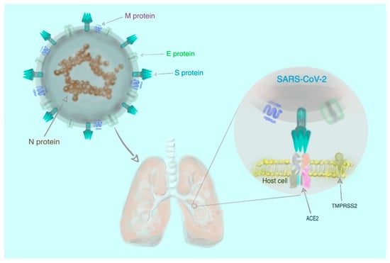
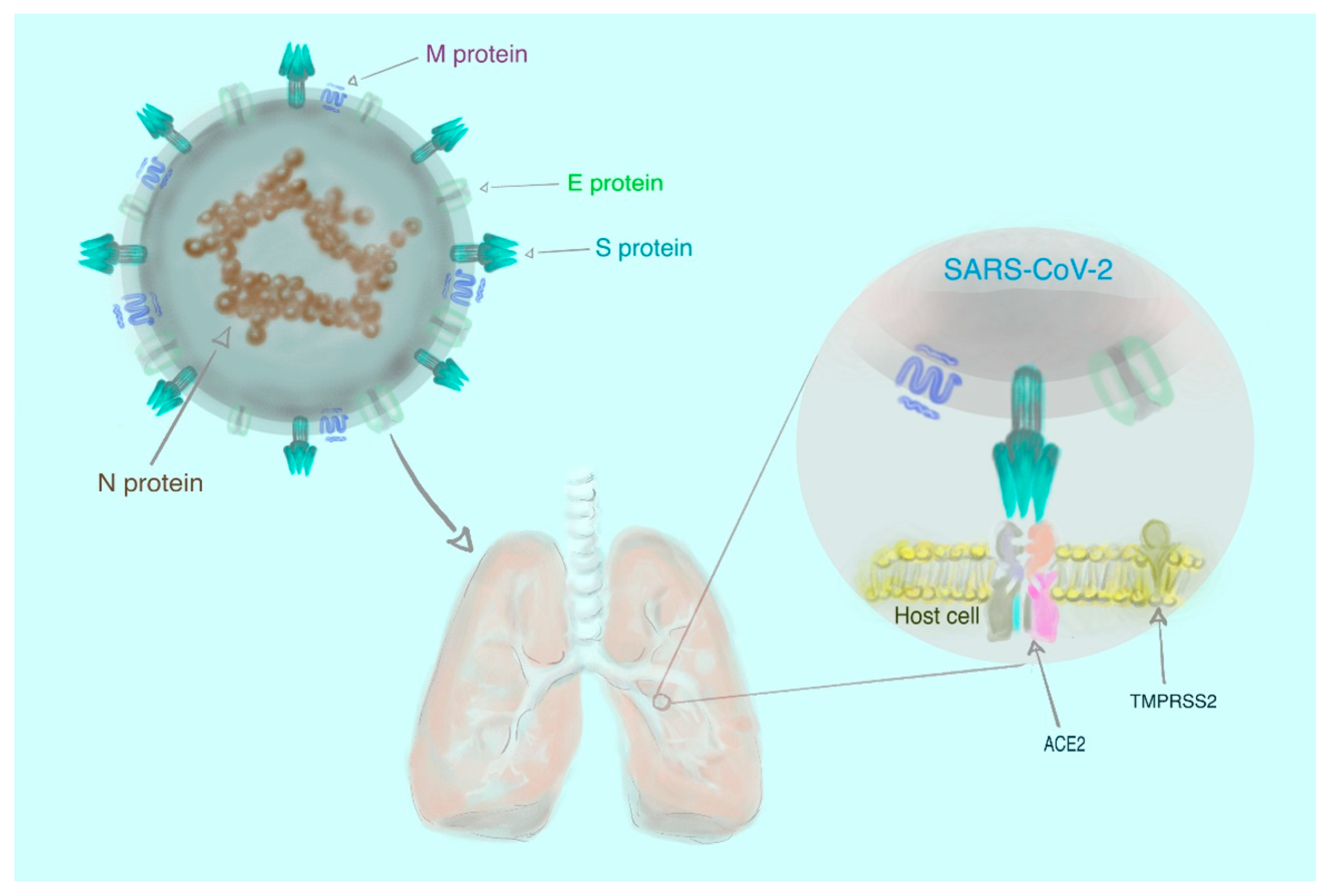
Molecular and phylogenetic analyses of clinical samples from the respiratory tracts of infected patients have revealed that SARS–CoV–2 resembles two bat–borne SARS–like CoVs that had been previously isolated in China [7,8,9]. However, the assertion that animals are the exclusive source of infection is not entirely accurate, since the virus has spread rapidly to all continents globally [3] as a result of human-to-human transmission [16]. There is experimental evidence that human transmembrane angiotensin–converting enzyme 2 (ACE2) serves as a receptor for SARS–CoV–2 [17,18,19,20]. Briefly, the receptor–binding spike (S) glycoproteins, located on the surface of the virus, attach to the host cell through ACE2 and, consequently, the viral RNA may enter the host cell and replicate [18,19,20,21]. This process also is quite likely mediated by transmembrane serine protease 2 (TMPRSS2) [22]. The simplified structure of SARS–CoV–2 is shown in Figure 1.
Figure 1.
The structure of severe acute respiratory syndrome coronavirus 2 (SARS–CoV–2) comprising four different proteins: spike (S), membrane (M), envelope (E) and nucleocapsid (N). The first three proteins (S, M, and E) are the components of the viral envelope, while the N protein (enclosing RNA) forms a part of the genome. To enter the cell, the virus requires the interaction of the S protein with human transmembrane angiotensin–converting enzyme 2 (ACE2) and transmembrane serine protease 2 (TMPRSS2) [22,23].
Understanding the mechanism of interaction between virus and host cells is essential to the development of anti–SARS–CoV–2 drugs and vaccines [21].
1.2. Coronavirus Disease 2019 (COVID–19) at a Glance
Research concerning coronavirus disease 2019 (COVID–19) disease is in its infancy and the current global understanding of the pathophysiology, nosology, and symptomology of COVID–19 lacks both depth and universal agreement. Consequently, several diverse classifications have been proposed. Lai et al., [24] distinguished between three forms of COVID–19 manifestation, including (a) asymptomatic carrier; (b) acute respiratory disease (ARD) and (c) pneumonia of variable severity. Regarding the first group of patients, no clinical symptoms or radiographic changes are observed and most of these patients will not require hospitalization; however, they may still be a source of infection for other individuals [25,26,27]. A minor subgroup of hospitalized COVID–19 patients is diagnosed with ARD and experience typical respiratory symptoms without radiological signs of pneumonia [24]. The last minor subgroup of patients exhibits respiratory symptoms and radiological evidence of pneumonia, typically requiring hospitalization [24]. Yuki et al., [28] classified the disease manifestation into five separate categories, namely: (a) asymptomatic; (b) disease with mild symptoms (including fever, fatigue, myalgia, cough, sore throat, runny nose, sneezing, nausea, vomiting, abdominal pain and diarrhea); (c) moderate disease (pneumonia with frequent fever, cough without obvious hypoxemia and computed tomography (CT) of the chest showing lesions); (d) severe disease (pneumonia with hypoxemia and peripheral oxygen saturation, SpO2 < 92%); and (e) critical disease (associated with ARD, shock, encephalopathy, myocardial injury, heart failure, coagulation dysfunction and acute kidney injury). More recently, three main stages of the disease have been defined: stage I (mild symptoms observed) and stage II (pulmonary involvement detected) both lasting 5–7 days, with stage II being further divided into two substages, II A (no hypoxia) and II B (with hypoxia). The most severe stage, stage III (systemic inflammation) is attained by approximately 10–15% of patients [29].
Additionally, it has been suggested that patients having recovered from COVID–19 would have experienced biphasic disease: during the first phase, which lasts 7–10 days, worsening of clinical and radiological symptoms associated with intense virus replication would have been observed [30]. The second phase would have featured clinical and radiological improvement, accompanying reduction of viremia [30]. Furthermore, division of COVID–19 diagnoses into four categories, based primarily on the radiological results of the patients, has been proposed [31]. Despite these various classification criteria, most studies still only distinguish between mild and severe (intensive care requiring) disease manifestation, aiming to identify factors which may predict the severity of disease and be applicable to COVID–19 diagnosis and monitoring of treatment.
1.3. The Aim of the Present Review
The purpose of this short review is to familiarize readers with the primary hematological manifestations of severe acute respiratory syndrome coronavirus 2 (SARS–CoV–2) infection. Although diagnosis of coronavirus disease 2019 (COVID–19) is challenging in the early stages due to non–obvious manifestations, hematological signs and symptoms provide clues to aid diagnosis. We separately address the effects on white blood cells (WBCs), red blood cells (RBCs), platelets (PLTs) and coagulation.
6. Conclusions and Further Directions
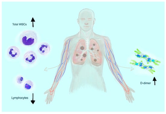
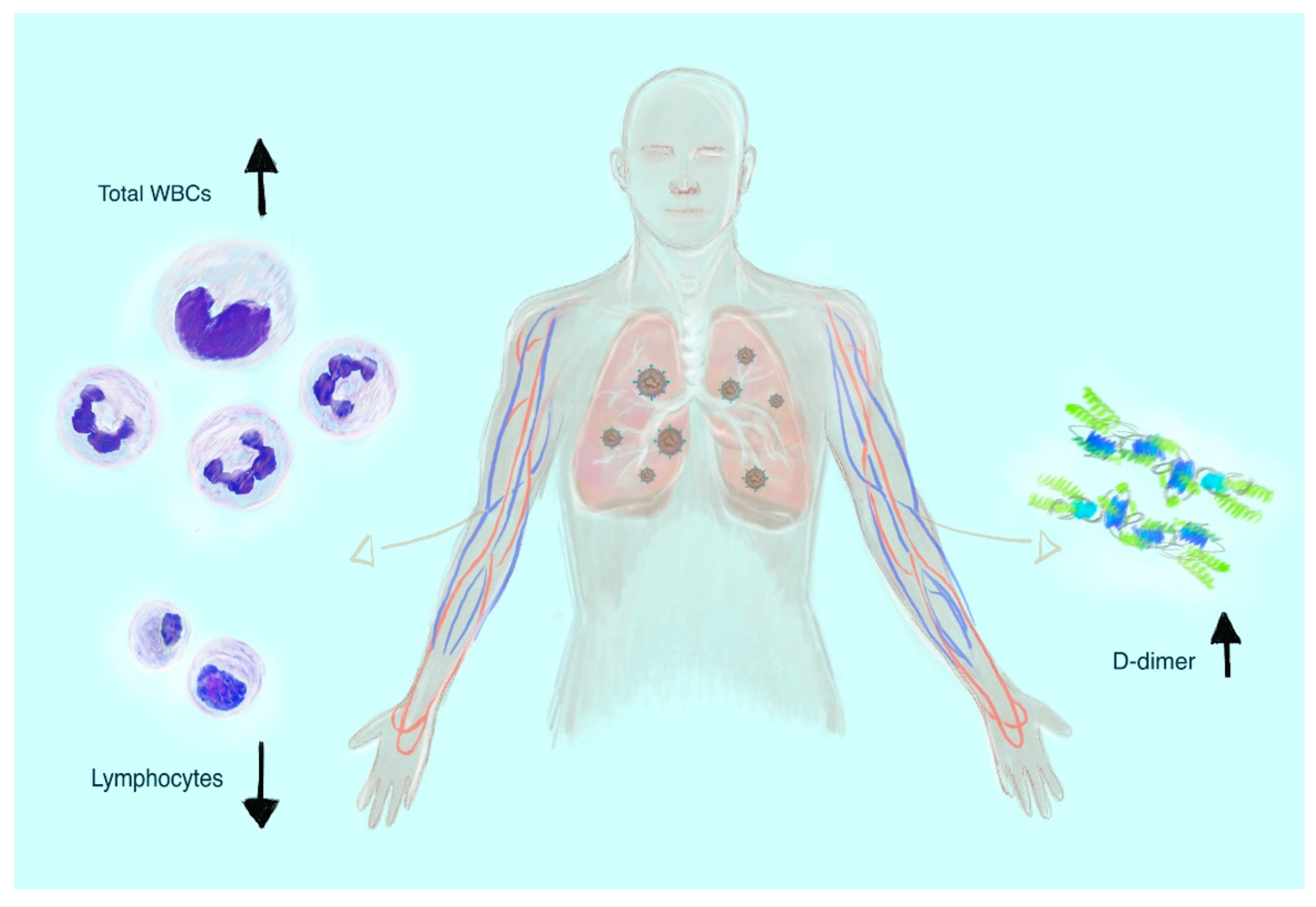
Coronavirus disease 2019 (COVID–19) is a rapidly evolving viral disorder and early stage diagnosis poses a great challenge to physicians. Insights drawn from studies to date provide compounding evidence regarding the significance of leukocytosis, lymphocytopenia and increased D–dimer levels in the prognosis facing severe acute respiratory syndrome coronavirus 2 (SARS–CoV–2) infected patients (Figure 2).
Figure 2.
The most common hematological laboratory symptoms of severe coronavirus disease 2019 (COVID–19) manifestation: leukocytosis, lymphocytopenia, and increased D–dimer levels.
Despite progress in the elucidation of COVID–19 disease mechanisms, many aspects remain unclear and continue to present an imposing challenge to the scientific community [135]. Taking a pathophysiological point of view, the mechanisms responsible for the infection and deterioration of the patient’s clinical condition are still poorly understood. This particularly concerns the immune response [136], including the role of cells such as monocytes/macrophages [137] and other cytokines such as interleukin 17A (IL–17A) [138]. The prevalence of thrombocytopenia [139] and the absence of laboratory signs of fibrinolysis in viscoelastic methods emphasize the importance of blood coagulation in the course of acute COVID–19. Furthermore, the long–term effects of infection on the hematological system are not evident yet. This will facilitate a deeper understanding of the role of the hematological system in the disease pathophysiology and progression. Taking a clinical point of view, development of effective treatment and vaccine regimens is highly dependent on a greater understanding of the pathomechanisms of COVID–19 and its associated complications. Due to the novelty of COVID–19 as a pandemic–level disease, many studies offer preliminary findings (often in pre–print form) and presently lack longitudinal and cross–sectional data, justifying the tentative interpretation of their conclusions [140].
Author Contributions
A.S. performed literature search, wrote the manuscript, prepared tables, and designed figures. M.K. and E.Ż. developed the concept of the manuscript, supervised, and critically read the manuscript. All the authors contributed to the review and revision of the manuscript, and all authors read and approved the final manuscript. All authors have read and agreed to the published version of the manuscript.
Acknowledgments
We are remarkably grateful to John Eikelboom (Hamilton, Canada) for comments that greatly improved the manuscript.
Conflicts of Interest
The authors declare that they have no competing interests.
References
- Available online: https://www.who.int/csr/don/12–january–2020–novel–coronavirus–china/en/ (accessed on 3 May 2020).
- Zhou, G.; Chen, S.; Chen, Z. Back to the Spring of 2020: Facts and Hope of COVID–19 Outbreak. Front. Med. 2020, 14, 113–116. [Google Scholar] [CrossRef] [Scilit] [PubMed]
- Whitworth, J. COVID–19: A Fast Evolving Pandemic. Trans. R. Soc. Trop. Med. Hyg. 2020, 114, 241–248. [Google Scholar] [CrossRef] [Scilit] [PubMed]
- Tu, H.; Tu, S.; Gao, S.; Shao, A.; Sheng, J. Current Epidemiological and Clinical Features of COVID–19; A Global Perspective from China. J. Infect. 2020, 81, 1–9. [Google Scholar] [CrossRef] [Scilit]
- Jiang, S.; Shi, Z.; Shu, Y.; Song, J.; Gao, G.F.; Tan, W.; Guo, D. A Distinct Name Is Needed for the New Coronavirus. Lancet 2020, 395, 949. [Google Scholar] [CrossRef] [Scilit]
- Coronaviridae Study Group of the International Committee on Taxonomy of Viruses. The Species Severe Acute Respiratory Syndrome–Related Coronavirus: Classifying 2019–nCoV and Naming It SARS–CoV–2. Nat. Microbiol. 2020, 5, 536–544. [Google Scholar] [CrossRef] [Scilit] [PubMed]
- Lu, R.; Zhao, X.; Li, J.; Niu, P.; Yang, B.; Wu, H.; Wang, W.; Song, H.; Huang, B.; Zhu, N.; et al. Genomic Characterisation and Epidemiology of 2019 Novel Coronavirus: Implications for Virus Origins and Receptor Binding. Lancet 2020, 395, 565–574. [Google Scholar] [CrossRef] [Scilit]
- Zhu, N.; Zhang, D.; Wang, W.; Li, X.; Yang, B.; Song, J.; Zhao, X.; Huang, B.; Shi, W.; Lu, R.; et al. A Novel Coronavirus from Patients with Pneumonia in China, 2019. N. Engl. J. Med. 2020, 382, 727–733. [Google Scholar] [CrossRef] [Scilit]
- Wu, F.; Zhao, S.; Yu, B.; Chen, Y.M.; Wang, W.; Song, Z.G.; Hu, Y.; Tao, Z.W.; Tian, J.H.; Pei, Y.Y.; et al. A New Coronavirus Associated with Human Respiratory Disease in China. Nature 2020, 579, 265–269. [Google Scholar] [CrossRef] [Scilit]
- Lei, J.; Hilgenfeld, R. RNA–virus Proteases Counteracting Host Innate Immunity. FEBS Lett. 2017, 591, 3190–3210. [Google Scholar] [CrossRef] [Scilit]
- Corman, V.M.; Muth, D.; Niemeyer, D.; Drosten, C. Hosts and Sources of Endemic Human Coronaviruses. Adv. Virus Res. 2018, 100, 163–188. [Google Scholar]
- Cui, J.; Li, F.; Shi, Z.L. Origin and Evolution of Pathogenic Coronaviruses. Nat. Rev. Microbiol. 2019, 17, 181–192. [Google Scholar] [CrossRef] [Scilit] [PubMed]
- Khan, S.; Siddique, R.; Shereen, M.A.; Ali, A.; Liu, J.; Bai, Q.; Bashir, N.; Xue, M. Emergence of a Novel Coronavirus, Severe Acute Respiratory Syndrome Coronavirus 2: Biology and Therapeutic Options. J. Clin. Microbiol. 2020, 58, e00187-20. [Google Scholar] [CrossRef] [Scilit] [PubMed]
- Drosten, C.; Günther, S.; Preiser, W.; van der Werf, S.; Brodt, H.R.; Becker, S.; Rabenau, H.; Panning, M.; Kolesnikova, L.; Fouchier, R.A.; et al. Identification of a Novel Coronavirus in Patients with Severe Acute Respiratory Syndrome. N. Engl. J. Med. 2003, 348, 1967–1976. [Google Scholar] [CrossRef] [Scilit] [PubMed]
- Zaki, A.M.; van Boheemen, S.; Bestebroer, T.M.; Osterhaus, A.D.; Fouchier, R.A. Isolation of a Novel Coronavirus from a Man with Pneumonia in Saudi Arabia. N. Engl. J. Med. 2012, 367, 1814–1820. [Google Scholar] [CrossRef] [Scilit]
- Chan, J.F.; Yuan, S.; Kok, K.H.; To, K.K.; Chu, H.; Yang, J.; Xing, F.; Liu, J.; Yip, C.C.; Poon, R.W.; et al. A Familial Cluster of Pneumonia Associated with the 2019 Novel Coronavirus Indicating Person–To–Person Transmission: A Study of a Family Cluster. Lancet 2020, 395, 514–523. [Google Scholar] [CrossRef] [Scilit]
- Zhou, P.; Yang, X.L.; Wang, X.G.; Hu, B.; Zhang, L.; Zhang, W.; Si, H.R.; Zhu, Y.; Li, B.; Huang, C.L.; et al. A Pneumonia Outbreak Associated with a New Coronavirus of Probable Bat Origin. Nature 2020, 579, 270–273. [Google Scholar] [CrossRef] [Scilit]
- Letko, M.; Marzi, A.; Munster, V. Functional Assessment of Cell Entry and Receptor Usage for SARS–CoV–2 and Other Lineage B Betacoronaviruses. Nat. Microbiol. 2020, 5, 562–569. [Google Scholar] [CrossRef] [Scilit]
- Wrapp, D.; Wang, N.; Corbett, K.S.; Goldsmith, J.A.; Hsieh, C.L.; Abiona, O.; Graham, B.S.; McLellan, J.S. Cryo–EM Structure of the 2019–nCoV Spike in the Prefusion Conformation. Science 2020, 367, 1260–1263. [Google Scholar] [CrossRef] [Scilit]
- Wan, Y.; Shang, J.; Graham, R.; Baric, R.S.; Li, F. Receptor Recognition by the Novel Coronavirus from Wuhan: An Analysis Based on Decade–Long Structural Studies of SARS Coronavirus. J. Virol. 2020, 94, e00127-20. [Google Scholar] [CrossRef] [Scilit]
- Walls, A.C.; Park, Y.J.; Tortorici, M.A.; Wall, A.; McGuire, A.T.; Veesler, D. Structure, Function, and Antigenicity of the SARS–CoV–2 Spike Glycoprotein. Cell 2020, 181, 281–292. [Google Scholar] [CrossRef] [Scilit]
- Hoffmann, M.; Kleine–Weber, H.; Schroeder, S.; Krüger, N.; Herrler, T.; Erichsen, S.; Schiergens, T.S.; Herrler, G.; Wu, N.H.; Nitsche, A.; et al. SARS–CoV–2 Cell Entry Depends on ACE2 and TMPRSS2 and Is Blocked by a Clinically Proven Protease Inhibitor. Cell 2020, 181, 271–280. [Google Scholar] [CrossRef] [Scilit] [PubMed]
- Wu, C.; Liu, Y.; Yang, Y.; Zhang, P.; Zhong, W.; Wang, Y.; Wang, Q.; Xu, Y.; Li, M.; Li, X.; et al. Analysis of Therapeutic Targets for SARS–CoV–2 and Discovery of Potential Drugs by Computational Methods. Acta Pharm. Sin. B 2020, 10, 766–788. [Google Scholar] [CrossRef] [Scilit] [PubMed]
- Lai, C.C.; Liu, Y.H.; Wang, C.Y.; Wang, Y.H.; Hsueh, S.C.; Yen, M.Y.; Ko, W.C.; Hsueh, P.R. Asymptomatic Carrier State, Acute Respiratory Disease, and Pneumonia Due to Severe Acute Respiratory Syndrome Coronavirus 2 (SARS–CoV–2): Facts and Myths. J. Microbiol. Immunol. Infect. 2020, 53, 404–412. [Google Scholar] [CrossRef] [Scilit]
- Bai, Y.; Yao, L.; Wei, T.; Tian, F.; Jin, D.Y.; Chen, L.; Wang, M. Presumed Asymptomatic Carrier Transmission of COVID–19. JAMA 2020, 323, 1406–1407. [Google Scholar] [CrossRef] [Scilit] [PubMed]
- Ye, F.; Xu, S.; Rong, Z.; Xu, R.; Liu, X.; Deng, P.; Liu, H.; Xu, X. Delivery of Infection from Asymptomatic Carriers of COVID–19 in a Familial Cluster. Int. J. Infect. Dis. 2020, 94, 133–138. [Google Scholar] [CrossRef] [Scilit] [PubMed]
- Verity, R.; Okell, L.C.; Dorigatti, I.; Winskill, P.; Whittaker, C.; Imai, N.; Cuomo–Dannenburg, G.; Thompson, H.; Walker, P.; Fu, H.; et al. Estimates of the Severity of Coronavirus Disease 2019: A Model–Based Analysis. Lancet Infect. Dis. 2020, 20, 669–677. [Google Scholar] [CrossRef] [Scilit]
- Yuki, K.; Fujiogi, M.; Koutsogiannaki, S. COVID–19 Pathophysiology: A Review. Clin. Immunol. 2020, 215, 108427. [Google Scholar] [CrossRef] [Scilit]
- Aguilar, R.B.; Hardigan, P.; Mayi, B.; Sider, D.; Piotrkowski, J.; Mehta, J.; Dev, J.; Seijo, Y.; Camargo, A.L.; Andux, L.; et al. Current Understanding of COVID–19 Clinical Course and Investigational Treatments. medRxiv 2020. [Google Scholar] [CrossRef] [Scilit]
- Chen, J.; Qi, T.; Liu, L.; Ling, Y.; Qian, Z.; Li, T.; Li, F.; Xu, Q.; Zhang, Y.; Xu, S.; et al. Clinical Progression of Patients with COVID–19 in Shanghai, China. J. Infect. 2020, 80, e1–e6. [Google Scholar] [CrossRef] [Scilit]
- Pan, F.; Ye, T.; Sun, P.; Gui, S.; Liang, B.; Li, L.; Zheng, D.; Wang, J.; Hesketh, R.L.; Yang, L.; et al. Time Course of Lung Changes at Chest CT During Recovery from Coronavirus Disease 2019 (COVID–19). Radiology 2020, 295, 715–721. [Google Scholar] [CrossRef] [Scilit]
- Huang, C.; Wang, Y.; Li, X.; Ren, L.; Zhao, J.; Hu, Y.; Zhang, L.; Fan, G.; Xu, J.; Gu, X.; et al. Clinical Features of Patients Infected with 2019 Novel Coronavirus in Wuhan, China. Lancet 2020, 395, 497–506. [Google Scholar] [CrossRef] [Scilit]
- Wang, D.; Hu, B.; Hu, C.; Zhu, F.; Liu, X.; Zhang, J.; Wang, B.; Xiang, H.; Cheng, Z.; Xiong, Y.; et al. Clinical Characteristics of 138 Hospitalized Patients with 2019 Novel Coronavirus–Infected Pneumonia in Wuhan, China. JAMA 2020, 323, 1061–1069. [Google Scholar] [CrossRef] [Scilit] [PubMed]
- Liu, K.; Fang, Y.Y.; Deng, Y.; Liu, W.; Wang, M.F.; Ma, J.P.; Xiao, W.; Wang, Y.N.; Zhong, M.H.; Li, C.H.; et al. Clinical Characteristics of Novel Coronavirus Cases in Tertiary Hospitals in Hubei Province. Chin. Med. J. (Engl.) 2020, 133, 1025–1031. [Google Scholar] [CrossRef] [Scilit]
- Guan, W.J.; Ni, Z.Y.; Hu, Y.; Liang, W.H.; Ou, C.Q.; He, J.X.; Liu, L.; Shan, H.; Lei, C.L.; Hui, D.S.C.; et al. Clinical Characteristics of Coronavirus Disease 2019 in China. N. Engl. J. Med. 2020, 382, 1708–1720. [Google Scholar] [CrossRef] [Scilit] [PubMed]
- Chang, D.; Lin, M.; Wei, L.; Xie, L.; Zhu, G.; Dela Cruz, C.S.; Sharma, L. Epidemiologic and Clinical Characteristics of Novel Coronavirus Infections Involving 13 Patients Outside Wuhan, China. JAMA 2020, 323, 1092–1093. [Google Scholar] [CrossRef] [Scilit] [PubMed]
- Xu, X.W.; Wu, X.X.; Jiang, X.G.; Xu, K.J.; Ying, L.J.; Ma, C.L.; Li, S.B.; Wang, H.Y.; Zhang, S.; Gao, H.N.; et al. Clinical Findings in a Group of Patients Infected with the 2019 Novel Coronavirus (SARS–Cov–2) Outside of Wuhan, China: Retrospective Case Series. BMJ 2020, 368, m606. [Google Scholar] [CrossRef]
- Zhang, J.J.; Dong, X.; Cao, Y.Y.; Yuan, Y.D.; Yang, Y.B.; Yan, Y.Q.; Akdis, C.A.; Gao, Y.D. Clinical Characteristics of 140 Patients Infected with SARS–CoV–2 in Wuhan, China. Allergy 2020. [Google Scholar] [CrossRef] [Scilit]
- Li, Q.; Ding, X.; Xia, G.; Geng, Z.; Chen, F.; Wang, L.; Wang, Z. A Simple Laboratory Parameter Facilitates Early Identification of COVID–19 Patients. medRxiv 2020. [Google Scholar] [CrossRef] [Scilit]
- Zini, G.; Bellesi, S.; Ramundo, F.; d’Onofrio, G. Morphological Anomalies of Circulating Blood Cells in COVID–19. Am. J. Hematol. 2020, 95, 870–872. [Google Scholar] [CrossRef] [Scilit]
- Mitra, A.; Dwyre, D.M.; Schivo, M.; Thompson, G.R., 3rd; Cohen, S.H.; Ku, N.; Graff, J.P. Leukoerythroblastic Reaction in a Patient with COVID–19 Infection. Am. J. Hematol. 2020. [Google Scholar] [CrossRef] [Scilit]
- Cantu, M.D.; Towne, W.S.; Emmons, F.N.; Mostyka, M.; Borczuk, A.; Salvatore, S.P.; Yang, H.S.; Zhao, Z.; Vasovic, L.V.; Racine–Brzostek, S.E. Clinical Significance of Blue–Green Neutrophil and Monocyte Cytoplasmic Inclusions in SARS–CoV–2 Positive Critically Ill Patients. Br. J. Haematol. 2020. [Google Scholar] [CrossRef] [Scilit] [PubMed]
- Singh, A.; Sood, N.; Narang, V.; Goyal, A. Morphology of COVID–19–affected Cells in Peripheral Blood Film. BMJ Case Rep. 2020, 13, e236117. [Google Scholar] [CrossRef] [Scilit] [PubMed]
- Chen, G.; Wu, D.; Guo, W.; Cao, Y.; Huang, D.; Wang, H.; Wang, T.; Zhang, X.; Chen, H.; Yu, H.; et al. Clinical and Immunological Features of Severe and Moderate Coronavirus Disease 2019. J. Clin. Investig. 2020, 130, 2620–2629. [Google Scholar] [CrossRef] [Scilit] [PubMed]
- Liu, J.; Li, S.; Liu, J.; Liang, B.; Wang, X.; Wang, H.; Li, W.; Tong, Q.; Yi, J.; Zhao, L.; et al. Longitudinal Characteristics of Lymphocyte Responses and Cytokine Profiles in the Peripheral Blood of SARS–CoV–2 Infected Patients. EBioMedicine 2020, 55, 102763. [Google Scholar] [CrossRef] [Scilit] [PubMed]
- Fan, B.E.; Chong, V.; Chan, S.; Lim, G.H.; Lim, K.; Tan, G.B.; Mucheli, S.S.; Kuperan, P.; Ong, K.H. Hematologic Parameters in Patients with COVID–19 Infection. Am. J. Hematol. 2020, 95, E131–E134. [Google Scholar] [CrossRef] [Scilit]
- Liu, J.; Liu, Y.; Xiang, P.; Pu, L.; Xiong, H.; Li, C.; Zhang, M.; Tan, J.; Xu, Y.; Song, R.; et al. Neutrophil–to–lymphocyte Ratio Predicts Critical Illness Patients with 2019 Coronavirus Disease in the Early Stage. J. Transl. Med. 2020, 18, 206. [Google Scholar] [CrossRef] [Scilit]
- Zhang, B.; Zhou, X.; Zhu, C.; Feng, F.; Qiu, Y.; Feng, J.; Joa, Q.; Song, Q.; Zhu, B.; Wang, J. Immune Phenotyping Based on Neutrophil–to–lymphocyte Ratio and IgG Predicts Disease Severity and Outcome for Patients with COVID–19. medRxiv 2020. [Google Scholar] [CrossRef] [Scilit]
- Yang, A.P.; Liu, J.P.; Tao, W.Q.; Li, H.M. The Diagnostic and Predictive Role of NLR, d–NLR and PLR in COVID–19 Patients. Int. Immunopharmacol. 2020, 84, 106504. [Google Scholar] [CrossRef] [Scilit]
- Lagunas–Rangel, F.A. Neutrophil–to–lymphocyte Ratio and lymphocyte–to–C–reactive Protein Ratio in Patients with Severe Coronavirus Disease 2019 (COVID–19): A Meta–Analysis. J. Med. Virol. 2020. [Google Scholar] [CrossRef] [Scilit]
- Ludvigsson, J.F. Systematic Review of COVID–19 in Children Shows Milder Cases and a Better Prognosis Than Adults. Acta Paediatr. 2020, 109, 1088–1095. [Google Scholar] [CrossRef] [Scilit]
- Tan, L.; Wang, Q.; Zhang, D.; Ding, J.; Huang, Q.; Tang, Y.Q.; Wang, Q.; Miao, H. Lymphopenia Predicts Disease Severity of COVID–19: A Descriptive and Predictive Study. Signal Transduct. Target. Ther. 2020, 5, 33. [Google Scholar] [CrossRef] [Scilit] [PubMed]
- Li, H.; Xiang, X.; Ren, H.; Xu, L.; Zhao, L.; Chen, X.; Long, H.; Wang, Q.; Wu, Q. Serum Amyloid A Is a Biomarker of Severe Coronavirus Disease and Poor Prognosis. J. Infect. 2020, 80, 646–655. [Google Scholar] [CrossRef] [Scilit] [PubMed]
- Li, T.; Qiu, Z.; Zhang, L.; Han, Y.; He, W.; Liu, Z.; Ma, X.; Fan, H.; Lu, W.; Xie, J.; et al. Significant Changes of Peripheral T Lymphocyte Subsets in Patients with Severe Acute Respiratory Syndrome. J. Infect. Dis. 2004, 189, 648–651. [Google Scholar] [CrossRef] [Scilit]
- Ko, J.H.; Park, G.E.; Lee, J.Y.; Lee, J.Y.; Cho, S.Y.; Ha, Y.E.; Kang, C.I.; Kang, J.M.; Kim, Y.J.; Huh, H.J.; et al. Predictive Factors for Pneumonia Development and Progression to Respiratory Failure in MERS–CoV Infected Patients. J. Infect. 2016, 73, 468–475. [Google Scholar] [CrossRef] [Scilit]
- Lin, L.; Lu, L.; Cao, W.; Li, T. Hypothesis for Potential Pathogenesis of SARS–CoV–2 Infection–A Review of Immune Changes in Patients with Viral Pneumonia. Emerg. Microbes. Infect. 2020, 9, 727–732. [Google Scholar] [CrossRef] [Scilit] [PubMed]
- Wang, X.; Xu, W.; Hu, G.; Xia, S.; Sun, Z.; Liu, Z.; Xie, Y.; Zhang, R.; Jiang, S.; Lu, L. SARS–CoV–2 Infects T Lymphocytes Through Its Spike Protein–Mediated Membrane Fusion. Cell. Mol. Immunol. 2020, 1–3. [Google Scholar] [CrossRef] [Scilit] [PubMed]
- Zheng, M.; Gao, Y.; Wang, G.; Song, G.; Liu, S.; Sun, D.; Xu, Y.; Tian, Z. Functional Exhaustion of Antiviral Lymphocytes in COVID–19 Patients. Cell. Mol. Immunol. 2020, 17, 533–535. [Google Scholar] [CrossRef] [Scilit]
- Yao, Z.; DuBois, D.C.; Almon, R.R.; Jusko, W.J. Pharmacokinetic/pharmacodynamic Modeling of Corticosterone Suppression and Lymphocytopenia by Methylprednisolone in Rats. J. Pharm. Sci. 2008, 97, 2820–2832. [Google Scholar] [CrossRef] [Scilit]
- Veronese, N.; Demurtas, J.; Yang, L.; Tonelli, R.; Barbagallo, M.; Lopalco, P.; Lagolio, E.; Celotto, S.; Pizzol, D.; Zou, L. Use of Corticosteroids in Coronavirus Disease 2019 Pneumonia: A Systematic Review of the Literature. Front. Med. (Lausanne) 2020, 7, 170. [Google Scholar] [CrossRef] [Scilit]
- Russell, C.D.; Millar, J.E.; Baillie, J.K. Clinical Evidence Does Not Support Corticosteroid Treatment for 2019–nCoV Lung Injury. Lancet 2020, 395, 473–475. [Google Scholar] [CrossRef] [Scilit]
- Tang, C.; Wang, Y.; Lv, H.; Guan, Z.; Gu, J. Caution Against Corticosteroid–Based COVID–19 Treatment. Lancet 2020, 395, 1759–1760. [Google Scholar] [CrossRef] [Scilit]
- Cai, Q.; Huang, D.; Ou, P.; Yu, H.; Zhu, Z.; Xia, Z.; Su, Y.; Ma, Z.; Zhang, Y.; Li, Z.; et al. COVID–19 in a Designated Infectious Diseases Hospital Outside Hubei Province, China. Allergy 2020. [Google Scholar] [CrossRef] [Scilit] [PubMed]
- Lippi, G.; Mattiuzzi, C. Hemoglobin Value May Be Decreased in Patients with Severe Coronavirus Disease 2019. Hematol. Transfus. Cell Ther. 2020, 42, 116–117. [Google Scholar] [CrossRef] [Scilit]
- Northrop–Clewes, C.A. Interpreting Indicators of Iron Status During an Acute Phase Response––Lessons from Malaria and Human Immunodeficiency Virus. Ann. Clin. Biochem. 2008, 45, 18–32. [Google Scholar] [CrossRef] [Scilit] [PubMed]
- Colafrancesco, S.; Alessandri, C.; Conti, F.; Priori, R. COVID–19 Gone Bad: A New Character in the Spectrum of the Hyperferritinemic Syndrome? Autoimmun. Rev. 2020, 19, 102573. [Google Scholar] [CrossRef] [Scilit] [PubMed]
- Zhang, H.; Wang, X.; Fu, Z.; Luo, M.; Zhang, Z.; Zhang, K.; He, Y.; Wan, D.; Zhang, L.; Wang, J.; et al. Potential Factors for Prediction of Disease Severity of COVID–19 Patients. medRxiv 2020. [Google Scholar] [CrossRef] [Scilit]
- Wang, Z.; Yang, B.; Li, Q.; Wen, L.; Zhang, R. Clinical Features of 69 Cases with Coronavirus Disease 2019 in Wuhan, China. Clin. Infect. Dis. 2020. [Google Scholar] [CrossRef] [Scilit]
- Lapić, I.; Rogić, D.; Plebani, M. Erythrocyte Sedimentation Rate Is Associated with Severe Coronavirus Disease 2019 (COVID–19): A Pooled Analysis. Clin. Chem. Lab. Med. 2020, 58, 1146–1148. [Google Scholar] [CrossRef] [Scilit]
- Zhao, J.; Yang, Y.; Huang, H.; Li, D.; Gu, D.; Lu, X.; Zhang, Z.; Liu, L.; Liu, T.; Liu, Y.; et al. Relationship between the ABO Blood Group and the COVID–19 Susceptibility. medRxiv 2020. [Google Scholar] [CrossRef] [Scilit]
- Zietz, M.; Tatonetti, N.P. Testing the Association between Blood Type and COVID–19 Infection, Intubation, and Death. medRxiv 2020. [Google Scholar] [CrossRef] [Scilit]
- Handtke, S.; Thiele, T. Large and Small platelets–(When) Do They Differ? J. Thromb. Haemost. 2020, 18, 1256–1267. [Google Scholar] [CrossRef] [Scilit] [PubMed]
- Lippi, G.; Plebani, M.; Henry, B.M. Thrombocytopenia Is Associated with Severe Coronavirus Disease 2019 (COVID–19) Infections: A Meta–Analysis. Clin. Chim. Acta 2020, 506, 145–148. [Google Scholar] [CrossRef] [Scilit]
- Thachil, J.; Cushman, M.; Srivastava, A. A Proposal for Staging COVID-19 Coagulopathy. Res. Pract. Thromb. Haemost. 2020. [Google Scholar] [CrossRef] [Scilit]
- Xu, P.; Zhou, Q.; Xu, J. Mechanism of Thrombocytopenia in COVID–19 Patients. Ann. Hematol. 2020, 99, 1205–1208. [Google Scholar] [CrossRef] [Scilit] [PubMed]
- Amgalan, A.; Othman, M. Hemostatic Laboratory Derangements in COVID–19 with a Focus on Platelet Count. Platelets 2020, 1–6. [Google Scholar] [CrossRef] [Scilit]
- Thachil, J. All Those D–dimers in COVID–19. J. Thromb. Haemost. 2020. [Google Scholar] [CrossRef] [Scilit]
- Lippi, G.; Favaloro, E.J. D–dimer Is Associated with Severity of Coronavirus Disease 2019: A Pooled Analysis. Thromb. Haemost. 2020, 120, 876–878. [Google Scholar] [CrossRef] [Scilit]
- Han, H.; Yang, L.; Liu, R.; Liu, F.; Wu, K.L.; Li, J.; Liu, X.H.; Zhu, C.L. Prominent Changes in Blood Coagulation of Patients with SARS–CoV–2 Infection. Clin. Chem. Lab. Med. 2020, 58, 1116–1120. [Google Scholar] [CrossRef] [Scilit]
- Tang, N.; Li, D.; Wang, X.; Sun, Z. Abnormal Coagulation Parameters Are Associated with Poor Prognosis in Patients with Novel Coronavirus Pneumonia. J. Thromb. Haemost. 2020, 18, 844–847. [Google Scholar] [CrossRef] [Scilit]
- Weitz, J.I.; Fredenburgh, J.C.; Eikelboom, J.W. A Test in Context: D–Dimer. J. Am. Coll. Cardiol. 2017, 70, 2411–2420. [Google Scholar] [CrossRef] [Scilit]
- Favresse, J.; Lippi, G.; Roy, P.M.; Chatelain, B.; Jacqmin, H.; Ten Cate, H.; Mullier, F. D–dimer: Preanalytical, Analytical, Postanalytical Variables, and Clinical Applications. Crit. Rev. Clin. Lab. Sci. 2018, 55, 548–577. [Google Scholar] [CrossRef] [Scilit]
- Goeijenbier, M.; van Wissen, M.; van de Weg, C.; Jong, E.; Gerdes, V.E.; Meijers, J.C.; Brandjes, D.P.; van Gorp, E.C. Review: Viral Infections and Mechanisms of Thrombosis and Bleeding. J. Med. Virol. 2012, 84, 1680–1696. [Google Scholar] [CrossRef] [Scilit]
- Escher, R.; Breakey, N.; Lämmle, B. Severe COVID–19 Infection Associated with Endothelial Activation. Thromb. Res. 2020, 190, 62. [Google Scholar] [CrossRef] [Scilit] [PubMed]
- Escher, R.; Breakey, N.; Lämmle, B. ADAMTS13 Activity, Von Willebrand Factor, Factor VIII and D–dimers in COVID–19 Inpatients. Thromb. Res. 2020, 192, 174–175. [Google Scholar] [CrossRef] [Scilit] [PubMed]
- Giardini, V.; Carrer, A.; Casati, M.; Contro, E.; Vergani, P.; Gambacorti–Passerini, C. Increased sFLT1/PlGF Ratio in COVID–19: A Novel Link to Angiotensin II–mediated Endothelial Dysfunction. Am. J. Hematol. 2020. [Google Scholar] [CrossRef] [Scilit]
- Varga, Z.; Flammer, A.J.; Steiger, P.; Haberecker, M.; Andermatt, R.; Zinkernagel, A.S.; Mehra, M.R.; Schuepbach, R.A.; Ruschitzka, F.; Moch, H. Endothelial Cell Infection and Endotheliitis in COVID–19. Lancet 2020, 395, 1417–1418. [Google Scholar] [CrossRef] [Scilit]
- Panigada, M.; Bottino, N.; Tagliabue, P.; Grasselli, G.; Novembrino, C.; Chantarangkul, V.; Pesenti, A.; Peyvandi, F.; Tripodi, A. Hypercoagulability of COVID–19 Patients in Intensive Care Unit. A Report of Thromboelastography Findings and Other Parameters of Hemostasis. J. Thromb. Haemost. 2020. [Google Scholar] [CrossRef] [Scilit]
- Maier, C.L.; Truong, A.D.; Auld, S.C.; Polly, D.M.; Tanksley, C.L.; Duncan, A. COVID–19–associated Hyperviscosity: A Link between Inflammation and Thrombophilia? Lancet 2020, 395, 1758–1759. [Google Scholar] [CrossRef] [Scilit]
- Spiezia, L.; Boscolo, A.; Poletto, F.; Cerruti, L.; Tiberio, I.; Campello, E.; Navalesi, P.; Simioni, P. COVID–19–Related Severe Hypercoagulability in Patients Admitted to Intensive Care Unit for Acute Respiratory Failure. Thromb. Haemost. 2020, 120, 998–1000. [Google Scholar]
- Pavoni, V.; Gianesello, L.; Pazzi, M.; Stera, C.; Meconi, T.; Frigieri, F.C. Evaluation of Coagulation Function by Rotation Thromboelastometry in Critically Ill Patients with Severe COVID–19 Pneumonia. J. Thromb. Thrombolysis 2020, 1–6. [Google Scholar] [CrossRef] [Scilit]
- Madathil, R.; Tabatabai, A.; Rabin, J.; Menne, A.R.; Henderson, R.; Mazzeffi, M.; Scalea, T.; Tanaka, K. Thromboelastometry and D–dimer Elevation in COVID–19. J Cardiothorac. Vasc. Anesth. 2020. [Google Scholar] [CrossRef] [Scilit]
- Fraissé, M.; Logre, E.; Pajot, O.; Mentec, H.; Plantefève, G.; Contou, D. Thrombotic and Hemorrhagic Events in Critically Ill COVID–19 Patients: A French Monocenter Retrospective Study. Crit. Care 2020, 24, 275. [Google Scholar] [CrossRef] [Scilit] [PubMed]
- Cui, S.; Chen, S.; Li, X.; Liu, S.; Wang, F. Prevalence of Venous Thromboembolism in Patients with Severe Novel Coronavirus Pneumonia. J. Thromb. Haemost. 2020, 18, 1421–1424. [Google Scholar] [CrossRef] [Scilit] [PubMed]
- Llitjos, J.F.; Leclerc, M.; Chochois, C.; Monsallier, J.M.; Ramakers, M.; Auvray, M.; Merouani, K. High Incidence of Venous Thromboembolic Events in Anticoagulated Severe COVID–19 Patients. J. Thromb. Haemost. 2020. [Google Scholar] [CrossRef] [Scilit]
- Helms, J.; Tacquard, C.; Severac, F.; Leonard–Lorant, I.; Ohana, M.; Delabranche, X.; Merdji, H.; Clere–Jehl, R.; Schenck, M.; Fagot Gandet, F.; et al. High Risk of Thrombosis in Patients with Severe SARS–CoV–2 Infection: A Multicenter Prospective Cohort Study. Intensive Care Med. 2020, 46, 1089–1098. [Google Scholar] [CrossRef] [Scilit]
- Klok, F.A.; Kruip, M.; van der Meer, N.; Arbous, M.S.; Gommers, D.; Kant, K.M.; Kaptein, F.; van Paassen, J.; Stals, M.; Huisman, M.V. Incidence of Thrombotic Complications in Critically Ill ICU Patients with COVID–19. Thromb. Res. 2020, 191, 145–147. [Google Scholar] [CrossRef] [Scilit]
- Nahum, J.; Morichau–Beauchant, T.; Daviaud, F.; Echegut, P.; Fichet, J.; Maillet, J.M.; Thierry, S. Venous Thrombosis Among Critically Ill Patients with Coronavirus Disease 2019 (COVID–19). JAMA Netw. Open 2020, 3, e2010478. [Google Scholar] [CrossRef] [Scilit]
- Khan, I.H.; Savarimuthu, S.; Leung, M.S.T.; Harky, A. The Need to Manage the Risk of Thromboembolism in COVID–19 Patients. J. Vasc. Surg. 2020. [Google Scholar] [CrossRef] [Scilit]
- Bikdeli, B.; Madhavan, M.V.; Gupta, A.; Jimenez, D.; Burton, J.R.; Der Nigoghossian, C.; Chuich, T.; Nouri, S.N.; Dreyfus, I.; Driggin, E.; et al. Pharmacological Agents Targeting Thromboinflammation in COVID–19: Review and Implications for Future Research. Thromb. Haemost. 2020. [Google Scholar] [CrossRef] [Scilit]
- Thachil, J.; Tang, N.; Gando, S.; Falanga, A.; Cattaneo, M.; Levi, M.; Clark, C.; Iba, T. ISTH Interim Guidance on Recognition and Management of Coagulopathy in COVID–19. J. Thromb. Haemost. 2020, 18, 1023–1026. [Google Scholar] [CrossRef] [Scilit]
- Bohn, M.K.; Lippi, G.; Horvath, A.; Sethi, S.; Koch, D.; Ferrari, M.; Wang, C.B.; Mancini, N.; Steele, S.; Adeli, K. Molecular, Serological, and Biochemical Diagnosis and Monitoring of COVID–19: IFCC Taskforce Evaluation of the Latest Evidence. Clin. Chem. Lab. Med. 2020, 58, 1037–1052. [Google Scholar] [CrossRef] [Scilit] [PubMed]
- Available online: https://www.escardio.org/Education/COVID–19–and–Cardiology/ESC–COVID–19–Guidance (accessed on 5 June 2020).
- Marietta, M.; Ageno, W.; Artoni, A.; De Candia, E.; Gresele, P.; Marchetti, M.; Marcucci, R.; Tripodi, A. COVID–19 and Haemostasis: A Position Paper from Italian Society on Thrombosis and Haemostasis (SISET). Blood Transfus. 2020, 18, 167–169. [Google Scholar] [PubMed]
- Flisiak, R.; Horban, A.; Jaroszewicz, J.; Kozielewicz, D.; Pawłowska, M.; Parczewski, M.; Piekarska, A.; Simon, K.; Tomasiewicz, K.; Zarębska–Michaluk, D. Management of SARS–CoV–2 Infection: Recommendations of the Polish Association of Epidemiologists and Infectiologists as of March 31, 2020. Pol. Arch. Int. Med. 2020, 130, 352–357. [Google Scholar]
- Lippi, G.; Plebani, M. The Critical Role of Laboratory Medicine During Coronavirus Disease 2019 (COVID–19) and Other Viral Outbreaks. Clin. Chem. Lab. Med. 2020, 58, 1063–1069. [Google Scholar] [CrossRef] [Scilit]
- Favaloro, E.J.; Lippi, G. Recommendations for Minimal Laboratory Testing Panels in Patients with COVID–19: Potential for Prognostic Monitoring. Semin. Thromb. Hemost. 2020, 46, 379–382. [Google Scholar] [CrossRef] [Scilit] [PubMed]
- Lippi, G.; Mattiuzzi, C.; Bovo, C.; Plebani, M. Current Laboratory Diagnostics of Coronavirus Disease 2019 (COVID–19). Acta Biomed. 2020, 91, 137–145. [Google Scholar]
- Galimberti, S.; Baldini, C.; Baratè, C.; Ricci, F.; Balducci, S.; Grassi, S.; Ferro, F.; Buda, G.; Benedetti, E.; Fazzi, R.; et al. The CoV–2 Outbreak: How Hematologists Could Help to Fight Covid–19. Pharmacol. Res. 2020, 157, 104866. [Google Scholar] [CrossRef] [Scilit]
- Jose, R.J.; Manuel, A. COVID–19 Cytokine Storm: The Interplay between Inflammation and Coagulation. Lancet Respir. Med. 2020, 8, e46–e47. [Google Scholar] [CrossRef] [Scilit]
- Kowalewski, M.; Fina, D.; Słomka, A.; Raffa, G.M.; Martucci, G.; Lo Coco, V.; De Piero, M.E.; Ranucci, M.; Suwalski, P.; Lorusso, R. COVID–19 and ECMO: The Interplay between Coagulation and Inflammation—A Narrative Review. Crit. Care 2020, 24, 205. [Google Scholar] [CrossRef] [Scilit]
- Coperchini, F.; Chiovato, L.; Croce, L.; Magri, F.; Rotondi, M. The Cytokine Storm in COVID–19: An Overview of the Involvement of the chemokine/chemokine–receptor System. Cytokine Growth Factor Rev. 2020, 53, 25–32. [Google Scholar] [CrossRef] [Scilit]
- Chau, V.Q.; Oliveros, E.; Mahmood, K.; Singhvi, A.; Lala, A.; Moss, N.; Gidwani, U.; Mancini, D.M.; Pinney, S.P.; Parikh, A. The Imperfect Cytokine Storm: Severe COVID–19 with ARDS in Patient on Durable LVAD Support. JACC Case Rep. 2020. [Google Scholar] [CrossRef] [Scilit] [PubMed]
- Chhetri, S.; Khamis, F.; Pandak, N.; Al Khalili, H.; Said, E.; Petersen, E. A Fatal Case of COVID–19 Due to Metabolic Acidosis Following Dysregulate Inflammatory Response (Cytokine Storm). IDCases 2020, 21, e00829. [Google Scholar] [CrossRef] [Scilit] [PubMed]
- Mo, P.; Xing, Y.; Xiao, Y.; Deng, L.; Zhao, Q.; Wang, H.; Xiong, Y.; Cheng, Z.; Gao, S.; Liang, K.; et al. Clinical Characteristics of Refractory COVID–19 Pneumonia in Wuhan, China. Clin. Infect. Dis. 2020. [Google Scholar] [CrossRef] [Scilit] [PubMed]
- Chen, T.; Wu, D.; Chen, H.; Yan, W.; Yang, D.; Chen, G.; Ma, K.; Xu, D.; Yu, H.; Wang, H.; et al. Clinical Characteristics of 113 Deceased Patients with Coronavirus Disease 2019: Retrospective Study. BMJ 2020, 368, m1091. [Google Scholar] [CrossRef] [Scilit]
- Ulhaq, Z.S.; Soraya, G.V. Interleukin–6 as a Potential Biomarker of COVID–19 Progression. Med. Mal. Infect. 2020, 50, 382–383. [Google Scholar] [CrossRef] [Scilit]
- Aziz, M.; Fatima, R.; Assaly, R. Elevated interleukin–6 and Severe COVID–19: A Meta–Analysis. J. Med. Virol. 2020. [Google Scholar] [CrossRef] [Scilit]
- Feldmann, M.; Maini, R.N.; Woody, J.N.; Holgate, S.T.; Winter, G.; Rowland, M.; Richards, D.; Hussell, T. Trials of Anti–Tumour Necrosis Factor Therapy for COVID–19 Are Urgently Needed. Lancet 2020, 395, 1407–1409. [Google Scholar] [CrossRef] [Scilit]
- Xu, X.; Han, M.; Li, T.; Sun, W.; Wang, D.; Fu, B.; Zhou, Y.; Zheng, X.; Yang, Y.; Li, X.; et al. Effective Treatment of Severe COVID–19 Patients with Tocilizumab. Proc. Natl. Acad. Sci. USA 2020, 117, 10970–10975. [Google Scholar] [CrossRef] [Scilit]
- Luo, P.; Liu, Y.; Qiu, L.; Liu, X.; Liu, D.; Li, J. Tocilizumab Treatment in COVID–19: A Single Center Experience. J. Med. Virol. 2020, 92, 814–818. [Google Scholar] [CrossRef] [Scilit]
- Day, J.W.; Fox, T.A.; Halsey, R.; Carpenter, B.; Kottaridis, P.D. Interleukin–1 Blockade with Anakinra in Acute Leukaemia Patients with Severe COVID–19 Pneumonia Appears Safe and May Result in Clinical Improvement. Br. J. Haematol. 2020. [Google Scholar] [CrossRef] [Scilit]
- Cavalli, G.; De Luca, G.; Campochiaro, C.; Della–Torre, E.; Ripa, M.; Canetti, D.; Oltolini, C.; Castiglioni, B.; Tassan Din, C.; Boffini, N.; et al. Interleukin–1 Blockade with High–Dose Anakinra in Patients with COVID–19, Acute Respiratory Distress Syndrome, and Hyperinflammation: A Retrospective Cohort Study. Lancet Rheumatol. 2020, 2, e325–e331. [Google Scholar] [CrossRef] [Scilit]
- Iba, T.; Levy, J.H.; Levi, M.; Connors, J.M.; Thachil, J. Coagulopathy of Coronavirus Disease 2019. Crit. Care Med. 2020. [Google Scholar] [CrossRef] [Scilit] [PubMed]
- van der Poll, T.; Herwald, H. The Coagulation System and Its Function in Early Immune Defense. Thromb. Haemost. 2014, 112, 640–648. [Google Scholar] [CrossRef] [Scilit] [PubMed]
- Foley, J.H.; Conway, E.M. Cross Talk Pathways between Coagulation and Inflammation. Circ. Res. 2016, 118, 1392–1408. [Google Scholar] [CrossRef] [Scilit] [PubMed]
- Lordan, R.; Tsoupras, A.; Zabetakis, I. Platelet Activation and Prothrombotic Mediators at the Nexus of Inflammation and Atherosclerosis: Potential Role of Antiplatelet Agents. Blood Rev. 2020, 100694. [Google Scholar] [CrossRef] [Scilit] [PubMed]
- de Lusignan, S.; Dorward, J.; Correa, A.; Jones, N.; Akinyemi, O.; Amirthalingam, G.; Andrews, N.; Byford, R.; Dabrera, G.; Elliot, A.; et al. Risk Factors for SARS–CoV–2 Among Patients in the Oxford Royal College of General Practitioners Research and Surveillance Centre Primary Care Network: A Cross–Sectional Study. Lancet Infect. Dis. 2020. [Google Scholar] [CrossRef] [Scilit]
- Jin, J.M.; Bai, P.; He, W.; Wu, F.; Liu, X.F.; Han, D.M.; Liu, S.; Yang, J.K. Gender Differences in Patients with COVID–19: Focus on Severity and Mortality. Front. Public Health 2020, 8, 152. [Google Scholar] [CrossRef] [Scilit]
- Serge, R.; Vandromme, J.; Charlotte, M. Are We Equal in Adversity? Does Covid–19 Affect Women and Men Differently? Maturitas 2020. [Google Scholar] [CrossRef] [Scilit]
- Dangis, A.; De Brucker, N.; Heremans, A.; Gillis, M.; Frans, J.; Demeyere, A.; Symons, R. Impact of Gender on Extent of Lung Injury in COVID–19. Clin. Radiol. 2020, 75, 554–556. [Google Scholar] [CrossRef] [Scilit]
- Sharma, G.; Volgman, A.S.; Michos, E.D. Sex Differences in Mortality from COVID–19 Pandemic: Are Men Vulnerable and Women Protected? JACC Case Rep. 2020. [Google Scholar] [CrossRef] [Scilit]
- Swärd, P.; Edsfeldt, A.; Reepalu, A.; Jehpsson, L.; Rosengren, B.E.; Karlsson, M.K. Age and Sex Differences in Soluble ACE2 May Give Insights for COVID–19. Crit. Care 2020, 24, 221. [Google Scholar] [CrossRef] [Scilit]
- Harrison, M. Erythrocyte Sedimentation Rate and C–reactive Protein. Aust. Prescr. 2015, 38, 93–94. [Google Scholar] [CrossRef] [Scilit] [PubMed]
- Welsby, P.D. COVID–19: Some Unanswered Questions. Postgrad. Med J. 2020. [Google Scholar] [CrossRef] [Scilit] [PubMed]
- Maggi, E.; Canonica, G.W.; Moretta, L. COVID–19: Unanswered Questions on Immune Response and Pathogenesis. J. Allergy Clin. Immunol. 2020. [Google Scholar] [CrossRef] [Scilit] [PubMed]
- Merad, M.; Martin, J.C. Pathological Inflammation in Patients with COVID–19: A Key Role for Monocytes and Macrophages. Nat. Rev. Immunol. 2020, 20, 355–362. [Google Scholar] [CrossRef] [Scilit] [PubMed]
- Raucci, F.; Mansour, A.A.; Casillo, G.M.; Saviano, A.; Caso, F.; Scarpa, R.; Mascolo, N.; Iqbal, A.J.; Maione, F. Interleukin–17A (IL–17A), a Key Molecule of Innate and Adaptive Immunity, and Its Potential Involvement in COVID–19–related Thrombotic and Vascular Mechanisms. Autoimmun. Rev. 2020, 19, 102572. [Google Scholar] [CrossRef] [Scilit] [PubMed]
- Amgalan, A.; Othman, M. Exploring Possible Mechanisms for COVID–19 Induced Thrombocytopenia: Unanswered Questions. J. Thromb. Haemost. 2020, 18, 1514–1516. [Google Scholar] [CrossRef] [Scilit]
- Favaloro, E.J.; Thachil, J. Reporting of D–dimer Data in COVID–19: Some Confusion and Potential for Misinformation. Clin. Chem. Lab. Med. 2020. [Google Scholar] [CrossRef] [Scilit]

© 2020 by the authors. Licensee MDPI, Basel, Switzerland. This article is an open access article distributed under the terms and conditions of the Creative Commons Attribution (CC BY) license (http://creativecommons.org/licenses/by/4.0/).