Prevention and Treatment of Staphylococcus aureus Biofilms Using Promising Agr-QS-Targeting Anti-Biofilm Agents

 and
and
Abstract
1. Introduction
2. Prevalence of Staphylococcus aureus

| Infection | Antibiotic | Clinical Implications | Major Therapeutic Limitations | Emerging Resistance | References |
|---|---|---|---|---|---|
| Endocarditis | Linezolid | To treat MRSA-like cases when IV access or oral step-down is needed | Myelosuppression, especially thrombocytopenia, dose- and duration-dependent toxicity | Linezolid resistance | [24,25,26,27] |
| Severe community- acquired pneumonia | Daptomycin | To treat MRSA, but generally not recommended for pneumonia | Inactivated by pulmonary surfactant | Increased daptomycin resistance | [26,28,29] |
| Bacteremia and vertebral osteomyelitis | To treat MRSA bacteremia and deep infections | Increased CPK levels, myopathy risk, need for higher dosing in severe infection | In vivo-acquired resistance and treatment failures in MRSA bacteremia | [30,31,32] | |
| Sepsis and septic shock | Vancomycin | Standard therapy for severe MRSA infection | Nephrotoxicity; infusion reactions (red man syndrome) | Increasing vancomycin-intermediate S. aureus (VISA) prevalence and rare VRSA. Recent sepsis guidelines highlighted reduced efficacy with elevated MICs | [33,34,35] |
| Chronic skin ulcers | Used for susceptible isolates but often limited in biofilm-rich chronic disease | Toxicity limits prolonged courses, poor penetration, and tolerance in biofilms | Increasing prevalence of VISA/VRSA along with reduced vancomycin susceptibility | [35,36,37] | |
| Skin and soft tissue infections | Clindamycin | Commonly used for susceptible MSSA/MRSA SSTIs | Gastrointestinal toxicity kills beneficial bacteria and allows harmful C. difficile to grow | Substantial inducible clindamycin resistance in MRSA and MSSA | [38,39,40] |
| Mixed (samples taken from hospital) | Quinupristin– dalfopristin | Used for highly resistant Gram-positive infections only | Infusion-related pain, arthralgia/myalgia, limited routine use | Plasmid-mediated resistance in S. aureus and other staphylococci | [26,41,42] |
| Mixed (samples taken from hospital) | Trimethoprim–sulfamethoxazole (TMP-SMX) | Useful for selected MRSA SSTIs and some invasive infections | Hypersensitivity; hematologic toxicity | TMP–SMX resistance in MRSA and MSSA | [42,43] |
| Mixed (samples taken from hospital) | Tigecycline | Alternative for complicated skin/soft tissue and intra-abdominal infections | Nausea/vomiting, low serum levels unsuitable for bacteremia | Emergence of tigecycline resistance in S. aureus and other Gram-positive pathogens | [26] |
3. Virulence Potential of Staphylococcus aureus
4. Biofilm Formation by Staphylococcus aureus
Biofilm Development—Attachment, Proliferation, and Detachment
5. Biofilm Development and Its Relation to Quorum Sensing
5.1. Role of Agr System in Biofilm Dispersal
| Agr Variant | Characteristic Features | Biofilm Regulation | Reference |
|---|---|---|---|
| agrI | Predominantly reported in CA-MRSA and methicillin-resistant bovine isolates | Regulates a broad set of toxins, proteases, and adhesins; agrI dysfunction is linked to prolonged bacteremia and altered biofilm behavior. | [75,76] |
| High prevalence of resistance towards beta-lactamase, glycopeptides, fluoroquinolones, aminoglycosides, tetracyclines, macrolides, lincomycins, and sulphonamides | [77] | ||
| agrII | Predominantly causes nosocomial MRSA and MRSA bloodstream infections | agrII activity influences biofilm maturation and dispersal. It is associated with distinct toxin/adhesin profiles. | [54,75] |
| Toxic shock syndrome | |||
| Prolific biofilm producers | [78] | ||
| agrIII | Predominantly causes CA-MRSA | Linked with toxin-mediated virulence, agrIII mutants show altered biofilm and persistence phenotypes. | [75,79] |
| Potent biofilm producers | [80] | ||
| agrIV | Predominantly reported in swine farm isolates and generalized exfoliative syndromes | Regulates virulence and biofilm, but detailed functional data are limited. | [54,77] |
| High prevalence of resistance to fluoroquinolones, aminoglycosides, tetracyclines, macrolides, lincomycins, and sulphonamides | [77] | ||
| High prevalence of enterotoxin genes | [54] |
5.2. Alternative Anti-Biofilm Strategies
5.3. Green Alternatives as Potential Medicinal Therapeutic Agents
5.4. Camellia sinensis
5.5. Moringa oleifera
5.6. Rosmarinus officinalis
5.7. Psidium guajava
5.8. Eucalyptus
5.9. Azadirachta indica
5.10. Curcuma longa
5.11. Sanguisorba officinalis
5.12. Others
5.13. Translational Challenges in the Clinical Development of Natural Anti-Biofilm Agents
6. Conclusions and Future Prospects
Author Contributions
Funding
Institutional Review Board Statement
Informed Consent Statement
Data Availability Statement
Conflicts of Interest
Abbreviations
| MRSA | Methicillin-resistant S. aureus |
| MSSA | Methicillin-susceptible S. aureus |
| CPK | Creatine phosphokinase |
| PVL | Panton–Valentine leucocidin |
| EVD | External ventricular drains |
| MSCRAMMs | Microbial surface components recognizing adhesive matrix molecules |
| TSST-1 | Toxic shock syndrome toxin 1 |
| ETs | Exfoliative toxins |
| MTSS | Menstrual toxic shock syndrome |
| NMTSS | Non-menstrual TSS |
| SSSS | Staphylococcal scalded skin syndrome |
| SFD | Staphylococcal food-borne diseases |
| EPSs | Exopolysaccharides |
| PIA | Polysaccharide intercellular adhesion |
| QS | Quorum sensing |
| AIPs | Auto-inducing peptides |
| PSMs | Phenol soluble modulins |
| ORFs | Open reading frames |
| LA-MRSA | Livestock-associated methicillin-resistant S. aureus |
| CA-MRSA | Community-associated methicillin-resistant S. aureus |
| VRSA | Vancomycin-resistant S. aureus |
| CAGR | Compound annual growth rate |
| EDTA | Ethylenediamine tetraacetic acid |
| EGTA | Ethylene glycol tetra acetic acid |
| TSC | Tri-sodium citrate |
| DTT | Dithiothreitol |
| EC | Epicatechin |
| EGC | Epigallocatechin |
| ECG | Epicatechin gallate |
| EGCG | Epigallocatechin gallate |
| PVC | Polyvinyl Chloride plastic |
| GC-MS | Gas chromatography–mass spectrometry |
| MIC | Minimum inhibitory concentration |
| NMR | Nuclear magnetic resonance |
| UPLC | Ultra-performance liquid chromatography |
| BIC | Benzyl isocyanate |
| LC-FTMS | Liquid chromatography Fourier transform mass spectrometry |
| SEM | Scanning electron microscopy |
References
- Li, J.; Liu, D.; Tian, X.; Koseki, S.; Chen, S.; Ye, X.; Ding, T. Novel antibacterial modalities against methicillin resistant Staphylococcus aureus derived from plants. Crit. Rev. Food Sci. Nutr. 2019, 59, S153–S161. [Google Scholar] [CrossRef] [Scilit] [PubMed]
- Sollid, J.U.E.; Furberg, A.; Hanssen, A.; Johannessen, M. Staphylococcus aureus: Determinants of human carriage. Infect. Genet. Evol. 2014, 21, 531–541. [Google Scholar] [CrossRef] [Scilit] [PubMed]
- Hindieh, P.; Yaghi, J.; Assaf, J.C.; Chokr, A.; Atoui, A.; Tzenios, N.; Louka, N.; Khoury, A.E. Emerging Multimodal Strategies for Bacterial Biofilm Eradication: A Comprehensive Review. Microorganisms 2025, 13, 2796. [Google Scholar] [CrossRef] [Scilit] [PubMed]
- Kiedrowski, M.R.; Horswill, A.R. New approaches for treating staphylococcal biofilm infections. Ann. N. Y. Acad. Sci. 2011, 1241, 104–121. [Google Scholar] [CrossRef] [Scilit] [PubMed]
- Høiby, N.; Ciofu, O.; Johansen, H.K.; Song, Z.j.; Moser, C.; Jensen, P.Ø.; Molin, S.; Givskov, M.; Tolker-Nielsen, T.; Bjarnsholt, T. The clinical impact of bacterial biofilms. Int. J. Oral Sci. 2011, 3, 55–65. [Google Scholar] [CrossRef] [Scilit] [PubMed]
- Jamal, M.; Ahmad, W.; Andleeb, S.; Jalil, F.; Imran, M.; Nawaz, M.A.; Hussain, T.; Ali, M.; Rafiq, M.; Kamil, M.A. Bacterial biofilm and associated infections. J. Chin. Med. Assoc. 2018, 81, 7–11. [Google Scholar] [CrossRef] [Scilit] [PubMed]
- Tuchscherr, L.; Löffler, B.; Buzzola, F.R.; Sordelli, D.O. Staphylococcus aureus adaptation to the host and persistence: Role of loss of capsular polysaccharide expression. Future Med. 2010, 5, 1823–1832. [Google Scholar] [CrossRef] [Scilit] [PubMed]
- Zaatout, N.; Ayachi, A.; Kecha, M. Interaction of primary mammary bovine epithelial cells with biofilm-forming staphylococci associated with subclinical bovine mastitis. Iran. J. Vet. Res. 2019, 20, 27–32. [Google Scholar] [PubMed]
- Pietrocola, G.; Nobile, G.; Rindi, S.; Speziale, P. Staphylococcus aureus manipulates innate immunity through own and host-expressed proteases. Front. Cell. Infect. Microbiol. 2017, 7, 166. [Google Scholar] [CrossRef] [Scilit] [PubMed]
- Parvizi, J.; Pawasarat, I.M.; Azzam, K.A.; Joshi, A.; Hansen, E.N.; Bozic, K.J. Periprosthetic joint infection: The economic impact of methicillin-resistant infections. J. Arthroplast. 2010, 25, 103–107. [Google Scholar] [CrossRef] [Scilit] [PubMed]
- Song, X.; Perencevich, E.; Campos, J.; Short, B.L.; Singh, N. Clinical and economic impact of methicillin-resistant Staphylococcus aureus colonization or infection on neonates in intensive care units. Infect. Control Hosp. Epidemiol. 2010, 31, 177–182. [Google Scholar] [CrossRef] [Scilit] [PubMed]
- Pesavento, G.; Maggini, V.; Maida, I.; Nostro, A.L.; Calonico, C.; Sassoli, C.; Perrin, E.; Fondi, M.; Mengoni, A.; Chiellini, C.; et al. Essential oil from Origanum vulgare completely inhibits the growth of multidrug-resistant cystic fibrosis pathogens. Nat. Prod. Commun. 2016, 11, 1934578X1601100641. [Google Scholar] [CrossRef] [Scilit]
- Solórzano-Santos, F.; Miranda-Novales, M.G. Essential oils from aromatic herbs as antimicrobial agents. Curr. Opin. Biotechnol. 2012, 23, 136–141. [Google Scholar] [CrossRef] [Scilit] [PubMed]
- Tuffs, S.W.; Haeryfar, S.M.M.; McCormick, J.K. Manipulation of innate and adaptive immunity by Staphylococcal superantigens. Pathogens 2018, 7, 53. [Google Scholar] [CrossRef] [Scilit] [PubMed]
- Kadariya, J.; Smith, T.C.; Thapaliya, D. Staphylococcus aureus and staphylococcal food-borne disease: An ongoing challenge in public health. BioMed Res. Int. 2014, 2014, 827965. [Google Scholar] [CrossRef] [Scilit] [PubMed]
- Hennekinne, J.-A.; De Buyser, M.-L.; Dragacci, S. Staphylococcus aureus and its food poisoning toxins: Characterization and outbreak investigation. FEMS Microbiol. Rev. 2012, 36, 815–836. [Google Scholar] [CrossRef] [Scilit] [PubMed]
- Rudd, K.E.; Johnson, S.C.; Agesa, K.M.; Shackelford, K.A.; Tsoi, D.; Kievlan, D.R.; Colombara, D.V.; Ikuta, K.S.; Kissoon, N.; Finfer, S.; et al. Global, regional, and national sepsis incidence and mortality, 1990–2017: Analysis for the Global Burden of Disease Study. Lancet 2020, 395, 200–211. [Google Scholar] [CrossRef] [Scilit] [PubMed]
- WHO. Sepsis Fact Sheet 2024. Available online: https://www.who.int/news-room/fact-sheets/detail/sepsis (accessed on 12 July 2026).
- Tobin, E.H.; Jogu, P.; Koirala, J. Methicillin-Resistant Staphylococcus aureus. Available online: https://www.ncbi.nlm.nih.gov/books/NBK482221/ (accessed on 11 January 2026).
- Shoaib, M.; Aqib, A.I.; Muzammil, I.; Majeed, N.; Bhutta, Z.A.; Kulyar, M.F.-e.-A.; Fatima, M.; Zaheer, C.-N.F.; Muneer, A.; Murtaza, M.; et al. MRSA compendium of epidemiology, transmission, pathophysiology, treatment, and prevention within one health framework. Front. Microbiol. 2023, 13, 1067284. [Google Scholar] [CrossRef] [Scilit] [PubMed]
- Liu, C.; Bayer, A.; Cosgrove, S.E.; Daum, R.S.; Fridkin, S.K.; Gorwitz, R.J.; Kaplan, S.L.; Karchmer, A.W.; Levine, D.P.; Murray, B.E.; et al. Clinical Practice Guidelines by the Infectious Diseases Society of America for the Treatment of Methicillin-Resistant Staphylococcus aureus Infections in Adults and Children. Clin. Infect. Dis. 2011, 52, e18–e55. [Google Scholar] [CrossRef] [Scilit] [PubMed]
- Britt, N.S.; Tirmizi, S.; Ritchie, D.J.; Topal, J.E.; McManus, D.; Nizet, V.; Casabar, E.; Sakoulas, G. Telavancin for refractory MRSA bacteraemia in intermittent haemodialysis recipients. J. Antimicrob. Chemother. 2018, 73, 764–767. [Google Scholar] [CrossRef] [Scilit] [PubMed]
- Vanderhaeghen, W.; Cerpentier, T.; Adriaensen, C.; Vicca, J.; Hermans, K.; Butaye, P. Methicillin-resistant Staphylococcus aureus (MRSA) ST398 associated with clinical and subclinical mastitis in Belgian cows. Vet. Microbiol. 2010, 144, 166–171. [Google Scholar] [CrossRef] [Scilit] [PubMed]
- Ha, K.N.Y.; Vu, T.T.T.; Nguyen, T.T.T.; Nguyen, H.Q.N.; Dang, T.N.D. A retrospective study of the risk factors for linezolid-induced thrombocytopenia in hospitalized patients. MedPharmRes 2025, 9, 207–219. [Google Scholar] [CrossRef] [Scilit]
- Muñoz, P.; De la Villa, S.; Martínez-Sellés, M.; Goenaga, M.A.; Reviejo-Jaka, K.; Revillas, F.A.L.; García-Cuello, L.; Hidalgo-Tenorio, C.; Rodríguez-Esteban, M.A.; Antorrena, I.; et al. Linezolid for infective endocarditis: A structured approach based on a national database experience. Medicine 2021, 100, e27597. [Google Scholar] [CrossRef] [Scilit] [PubMed]
- Shariati, A.; Dadashi, M.; Chegini, Z.; van Belkum, A.; Mirzaii, M.; Khoramrooz, S.S.; Darban-Sarokhalil, D. The global prevalence of Daptomycin, Tigecycline, Quinupristin/Dalfopristin, and Linezolid-resistant Staphylococcus aureus and coagulase-negative staphylococci strains: A systematic review and meta-analysis. Antimicrob. Resist. Infect. Control 2020, 9, 56. [Google Scholar] [CrossRef] [Scilit] [PubMed]
- Kobayashi, T.; Ando, T.; Streit, J.; Sekar, P. Current Evidence on Oral Antibiotics for Infective Endocarditis: A Narrative Review. Cardiol. Ther. 2019, 8, 167–177. [Google Scholar] [CrossRef] [Scilit] [PubMed]
- Skiest, D.J. Treatment failure resulting from resistance of Staphylococcus aureus to daptomycin. J. Clin. Microbiol. 2006, 44, 655–656. [Google Scholar] [CrossRef] [Scilit] [PubMed]
- Fiore, M.; Alfieri, A.; Fiore, D.; Iuliano, P.; Spatola, F.G.; Limone, A.; Pezone, I.; Leone, S. Use of Daptomycin to Manage Severe MRSA Infections in Humans. Medicine 2025, 14, 617. [Google Scholar] [CrossRef] [Scilit] [PubMed]
- Bhavnani, S.M.; Rubino, C.M.; Ambrose, P.G.; Drusano, G.L. Daptomycin exposure and the probability of elevations in the creatine phosphokinase level: Data from a randomized trial of patients with bacteremia and endocarditis. Clin. Infect. Dis. Off. Publ. Infect. Dis. Soc. Am. 2010, 50, 1568–1574. [Google Scholar] [CrossRef] [Scilit] [PubMed]
- Tally, F.P.; DeBruin, M.F. Development of daptomycin for gram-positive infections. J. Antimicrob. Chemother. 2000, 46, 523–526. [Google Scholar] [CrossRef] [Scilit] [PubMed]
- Marty, F.M.; Yeh, W.W.; Wennersten, C.B.; Venkataraman, L.; Albano, E.; Alyea, E.P.; Gold, H.S.; Baden, L.R.; Pillai, S.K. Emergence of a clinical daptomycin-resistant Staphylococcus aureus isolate during treatment of methicillin-resistant Staphylococcus aureus bacteremia and osteomyelitis. J. Clin. Microbiol. 2006, 44, 595–597. [Google Scholar] [CrossRef] [Scilit] [PubMed]
- Zamoner, W.; Prado, I.R.S.; Balbi, A.L.; Ponce, D. Vancomycin dosing, monitoring and toxicity: Critical review of the clinical practice. Clin. Exp. Pharmacol. Physiol. 2019, 46, 292–301. [Google Scholar] [CrossRef] [Scilit] [PubMed]
- Niforatos, J.D.; Hinson, J.S.; Rothman, R.E.; Cosgrove, S.E.; Dzintars, K.; Klein, E.Y. Methicillin-resistant Staphylococcus aureus and Vancomycin Prescribing in the Emergency Department: A Single-center Study Assessing Antibiotic Prescribing. J. Am. Coll. Emerg. Phys. Open 2025, 6, 100021. [Google Scholar] [CrossRef] [Scilit] [PubMed]
- Keikha, M.; Karbalaei, M. Global distribution of heterogeneous vancomycin-intermediate Staphylococcus aureus strains (1997–2021): A systematic review and meta-analysis. J. Glob. Antimicrob. Resist. 2024, 37, 11–21. [Google Scholar] [CrossRef] [Scilit] [PubMed]
- Sievert, D.M.; Rudrik, J.T.; Patel, J.B.; McDonald, L.C.; Wilkins, M.J.; Hageman, J.C. Vancomycin-resistant Staphylococcus aureus in the United States, 2002–2006. Clin. Infect. Dis. Off. Publ. Infect. Dis. Soc. Am. 2008, 46, 668–674. [Google Scholar] [CrossRef] [Scilit] [PubMed]
- Zhang, S.; Sun, X.; Chang, W.; Dai, Y.; Ma, X. Systematic review and meta-analysis of the epidemiology of vancomycin-intermediate and heterogeneous vancomycin-intermediate Staphylococcus aureus isolates. PLoS ONE 2015, 10, e0136082. [Google Scholar] [CrossRef] [Scilit] [PubMed]
- Deotale, V.; Mendiratta, D.K.; Raut, U.; Narang, P. Inducible clindamycin resistance in Staphylococcus aureus isolated from clinical samples. Indian J. Med. Microbiol. 2010, 28, 124–126. [Google Scholar] [CrossRef] [Scilit] [PubMed]
- Fomda, B.A.; Peer, M.A.; Zahoor, D.; Thokar, M.A.; Nasir, R.A. Phenotypic detection of constitutive and inducible clindamycin resistance in clinical isolates of Staphylococcus aureus and coagulase negative Staphylococcus on routine susceptibility plate. J. Commun. Dis. 2010, 42, 19–26. [Google Scholar] [PubMed]
- Addis, Z.; Aschale, Y.; Fenta, A.; Teffera, Z.H.; Melkamu, A.; Tigab, A.; Dilnessa, T. Methicillin and inducible clindamycin resistance in clinical Staphylococcus aureus isolates: A cross-sectional study from Northwest Ethiopia. Front. Microbiol. 2025, 16, 1569242. [Google Scholar] [CrossRef] [Scilit] [PubMed]
- Werner, G.; Cuny, C.; Schmitz, F.J.; Witte, W. Methicillin-resistant, quinupristin-dalfopristin-resistant Staphylococcus aureus with reduced sensitivity to glycopeptides. J. Clin. Microbiol. 2001, 39, 3586–3590. [Google Scholar] [CrossRef] [Scilit] [PubMed]
- Pharm, U. ToExamTable Blog Summary with Recent VISA/VRSA Data and Therapeutic Options: “Vancomycin-Resistant Staphylococcus aureus: Emergence and Treatment Options”. 2022. Available online: https://blog.unmc.edu/infectious-disease/2022/11/03/pharmtoexamtable-vancomycin-resistant-staphylococcus-aureus-emergence-and-treatment/ (accessed on 8 June 2026).
- Coelho, C.; de Lencastre, H.; Aires-de-Sousa, M. Frequent occurrence of trimethoprim-sulfamethoxazole hetero-resistant Staphylococcus aureus isolates in different African countries. Eur. J. Clin. Microbiol. Infect. Dis. Off. Publ. Eur. Soc. Clin. Microbiol. 2017, 36, 1243–1252. [Google Scholar] [CrossRef] [Scilit] [PubMed]
- Abd El-Hamid, M.I.; Bendary, M.M.; Merwad, A.M.A.; Elsohaby, I.; Mohammad Ghaith, D.; Alshareef, W.A. What is behind phylogenetic analysis of hospital-, community- and livestock-associated methicillin-resistant Staphylococcus aureus? Transbound. Emerg. Dis. 2019, 66, 1506–1517. [Google Scholar] [CrossRef] [Scilit]
- Van Acker, H.; Van Dijck, P.; Coenye, T. Molecular mechanisms of antimicrobial tolerance and resistance in bacterial and fungal biofilms. Trends Microbiol. 2014, 22, 326–333. [Google Scholar] [CrossRef] [Scilit] [PubMed]
- McCarthy, H.; Rudkin, J.K.; Black, N.S.; Gallagher, L.; O’Neill, E.; O’Gara, J.P. Methicillin resistance and the biofilm phenotype in Staphylococcus aureus. Front. Cell. Infect. Microbiol. 2015, 5, 1. [Google Scholar] [CrossRef] [Scilit] [PubMed]
- Lewis, K. Multidrug tolerance of biofilms and persister cells. Curr. Top. Microbiol. Immunol. 2008, 322, 107–131. [Google Scholar] [CrossRef] [Scilit] [PubMed]
- Otto, M. Physical stress and bacterial colonization. FEMS Microbiol. Rev. 2014, 38, 1250–1270. [Google Scholar] [CrossRef] [Scilit] [PubMed]
- Zaatout, N.; Ayachi, A.; Kecha, M. Staphylococcus aureus persistence properties associated with bovine mastitis and alternative therapeutic modalities. J. Appl. Microbiol. 2020, 129, 1102–1119. [Google Scholar] [CrossRef] [Scilit] [PubMed]
- Archer, N.K.; Mazaitis, M.J.; Costerton, J.W.; Leid, J.G.; Powers, M.E.; Shirtliff, M.E. Staphylococcus aureus biofilms: Properties, regulation, and roles in human disease. Virulence 2011, 2, 445–459. [Google Scholar] [CrossRef] [Scilit] [PubMed]
- Kraushaar, B.; Fetsch, A. First description of PVL-positive methicillin-resistant Staphylococcus aureus (MRSA) in wild boar meat. Int. J. Food Microbiol. 2014, 186, 68–73. [Google Scholar] [CrossRef] [Scilit] [PubMed]
- Arciola, C.R.; Campoccia, D.; Ravaioli, S.; Montanaro, L. Polysaccharide intercellular adhesin in biofilm: Structural and regulatory aspects. Front. Cell. Infect. Microbiol. 2015, 5, 7. [Google Scholar] [CrossRef] [Scilit] [PubMed]
- Boles, B.R.; Horswill, A.R. Agr-mediated dispersal of Staphylococcus aureus biofilms. PLoS Pathog. 2008, 4, e1000052. [Google Scholar] [CrossRef] [Scilit] [PubMed]
- Tan, L.; Li, S.R.; Jiang, B.; Hu, X.M.; Li, S. Therapeutic targeting of the Staphylococcus aureus accessory gene regulator (agr) system. Front. Microbiol. 2018, 9, 55. [Google Scholar] [CrossRef] [Scilit] [PubMed]
- Traber, K.E.; Lee, E.; Benson, S.; Corrigan, R.; Cantera, M.; Shopsin, B.; Novick, R.P. Agr function in clinical Staphylococcus aureus isolates. Microbiology 2008, 154, 2265–2274. [Google Scholar] [CrossRef] [Scilit] [PubMed]
- Le, K.Y.; Otto, M. Quorum-sensing regulation in staphylococci—An overview. Front. Microbiol. 2015, 6, 1174. [Google Scholar] [CrossRef] [Scilit] [PubMed]
- Reffuveille, F.; Josse, J.; Vallé, Q.; Gangloff, C.; Gangloff, S.C. Staphylococcus aureus biofilms and their impact on the medical field. Rise Virulence Antibiot. Resist. Staphylococcus Aureus 2017, 11, 187. [Google Scholar]
- Josse, J.; Velard, F.; Gangloff, S.C. Staphylococcus aureus vs. osteoblast: Relationship and consequences in osteomyelitis. Front. Cell. Infect. Microbiol. 2015, 5, 85. [Google Scholar] [CrossRef] [Scilit] [PubMed]
- Otto, M. Staphylococcal infections: Mechanisms of biofilm maturation and detachment as critical determinants of pathogenicity. Annu. Rev. Med. 2013, 64, 175–188. [Google Scholar] [CrossRef] [Scilit] [PubMed]
- Nguyen, H.T.T.; Nguyen, T.H.; Otto, M. The staphylococcal exopolysaccharide PIA—Biosynthesis and role in biofilm formation, colonization, and infection. Comput. Struct. Biotechnol. J. 2020, 18, 3324–3334. [Google Scholar] [CrossRef] [Scilit] [PubMed]
- Lister, J.L.; Horswill, A.R. Staphylococcus aureus biofilms: Recent developments in biofilm dispersal. Front. Cell. Infect. Microbiol. 2014, 4, 178. [Google Scholar] [CrossRef] [Scilit] [PubMed]
- Sugimoto, S.; Sato, F.; Miyakawa, R.; Chiba, A.; Onodera, S.; Hori, S.; Mizunoe, Y. Broad impact of extracellular DNA on biofilm formation by clinically isolated Methicillin-resistant and -sensitive strains of Staphylococcus aureus. Sci. Rep. 2018, 8, 2254. [Google Scholar] [CrossRef] [Scilit] [PubMed]
- Rohde, H.; Burandt, E.C.; Siemssen, N.; Frommelt, L.; Burdelski, C.; Wurster, S.; Scherpe, S.; Davies, A.P.; Harris, L.G.; Horstkotte, M.A.; et al. Polysaccharide intercellular adhesin or protein factors in biofilm accumulation of Staphylococcus epidermidis and Staphylococcus aureus isolated from prosthetic hip and knee joint infections. Biomaterials 2007, 28, 1711–1720. [Google Scholar] [CrossRef] [Scilit] [PubMed]
- O’Neill, E.; Pozzi, C.; Houston, P.; Humphreys, H.; Robinson, D.A.; Loughman, A.; Foster, T.J.; O’Gara James, P. A Novel Staphylococcus aureus Biofilm Phenotype Mediated by the Fibronectin-Binding Proteins, FnBPA and FnBPB. J. Bacteriol. 2008, 190, 3835–3850. [Google Scholar] [CrossRef] [Scilit] [PubMed]
- Geoghegan, J.A.; Corrigan, R.M.; Gruszka, D.T.; Speziale, P.; O’Gara, J.P.; Potts, J.R.; Foster, T.J. Role of Surface Protein SasG in Biofilm Formation by Staphylococcus aureus. J. Bacteriol. 2010, 192, 5663–5673. [Google Scholar] [CrossRef] [Scilit] [PubMed]
- Speziale, P.; Pietrocola, G.; Foster, T.J.; Geoghegan, J.A. Protein-based biofilm matrices in Staphylococci. Front. Cell. Infect. Microbiol. 2014, 4, 171. [Google Scholar] [CrossRef] [Scilit] [PubMed]
- Kavanaugh Jeffrey, S.; Flack Caralyn, E.; Lister, J.; Ricker Erica, B.; Ibberson Carolyn, B.; Jenul, C.; Moormeier Derek, E.; Delmain Elizabeth, A.; Bayles Kenneth, W.; Horswill Alexander, R. Identification of Extracellular DNA-Binding Proteins in the Biofilm Matrix. mBio 2019, 10, 10-1128. [Google Scholar] [CrossRef] [Scilit] [PubMed]
- Lasa, I.; Penadés, J.R. Bap: A family of surface proteins involved in biofilm formation. Res. Microbiol. 2006, 157, 99–107. [Google Scholar] [CrossRef] [Scilit] [PubMed]
- Cue, D.; Lei, M.G.; Luong, T.T.; Kuechenmeister, L.; Dunman, P.M.; O’Donnell, S.; Rowe, S.; O’Gara, J.P.; Lee, C.Y. Rbf promotes biofilm formation by Staphylococcus aureus via repression of icaR, a negative regulator of icaADBC. J. Bacteriol. 2009, 191, 6363–6373. [Google Scholar] [CrossRef] [Scilit] [PubMed]
- Abisado, R.G.; Benomar, S.; Klaus, J.R.; Dandekar, A.A.; Chandler, J.R. Bacterial quorum sensing and microbial community interactions. mBio 2018, 9, e02331-17. [Google Scholar] [CrossRef] [Scilit] [PubMed]
- Passos da Silva, D.; Schofield, M.C.; Parsek, M.R.; Tseng, B.S. An update on the sociomicrobiology of quorum sensing in gram-negative biofilm development. Pathogens 2017, 6, 51. [Google Scholar] [CrossRef] [Scilit] [PubMed]
- Xie, Y.; He, Y.; Gehring, A.; Hu, Y.; Li, Q.; Tu, S.-I.; Shi, X. Genotypes and toxin gene profiles of Staphylococcus aureus clinical isolates from China. PLoS ONE 2011, 6, e28276. [Google Scholar] [CrossRef] [Scilit] [PubMed]
- Bardiau, M.; Caplin, J.; Detilleux, J.; Graber, H.; Moroni, P.; Taminiau, B.; Mainil, J.G. Existence of two groups of Staphylococcus aureus strains isolated from bovine mastitis based on biofilm formation, intracellular survival, capsular profile and agr-typing. Vet. Microbiol. 2016, 185, 1–6. [Google Scholar] [CrossRef] [Scilit] [PubMed]
- Fechter, P.; Caldelari, I.; Lioliou, E.; Romby, P. Novel aspects of RNA regulation in Staphylococcus aureus. FEBS Lett. 2014, 588, 2523–2529. [Google Scholar] [CrossRef] [Scilit] [PubMed]
- Nichol, K.A.; Adam, H.J.; Hussain, Z.; Mulvey, M.R.; McCracken, M.; Mataseje, L.F.; Thompson, K.; Kost, S.; Lagacé-Wiens, P.R.; Hoban, D.J.; et al. Comparison of community-associated and health care-associated methicillin-resistant Staphylococcus aureus in Canada: Results of the CANWARD 2007-2009 study. Diagn. Microbiol. Infect. Dis. 2011, 69, 320–325. [Google Scholar] [CrossRef] [Scilit] [PubMed]
- Mohsenzadeh, M.; Ghazvini, K.; Azimian, A. Frequency of specific agr groups and antibiotic resistance in Staphylococcus aureus isolated from bovine mastitis in the northeast of Iran. Vet. Res. Forum Int. Q. J. 2015, 6, 295–299. [Google Scholar]
- Zhang, Y.; Xu, D.; Shi, L.; Cai, R.; Li, C.; Yan, H. Association between agr type, virulence factors, biofilm formation and antibiotic resistance of Staphylococcus aureus isolates from pork production. Front. Microbiol. 2018, 9, 1876. [Google Scholar] [CrossRef] [Scilit] [PubMed]
- Cafiso, V.; Bertuccio, T.; Santagati, M.; Demelio, V.; Spina, D.; Nicoletti, G.; Stefani, S. Agr-Genotyping and transcriptional analysis of biofilm-producing Staphylococcus aureus. FEMS Immunol. Med. Microbiol. 2007, 51, 220–227. [Google Scholar] [CrossRef] [Scilit] [PubMed]
- Ikonomidis, A.; Vasdeki, A.; Kristo, I.; Maniatis, A.N.; Tsakris, A.; Malizos, K.N.; Pournaras, S. Association of biofilm formation and methicillin-resistance with accessory gene regulator (agr) loci in Greek Staphylococcus aureus clones. Microb. Pathog. 2009, 47, 341–344. [Google Scholar] [CrossRef] [Scilit] [PubMed]
- Khoramrooz, S.S.; Mansouri, F.; Marashifard, M.; Malek Hosseini, S.A.; Akbarian Chenarestane-Olia, F.; Ganavehei, B.; Gharibpour, F.; Shahbazi, A.; Mirzaii, M.; Darban-Sarokhalil, D. Detection of biofilm related genes, classical enterotoxin genes and agr typing among Staphylococcus aureus isolated from bovine with subclinical mastitis in southwest of Iran. Microb. Pathog. 2016, 97, 45–51. [Google Scholar] [CrossRef] [Scilit] [PubMed]
- Ventola, C.L. The antibiotic resistance crisis: Part 1: Causes and threats. Pharm. Ther. 2015, 40, 277–283. [Google Scholar]
- Seattle. Methicillin-Resistant Staphylococcus aureus Market Size Is Estimated to Reach US$ 1,327.9 Million by 2027-Press Release. Available online: https://www.pharmiweb.com/press-release/2021-03-25/methicillin-resistant-staphylococcus-aureus-market-size-is-estimated-to-reach-us-1-3279-million-by (accessed on 22 April 2026).
- Manoharadas, S.; Altaf, M.; Alrefaei, A.F.; Devasia, R.M.; Badjah Hadj, A.Y.M.; Abuhasil, M.S.A. Concerted dispersion of Staphylococcus aureus biofilm by bacteriophage and ‘green synthesized’ silver nanoparticles. RSC Adv. 2021, 11, 1420–1429. [Google Scholar] [CrossRef] [Scilit] [PubMed]
- Wypij, M.; Jędrzejewski, T.; Trzcińska-Wencel, J.; Ostrowski, M.; Rai, M.; Golińska, P. Green synthesized silver nanoparticles: Antibacterial and anticancer activities, biocompatibility, and analyses of surface-attached proteins. Front. Microbiol. 2021, 12, 632505. [Google Scholar] [CrossRef] [Scilit] [PubMed]
- Zhou, Y.; Zhao, R.; Ma, B.; Gao, H.; Xue, X.; Qu, D.; Li, M.; Meng, J.; Luo, X.; Hou, Z. Oligomerization of RNAIII-Inhibiting Peptide Inhibits Adherence and Biofilm Formation of Methicillin-Resistant Staphylococcus aureus In Vitro and In Vivo. Microb. Drug Resist. 2016, 22, 193–201. [Google Scholar] [CrossRef] [Scilit] [PubMed]
- Ma, B.; Zhou, Y.; Li, M.; Yu, Q.; Xue, X.; Li, Z.; Da, F.; Hou, Z.; Luo, X. RIP-V improves murine survival in a sepsis model by down-regulating RNAIII expression and α-hemolysin release of methicillin-resistant Staphylococcus aureus. Die Pharm. 2015, 70, 81–87. [Google Scholar] [CrossRef] [Scilit]
- Salam, A.M.; Quave, C.L. Targeting virulence in Staphylococcus aureus by chemical inhibition of the accessory gene regulator system in vivo. mSphere 2018, 3, e00500–e00517. [Google Scholar] [CrossRef] [Scilit] [PubMed]
- Sully, E.K.; Malachowa, N.; Elmore, B.O.; Alexander, S.M.; Femling, J.K.; Gray, B.M.; DeLeo, F.R.; Otto, M.; Cheung, A.L.; Edwards, B.S.; et al. Selective chemical inhibition of agr quorum sensing in Staphylococcus aureus promotes host defense with minimal impact on resistance. PLoS Pathog. 2014, 10, e1004174. [Google Scholar] [CrossRef] [Scilit] [PubMed]
- Pant, N.; Miranda-Hernandez, S.; Rush, C.; Warner, J.; Eisen, D.P. Effect of savirin in the prevention of biofilm-related Staphylococcus aureus prosthetic joint infection. Front. Pharmacol. 2022, 13, 989417. [Google Scholar] [CrossRef] [Scilit] [PubMed]
- Wallis, J.K.; Krömker, V.; Paduch, J.-H. Biofilm challenge: Lactic acid bacteria isolated from bovine udders versus Staphylococci. Foods 2019, 8, 79. [Google Scholar] [CrossRef] [Scilit] [PubMed]
- Bouchard, D.S.; Rault, L.; Berkova, N.; Le Loir, Y.; Even, S. Inhibition of Staphylococcus aureus invasion into bovine mammary epithelial cells by contact with live Lactobacillus casei. Appl. Environ. Microbiol. 2013, 79, 877–885. [Google Scholar] [CrossRef] [Scilit] [PubMed]
- Meroni, G.; Panelli, S.; Zuccotti, G.; Bandi, C.; Drago, L.; Pistone, D. Probiotics as Therapeutic Tools against Pathogenic Biofilms: Have We Found the Perfect Weapon? Microbiol. Res. 2021, 12, 916–937. [Google Scholar] [CrossRef] [Scilit]
- Asli, A.; Brouillette, E.; Ster, C.; Ghinet, M.G.; Brzezinski, R.; Lacasse, P.; Jacques, M.; Malouin, F. Antibiofilm and antibacterial effects of specific chitosan molecules on Staphylococcus aureus isolates associated with bovine mastitis. PLoS ONE 2017, 12, e0176988. [Google Scholar] [CrossRef] [Scilit] [PubMed]
- Canovas, J.; Baldry, M.; Bojer, M.S.; Andersen, P.S.; Grzeskowiak, P.K.; Stegger, M.; Damborg, P.; Olsen, C.A.; Ingmer, H. Cross-talk between Staphylococcus aureus and other Staphylococcal species via the agr quorum sensing system. Front. Microbiol. 2016, 7, 1733. [Google Scholar] [CrossRef] [Scilit] [PubMed]
- Wang, B.; Muir, T.W. Regulation of Virulence in Staphylococcus aureus: Molecular Mechanisms and Remaining Puzzles. Cell Chem. Biol. 2016, 23, 214–224. [Google Scholar] [CrossRef] [Scilit] [PubMed]
- O’Flaherty, S.; Ross, R.P.; Flynn, J.; Meaney, W.J.; Fitzgerald, G.F.; Coffey, A. Isolation and characterization of two anti-staphylococcal bacteriophages specific for pathogenic Staphylococcus aureus associated with bovine infections. Lett. Appl. Microbiol. 2005, 41, 482–486. [Google Scholar] [CrossRef] [Scilit] [PubMed]
- Iwano, H.; Inoue, Y.; Takasago, T.; Kobayashi, H.; Furusawa, T.; Taniguchi, K.; Fujiki, J.; Yokota, H.; Usui, M.; Tanji, Y.; et al. Bacteriophage ΦSA012 has a broad host range against Staphylococcus aureus and effective lytic capacity in a mouse mastitis model. Biology 2018, 7, 8. [Google Scholar] [CrossRef] [Scilit] [PubMed]
- Lungren, M.P.; Christensen, D.; Kankotia, R.; Falk, I.; Paxton, B.E.; Kim, C.Y. Bacteriophage K for reduction of Staphylococcus aureus biofilm on central venous catheter material. Bacteriophage 2013, 3, e26825. [Google Scholar] [CrossRef] [Scilit] [PubMed]
- Herren, S.C.; Karau, M.J.; Guarin Perez, S.F.; Koscianski, C.A.; Mandrekar, J.; Patel, R.; Bedard, N.A. Activity of Phage and Vancomycin in a Murine Staphylococcus aureus Periprosthetic Joint Infection Model Managed With Debridement and Implant Retention. J. Orthop. Res. 2025, 43, 1767–1775. [Google Scholar] [CrossRef] [Scilit] [PubMed]
- Alves, D.R.; Gaudion, A.; Bean, J.E.; Perez Esteban, P.; Arnot, T.C.; Harper, D.R.; Kot, W.; Hansen, L.H.; Enright, M.C.; Jenkins, A.T.A. Combined use of bacteriophage K and a novel bacteriophage to reduce Staphylococcus aureus biofilm formation. Appl. Environ. Microbiol. 2014, 80, 6694–6703. [Google Scholar] [CrossRef] [Scilit] [PubMed]
- Klein, R.C.; Fabres-Klein, M.H.; de Oliveira, L.L.; Feio, R.N.; Malouin, F.; Ribon Ade, O. A C-type lectin from Bothrops jararacussu venom disrupts Staphylococcal biofilms. PLoS ONE 2015, 10, e0120514. [Google Scholar] [CrossRef] [Scilit] [PubMed]
- Mihu, M.R.; Cabral, V.; Pattabhi, R.; Tar, M.T.; Davies, K.P.; Friedman, A.J.; Martinez, L.R.; Nosanchuk, J.D. Sustained nitric oxide-releasing nanoparticles interfere with methicillin-resistant Staphylococcus aureus adhesion and biofilm formation in a rat central venous catheter model. Antimicrob. Agents Chemother. 2016, 61, e02020-16. [Google Scholar] [CrossRef] [Scilit] [PubMed]
- Masurkar, S.A.; Chaudhari, P.R.; Shidore, V.B.; Kamble, S.P. Effect of biologically synthesised silver nanoparticles on Staphylococcus aureus biofilm quenching and prevention of biofilm formation. IET Nanobiotechnol. 2012, 6, 110–114. [Google Scholar] [CrossRef] [Scilit] [PubMed]
- Mu, H.; Tang, J.; Liu, Q.; Sun, C.; Wang, T.; Duan, J. Potent Antibacterial Nanoparticles against Biofilm and Intracellular Bacteria. Sci. Rep. 2016, 6, 18877. [Google Scholar] [CrossRef] [Scilit] [PubMed]
- Harris, M.A.; Beenken, K.E.; Smeltzer, M.S.; Haggard, W.O.; Jennings, J.A. Phosphatidylcholine coatings deliver local antimicrobials and reduce infection in a murine model: A preliminary study. Clin. Orthop. Relat. Res.® 2017, 475, 1847–1853. [Google Scholar] [CrossRef] [Scilit] [PubMed]
- Lellouche, J.; Friedman, A.; Gedanken, A.; Banin, E. Antibacterial and antibiofilm properties of yttrium fluoride nanoparticles. Int. J. Nanomed. 2012, 7, 5611–5624. [Google Scholar] [CrossRef] [Scilit] [PubMed]
- Lellouche, J.; Friedman, A.; Lahmi, R.; Gedanken, A.; Banin, E. Antibiofilm surface functionalization of catheters by magnesium fluoride nanoparticles. Int. J. Nanomed. 2012, 7, 1175–1188. [Google Scholar] [CrossRef] [Scilit] [PubMed]
- Medina-Estrada, I.; López-Meza, J.E.; Ochoa-Zarzosa, A. Anti-inflammatory and antimicrobial effects of estradiol in bovine mammary epithelial cells during Staphylococcus aureus internalization. Mediat. Inflamm. 2016, 2016, 6120509. [Google Scholar] [CrossRef] [Scilit]
- Wu, X.; Wang, X.; Kang, J.; Dong, A.; Yang, Y.-W. Autoinducing Peptide-Mediated Bacterial Keratitis Therapy by Camouflaged Staphylococcus aureus. ACS Nano 2025, 19, 33857–33867. [Google Scholar] [CrossRef] [Scilit] [PubMed]
- Wang, J.; Nong, X.H.; Zhang, X.Y.; Xu, X.Y.; Amin, M.; Qi, S.H. Screening of anti-biofilm compounds from marine-derived fungi and the effects of Secalonic acid D on Staphylococcus aureus biofilm. J. Microbiol. Biotechnol. 2017, 27, 1078–1089. [Google Scholar] [CrossRef] [Scilit] [PubMed]
- Craigen, B.; Dashiff, A.; Kadouri, D.E. The use of commercially available alpha-amylase compounds to inhibit and remove Staphylococcus aureus biofilms. Open Microbiol. J. 2011, 5, 21–31. [Google Scholar] [CrossRef] [Scilit] [PubMed]
- Kokai-Kun, J.F.; Chanturiya, T.; Mond, J.J. Lysostaphin eradicates established Staphylococcus aureus biofilms in jugular vein catheterized mice. J. Antimicrob. Chemother. 2009, 64, 94–100. [Google Scholar] [CrossRef] [Scilit] [PubMed]
- Index, N. Lysostaphin Applications for Staphylococcus aureus Infections. Available online: https://www.nature.com/nature-index/topics/l4/lysostaphin-applications-for-staphylococcus-aureus-infections (accessed on 22 June 2026).
- Zeng, P.; Zhang, P.; Chan, H.W.; Chow, S.F.; Lam, J.K.W.; Ip, M.; Leung, S.S.Y. Storage stability of lysostaphin solution and its pulmonary delivery. Drug Deliv. Transl. Res. 2024, 14, 2433–2443. [Google Scholar] [CrossRef] [Scilit] [PubMed]
- Ibberson, C.B.; Parlet, C.P.; Kwiecinski, J.; Crosby, H.A.; Meyerholz, D.K.; Horswill, A.R. Hyaluronan modulation impacts Staphylococcus aureus biofilm infection. Infect. Immun. 2016, 84, 1917–1929. [Google Scholar] [CrossRef] [Scilit] [PubMed]
- Fenton, M.; Keary, R.; McAuliffe, O.; Ross, R.P.; O’Mahony, J.; Coffey, A. Bacteriophage-Derived Peptidase CHAPK Eliminates and Prevents Staphylococcal Biofilms. Int. J. Microbiol. 2013, 2013, 625341. [Google Scholar] [CrossRef] [Scilit] [PubMed]
- Gutiérrez, D.; Ruas-Madiedo, P.; Martínez, B.; Rodríguez, A.; García, P. Effective Removal of Staphylococcal Biofilms by the Endolysin LysH5. PLoS ONE 2014, 9, e107307. [Google Scholar] [CrossRef] [Scilit] [PubMed]
- Nasiri Shoeibi, Z.; Rafiee, F.; Akrami, S.; Ghandehari, Z.; Feizabadi, M.M. Endolysins, Depolymerases, Holins, and VALs: Phage-Derived Proteins for Combating Antimicrobial Resistance. Microb. Drug Resist. 2026, 10766294261441705. [Google Scholar] [CrossRef] [Scilit] [PubMed]
- Suresh, M.K.; Biswas, R.; Biswas, L. An update on recent developments in the prevention and treatment of Staphylococcus aureus biofilms. Int. J. Med. Microbiol. 2019, 309, 1–12. [Google Scholar] [CrossRef] [Scilit] [PubMed]
- Mootz, J.M.; Malone, C.L.; Shaw, L.N.; Horswill, A.R. Staphopains modulate Staphylococcus aureus biofilm integrity. Infect. Immun. 2013, 81, 3227–3238. [Google Scholar] [CrossRef] [Scilit] [PubMed]
- Loughran, A.J.; Atwood, D.N.; Anthony, A.C.; Harik, N.S.; Spencer, H.J.; Beenken, K.E.; Smeltzer, M.S. Impact of individual extracellular proteases on Staphylococcus aureus biofilm formation in diverse clinical isolates and their isogenic sarA mutants. Microbiologyopen 2014, 3, 897–909. [Google Scholar] [CrossRef] [Scilit] [PubMed]
- Kaplan, J.B.; LoVetri, K.; Cardona, S.T.; Madhyastha, S.; Sadovskaya, I.; Jabbouri, S.; Izano, E.A. Recombinant human DNase I decreases biofilm and increases antimicrobial susceptibility in staphylococci. J. Antibiot. 2012, 65, 73–77. [Google Scholar] [CrossRef] [Scilit] [PubMed]
- Deng, W.; Lei, Y.; Tang, X.; Li, D.; Liang, J.; Luo, J.; Liu, L.; Zhang, W.; Ye, L.; Kong, J.; et al. DNase inhibits early biofilm formation in Pseudomonas aeruginosa- or Staphylococcus aureus-induced empyema models. Front. Cell. Infect. Microbiol. 2022, 12, 917038. [Google Scholar] [CrossRef] [Scilit] [PubMed]
- Kaplan, J.B.; Sukhishvili, S.A.; Sailer, M.; Kridin, K.; Ramasubbu, N. Aggregatibacter actinomycetemcomitans Dispersin B: The Quintessential Antibiofilm Enzyme. Pathogens 2024, 13, 668. [Google Scholar] [CrossRef] [Scilit] [PubMed]
- Elchinger, P.H.; Delattre, C.; Faure, S.; Roy, O.; Badel, S.; Bernardi, T.; Taillefumier, C.; Michaud, P. Effect of proteases against biofilms of Staphylococcus aureus and Staphylococcus epidermidis. Lett. Appl. Microbiol. 2014, 59, 507–513. [Google Scholar] [CrossRef] [Scilit] [PubMed]
- Shu, Q.; Wei, T.; Lu, H.; Niu, Y.; Chen, Q. Mannosylerythritol lipids: Dual inhibitory modes against Staphylococcus aureus through membrane-mediated apoptosis and biofilm disruption. Appl. Microbiol. Biotechnol. 2020, 104, 5053–5064. [Google Scholar] [CrossRef] [Scilit] [PubMed]
- Abraham, N.M.; Lamlertthon, S.; Fowler, V.G.; Jefferson, K.K. Chelating agents exert distinct effects on biofilm formation in Staphylococcus aureus depending on strain background: Role for clumping factor B. J. Med. Microbiol. 2012, 61, 1062–1070. [Google Scholar] [CrossRef] [Scilit] [PubMed]
- Wu, X.; Wang, Y.; Tao, L. Sulfhydryl compounds reduce Staphylococcus aureus biofilm formation by inhibiting PIA biosynthesis. FEMS Microbiol. Lett. 2011, 316, 44–50. [Google Scholar] [CrossRef] [Scilit] [PubMed]
- Mondal, A.; Gandhi, A.; Fimognari, C.; Atanasov, A.G.; Bishayee, A. Alkaloids for cancer prevention and therapy: Current progress and future perspectives. Eur. J. Pharmacol. 2019, 858, 172472. [Google Scholar] [CrossRef] [Scilit] [PubMed]
- Gomes, F.; Martins, N.; Ferreira, I.C.F.R.; Henriques, M. Anti-biofilm activity of hydromethanolic plant extracts against Staphylococcus aureus isolates from bovine mastitis. Heliyon 2019, 5, e01728. [Google Scholar] [CrossRef] [Scilit] [PubMed]
- Zacchino, S.A.; Butassi, E.; Liberto, M.D.; Raimondi, M.; Postigo, A.; Sortino, M. Plant phenolics and terpenoids as adjuvants of antibacterial and antifungal drugs. Phytomedicine 2017, 37, 27–48. [Google Scholar] [CrossRef] [Scilit] [PubMed]
- Swolana, D.; Kępa, M.; Kabała-Dzik, A.; Dzik, R.; Wojtyczka, R.D. Sensitivity of Staphylococcal biofilm to selected compounds of plant origin. Antibiotics 2021, 10, 607. [Google Scholar] [CrossRef] [Scilit] [PubMed]
- Dong, G.; Liu, H.; Yu, X.; Zhang, X.; Lu, H.; Zhou, T.; Cao, J. Antimicrobial and anti-biofilm activity of tannic acid against Staphylococcus aureus. Nat. Prod. Res. 2018, 32, 2225–2228. [Google Scholar] [CrossRef] [Scilit] [PubMed]
- Maisetta, G.; Batoni, G.; Caboni, P.; Esin, S.; Rinaldi, A.C.; Zucca, P. Tannin profile, antioxidant properties, and antimicrobial activity of extracts from two Mediterranean species of parasitic plant Cytinus. BMC Complement. Altern. Med. 2019, 19, 82. [Google Scholar] [CrossRef] [Scilit] [PubMed]
- Schestakow, A.; Guth, M.S.; Eisenmenger, T.A.; Hannig, M. Evaluation of anti-biofilm activity of mouthrinses containing tannic acid or chitosan on dentin in situ. Molecules 2021, 26, 1351. [Google Scholar] [CrossRef] [Scilit] [PubMed]
- Ze-Feng, W.; Jia, L.; Yong-An, Y.; Hai-Liang, Z. A Review: The anti-inflammatory, anticancer and antibacterial properties of four kinds of licorice flavonoids isolated from licorice. Curr. Med. Chem. 2020, 27, 1997–2011. [Google Scholar] [CrossRef] [Scilit] [PubMed]
- Feng, D.; Zhang, A.; Yang, Y.; Yang, P. Coumarin-containing hybrids and their antibacterial activities. Arch. Pharm. 2020, 353, 1900380. [Google Scholar] [CrossRef] [Scilit] [PubMed]
- Sayout, A.; Ouarhach, A.; Rabie, R.; Dilagui, I.; Soraa, N.; Romane, A. Evaluation of antibacterial activity of Lavandulapedunculata subsp. atlantica (Braun-Blanq.) romo essential oil and selected terpenoids against resistant bacteria strains–structure–activity relationships. Biochem. Biodivers. 2020, 17, e1900496. [Google Scholar] [CrossRef] [Scilit] [PubMed]
- Wu, S.-C.; Liu, F.; Zhu, K.; Shen, J.-Z. Natural Products That Target Virulence Factors in Antibiotic-Resistant Staphylococcus aureus. J. Agric. Food Chem. 2019, 67, 13195–13211. [Google Scholar] [CrossRef] [Scilit] [PubMed]
- Knidel, C.; Pereira, M.F.; Barcelos, D.H.F.; Gomes, D.C.d.O.; Guimarães, M.C.C.; Schuenck, R.P. Epigallocatechin gallate has antibacterial and antibiofilm activity in methicillin resistant and susceptible Staphylococcus aureus of different lineages in non-cytotoxic concentrations. Nat. Prod. Res. 2021, 35, 4643–4647. [Google Scholar] [PubMed]
- Shinde, S.; Lee, L.H.; Chu, T. Inhibition of Biofilm Formation by the Synergistic Action of EGCG-S and Antibiotics. Antibiotics 2021, 10, 102. [Google Scholar] [CrossRef] [Scilit] [PubMed]
- Hengge, R. Targeting Bacterial Biofilms by the Green Tea Polyphenol EGCG. Molecules 2019, 24, 2403. [Google Scholar] [CrossRef] [Scilit] [PubMed]
- Nunthanawanich, P.; Sompong, W.; Sirikwanpong, S.; Mäkynen, K.; Adisakwattana, S.; Dahlan, W.; Ngamukote, S. Moringa oleifera aqueous leaf extract inhibits reducing monosaccharide-induced protein glycation and oxidation of bovine serum albumin. Springerplus 2016, 5, 1098. [Google Scholar] [CrossRef] [Scilit] [PubMed]
- Menon, L.; Chouhan, O.; Walke, R.; Shah, S.; Damare, S.; Biswas, S. Disruption of Staphylococcus aureus biofilms with purified Moringa Oleifera leaf extract protein. Protein Pept. Lett. 2023, 30, 116–125. [Google Scholar] [CrossRef] [Scilit] [PubMed]
- Kim, S.; Kim, T.J. Inhibitory Effect of Moringa oleifera Seed Extract and Its Behenic Acid Component on Staphylococcus aureus Biofilm Formation. Antibiotics 2024, 14, 19. [Google Scholar] [CrossRef] [Scilit] [PubMed]
- Ali, M.K.; Galut, H.S.; Matar, E.; Mamand, S.F.; Sabrie, L.S.; Mala, S.F.; Jghef, M.M. Exploring the antibacterial and anti-biofilm properties of Rosmarinus officinalis extracts: A natural strategy against methicillin-resistant Staphylococcus aureus. Open Vet. J. 2025, 15, 5549–5561. [Google Scholar] [CrossRef] [Scilit] [PubMed]
- Iobbi, V.; Parisi, V.; Bernabè, G.; De Tommasi, N.; Bisio, A.; Brun, P. Anti-Biofilm Activity of Carnosic Acid from Salvia rosmarinus against Methicillin-Resistant Staphylococcus aureus. Plants 2023, 12, 3679. [Google Scholar] [CrossRef] [Scilit] [PubMed]
- Nakagawa, S.; Hillebrand, G.G.; Nunez, G. Rosmarinus officinalis L. (Rosemary) Extracts Containing Carnosic Acid and Carnosol are Potent Quorum Sensing Inhibitors of Staphylococcus aureus Virulence. Antibiotics 2020, 9, 149. [Google Scholar] [CrossRef] [Scilit] [PubMed]
- Khan, M.A.; Celik, I.; Khan, H.M.; Shahid, M.; Shahzad, A.; Kumar, S.; Ahmed, B. Antibiofilm and anti-quorum sensing activity of Psidium guajava L. leaf extract: In vitro and in silico approach. PLoS ONE 2023, 18, e0295524. [Google Scholar] [CrossRef] [Scilit] [PubMed]
- Akter, F.; Ahmed, S.M.; Jowarder, R.J.; Nahar, S.; Razia, S.; Ferdous, J.; Afrin, M.; Manju, A.M.; Nishat, E.F. Antibacterial Activities of Guava (Psidium guajava) Leaf Aqueous Extract Against Staphylococcus aureus and Escherichia coli. Mymensingh Med. J. 2026, 35, 685–691. [Google Scholar] [PubMed]
- Dutta, K.; Karmakar, A.; Jana, D.; Ballav, S.; Shityakov, S.; Panda, A.K.; Ghosh, C. Benzyl isocyanate isolated from the leaves of Psidium guajava inhibits Staphylococcus aureus biofilm formation. Biofouling 2020, 36, 1000–1017. [Google Scholar] [CrossRef] [Scilit] [PubMed]
- Al-Rimawi, F.; Imtara, H.; Abbadi, J.; Sawalha, S.; Atiya, M.; Mudalal, S.; Mauriello, G. Evaluation of antimicrobial efficacy of Psidium guajava L. leaf extract in ketoconazole shampoo. Sci. Rep. 2025, 15, 39460. [Google Scholar] [CrossRef] [Scilit] [PubMed]
- Tabti, S.; Boukraâ, D.; Itatahine, A.; Truchado, P.; Miloudi, K.; Erenler, R.; Sami, R.; Abduljawad, S.H.; Basri, A.; Alsulaimani, F.; et al. Targeting QS-mediated virulence in Staphylococcus aureus using Eucalyptus globulus essential oil: From chemistry to molecular docking. Open Chem. 2026, 24, 20250201. [Google Scholar] [CrossRef] [Scilit]
- Elangovan, S.; Mudgil, P. Antibacterial Properties of Eucalyptus globulus Essential Oil against MRSA: A Systematic Review. Antibiotics 2023, 12, 474. [Google Scholar] [CrossRef] [Scilit] [PubMed]
- Zhang, H.; Zhang, Z.; Li, J.; Qin, G. New Strategies for Biocontrol of Bacterial Toxins and Virulence: Focusing on Quorum-Sensing Interference and Biofilm Inhibition. Toxins 2023, 15, 570. [Google Scholar] [CrossRef] [Scilit] [PubMed]
- Okba, M.M.; El-Shiekh, R.A.; Abu-Elghait, M.; Sobeh, M.; Ashour, R.M.S. HPLC-PDA-ESI-MS/MS profiling and anti-biofilm potential of Eucalyptussideroxylon flowers. Antibiotics 2021, 10, 761. [Google Scholar] [CrossRef] [Scilit] [PubMed]
- Ashour, R.M.S.; Okba, M.M.; Menze, E.T.; El Gedaily, R.A. Eucalyptus Sideroxylon Bark anti-inflammatory potential, its UPLC-PDA-ESI-qTOF-MS profiling, and isolation of a new phloroglucinol. J. Chromatogr. Sci. 2019, 57, 565–574. [Google Scholar] [CrossRef] [Scilit] [PubMed]
- Quelemes, P.V.; Perfeito, M.L.G.; Guimarães, M.A.; dos Santos, R.C.; Lima, D.F.; Nascimento, C.; Silva, M.P.N.; Soares, M.J.d.S.; Ropke, C.D.; Eaton, P.; et al. Effect of neem (Azadirachta indica A. Juss) leaf extract on resistant Staphylococcus aureus biofilm formation and Schistosoma mansoni worms. J. Ethnopharmacol. 2015, 175, 287–294. [Google Scholar] [CrossRef] [Scilit] [PubMed]
- Shil, A.; Shuvo, M.N.; Sultana, H.; Alam, N.; Himel, M.K. Impact of Neem (Azadirachta indica) and Papaya (Carica papaya) Leaves on Staphylococcus aureus Virulence Factors. Res. Perspect. Biol. Sci. 2026, 11, 110–124. [Google Scholar] [CrossRef] [Scilit]
- Ziegert, Z.; Dietz, M.; Hill, M.; McBride, M.; Painter, E.; Elias, M.H.; Staley, C. Targeting quorum sensing for manipulation of commensal microbiota. BMC Biotechnol. 2024, 24, 106. [Google Scholar] [CrossRef] [Scilit] [PubMed]
- Packiavathy, I.A.; Priya, S.; Pandian, S.K.; Ravi, A.V. Inhibition of biofilm development of uropathogens by curcumin—An anti-quorum sensing agent from Curcuma longa. Food Chem. 2014, 148, 453–460. [Google Scholar] [CrossRef] [Scilit] [PubMed]
- Adamczak, A.; Ożarowski, M.; Karpiński, T.M. Curcumin, a Natural Antimicrobial Agent with Strain-Specific Activity. Pharmaceuticals 2020, 13, 153. [Google Scholar] [CrossRef] [Scilit] [PubMed]
- Pu, Z.; Tang, H.; Long, N.; Qiu, M.; Gao, M.; Sun, F.; Dai, M. Assessment of the anti-virulence potential of extracts from four plants used in traditional Chinese medicine against multidrug-resistant pathogens. BMC Complement. Med. Ther. 2020, 20, 318. [Google Scholar] [CrossRef] [Scilit] [PubMed]
- Silva, E.; Teixeira, J.A.; Pereira, M.O.; Rocha, C.M.R.; Sousa, A.M. Evolving biofilm inhibition and eradication in clinical settings through plant-based antibiofilm agents. Phytomedicine 2023, 119, 154973. [Google Scholar] [CrossRef] [Scilit] [PubMed]
- Nair, A.; Debprasad, C.; Bhaskar, S. Plant-derived immunomodulators. In New Look to Phytomedicine; Academic Press: Cambridge, MA, USA, 2019; pp. 435–499. [Google Scholar]
- Mahmood, M.S.; Mártinez, J.L.; Aslam, A.; Rafique, A.; Vinet, R.; Laurido, C.; Hussain, I.; Abbas, R.Z.; Khan, A.; Ali, S. Antiviral effects of green tea (Camellia sinensis) against pathogenic viruses in human and animals (a mini-review). Afr. J. Tradit. Complement. Altern. Med. 2016, 13, 176–184. [Google Scholar] [CrossRef] [Scilit]
- Cui, Y.; Oh, Y.J.; Lim, J.; Youn, M.; Lee, I.; Pak, H.K.; Park, W.; Jo, W.; Park, S. AFM study of the differential inhibitory effects of the green tea polyphenol (-)-epigallocatechin-3-gallate (EGCG) against Gram-positive and Gram-negative bacteria. Food Microbiol. 2012, 29, 80–87. [Google Scholar] [CrossRef] [Scilit] [PubMed]
- Tamfu, A.N.; Ceylan, O.; Kucukaydin, S.; Duru, M.E. HPLC-DAD phenolic profiles, antibiofilm, anti-quorum sensing and enzyme inhibitory potentials of Camellia sinensis (L.) O. Kuntze and Curcuma longa L. LWT 2020, 133, 110150. [Google Scholar] [CrossRef] [Scilit]
- Armstrong, L.; Araújo Vieira do Carmo, M.; Wu, Y.; Antônio Esmerino, L.; Azevedo, L.; Zhang, L.; Granato, D. Optimizing the extraction of bioactive compounds from pu-erh tea (Camellia sinensis var. assamica) and evaluation of antioxidant, cytotoxic, antimicrobial, antihemolytic, and inhibition of α-amylase and α-glucosidase activities. Food Res. Int. 2020, 137, 109430. [Google Scholar] [CrossRef] [Scilit] [PubMed]
- Yadav, N.R.; Jain, M.; Sharma, A.; Aggarwal, A.; Pahuja, M.; Mehta, A.; Rawal, A.; Jain, V. Role of a Miracle Tree (Moringa oleifera) in Healthcare. J. Evol. Med. Dent. Sci. 2021, 10, 1628. [Google Scholar] [CrossRef] [Scilit]
- de Oliveira, A.M.; de Abreu Filho, B.A.; de Jesus Bassetti, F.; Bergamasco, R.; Gomes, R.G. Natural extract of moringa oleifera leaves promoting control of Staphylococcus aureus strains biofilm on PVC surface. Food Bioprocess Technol. 2020, 13, 1817–1832. [Google Scholar] [CrossRef] [Scilit]
- Enan, G.; Al-Mohammadi, A.-R.; Mahgoub, S.; Abdel-Shafi, S.; Askar, E.; Ghaly, M.F.; Taha, M.A.; El-Gazzar, N. Inhibition of Staphylococcus aureus LC 554891 by Moringa oleifera seed extract either singly or in combination with antibiotics. Molecules 2020, 25, 4583. [Google Scholar] [CrossRef] [Scilit] [PubMed]
- de Oliveira, J.R.; Camargo, S.E.A.; de Oliveira, L.D. Rosmarinus officinalis L. (rosemary) as therapeutic and prophylactic agent. J. Biomed. Sci. 2019, 26, 5. [Google Scholar] [CrossRef] [Scilit] [PubMed]
- Jardak, M.; Elloumi-Mseddi, J.; Aifa, S.; Mnif, S. Chemical composition, anti-biofilm activity and potential cytotoxic effect on cancer cells of Rosmarinus officinalis L. essential oil from Tunisia. Lipids Health Dis. 2017, 16, 190. [Google Scholar] [CrossRef] [Scilit] [PubMed]
- de Oliveira, J.R.; de Jesus, D.; Figueira, L.W.; de Oliveira, F.E.; Pacheco Soares, C.; Camargo, S.E.A.; Jorge, A.O.C.; de Oliveira, L.D. Biological activities of Rosmarinus officinalis L. (rosemary) extract as analyzed in microorganisms and cells. Exp. Biol. Med. 2017, 242, 625–634. [Google Scholar] [CrossRef] [Scilit] [PubMed]
- Nasr-Eldin, M.A.; Abdelhamid, A.; Baraka, D. Antibiofilm and antiviral potential of leaf extracts from Moringa oleifera and rosemary (Rosmarinus officinalis Lam.). Egypt. J. Microbiol. 2017, 52, 129–139. [Google Scholar]
- Khin, M.; Knowles, S.L.; Crandall, W.J.; Jones, D.D.; Oberlies, N.H.; Cech, N.B.; Houriet, J. Capturing the antimicrobial profile of Rosmarinus officinalis against methicillin-resistant Staphylococcus aureus (MRSA) with bioassay-guided fractionation and bioinformatics. J. Pharm. Biomed. Anal. 2021, 197, 113965. [Google Scholar] [CrossRef] [Scilit] [PubMed]
- S., L.P.; A, U.; S.J, G.F. Investigation on the biofilm eradication potential of selected medicinal plants against methicillin-resistant Staphylococcus aureus. Biotechnol. Rep. 2020, 28, e00523. [Google Scholar] [CrossRef] [Scilit] [PubMed]
- Rasool, M.; Malik, A.; Arooj, M.; Alam, M.Z.; Alam, Q.; Awan, M.; Asif, M.; Qazi, M.H.; Zaheer, A.; Khan, S.U. Evaluation of antimicrobial activity of ethanolic extracts of Azadirachta indica and Psidium guajava against clinically important bacteria at varying pH and temperature. Biomed. Res.-India 2017, 28, 134–139. [Google Scholar]
- Qaralleh, H.; Al-Limoun, M.; Khlaifat, A.; Khleifat, K.; Al-Tawarah, N.; Alsharafa, K.; Abu-Harirah, H. Antibacterial and antibiofilm activities of a traditional herbal formula against respiratory infection causing bacteria. arXiv 2021, arXiv:2102.04301. [Google Scholar]
- Dildar, N.D.; Ali, S.N.; Begum, S.; Andleeb, S.; SIDDIQUI, B.S. Antibiofilm activity and composition of the petroleum ether soluble fraction of Psidium guajava Linn. FUUAST J. Biol. 2020, 10, 71–75. [Google Scholar]
- dos Santos, R.M.; Costa, G.; Cerávolo, I.P.; Dias-Souza, M.V. Antibiofilm potential of Psidium guajava and Passiflora edulis pulp extracts against Staphylococcus aureus, cytotoxicity, and interference on the activity of antimicrobial drugs. Future J. Pharm. Sci. 2020, 6, 48. [Google Scholar] [CrossRef] [Scilit]
- Penesyan, A.; Nagy, S.S.; Kjelleberg, S.; Gillings, M.R.; Paulsen, I.T. Rapid microevolution of biofilm cells in response to antibiotics. npj Biofilms Microbiomes 2019, 5, 34. [Google Scholar] [CrossRef] [Scilit] [PubMed]
- Mohamed, G.A.; Ibrahim, S.R. Eucalyptone G, a new phloroglucinol derivative and other constituents from Eucalyptus Globulus Labill. Arkivoc 2007, 15, 281–291. [Google Scholar] [CrossRef] [Scilit]
- Brooker, M.I.H. Eucalyptus globulus. Royal Botanic Gardens Victoria. Available online: https://vicflora.rbg.vic.gov.au/flora/taxon/1dfdd922-7fa3-4fc4-bb6e-96cfa112337a (accessed on 18 July 2019).
- Mota, V.d.S.; Turrini, R.N.; Poveda, V.d.B. Antimicrobial activity of Eucalyptus globulus oil, xylitol and papain: A pilot study. Rev. Esc. Enferm. USP 2015, 49, 216–220. [Google Scholar] [CrossRef] [Scilit] [PubMed]
- Merghni, A.; Noumi, E.; Hadded, O.; Dridi, N.; Panwar, H.; Ceylan, O.; Mastouri, M.; Snoussi, M. Assessment of the antibiofilm and antiquorum sensing activities of Eucalyptus globulus essential oil and its main component 1,8-cineole against methicillin-resistant Staphylococcus aureus strains. Microb. Pathog. 2018, 118, 74–80. [Google Scholar] [CrossRef] [Scilit] [PubMed]
- Kaur, S.; Sharma, N.; Aanchal, A.G.; Sharma, A.; Sharma, A.; Sharma, V. Anti-biofilm potential of aqueous Eucalyptus leaf extract against nosocomial pathogens: Staphylococcus and Pseudomonas aeruginosa. Pharma Innovtion 2018, 7, 425–432. [Google Scholar]
- Santos, B.M.d.; Zibrandtsen, J.F.S.; Gunbilig, D.; Sørensen, M.; Cozzi, F.; Boughton, B.A.; Heskes, A.M.; Neilson, E.H.J. Quantification and localization of formylated phloroglucinol compounds (FPCs) in Eucalyptus species. Front. Plant Sci. 2019, 10, 186. [Google Scholar] [CrossRef] [Scilit] [PubMed]
- Mostafa, I.; Abbas, H.A.; Ashour, M.L.; Yasri, A.; El-Shazly, A.M.; Wink, M.; Sobeh, M. Polyphenols from Salix tetrasperma impair virulence and inhibit quorum sensing of Pseudomonas aeruginosa. Molecules 2020, 25, 1341. [Google Scholar] [CrossRef] [Scilit] [PubMed]
- Alzohairy, M.A. Therapeutics role of Azadirachta indica (Neem) and their active constituents in diseases prevention and treatment. Evid.-Based Complement. Altern. Med. 2016, 2016, 7382506. [Google Scholar] [CrossRef] [Scilit]
- Zihadi, M.A.H.; Rahman, M.; Talukder, S.; Hasan, M.M.; Nahar, S.; Sikder, M.H. Antibacterial efficacy of ethanolic extract of Camellia sinensis and Azadirachta indica leaves on methicillin-resistant Staphylococcus aureus and shiga-toxigenic Escherichia coli. J. Adv. Vet. Anim. Res. 2019, 6, 247–252. [Google Scholar] [CrossRef] [Scilit] [PubMed]
- Sood, S.; Davis, J. Anti-biofilm activity of Azadirachta indica and Ocimum sanctum aqueous extract combination against MRSA. Int. J. Pharm. Res. 2020, 12, 514–520. [Google Scholar] [CrossRef] [Scilit]
- Omosa, L.K.; Midiwo, J.O.; Kuete, V. Curcuma longa. In Medicinal Spices and Vegetables from Africa; Elsevier: Amsterdam, The Netherlands, 2017; pp. 425–435. [Google Scholar]
- Kamran, A.; Sadia, S. A comprehensive review on Curcuma longa Linn.: Phytochemical, pharmacological, and molecular study. Int. J. Green Pharm. 2017, 11, S1–S15. [Google Scholar]
- Tilak, J.C.; Banerjee, M.; Mohan, H.; Devasagayam, T.P. Antioxidant availability of turmeric in relation to its medicinal and culinary uses. Phytother. Res. PTR 2004, 18, 798–804. [Google Scholar] [CrossRef] [Scilit] [PubMed]
- Ribeiro, A.P.D.; Pavarina, A.C.; Dovigo, L.N.; Brunetti, I.L.; Bagnato, V.S.; Vergani, C.E.; de Souza Costa, C.A. Phototoxic effect of curcumin on methicillin-resistant Staphylococcus aureus and L929 fibroblasts. Lasers Med. Sci. 2013, 28, 391–398. [Google Scholar] [PubMed]
- Ye, Y.; Li, Y.; Fang, F. Upconversion nanoparticles conjugated with curcumin as a photosensitizer to inhibit methicillin-resistant Staphylococcus aureus in lung under near infrared light. Int. J. Nanomed. 2014, 9, 5157. [Google Scholar] [CrossRef] [Scilit]
- Sanguisorba officinalis L. Plants of the World Online. Available online: https://powo.science.kew.org/taxon/urn:lsid:ipni.org:names:741402-1 (accessed on 4 April 2025).
- Wang, S.; Luo, J.; Liu, X.-Q.; Kang, O.-H.; Kwon, D.-Y. Antibacterial activity and synergy of antibiotics with sanguisorbigenin isolated from Sanguisorba officinalis L. against methicillin-resistant Staphylococcus aureus. Lett. Appl. Microbiol. 2021, 72, 238–244. [Google Scholar] [CrossRef] [Scilit] [PubMed]
- Zhu, H.-l.; Chen, G.; Chen, S.-n.; Wang, Q.-r.; Wan, L.; Jian, S.-p. Characterization of polyphenolic constituents from Sanguisorba officinalis L. and its antibacterial activity. Eur. Food Res. Technol. 2019, 245, 1487–1498. [Google Scholar] [CrossRef] [Scilit]
- Jang, Y.S.; Mosolygó, T. Inhibition of bacterial biofilm formation by phytotherapeutics with focus on overcoming antimicrobial resistance. Curr. Pharm. Des. 2020, 26, 2807–2816. [Google Scholar] [CrossRef] [Scilit] [PubMed]
- Chen, X.; Shang, F.; Meng, Y.; Li, L.; Cui, Y.; Zhang, M.; Qi, K.; Xue, T. Ethanol extract of Sanguisorba officinalis L. inhibits biofilm formation of methicillin-resistant Staphylococcus aureus in an ica-dependent manner. J. Dairy Sci. 2015, 98, 8486–8491. [Google Scholar] [CrossRef] [Scilit] [PubMed]
- Jang, E.; Inn, K.-S.; Jang, Y.P.; Lee, K.-T.; Lee, J.-H. Phytotherapeutic activities of Sanguisorba officinalis and its chemical constituents: A Review. Am. J. Chin. Med. 2018, 46, 299–318. [Google Scholar] [CrossRef] [Scilit] [PubMed]
- Manilal, A.; Sabu, K.R.; Shewangizaw, M.; Aklilu, A.; Seid, M.; Merdekios, B.; Tsegaye, B. In vitro antibacterial activity of medicinal plants against biofilm-forming methicillin-resistant Staphylococcus aureus: Efficacy of Moringa stenopetala and Rosmarinus officinalis extracts. Heliyon 2020, 6, e03303. [Google Scholar] [CrossRef] [Scilit] [PubMed]
- Mohamed, E.H.; Abdel-Hafez, S.M.; Soliman, M.M.; Alotaibi, S.H.; Alkhedaide, A.; Mostafa, M.A. Characterization and identification of methicillin-resistant Staphylococcus aureus (MRSA) producing biofilm: Impacts of garlic extract and Lactobacillus biosurfactants. Biomed. Pharmacol. J. 2020, 13, 1103–1112. [Google Scholar] [CrossRef] [Scilit]
- Tamfu, A.N.; Ceylan, O.; Fru, G.C.; Ozturk, M.; Duru, M.E.; Shaheen, F. Antibiofilm, antiquorum sensing and antioxidant activity of secondary metabolites from seeds of Annona senegalensis, Persoon. Microb. Pathog. 2020, 144, 104191. [Google Scholar] [CrossRef] [Scilit] [PubMed]
- Utami, R.; Aini, S.R.; Wirasisya, D.G. The Antibiofilm Activity of Jatropha curcas L. Latex on Staphylococcus aureus ATCC 25923. Nat. B J. Health Environ. Sci. 2019, 5, 7–12. [Google Scholar]
- Kim, J.-H.; Han, S.-Y.; Kwon, J.-H.; Lee, D.-S. Orostachys japonicus ethyl acetate fraction suppresses MRSA biofilm formation. Asian Pac. J. Trop. Med. 2020, 13, 38–45. [Google Scholar] [CrossRef] [Scilit]
- Hadis, M.; Gebreyohannes, Y.; Gemeda, N. Potential therapeutic uses of Moringa stenopetala: A scoping review. J. Glob. Health Sci. 2020, 2, e26. [Google Scholar] [CrossRef] [Scilit]
- Uc-Cachón, A.H.; Dzul-Beh, A.d.J.; Palma-Pech, G.A.; Jiménez-Delgadillo, B.; Flores-Guido, J.S.; Gracida-Osorno, C.; Molina-Salinas, G.M. Antibacterial and antibiofilm activities of Mayan medicinal plants against methicillin-susceptible and -resistant strains of Staphylococcus aureus. J. Ethnopharmacol. 2021, 279, 114369. [Google Scholar] [CrossRef] [Scilit] [PubMed]
- Acquaviva, R.; D’Angeli, F.; Malfa, G.A.; Ronsisvalle, S.; Garozzo, A.; Stivala, A.; Ragusa, S.; Nicolosi, D.; Salmeri, M.; Genovese, C. Antibacterial and anti-biofilm activities of walnut pellicle extract (Juglans regia L.) against coagulase-negative staphylococci. Nat. Prod. Res. 2021, 35, 2076–2081. [Google Scholar] [CrossRef] [Scilit] [PubMed]
- M, Z. Antimicrobial and anti-biofilm activities of Citrus sinensis and Moringa oleifera against the pathogenic Pseudomonas aeruginosa and Staphylococcus aureus. Cureus 2020, 12, e12337. [Google Scholar] [CrossRef] [Scilit] [PubMed]
- Parai, D.; Islam, E.; Mitra, J.; Mukherjee, S.K. Effect of Bacoside A on growth and biofilm formation by Staphylococcus aureus and Pseudomonas aeruginosa. Can. J. Microbiol. 2017, 63, 169–178. [Google Scholar] [CrossRef] [Scilit] [PubMed]
- Diniz, R.C.; Soares, L.W.; Nascimento da Silva, L.C. Virtual screening for the development of new effective compounds against Staphylococcus aureus. Curr. Med. Chem. 2018, 25, 5975–5985. [Google Scholar] [PubMed]
- Huda, N.; Dwiyanti, R.D.; Thuraidah, A. Effectiveness of Cinnamon (Cinnamomum burmannii) ethanol extract against Staphylococcus aureus growth. Trop. Health Med. Res. 2019, 1, 39–43. [Google Scholar] [CrossRef] [Scilit]
- Wijesinghe, G.K.; Feiria, S.B.; Maia, F.C.; Oliveira, T.R.; Joia, F.; Barbosa, J.P.; Boni, G.C.; HÖfling, J.F. In-vitro antibacterial and antibiofilm activity of Cinnamomum verum leaf oil against Pseudomonas aeruginosa, Staphylococcus aureus and Klebsiella pneumoniae. An. DA Acad. Bras. DE Ciênc. 2021, 93, e20201507. [Google Scholar] [CrossRef] [Scilit]
- Nuță, D.C.; Limban, C.; Chiriță, C.; Chifiriuc, M.C.; Costea, T.; Ioniță, P.; Nicolau, I.; Zarafu, I. Contribution of essential oils to the fight against microbial biofilms—A review. Processes 2021, 9, 537. [Google Scholar] [CrossRef] [Scilit]
- Bazargani, M.M.; Rohloff, J. Antibiofilm activity of essential oils and plant extracts against Staphylococcus aureus and Escherichia coli biofilms. Food Control 2016, 61, 156–164. [Google Scholar] [CrossRef] [Scilit]
- Alexa, E.; Danciu, C.; Radulov, I.; Obistioiu, D.; Sumalan, R.M.; Morar, A.; Dehelean, C.A. Phytochemical screening and biological activity of Mentha × piperita L. and Lavandula angustifolia Mill. extracts. Anal. Cell. Pathol. 2018, 2018, 2678924. [Google Scholar] [CrossRef] [Scilit] [PubMed]
- Oral, N.B.; Vatansever, L.; Aydin, B.D.; Sezer, C.; Guven, A.; Gumez, M.; Kurkcuoglu, M. Effect of oregano essential oil on biofilms formed by Staphylococci and Escherichia coli. Kafkas Univ. Vet. Fak. Derg. 2010, 16, S23–S29. [Google Scholar]
- Quave, C.L.; Plano, L.R.W.; Pantuso, T.; Bennett, B.C. Effects of extracts from Italian medicinal plants on planktonic growth, biofilm formation and adherence of methicillin-resistant Staphylococcus aureus. J. Ethnopharmacol. 2008, 118, 418–428. [Google Scholar] [CrossRef] [Scilit] [PubMed]
- Przerwa, F.; Kukowka, A.; Uzar, I. Ballota nigra L.—An overview of pharmacological effects and traditional uses. Int. J. 2020, 66, 56–65. [Google Scholar] [CrossRef] [Scilit]
- Mordmuang, A.; Brouillette, E.; Voravuthikunchai, S.P.; Malouin, F. Evaluation of a Rhodomyrtus tomentosa ethanolic extract for its therapeutic potential on Staphylococcus aureus infections using in vitro and in vivo models of mastitis. Vet. Res. 2019, 50, 49. [Google Scholar] [CrossRef] [Scilit] [PubMed]
- Mohd Amin, I.; Mohamad Zain, N.; Ahmad, V.N.; Mat Daud, D.N.; Abdullah Sani, N. Antimicrobial and antibiofilm activities of the methanol extracts of Rosa damascena against S. aureus. In Proceedings of the 9th Dental Student’s Scientific Symposium; Universiti Teknologi MARA: Selangor, Malaysia, 2019; Volume 30163, pp. 1–4. [Google Scholar]
- Hemmati, F.; Salehi, R.; Ghotaslou, R.; Kafil, H.S.; Hasani, A.; Gholizadeh, P.; Rezaee, M.A. The assessment of antibiofilm activity of chitosan-zinc oxide-gentamicin nanocomposite on Pseudomonas aeruginosa and Staphylococcus aureus. Int. J. Biol. Macromol. 2020, 163, 2248–2258. [Google Scholar] [CrossRef] [Scilit] [PubMed]
- Quave, C.L.; Plano, L.R.; Bennett, B.C. Quorum sensing inhibitors of Staphylococcus aureus from Italian medicinal plants. Planta Medica 2011, 77, 188–195. [Google Scholar] [CrossRef] [Scilit] [PubMed]
- Mulat, M.; Khan, F.; Pandita, A. Biochemical composition, antibacterial and anti-biofilm activities of Indian medicinal plants. Anti-Infect. Agents 2021, 19, 91–102. [Google Scholar] [CrossRef] [Scilit]
- Utami, P.S.M.; Noorhamdani, N.; Rahayu, M. The extract of kemangi leaves as inhibitor of biofilm from Staphylococcus aureus in vitro. J. Agromedicine Med. Sci. 2020, 6, 168–173. [Google Scholar] [CrossRef] [Scilit]
- Mouwakeh, A.; Kincses, A.; Nové, M.; Mosolygó, T.; Mohácsi-Farkas, C.; Kiskó, G.; Spengler, G. Nigella sativa essential oil and its bioactive compounds as resistance modifiers against Staphylococcus aureus. Phytother. Res. 2019, 33, 1010–1018. [Google Scholar] [CrossRef] [Scilit] [PubMed]
- Gawron, G.; Krzyczkowski, W.; Lemke, K.; Ołdak, A.; Kadziński, L.; Banecki, B. Nigella sativa seed extract applicability in preparations against methicillin-resistant Staphylococcus aureus and effects on human dermal fibroblasts viability. J. Ethnopharmacol. 2019, 244, 112135. [Google Scholar] [CrossRef] [Scilit] [PubMed]
- Gómez-Sequeda, N.; Cáceres, M.; Stashenko, E.E.; Hidalgo, W.; Ortiz, C. Antimicrobial and antibiofilm activities of essential oils against Escherichia coli O157: H7 and methicillin-resistant Staphylococcus aureus (MRSA). Antibiotics 2020, 9, 730. [Google Scholar] [CrossRef] [Scilit] [PubMed]
- Kryvtsova, M.; Salamon, I.; Koscova, J.; Bucko, D.; Spivak, M. Antimicrobial, antibiofilm and biochemichal properties of Thymus vulgaris essential oil against clinical isolates of opportunistic infections. Biosyst. Divers. 2019, 27, 270–275. [Google Scholar] [CrossRef] [Scilit]
- de Oliveira, J.R.; Figueira, L.W.; Sper, F.L.; Meccatti, V.M.; Camargo, S.E.A.; de Oliveira, L.D. Thymus vulgaris L. and thymol assist murine macrophages (RAW 264.7) in the control of in vitro infections by Staphylococcus aureus, Pseudomonas aeruginosa, and Candida albicans. Immunol. Res. 2017, 65, 932–943. [Google Scholar] [CrossRef] [Scilit] [PubMed]
- Cech, N.B.; Junio, H.A.; Ackermann, L.W.; Kavanaugh, J.S.; Horswill, A.R. Quorum quenching and antimicrobial activity of goldenseal (Hydrastis canadensis) against methicillin-resistant Staphylococcus aureus (MRSA). Planta Medica 2012, 78, 1556–1561. [Google Scholar] [CrossRef] [Scilit] [PubMed]
- Pervaiz, A.; Khan, R.; Anwar, F.; Mushtaq, G.; A Kamal, M.; Khan, H. Alkaloids: An emerging antibacterial modality against methicillin resistant Staphylococcus aureus. Curr. Pharm. Des. 2016, 22, 4420–4429. [Google Scholar] [CrossRef] [Scilit] [PubMed]
- Chemsa, A.E.; Erol, E.; Öztürk, M.; Zellagui, A.; Özgür, C.; Gherraf, N.; Duru, M.E. Chemical constituents of essential oil of endemic Rhanterium suaveolens Desf. growing in Algerian Sahara with antibiofilm, antioxidant and anticholinesterase activities. Nat. Prod. Res. 2016, 30, 2120–2124. [Google Scholar] [CrossRef] [Scilit] [PubMed]
- Ben Abdallah, F.; Lagha, R.; Gaber, A. Biofilm inhibition and eradication properties of medicinal plant essential oils against methicillin-resistant Staphylococcus aureus clinical isolates. Pharmaceuticals 2020, 13, 369. [Google Scholar] [CrossRef] [Scilit] [PubMed]
- Endo, E.H.; Costa, G.M.; Makimori, R.Y.; Ueda-Nakamura, T.; Nakamura, C.V.; Dias Filho, B.P. Anti-biofilm activity of Rosmarinus officinalis, Punica granatum and Tetradenia riparia against methicillin-resistant Staphylococcus aureus (MRSA) and synergic interaction with penicillin. J. Herb. Med. 2018, 14, 48–54. [Google Scholar] [CrossRef] [Scilit]
- Kodi, M.K.; Hassan, M.H.; Nader, M.I. Effect of alcoholic extracts of Rosmarinus officinalis and Propolis on inhibition of Staphylococcus aureus and Klebsiella pneumoniae biofilm isolate from urinary tract infection. J. Univ. Anbar Pure Sci. 2018, 12, 40–53. [Google Scholar]
- Craft, K.M.; Nguyen, J.M.; Berg, L.J.; Townsend, S.D. Methicillin-resistant Staphylococcus aureus (MRSA): Antibiotic-resistance and the biofilm phenotype. MedChemComm 2019, 10, 1231–1241. [Google Scholar] [CrossRef] [Scilit] [PubMed]
- Quave, C.L.; Estévez-Carmona, M.; Compadre, C.M.; Hobby, G.; Hendrickson, H.; Beenken, K.E.; Smeltzer, M.S. Ellagic acid derivatives from Rubus ulmifolius inhibit Staphylococcus aureus biofilm formation and improve response to antibiotics. PLoS ONE 2012, 7, e28737. [Google Scholar] [CrossRef] [Scilit] [PubMed]
- Chung, P.Y.; Toh, Y.S. Anti-biofilm agents: Recent breakthrough against multi-drug resistant Staphylococcus aureus. Pathog. Dis. 2014, 70, 231–239. [Google Scholar] [CrossRef] [Scilit] [PubMed]
- Kou, J.; Xin, T.Y.; McCarron, P.; Gupta, G.; Dureja, H.; Satija, S.; Mehta, M.; Bakshi, H.A.; Tambuwala, M.M.; Collet, T.; et al. Going beyond antibiotics: Natural plant extracts as an emergent strategy to combat biofilm-associated infections. J. Environ. Pathol. Toxicol. Oncol. 2020, 39, 125–136. [Google Scholar] [CrossRef] [Scilit] [PubMed]
- Kačániová, M.; Galovičová, L.; Ivanišová, E.; Vukovic, N.L.; Štefániková, J.; Valková, V.; Borotová, P.; Žiarovská, J.; Terentjeva, M.; Felšöciová, S.; et al. Antioxidant, antimicrobial and antibiofilm activity of coriander (Coriandrum sativum L.) essential oil for its application in foods. Foods 2020, 9, 282. [Google Scholar] [CrossRef] [Scilit] [PubMed]
- Yincharoen, K.; Adekoya, A.E.; Chokpaisarn, J.; Kunworarath, N.; Jaisamut, P.; Limsuwan, S.; Chusri, S. Anti-infective effects of traditional household remedies described in the national list of essential medicines, Thailand, on important human pathogens. J. Herb. Med. 2021, 26, 100401. [Google Scholar] [CrossRef] [Scilit]
- Khan, M.F.; Tang, H.; Lyles, J.T.; Pineau, R.; Mashwani, Z.-u.-R.; Quave, C.L. Antibacterial properties of medicinal plants from Pakistan against multidrug-resistant ESKAPE pathogens. Front. Pharmacol. 2018, 9, 815. [Google Scholar] [CrossRef] [Scilit] [PubMed]
- Hassan, H.A.; Abdelwahab, S.F.; Desoukey, S.Y.; Mohamed, K.M.; Kamel, M.S. Comparative study of antimicrobial activity of seven ficus species cultivated in Egypt. Indian J. Public Health Res. Dev. 2019, 10, 1938–1943. [Google Scholar] [CrossRef] [Scilit]
- Awolola, G.V.; Koorbanally, N.A.; Chenia, H.; Shode, F.O.; Baijnath, H. Antibacterial and anti-biofilm activity of flavonoids and triterpenes isolated from the extracts of Ficus sansibarica Warb. subsp. sansibarica (Moraceae) extracts. Afr. J. Tradit. Complement. Altern. Med. 2014, 11, 124–131. [Google Scholar] [CrossRef] [Scilit] [PubMed]
- Haq, A.; Siddiqi, M.; Batool, S.Z.; Islam, A.; Khan, A.; Khan, D.; Khan, S.; Khan, H.; Shah, A.A.; Hasan, F.; et al. Comprehensive investigation on the synergistic antibacterial activities of Jatropha curcas pressed cake and seed oil in combination with antibiotics. AMB Express 2019, 9, 67. [Google Scholar] [CrossRef] [Scilit] [PubMed]
- Chusri, S.; Sinvaraphan, N.; Chaipak, P.; Luxsananuwong, A.; Voravuthikunchai, S.P. Evaluation of antibacterial activity, phytochemical constituents, and cytotoxicity effects of Thai household ancient remedies. J. Altern. Complement. Med. 2014, 20, 909–918. [Google Scholar] [CrossRef] [Scilit] [PubMed]
- Triveni, A.; Kumar, M.S.; T Shivannavar, C.; Gaddad, S.M. Antibacterial and antibiofilm activities of crude extracts of Lawsonia inermis against methicillin-resistant Staphylococcus aureus. Asian J. Pharm. Clin. Res. 2016, 9, 263–265. [Google Scholar] [CrossRef] [Scilit]
- Edziri, H.; Jaziri, R.; Chehab, H.; Verschaeve, L.; Flamini, G.; Boujnah, D.; Hammami, M.; Aouni, M.; Mastouri, M. A comparative study on chemical composition, antibiofilm and biological activities of leaves extracts of four Tunisian olive cultivars. Heliyon 2019, 5, e01604. [Google Scholar] [CrossRef] [Scilit] [PubMed]
- Sidkey, B.A.; Omran, A.H. Evaluation of the antibacterial effects of Eucalyptus camaldulensis L., Glycyrrhiza glabra L. and Morus nigra L. extracts against some pathogenic bacteria in vitro. Iraqi J. Sci. 2017, 58, 1371–1380. [Google Scholar] [CrossRef] [Scilit]
- Subramanian, R.; Chandra, M.; Yogapriya, S.; Aravindh, S.; Ponmurugan, K. Isolation of methyl gallate from mango twigs and its anti-biofilm activity. J. Biol. Act. Prod. Nat. 2016, 6, 383–392. [Google Scholar] [CrossRef] [Scilit]
- Al-Gbouri, N.; Hamzah, A. Evaluation of Phyllanthus emblica extract as antibacterial and antibiofilm against biofilm formation bacteria. Iraqi J. Agric. Sci. 2018, 49, 142–151. [Google Scholar] [CrossRef] [Scilit]
- Liu, T.; Wang, J.; Gong, X.; Wu, X.; Liu, L.; Chi, F. Rosemary and tea tree essential oils exert antibiofilm activities in vitro against Staphylococcus aureus and Escherichia coli. J. Food Prot. 2020, 83, 1261–1267. [Google Scholar] [CrossRef] [Scilit] [PubMed]
- Zhao, X.; Liu, Z.; Liu, Z.; Meng, R.; Shi, C.; Chen, X.; Bu, X.; Guo, N. Phenotype and RNA-seq-based transcriptome profiling of Staphylococcus aureus biofilms in response to tea tree oil. Microb. Pathog. 2018, 123, 304–313. [Google Scholar] [CrossRef] [Scilit] [PubMed]
- Li, K.; Han, X.; Li, R.; Xu, Z.; Pan, T.; Liu, J.; Li, B.; Wang, S.; Diao, Y.; Liu, X. Composition, antivirulence activity, and active property distribution of the fruit of Terminalia chebula Retz. J. Food Sci. 2019, 84, 1721–1729. [Google Scholar] [CrossRef] [Scilit] [PubMed]
- Ouyang, P.; He, X.; Yuan, Z.-W.; Yin, Z.-Q.; Fu, H.; Lin, J.; He, C.; Liang, X.; Lv, C.; Shu, G.; et al. Erianin against Staphylococcus aureus infection via inhibiting sortase A. Toxins 2018, 10, 385. [Google Scholar] [CrossRef] [Scilit] [PubMed]
- Anupam, R.; Bagde, S.; Mondal, R. Antibiofilm and synergistic antibacterial effect of Garlic (Allium sativum) against Methicillin resistant Staphylococcus aureus (MRSA). Int. J. Infect. Dis. 2020, 101, 52–53. [Google Scholar] [CrossRef] [Scilit]
- Zhang, H.; Wang, J. Constituents of the essential oils of garlic and citronella and their vapor-phase inhibition mechanism against S. aureus. Food Sci. Technol. Res. 2019, 25, 65–74. [Google Scholar] [CrossRef] [Scilit]
- Morán, A.; Gutiérrez, S.; Martínez-Blanco, H.; Ferrero, M.; Monteagudo-Mera, A.; Rodríguez-Aparicio, L. Non-toxic plant metabolites regulate Staphylococcus viability and biofilm formation: A natural therapeutic strategy useful in the treatment and prevention of skin infections. Biofouling 2014, 30, 1175–1182. [Google Scholar] [CrossRef] [Scilit] [PubMed]
- Zhang, Y.; Wang, J.F.; Dong, J.; Wei, J.Y.; Wang, Y.N.; Dai, X.H.; Wang, X.; Luo, M.J.; Tan, W.; Deng, X.M.; et al. Inhibition of α-toxin production by subinhibitory concentrations of naringenin controls Staphylococcus aureus pneumonia. Fitoterapia 2013, 86, 92–99. [Google Scholar] [CrossRef] [Scilit] [PubMed]
- Valliammai, A.; Sethupathy, S.; Ananthi, S.; Priya, A.; Selvaraj, A.; Nivetha, V.; Aravindraja, C.; Mahalingam, S.; Pandian, S.K. Proteomic profiling unveils citral modulating expression of IsaA, CodY and SaeS to inhibit biofilm and virulence in methicillin-resistant Staphylococcus aureus. Int. J. Biol. Macromol. 2020, 158, 208–221. [Google Scholar] [CrossRef] [Scilit]
- Fanin, M.; Fanin, É.L.B.B.; dos Santos, I.C.; de Lima, J.S.; Gonçalves, A.P.P.; de Almeida Martins, L.J.M.V. Métodos alternativos no tratamento de infecções causadas por Staphylococcus aureus. Med. Veterinária 2020, 14, 24–32. [Google Scholar] [CrossRef] [Scilit]
- Seshadri, V.D.; Vijayaraghavan, P.; Kim, Y.-O.; Kim, H.-J.; Al-Ghamdi, A.A.; Elshikh, M.S.; Al-Dosary, M.A.; Alsubaie, Q.D. In vitro antioxidant and cytotoxic activities of polyherbal extracts from Vetiveria zizanioides, Trichosanthes cucumerina, and Mollugo cerviana on HeLa and MCF-7 cell lines. Saudi J. Biol. Sci. 2020, 27, 1475–1481. [Google Scholar] [CrossRef] [Scilit]
- Kannappan, A.; Gowrishankar, S.; Srinivasan, R.; Pandian, S.K.; Ravi, A.V. Antibiofilm activity of Vetiveria zizanioides root extract against methicillin-resistant Staphylococcus aureus. Microb. Pathog. 2017, 110, 313–324. [Google Scholar] [CrossRef] [Scilit] [PubMed]
- Song, X.; Liu, T.; Wang, L.; Liu, L.; Li, X.; Wu, X. Antibacterial effects and mechanism of mandarin (Citrus reticulata L.) essential oil against Staphylococcus aureus. Molecules 2020, 25, 4956. [Google Scholar] [CrossRef] [Scilit] [PubMed]
- Junren, C.; Xiaofang, X.; Mengting, L.; Qiuyun, X.; Gangmin, L.; Huiqiong, Z.; Guanru, C.; Xin, X.; Yanpeng, Y.; Fu, P.; et al. Pharmacological activities and mechanisms of action of Pogostemon cablin Benth: A review. Chin. Med. 2021, 16, 5. [Google Scholar] [CrossRef] [Scilit] [PubMed]
- Shahid, A.; Rasool, M.; Akhter, N.; Aslam, B.; Hassan, A.; Sana, S.; Rasool, M.H.; Khurshid, M. Innovative strategies for the control of biofilm formation in clinical settings. In Bacterial Biofilms; IntechOpen: London, UK, 2019. [Google Scholar]
- Nadaf, N.H.; Parulekar, R.S.; Patil, R.S.; Gade, T.K.; Momin, A.A.; Waghmare, S.R.; Dhanavade, M.J.; Arvindekar, A.U.; Sonawane, K.D. Biofilm inhibition mechanism from extract of Hymenocallis littoralis leaves. J. Ethnopharmacol. 2018, 222, 121–132. [Google Scholar] [CrossRef] [Scilit] [PubMed]
- Gao, S.; Liu, G.; Li, J.; Chen, J.; Li, L.; Li, Z.; Zhang, X.; Zhang, S.; Thorne, R.F.; Zhang, S. Antimicrobial Activity of Lemongrass Essential Oil (Cymbopogon flexuosus) and Its Active Component Citral Against Dual-Species Biofilms of Staphylococcus aureus and Candida Species. Front. Cell. Infect. Microbiol. 2020, 10, 603858. [Google Scholar] [CrossRef] [Scilit] [PubMed]
- da Silva Gündel, S.; de Souza, M.E.; Quatrin, P.M.; Klein, B.; Wagner, R.; Gündel, A.; de Almeida Vaucher, R.; Santos, R.C.V.; Ourique, A.F. Nanoemulsions containing Cymbopogon flexuosus essential oil: Development, characterization, stability study and evaluation of antimicrobial and antibiofilm activities. Microb. Pathog. 2018, 118, 268–276. [Google Scholar] [CrossRef] [Scilit] [PubMed]
- Zheng, X.; Chen, L.; Zeng, W.; Liao, W.; Wang, Z.; Tian, X.; Fang, R.; Sun, Y.; Zhou, T. Antibacterial and anti-biofilm efficacy of Chinese Dragon’s Blood against Staphylococcus aureus isolated from infected wounds. Front. Microbiol. 2021, 12, 672943. [Google Scholar] [CrossRef] [Scilit] [PubMed]
- Thu, Z.M.; Myo, K.K.; Aung, H.T.; Armijos, C.; Vidari, G. Flavonoids and stilbenoids of the genera Dracaena and Sansevieria: Structures and bioactivities. Molecules 2020, 25, 2608. [Google Scholar] [CrossRef] [Scilit] [PubMed]
- Akhter, F.; Islam, Q.S.; Adib, M.; Al Faruq, A.; Ibrahim, M. Phytochemical screenings and bioactivities of Duabanga grandiflora (Roxb. ex DC.) Walp. Bangladesh Pharm. J. 2019, 22, 68–72. [Google Scholar] [CrossRef] [Scilit]
- Santiago, C.; Lim, K.-H.; Loh, H.-S.; Ting, K.N. Inhibitory effect of Duabanga grandiflora on MRSA biofilm formation via prevention of cell-surface attachment and PBP2a production. Molecules 2015, 20, 4473–4482. [Google Scholar] [CrossRef] [Scilit] [PubMed]
- Cunha, B.G.; Duque, C.; Caiaffa, K.S.; Massunari, L.; Catanoze, I.A.; dos Santos, D.M.; de Oliveira, S.H.P.; Guiotti, A.M. Cytotoxicity and antimicrobial effects of citronella oil (Cymbopogon nardus) and commercial mouthwashes on S. aureus and C. albicans biofilms in prosthetic materials. Arch. Oral Biol. 2020, 109, 104577. [Google Scholar] [CrossRef] [Scilit] [PubMed]
- Hidayah, A.N.; Wasito, E.B.; Debora, K.; Basori, A.; Isnaeni, I.; Utomo, B. Correlation between the bacteriostatic and bactericide effect with antibiofilm and anticolony spreading from javanese citronella oil on methicillin-resistant Staphylococcus aureus (MRSA). Folia Medica Indones. 2019, 55, 1–9. [Google Scholar] [CrossRef] [Scilit]
- Askarinia, M.; Ganji, A.; Jadidi-Niaragh, F.; Hasanzadeh, S.; Mohammadi, B.; Ghalamfarsa, F.; Ghalamfarsa, G.; Mahmoudi, H. A review on medicinal plant extracts and their active ingredients against methicillin-resistant and methicillin-sensitive Staphylococcus aureus. J. Herbmed Pharmacol. 2019, 8, 173–184. [Google Scholar] [CrossRef] [Scilit]
- Chimnoi, N.; Reuk-Ngam, N.; Chuysinuan, P.; Khlaychan, P.; Khunnawutmanotham, N.; Chokchaichamnankit, D.; Thamniyom, W.; Klayraung, S.; Mahidol, C.; Techasakul, S. Characterization of essential oil from Ocimum gratissimum leaves: Antibacterial and mode of action against selected gastroenteritis pathogens. Microb. Pathog. 2018, 118, 290–300. [Google Scholar] [CrossRef] [Scilit] [PubMed]
- Melo, R.S.; Albuquerque Azevedo Á, M.; Gomes Pereira, A.M.; Rocha, R.R.; Bastos Cavalcante, R.M.; Carneiro Matos, M.N.; Ribeiro Lopes, P.H.; Gomes, G.A.; Soares Rodrigues, T.H.; Santos, H.S.D.; et al. Chemical composition and antimicrobial effectiveness of Ocimum gratissimum L. essential oil against multidrug-resistant isolates of Staphylococcus aureus and Escherichia coli. Molecules 2019, 24, 3864. [Google Scholar] [CrossRef] [Scilit] [PubMed]
- Zhou, K.; Shi, M.; Chen, R.; Zhang, Y.; Sheng, Y.; Tong, C.; Cao, G.; Shou, D. Natural phytochemical-based strategies for antibiofilm applications. Chin. Med. 2025, 20, 96. [Google Scholar] [CrossRef] [Scilit] [PubMed]
- Vaou, N.; Stavropoulou, E.; Voidarou, C.; Tsigalou, C.; Bezirtzoglou, E. Towards Advances in Medicinal Plant Antimicrobial Activity: A Review Study on Challenges and Future Perspectives. Microorganisms 2021, 9, 2041. [Google Scholar] [CrossRef] [Scilit] [PubMed]
- Aljabali, A.A.A.; Obeid, M.A.; Bashatwah, R.M.; Qnais, E.; Gammoh, O.; Alqudah, A.; Mishra, V.; Mishra, Y.; Khan, M.A.; Parvez, S.; et al. Phytochemicals in Cancer Therapy: A Structured Review of Mechanisms, Challenges, and Progress in Personalized Treatment. Chem. Biodivers. 2025, 22, e202402479. [Google Scholar] [CrossRef] [Scilit] [PubMed]
- El-Saadony, M.T.T.; Saad, A.M.; Mohammed, D.M.; Korma, S.A.; Alshahrani, M.Y.; Ahmed, A.E.; Ibrahim, E.H.; Salem, H.M.; Alkafaas, S.S.; Saif, A.M.; et al. Medicinal plants: Bioactive compounds, biological activities, combating multidrug-resistant microorganisms, and human health benefits—A comprehensive review. Front. Immunol. 2025, 16, 1491777. [Google Scholar] [CrossRef] [Scilit] [PubMed]
- Touati, A.; Mairi, A.; Ibrahim, N.A.; Idres, T. Essential Oils for Biofilm Control: Mechanisms, Synergies, and Translational Challenges in the Era of Antimicrobial Resistance. Antibiotics 2025, 14, 503. [Google Scholar] [CrossRef] [Scilit] [PubMed]
- Idakwoji, P.A.; Tukur, U.; Onwukwe, B.O.; Agboola, J.B.; Onugwu, E.O.; Iyiola, A.T.; Amadi, B.E.; Onoja, A.O.; Ali, A.S.; Ojo, S.S.; et al. Phytochemicals as promising lead compounds for drug discovery: A comprehensive review of mechanisms and pharmacological potentials. Lett. Drug Des. Discov. 2026, 23, 100363. [Google Scholar] [CrossRef] [Scilit]


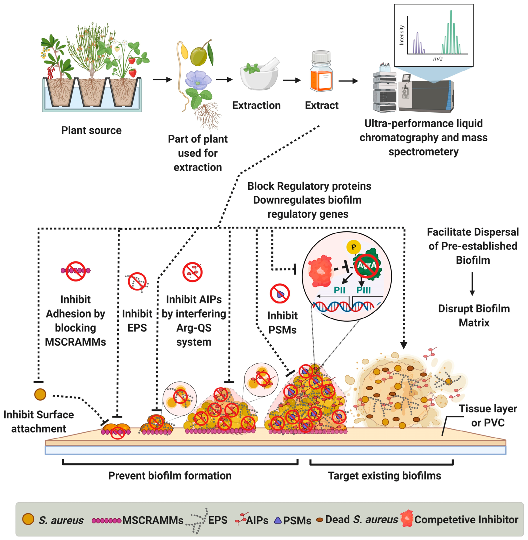
| Clinical Epidemiology | Population | Metric Reported | Approximate Affected Population | Biofilm Resistance | References |
|---|---|---|---|---|---|
| Staphylococcal food poisoning (SFP) | Outbreak of S. aureus enterotoxin-mediated foodborne disease | Individuals exposed to these specific outbreaks | Up to ~85% of exposed persons | Consumption of enterotoxin-producing S. aureus-contaminated food and contact surfaces contributes to persistence biofilms | [15,16] |
| Sepsis (all causes) | Global population | Annual sepsis cases and deaths, proportion of all global deaths | ~48.9 million cases and ~11 million deaths in 2017, ~20% of all deaths worldwide | MRSA leads to sepsis and bloodstream infection. It forms biofilms on intravascular devices and endovascular tissues | [17,18] |
| Healthcare-associated MRSA infections | Hospitalized patients | Proportion of healthcare-associated S. aureus infections due to MRSA | Commonly 20–50% depending on region | MRSA causes device-related and surgical site infections, frequently associated with biofilm and multidrug resistance | [19,20] |
| Community-associated MRSA skin and soft tissue infections | Patients with SSTIs in community settings | Proportion of purulent SSTIs caused by MRSA | Often >50% of purulent SSTIs (high variability reported) | MRSA SSTIs frequently involve biofilm on skin and soft tissue surfaces, complicating treatment and recurrence | [20,21] |
| Prosthetic joint and device-related infections | Patients with orthopedic implants and intravascular catheters | Proportion of device-related infections due to S. aureus | S. aureus commonly accounts for 20–40% of prosthetic joint infections and many catheter-related bloodstream infections | Biofilm formation on prosthetic materials and catheters is central to chronic, relapsing infection and antibiotic tolerance | [20] |
| Virulence Factors | Associated Genes | Function | Clinical Symptoms | References |
|---|---|---|---|---|
| MSCRAMMs | clfA, clfB, fnbA, fnbB, cna, sdr, bbp | Adhesion with host tissues | Endocarditis, osteomyelitis, endoprosthesis | [47,48] |
| Biofilm | Locus ica, arg system | Persistence in the host | Chronic infections | [49,50] |
| Leukocidins (e.g., PVL and toxin γ) | luks-PV, lukF-PV | Deceive the host immune response | Invasive skin infections, pneumonia, abscesses | [51] |
| Capsular polysaccharides, protein A, extracellular matrix binding protein | hlg, cap5, cap8, spa, eap | Deceive the host immune response | Invasive skin infections, pneumonia, abscesses | [52,53] |
| RNAIII-dependent proteases, lipases, and nucleases | V8, hysA, hla, plc, sepA | Penetration into host tissue | Tissue lesions | [54,55] |
| Toxins | TSST-1, ETs, and enterotoxins | Biofilm dispersal and virulence | MTSS, NMTSS, SSSS, Bullous impetigo, SFD | [56] |
| Category | Source | Targeted Action | Limitations | Level of Evidence | Reference |
|---|---|---|---|---|---|
| Synthetic Compounds | RNAIII-inhibiting protein and its derivatives | Inhibit the expression of agr and biofilm-producing genes | High production cost, limited sustainability | In vitro and animal models | [85,86] |
| Savirin | Inhibits auto-induction and quorum sensing | In vitro and animal models | [87,88,89] | ||
| Probiotics | Lactobacillus casei, Lactococcus lactis V7, Lactobacillus rhamnosus ATCC 7469 | Inhibit adhesion, invasion, and biofilm formation | Side effects on beneficial bacteria of the host and emergence of antimicrobial resistance. Variable colonization efficiency; delivery challenges | In vitro and animal models | [90,91,92] |
| Bacterial | Streptomyces sp. N174 | Antimicrobial and anti-biofilm properties | Survival of live cells during the gastrointestinal transit and their effective delivery to target tissues. Cross-species variability | In vitro | [93] |
| Staphylococcus schleiferi | Inhibits Agr expression | In vitro | [94] | ||
| Marine bacteria | Competitive inhibitor of AgrC | In vitro | [95] | ||
| 3-oxo-C12-HSL, (HQNO) from Pseudomonas aeruginosa | Inhibits auto-induction of AIPs | In vitro | |||
| Bacteriophages | Isolated from farmyard slurry, host: S. aureus DPC5246 | Inhibits biofilm proliferation | Bacterial resistance, immunogenicity, co-evolutionary dynamics, narrow host range, and difficulty in phage delivery to the target site. Host specificity | In vitro | [96] |
| ᶲ SA012 | In vitro | [97] | |||
| Polyvalent phage K | Inhibits biofilm formation | In vitro and animal models | [98,99] | ||
| Bacteriophage K and DRA88 | Reduced biofilm | In vitro | [100] | ||
| Snake venom lectins | Bothrops jararacussu | Biofilm disruption | Small-size peptide hinders its large-scale production and toxicity evaluation | In vitro | [101] |
| Nanoparticles | Silver and gold | Anti-biofilm properties | Expensive production, high doses exert cytotoxic and genotoxic effects | In vitro | [83,84] |
| Silver and Zinc oxide with nitric oxide | Inhibit biofilm formation | In vitro | [102] | ||
| Silver and Zinc oxide nanoparticles in combination with Antibiotics | Dispersion of biofilm | In vitro | [103] | ||
| Phosphatidylcholine-decorated gentamicin-loaded gold nanoparticles | Anti-biofilm | In vitro and animal models | [104,105] | ||
| Magnesium fluoride and yattrium fluoride nanomaterials | Reduce colonization | In vitro | [106,107] | ||
| Hormones | 17β-Estradiol | Invasion | High cost, less sustainability | In vitro | [108] |
| AIP and AIP derivatives | Truncated AIP-I, II, III, | Inhibits auto-induction of AIPs | High cost | In vitro and animal models | [59,109] |
| Fungal | 12 compounds from marine-derived fungi | Inhibit biofilm formation | High cost, less sustainability | In vitro | [110] |
| Enzymes | Lysostaphin | Disrupts biofilms | Poor retention, enzymatic stability, and activation of immune responses | In vitro, animal models, and early clinical evaluation | [111,112,113,114] |
| Cysteine histidine-dependent amidohydrolase/peptidase | Disrupts biofilms | In vitro | [115] | ||
| Endolysins | Disrupt biofilms | In vitro and animal models | [116,117,118] | ||
| V8 protease | Inhibits biofilm formation and promotes biofilm detachment | In vitro | [119] | ||
| Staphopains | Affects biofilm integrity | In vitro | [119,120] | ||
| Aureolysin | Inhibits biofilm formation and disperses preformed biofilms | In vitro | [121] | ||
| Cysteine proteases | Anti-biofilm activity | In vitro | [119] | ||
| DNases | Disrupting mature biofilms | In vitro and animal models | [122,123] | ||
| Dispersin B | Inhibits adherence and attachment | In vitro and animal models | [116,117,124] | ||
| Neutrase from Bacillus Amyloliquefaciens | Anti-biofilm activity | In vitro | [125] | ||
| Bio-surfactants | Mannosylerythritol lipids | Biofilm disruption | Expensive production | In vitro | [126] |
| Prokaryotes | Cochinmicin from Actinomycetes | Competitive inhibitor of AgrC | Survival of cells during the gastrointestinal transit and their effective delivery to target tissues upon ingestion | In vitro | [95] |
| Avellanin from sponges | In vitro | ||||
| Chelators | EDTA, EGTA, and TSC | Inhibit biofilm formation | Health hazards, cytotoxic, weakly genotoxic, intracellular metal accumulation, unsuitable for systemic applications | In vitro and limited clinical adjunct use | [127] |
| Sulfhydryl compounds | DTT, betamercaptoethanol, and cysteine | Inhibit biofilm formation | Skin irritation, organ toxicity, unsuitable for systemic applications | In vitro | [52,128] |
| Plant | Main Bioactive Compound(s) | Molecular Target | Anti-Biofilm Mechanism | Effect on Agr-QS | Experimental Model Used | Effective Concentration | Stage of Development | Reference |
|---|---|---|---|---|---|---|---|---|
| Camellia sinensis | Catechins, especially EGCG and its derivatives | Bacterial membrane, efflux pumps, amyloid-like biofilm matrix | Membrane disruption, anti-adhesion, anti-biofilm | Interferes with the AgrA response regulator and downregulates RNAIII | S. aureus and MRSA clinical isolates | Reported active at concentrations lower than MIC, 10 μg/mL to 60 μg/mL | Preclinical phase | [139,140,141,142] |
| Moringa oleifera | Phytochemicals, fatty acids, including palmitoleic, linolenic, and oleic acids | Quorum sensing and virulence | Inhibits biofilm formation, reduces CFU in biofilms, lowers MIC | Interferes with agr locus targeting AgrA or AgrC | S. aureus and MRSA from PVC-surface biofilm model | 0.5 to 2.0 mg/mL, resulting in up to 99% inhibition | In vitro and early animal stage (feed only) | [140,143,144,145] |
| Rosmarinus officinalis | Diterpene carnosic acid, camphor, micromeric acid, oleanolic acid, ursolic acid | Early attachment and biofilm formation | Inhibits initial attachment, formation, and promotes dispersal of preformed biofilms | Reduces AgrA and RNAIII expression | S. aureus and MRSA isolates | 0.05 mg/mL (0.1%) extract reported to inhibit biofilm development by 94% | Early preclinical translational stage | [146,147,148] |
| Psidium guajava | Benzyl isocyanate, phenolics, L-5-propylthiomethylhydantoin | Biofilm formation and Quorum sensing | Inhibits biofilm formation | Downregulates agr, icaAD, and sarA | MRSA, other S. aureus isolates, and BGM cell-line | 100 μg/mL to 1000 μg/mL (sub-minimal inhibitory concentration) | Preclinical phase | [149,150,151,152] |
| Eucalyptus globulus | Essential oil, 1,8-cineole | Early adhesion/attachment | Inhibits initial attachment and adhesion, decreases virulence and biofilm formation | Targets AgrA-AIP (auto-inducing peptide) | MRSA and S. aureus isolates | 2.5 mg/mL or less | Preclinical phase | [153,154,155] |
| Eucalyptus sideroxylon | Flower extract enriched in phloroglucinols | Biofilm formation and Quorum sensing | Inhibits biofilm formation | Targets Agr system | MRSA and S. aureus isolates | 0.05 mg/mL achieves up to 95.9% inhibition at sublethal doses | In vitro | [156,157] |
| Azadirachta indica | Crude and methanolic leaf extracts | Disrupting quorum sensing and downregulating virulence | Reduces biofilm formation | Repressing the Agr system | MRSA/MSSA and S. aureus isolates | 62.5 µg/mL to 125 µg/mL sub-minimum inhibitory concentrations | Preclinical phase | [158,159] |
| Curcuma longa | Curcumin; curcuminoids; diacetyl curcumin | Disrupting quorum sensing and inhibiting swimming and swarming ability | Suppresses biofilm formation, antibiotic synergy, anti-adhesive effects, and photodynamic killing with blue LED | Targets agrA and agrC operons and RNAIII | MRSA and S. aureus isolates | 100% inhibition at 20 μM Curcumin plus blue light, 62.5 μg/mL to 125 μg/mL sub-inhibitory concentrations | Preclinical phase | [160,161,162] |
| Sanguisorba officinalis | Triterpenoid saponins; tannins; polyphenolic root extract | Targets the ica locus and agr system genes | Reduces biofilm formation and density | Interacts with ica and agr system genes | MRSA and S. aureus isolates | 256 μg/mL half-maximal inhibitory concentration (IC50) | Preclinical phase | [163,164] |
| Scientific Name | Common Name | Part of a Plant | Bioactive Component | Phytochemical | Function | Mechanism | Reference |
|---|---|---|---|---|---|---|---|
| Bacopa monnieri (Plantaginaceae) | Brahmi (herb) | - | Bacoside A and saponins | Terpenoids | Inhibits microbial adhesion, biofilm formation, and ability to disrupt biofilms | Binding with IcaA | [214,215] |
| Cinnamomum spp. (Lauraceae) | Cinnamon | Essential oil and bark | - | - | Cellular shrinkages, cell wall damages, and decreased biofilm densities | - | [216,217] |
| Lavandula angustifolia (Lamiaceae) | Lavender | Essential oil | Camphor, caryophyllene, eucalyptol, lavendulyl acetate, limonene, linalool, linalyl acetate, cis-ocimene, α-pinene, transocimene, terpinen-4-ol | Terpenoids | Inhibits proliferation and biofilm formation | - | [218,219,220] |
| Origanum onites and Origanum vulgare (Lamiaceae) | Oregano | Seeds and essential oil | Carvacrol, γ-terpinene, p-cymene, and thymol | Terpenoids | Inhibits biofilm formation and disrupts pre-formed biofilms | - | [219,221] |
| Leopoldia comosa (Asparagaceae) | Tassel hyacinth | Bulb | - | - | Inhibits biofilm formation | - | [222] |
| Mentha × piperita (Lamiaceae) | Pepper mint | Leaves | HCAs, rosmarinic, 1,8-cineole, and menthol | Polyphenol and terpenoids | Inhibits biofilm formation | - | [219,220] |
| Ballota nigra (Lamiaceae) | Black horehound | Aerial parts | Phenylpropanoid glycosides and phenylpropanoid derivatives | Flavonoids, glycosides, and terpenes | Inhibits biofilm growth and adherence | Inhibits δ-hemolysin, a small peptide encoded by RNAIII transcript | [1,222,223] |
| Sanguisorba officinalis L. (Rosaceae) | Great burnet | Dried roots | Saponins | Terpenoids and tannins | Anti-biofilm activity | ica-dependent manner | [163,202,203] |
| Juglans regia L. (Juglandaceae) | Walnut | Immature fruits and leaves | Naphtoquinones | Quinones, polyphenols and flavonoids | Anti-biofilm activity | - | [222] |
| Rhodomyrtus tomentosa (Myrtaceae) | Kemunting, rose, myrtle | Leaf | Rhodomyrtone | Flavonoid | Inhibits adherence and biofilm formations | Possible cure for mastitis, even better than vancomycin | [224] |
| Rosa damascene (Rosaceae) | Damask rose | Flower | - | - | Eradicates biofilms | - | [225] |
| Rosa canina (Rosaceae) | Rosehip | Fruit | - | Flavonoids and polyphenols | Inhibits biofilm formation | Inhibition of exopolysaccharides | [222,226] |
| Sambucus nigra and Sambucus ebulus (Adoxaceae) | Elder and dwarf elder | Leaves and stems | - | Flavonoids | Inhibits biofilm formation | Inhibits δ-hemolysin, a small peptide encoded by RNAIII transcript | [1,227] |
| Cyclamen hederifolium (Myrsinaceae) | Ivy-leaved cyclamen | Tubers | Saponins | Terpenoids | Inhibits biofilm formation | Inhibits δ-hemolysin, a small peptide encoded by RNAIII transcript | [222,227] |
| Ocimum sanctum (Lamiaceae) | Basil | Leaves | - | Eugenol and tannins | Inhibits biofilm formation | - | [193,228,229] |
| Lonicera alpigena (Caprifoliaceae) | Alpine honeysuckle | Woody parts and leaves | - | - | Inhibits biofilm formation | Inhibits δ-hemolysin, a small peptide encoded by RNAIII transcript | [222,227] |
| Nigella sativa (Ranunculaceae) | Black cumin | Seed oil | Thymoquinone | Quinones | Inhibits biofilm formation | - | [230,231] |
| Castanea sativa (Fagaceae) | European chestnut | Leaves | Ursene and oleanene | Flavonoids and terpenoids | Inhibits biofilm formation | Inhibits the agr system | [222,227] |
| Malva sylvestris (Malvaceae) | Common mallow | Stems and flowers | Menthol and sorbitol | Terpene | Anti-biofilm activity | Used in commercial and mouth rinses | [222,227] |
| Thymus vulgaris (Lamiaceae) | Red thyme | Essential oils | Thymol | Terpenoids | Anti-biofilm activity | - | [232,233,234] |
| Alcea rosea L. (Malvaceae) | Hollyhock | Leaves, stems, flowers, and roots | Menthol | Terpene | Anti-biofilm activity | Inhibits δ-hemolysin, a small peptide encoded by the RNAIII transcript | [1,227] |
| Hydrastis canadensis (Ranunculaceae) | Goldenseal | Leaves | Berberine and Mycopyranone: A 8,8′-binaphthopyranone | Alkaloids | Anti-biofilm activity | Inhibits the agr QS system by blocking signal transduction of the AgrCA two-component system | [235,236] |
| Solanum nigrum (Solanacae) | Black nightshade | Leaves | - | - | Anti-biofilm activity | - | [228,230] |
| Rhanterium suaveolens (Asteraceae) | Arfej (shrub) | Essential oil | Carvacrol, linalool, and citrals | Polyphenols | Anti-biofilm activity | - | [237] |
| Rosmarinus officinalis (Lamiaceae) | Rosemary | Leaves | Pinene, camphor, micromeric acid, oleanolic acid, and ursolic acid | Terpenoids | Inhibits biofilm formation and disrupts pre-formed biofilms | - | [174,175,176,238,239,240] |
| Cananga odorata (Annonaceae) | Fragrant cananga | Essential oils | p-cresyl methyl ether, linalool, geranyl acetate, geraniol, eucalyptol | Polyphenols and terpenoids | Anti-biofilm activity | - | [218,232] |
| Rubus ulmifolius (Rosaceae) | Elm-leaf blackberry | Leaves, stem, and roots | Ellagic acid derivatives | Polyphenols and glycosides | Anti-biofilm activity | Inhibits δ-hemolysin, a small peptide encoded by RNAIII transcript | [222,227,241,242,243] |
| Melissa officinalis (Lamiaceae) | Lemon balm | Essential oil | Citrals (geranial + neral, citronellal, limonene, geraniol, β-caryophyllene, β-caryophyllene oxide, and germacrene D) | Terpenoids | Anti-biofilm activity | - | [218,219] |
| Cocculus trilobus (Menispermaceae) | Queen coralbead | Rhizome | Isoquinoline | Alkaloids and quinones | Inhibits microbial adhesion and biofilm formation | - | [244] |
| Coriandrum sativum L. (Apiaceae) | Coriander | Seeds and essential oil | p-cymene, g-terpinene, linalool, geranyl acetate | Terpenoids | Anti-biofilm activity | - | [219,220,245,246] |
| Zanthoxylum armatum (Rutaceae) | Winged prickly ash | Fruit | - | Alkaloids and others | Anti-biofilm activity | - | [247] |
| Ficus sansibarica (Moraceae) | Knobbly fig | Fruits, leaves, and stem bark | 5,7,4′-trihydroxyflavan-3-ol and isovitexin | Flavonoids and triterpenes | Inhibits microbial adhesion and biofilm formation | - | [248,249] |
| Marrubium vulgare (Lamiaceae) | White horehound | Roots, leaves, stem, and flowers | - | Terpenes, sterols, and flavonoids | Inhibits microbial adhesion and biofilm formation | Inhibits δ-hemolysin, a small peptide encoded by the RNAIII transcript | [222,227] |
| Jatropha curcas (Euphorbiaceae) | Purging nut | Pressed cake of whole plant and seed oil | Saponins, linoleic acid, and oleic acid | Alkaloids and polyphenols | Biofilm inhibition and degradation | - | [208,250] |
| Pimpinella anisum L. (Apiaceae) | Anise | Seeds and essential oil | (E)-anethole and estragol | Phenyl propanoids | Anti-biofilm activity | - | [219,220] |
| Dischidia rafflesiana (Apocynaceae) | Ant plant | Polyherbal formulations | - | - | Anti-biofilm activity | - | [246,251] |
| Krameria argentea (Krameriaceae) | Brazilian Rhatany | Roots | Chelerythrine, sanguinarine, dihydroxybenzofuran, and proanthocyanidin | Alkaloids and quinones | Anti-biofilm activity | Interferes with the arg-QS system | [243] |
| Lawsonia inermis (Lythraceae) | Henna tree | Leaves | Lawsone | Quinones | Anti-biofilm activity | - | [246,252] |
| Olea europaea L. (Oleaceae) | Olives | Leaves | Oleuropein | Polyphenols and flavonoids | Anti-biofilm activity | - | [253] |
| Glycyrrhiza glabra (Fabaceae) | Liquorice | Root | Glycyrrhizin, triterpinoid saponin, and glabridin | Terpenoids | Preventing biofilm formation and adherence | Interferes with the arg-QS system and inhibits exotoxin production | [246,254] |
| Leopoldia comosa (Hyacinthaceae) | Tassel grape hyacinth | Bulb | - | - | Preventing biofilm formation and adherence | - | [222] |
| Annona senegalensis (Annonaceae) | Wild custard apple | Seeds | N-cerotoyltryptamine, asimicin, and ent-19-carbomethoxykauran-17-oic acid | Polyphenols | Anti-biofilm activity | Interferes with the arg-QS system | [207] |
| Quercus cerris L., (Fagaceae) | Oak | Leaves, stem, and fruit | - | - | Anti-biofilm activity | Inhibits δ-hemolysin, a small peptide encoded by the RNAIII transcript | [222,227,255] |
| Orostachys japonicus (Crassulaceae) | Rock pine | Whole plant | - | Quinones | Inhibits cell-surface attachment | Downregulation of the psm-mec gene | [209] |
| Phyllanthus emblica (Phyllanthaceae) | Indian Gooseberry | Fruits | Gallic acid | Polyphenols | Inhibits cell-surface attachment | - | [246,256] |
| Melaleuca alternifolia (Myrtaceae) | Tea tree | Essential oil | 4-Terpineol and terpinolene | Terpenoids | Inhibits biofilm adhesion | Alters threonine, purine, pyrimidine, and amino acid biosynthesis pathways | [257,258] |
| Terminalia bellirica (Combretaceae) | Beleric nut tree (Baheda) | Fruit | Termilignan, thannilignan, and anolignan | Tannins | Inhibits biofilm formation | - | [246,259] |
| Vanilla planifolia (Orchidaceae) | Vanilla | Pods and essential oil | Ethylvanillin, 4-hydroxybenzaldehyde, methyl anisate, 4-hydroxybenzyl methyl ether, piperonal, vanillic acid, vanillin, carvacrol, and thymol | Terpenoids, flavonoids, and polyphenols | Inhibits biofilm formation | Interferes with the agr-QS system | [202,218] |
| Dendrobium chrysotoxum (Orchidaceae) | Fried-egg orchid | Whole plant | Erianin isovitexin and parthenolide | Flavonoids | Inhibits biofilm adhesion | Interferes with the agr-QS system | [260] |
| Allium sativum (Amaryllidaceae) | Garlic | Essential oil | Allicin | Alkaloids | Inhibits biofilm formation | Downregulates the expression of icaA and interferes with agr expression | [261,262] |
| Vaccinium macrocarpon (Ericaceae) | American cranberry | Leaves and fruit | Urell R and proanthocyanins | Quinones and polyphenols | Inhibits microbial adhesion and biofilm formation | Non-toxic, cyto- compatible metabolites | [243,263] |
| Myristica fragrans (Myristicaceae) | Nutmeg | Seed essential oil | Eugenol, isoelemicin, isoeugenol, methoxy eugenol, myristic acid, myristicin, kayeassamin A, surangin C, theraphin B, | Polyphenols and alkaloids | Inhibits biofilm formation | - | [246] |
| Arundo donax (Poaceae) | Giant reed | Reed nodes | Bufotenidine and gramine | Alkaloids | Inhibits biofilm formation and disrupts already established biofilms | - | [222] |
| Citrus × paradisi (Rutaceae) | Grapefruit | Seeds and essential oil | Naringenin | Flavonoid | Inhibits microbial adhesion and biofilm formation | Reduces agrA and hla | [264,265] |
| Spondias purpurea (Anacardiaceae) | Spanish Plum | Leaves and fruit juice pulp | - | Terpenoids and flavonoids, polyphenols | Inhibits biofilm formation | Interferes with the agr-QS system | [211,266] |
| Aesculus hippocastanum (Sapindaceae) | Horse chestnut | Whole plant | Chelerythrine, sanguinarine, umbelliferone-3, aesculetin, dihydroxybenzofuran, and proanthocyanidin | Quinones and polyphenols | Inhibits microbial adhesion and biofilm formation | - | [241,243] |
| Moringa stenopetala (Moringaceae) | Cabbage tree/ African horse radish tree | Leaves and seeds | - | - | Inhibits biofilm formation | - | [205,210] |
| Vetiveria zizanioides (Poaceae) | Vetiver (Khus) | Roots | β-vetivenene, vetiselinenol, isovalencenol, vetivenic acid, α-vetivone, and β-vetivone | Terpenoids and polyphenols | Inhibits microbial adhesion and biofilm formation | Inhibits EPS and α-hemolysin toxin production via the agr-QS system | [213,267,268] |
| Citrus sinensis (Rutaceae) | Sweet orange | Fruits and essential oils from the peel | Limonene, myrcene, α-farnesene, γ-terpinene, α-pinene, and sabinene | Terpenes | Inhibits biofilm formation | - | [213,269] |
| Pogostemon cablin (Lamiaceae) | Patchouli | Whole plant | α-guaiene, β-caryophyllene, δ-cadinene, pogostol, patchoulol, seychellene, α- and β-patchoulene | Terpenoids, flavonoids, glycosides | Biofilm eradication | Upregulates biofilm-related bacterial genes luxR (inhibitor for the arg-QS system) | [246,270] |
| Hymenocallis littoralis (Amaryllidaceae) | Spider lily | Leaves | 4-methylesculetin, methylisoeugenol, Quercetin 5,7,3′,4′-tetramethyl ether 3-rutinoside | Polyphenols and flavonoids | Inhibits microbial adhesion and biofilm formation | Blocks the active site residues of adhesion proteins | [271,272] |
| Cymbopogon flexuosus (Poaceae) | Lemon grass | Essential oil | Citral and β-Geranial | Aliphatic aldehydes | Inhibits microbial adhesion and proliferation to disrupt biofilm matrix | Inhibits PIA and arg-QS system | [273,274] |
| Dracaena cochinchinensis (Asparagaceae) | Chinese dragon’s blood (red resin) | Resin powder | Homo isoflavans and homo isoflavanones | Flavonoids | Disrupts biofilm | Downregulates biofilm regulatory genes saeR, saeS, and hla | [275,276] |
| Duabanga grandiflora (Lythraceae) | Duabanga | Leaves | F-10 fraction | Alkaloids, tannins, saponins, steroids, glycosides, and flavonoids | Inhibits cell-surface attachment and biofilm formation | Interfere with the agr-QS system and competitive inhibitor of PBP2a | [277,278] |
| Cymbopogon nardus (Poaceae) | Citronella grass | Essential oil | Geraniol and citronellal | Terpenes | Inhibits microbial adhesion and proliferation | Non-toxic, cytocompatible metabolites as an alternative for future mouthwashes formulations | [279,280] |
| Chelidonium majus (Papaveraceae) | Great celandine | Whole plant | Chelerythrine, sanguinarine, dihydroxybenzofuran, and proanthocyanidin | Alkaloids | Inhibits cell-surface attachment and biofilm formation | - | [236,243,281] |
| Ocimum gratissimum (Lamiaceae) | Clove basil | Essential oil and leaves | Eugenol, 1,8-cineole, α-terpineol, γ-terpinene | Alkaloids and terpenes | Inhibits cell-surface attachment and biofilm formation | [282,283] |
Disclaimer/Publisher’s Note: The statements, opinions and data contained in all publications are solely those of the individual author(s) and contributor(s) and not of MDPI and/or the editor(s). MDPI and/or the editor(s) disclaim responsibility for any injury to people or property resulting from any ideas, methods, instructions or products referred to in the content. |
© 2026 by the authors. Licensee MDPI, Basel, Switzerland. This article is an open access article distributed under the terms and conditions of the Creative Commons Attribution (CC BY) license.
Share and Cite
Sheikh, S.W.; Ali, A.; Ahsan, A.; Shang, F.; Xue, T.; Gollahon, L. Prevention and Treatment of Staphylococcus aureus Biofilms Using Promising Agr-QS-Targeting Anti-Biofilm Agents. Pathogens 2026, 15, 795. https://doi.org/10.3390/pathogens15080795
Sheikh SW, Ali A, Ahsan A, Shang F, Xue T, Gollahon L. Prevention and Treatment of Staphylococcus aureus Biofilms Using Promising Agr-QS-Targeting Anti-Biofilm Agents. Pathogens. 2026; 15(8):795. https://doi.org/10.3390/pathogens15080795
Chicago/Turabian StyleSheikh, Salma Waheed, Ahmad Ali, Asma Ahsan, Fei Shang, Ting Xue, and Lauren Gollahon. 2026. "Prevention and Treatment of Staphylococcus aureus Biofilms Using Promising Agr-QS-Targeting Anti-Biofilm Agents" Pathogens 15, no. 8: 795. https://doi.org/10.3390/pathogens15080795
APA StyleSheikh, S. W., Ali, A., Ahsan, A., Shang, F., Xue, T., & Gollahon, L. (2026). Prevention and Treatment of Staphylococcus aureus Biofilms Using Promising Agr-QS-Targeting Anti-Biofilm Agents. Pathogens, 15(8), 795. https://doi.org/10.3390/pathogens15080795