Article

Hemorrhagic Fever Disease in STAT-1 Knockout Mice Infected with Lujo Virus

1
Texas Biomedical Research Institute, San Antonio, TX 78227, USA
2
Sandia National Laboratories, Livermore, CA 94550, USA
3
Galveston National Laboratory, Department of Microbiology and Immunology, University of Texas Medical Branch, Galveston, TX 77555, USA
*
Author to whom correspondence should be addressed.
Pathogens 2026, 15(4), 394; https://doi.org/10.3390/pathogens15040394
Submission received: 20 March 2026 / Revised: 27 March 2026 / Accepted: 2 April 2026 / Published: 7 April 2026
(This article belongs to the Special Issue Antiviral Strategies and Vaccines Against Emerging RNA Viruses)

Abstract

Lujo virus (LUJV) is an arenavirus that causes Lujo Hemorrhagic Fever (LHF), a viral hemorrhagic fever that emerged in a 2007 outbreak in Zambia and South Africa with an 80% case fatality rate and evidence of human-to-human nosocomial transmission. There are no approved medical countermeasures for LHF, although several screens have identified lead antiviral compounds. The lack of accessible animal models limits the development of lead compounds and characterization of broadly protective anti-arenavirus compounds such as ribavirin for the treatment of LHF. Here, we present preliminary data characterizing the partially lethal disease caused by LUVJ in STAT-1 deficient mice. Several key hematological, clinical chemistry, and histologic findings common to LHF disease are recapitulated in this model. This work suggests that further characterization of LUJV infection in STAT-1 deficient mice may allow development of a model that would be instrumental in the development of medical countermeasures for LHF.

1. Introduction

Lujo mammarenavirus (LUJV) first appeared in an infected patient in the Lusaka region of Zambia in 2008. The patient was transferred to Johannesburg, South Africa for treatment which initiated a nosocomial outbreak of Lujo hemorrhagic fever (LHF), a viral hemorrhagic fever disease with an unusually high (4/5, 80%) case fatality rate [1,2]. LHF in humans is associated with acute respiratory distress, impaired renal function, thrombocytopenia, leukopenia, granulocytopenia, hepatocyte necrosis, and elevated (alanine and aspartate) transaminase levels [2]. Ribavirin, a broad-spectrum antiviral, was used as treatment for two patients, with one patient receiving an earlier course of both oral followed by intravenous treatment surviving LHF [2]. The use of ribavirin has been recommended for LHF; however, concerns of exacerbation of viral hemorrhagic fever diseases due to ribavirin’s renal and hepatic toxicity, along with the appropriate treatment window, remain a topic of debate [3,4,5,6].
LUJV is most closely related to other Old World arenaviruses, including Lassa mammarenavirus (LASV), the causative agent of Lassa Fever (LF), and Lymphocytic Choriomeningitis virus (LCMV), associated with disease in immunocompromised patients, as well as numerous other non-pathogenic African arenaviruses including Mopeia mammarenavirus (MOPV), Ippy mammarenavirus (IPPV), and Mobala mammarenavirus (MOBV) [7]. Although the LUJV glycoprotein cross reacts with LASV, LCMV, and MOBV, but not MOPV or IPPV immune sera [8], the entry receptor for LUJV appears unique among arenaviruses [9,10,11]. Despite surveillance efforts in Zambia, the (presumed rodent) reservoir remains unidentified [12,13,14].
LHF has been modeled in strain 13 guinea pigs with features that align to human disease. However, there are challenges with strain 13 models of arenavirus infection including the lack of commercially available strain 13 guinea pigs, limited immunological reagents for guinea pigs in general, and limited genetic information about this strain [15]. Additionally, strain 13 guinea pigs can show hemorrhagic fever disease signs when infected with mammarenaviruses that are thought to be apathogenic in humans [16]. LUJV infection of cynomolgus macaques resulted in a mild illness characterized by delayed inflammatory and apoptotic host responses [17]. STAT-1 constitutive knockout mice (STAT-1−/−) [18,19,20,21,22], interferon alpha receptor knockout [23,24] and interferon alpha receptor and interferon gamma receptor double knockout mice (IFrag−/−) [23,25,26] have been described as murine models of other arenavirus hemorrhagic fever diseases but have yet to be used in characterizing LUJV. The exploratory hypothesis of this research is that because immunocompromised mouse models were described for other arenaviruses, they may also be instrumental to the aim of developing a murine model of a LHF-like disease. Because of prior successes with model development for other arenavirus hemorrhagic fevers in STAT-1−/− and interferon deficient mice, these model systems were selected for preliminary study.
Murine models of arenavirus disease are instrumental in medical countermeasure development [27]. Medical countermeasure development for LHF has likely been hindered by the lack of accessible small animal models of disease. No vaccines have been developed for LHF [28], although the application of a LUJV reverse genetics reporter system [29] was key in the identification of several lead compounds for LHF drug development [30,31]. Here, we present data from LUJV infection of STAT-1−/− and IFrag−/− mice providing preliminary evidence of their potential utility as models of LUJV infection that could be used for the in vivo development of LHF medical countermeasures.

2. Materials and Methods

Four- to six-week-old STAT-1−/− mice (129S6/SvEv-Stat1tm1Rds, n = 10, 5 males and 5 females) were purchased from Taconic (Germantown, New York, NY, USA). IFrag−/− mice (B6.Cg-Ifngr1tm1Agt Ifnar1tm1.2Ees/J, n = 5, 3 males, 2 females, 65 days-old on the day of virus inoculation) were purchased from the Jackson Laboratory (Bar Harbor, ME, USA). Mice were housed at the University of Texas Medical Branch (UTMB) Galveston National Laboratory under ABSL4 conditions with up to 5 mice per cage segregated by strain and sex. Mice were housed in sterile Techniplast Green Line caging (West Chester, PA, USA) with corncob bedding, and shredded foraging paper and cardboard huts for enrichment. STAT-1−/− mice were acclimated to BSL4 for 4 days prior to inoculation; IFrag−/− were acclimated for 9 days. Implantable electronic identification and temperature transponders (Bio Medic Data Systems, Seaford, DE, USA) were implanted subcutaneously under isoflurane anesthetic the day prior to virus inoculation for STAT-1−/− mice and 5 days prior to virus inoculation for IFrag−/− mice. All mice were allocated to the LUJV challenge group and were inoculated intraperitoneally under isoflurane anesthetic on study day zero with 9.0 × 10³ plaque forming units (PFU) of LUJV strain Prototype Zambia (200809232). A target dose of 1.0 × 10⁴ was selected based on previously published work with LASV [18]. The actual challenge dose of 9.0 × 10³ was confirmed via plaque assay of the infectious inoculum on Vero 76 cells under 0.8% agarose overlay with the addition of neutral red 5 days following infection and plaque counting the following day, as previously described [32]. This study design precluded randomization and blinding, and would not be expected to be robust to cage effect bias. Mice were weighed and clinically scored daily following inoculation and were euthanized when they met humane endpoint criteria or at 28 days post-inoculation (DPI). All animal procedures were approved by the UTMB Institutional Animal Care and Use (IACUC) and Institutional Biosafety (IBC) committees.
At the time of euthanasia, blood was collected into MiniCollect K2EDTA plasma tubes (Greiner Bio-One, Monroe, NC, USA) and analyzed on a Vetscan HM5 (Zoetis, Parsippany, NJ, USA). Remaining blood was centrifuged and plasma was collected for analysis with a Piccolo BioChemistry Panel Plus (Abbott Laboratories, Abbott Park, IL, USA). Historical controls (HC) were included from a previously published study of STAT-1−/− mice which were processed similarly except a Vetscan VS2 Comprehensive Diagnostic Profile was used for clinical chemistry measurements [33].
Liver, spleen, kidney, lung, and brain tissues were collected and preserved by RNAlater (Thermo Fisher Scientific, Waltham, MA, USA) for quantitative reverse transcriptase PCR (qRT-PCR) viral load quantification, or in 10% neutral buffered formalin (Thermo Fisher Scientific, Waltham, MA, USA) for histology. RNA was isolated from RNAlater using an RNEasy kit (Qiagen, Hilden, Germany). Inactivated RNA was removed from the BSL4 according to UTMB IBC approved procedures. Quantitative reverse transcriptase PCR (qRT-PCR) was conducted on a CFX96 Real-Time PCR Detection System (Bio-Rad Laboratories, Berkeley, CA, USA) using a QuantiNova Probe RT-PCR kit (Qiagen, Hilden, Germany) with the following primer and probe set: Forward—5′-CTTTC GTTCT GTGGA GGTGT AG-3′; Reverse—5′-CATCT CTTCC AGAAC CAGCA TAA-3′; Probe—5′-6-FAM-CTGTG TTGT-ZEN-CCCAG GATCT GCCTT-3′-IABkFQ (Integrated DNA Technologies, Coralville, IA, USA). Results were normalized to a mouse β-actin primer-probe set (cat# Mm.PT.39a.22214843.g, Integrated DNA Technologies, Coralville, IA, USA) and quantified based on comparison to a standard curve generated with a synthetic DNA target: ctcac accca cagga aattg gtggg gggct tttgc tcttt cgttc tgtgg aggtg taggg tcctt tggag tcatat caatg actgt gttgt cccag gatct gcctt ttatg ctggt tctgg aagag atgta tggcc (Integrated DNA Technologies, Coralville, IA, USA).
Formalin fixed tissue was removed from the BSL4 according to University of Texas Medical Branch (UTMB) institutional biosafety committee approved procedures, embedded in paraffin, sectioned, stained with hematoxylin and eosin, and imaged on a Zeiss Axioscan slide scanner (Oberkochen, Germany).
Figures were made with Prism (version 10, GraphPad Software, Boston, MA, USA) with specific statistical analysis reported in the figure legends. Single mice were treated as individual experimental units (data points) for analysis. No data were excluded from analysis except when insufficient sample volume for clinical chemistry led to a single LUJV infected IFrag−/− being excluded from this analysis; and one LUJV infected STAT-1−/− did not return an alanine aminotransferase value due to sample hemolysis.

3. Results

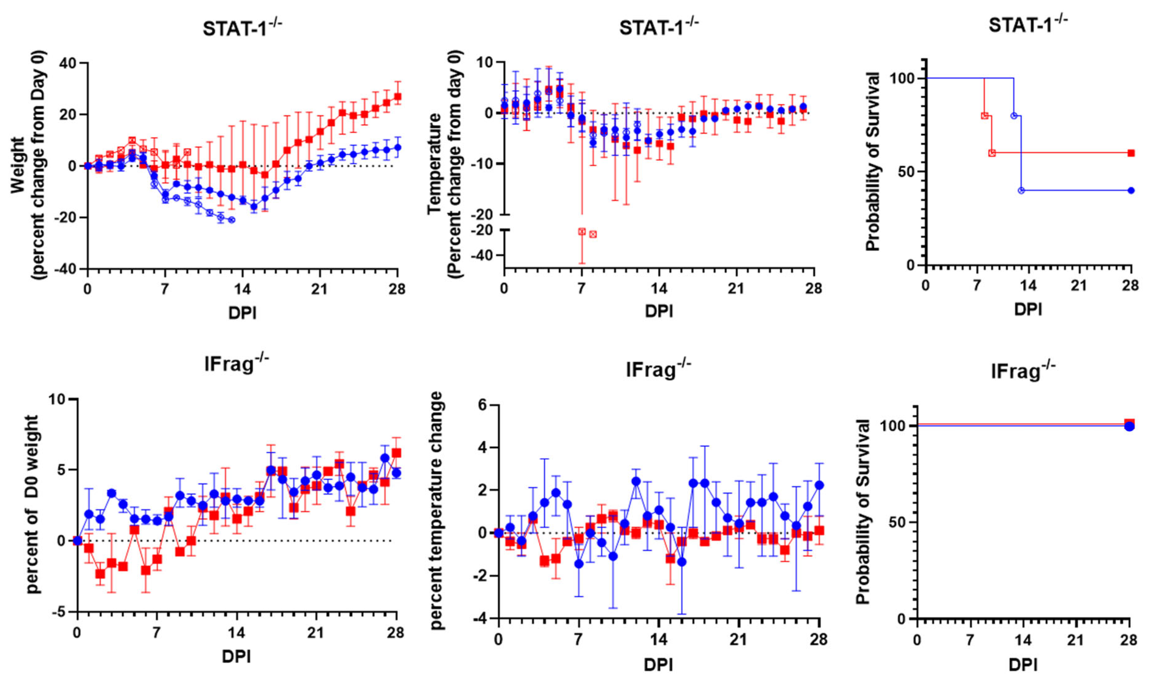
STAT-1−/− mice had progressive weight loss following LUJV inoculation starting five days post-infection (DPI). Survivors began to gain weight at about two weeks post-infection (Figure 1 top left). A fever was observed at 4 and 5 DPI in most subjects followed by hypothermia, with severe hypothermia observed in the final measurement from both female subjects meeting early euthanasia criteria (Figure 1 top middle). Half of the animals (5 of 10) met early euthanasia criteria by 13 days post-LUJV inoculation (Figure 1 top right). Three mice met early euthanasia criteria due to loss of more than 20% of their initial body weight. The other two meeting early euthanasia criteria both had severe hypothermia with one displaying ataxia and the loss of righting reflex, while the other was moribund. Of these, gross anatomical observation revealed necrotic pneumonia in 2 of 5 subjects, splenomegaly in 3 of 5 subjects, and a liver pallor in 1 of 5 subjects. No significant lesions were observed during the necropsy of STAT-1−/− mice surviving to the planned end of the study at 28 DPI.
IFrag−/− mice had transient weight loss lasting about a week followed by normal weight gain after LUJV infection (Figure 1 bottom left). Body temperatures varied throughout the experiment but did not show a detectable disease related pattern (Figure 1 bottom middle). All IFrag−/− mice survived until the planned end of the study 28 DPI (Figure 1 bottom right). Both female IFrag−/− had necrotic lesions on the distal spleen at necropsy, but otherwise no significant lesions were noted during necropsy.
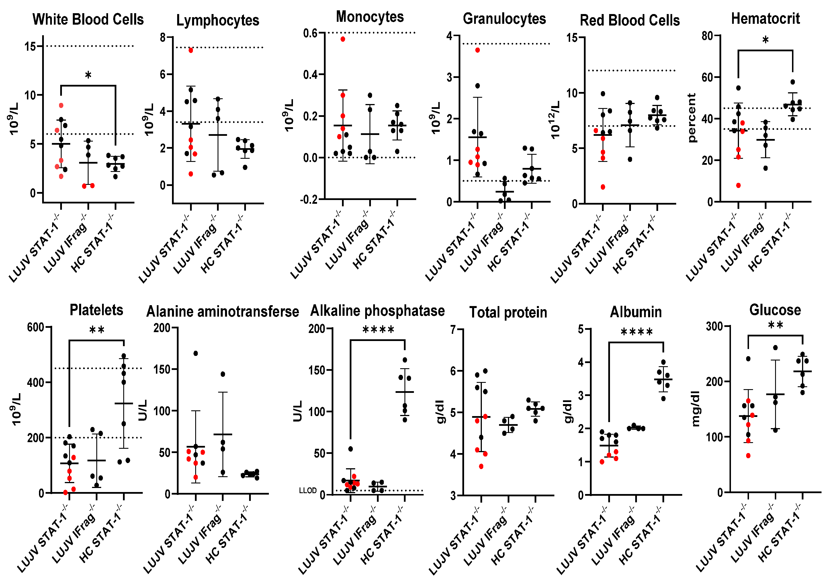
Hematologic values showed a trend towards leukocytosis, anemia, and thrombocytopenia consistent with viral hemorrhagic fever disease (Figure 2). Significantly elevated alanine transaminase levels suggest hepatic dysfunction, a typical finding during arenavirus hemorrhagic fever disease (Figure 2). Low albumin may suggest that subjects experienced systemic vascular failure and/or renal dysfunction as well (Figure 2).
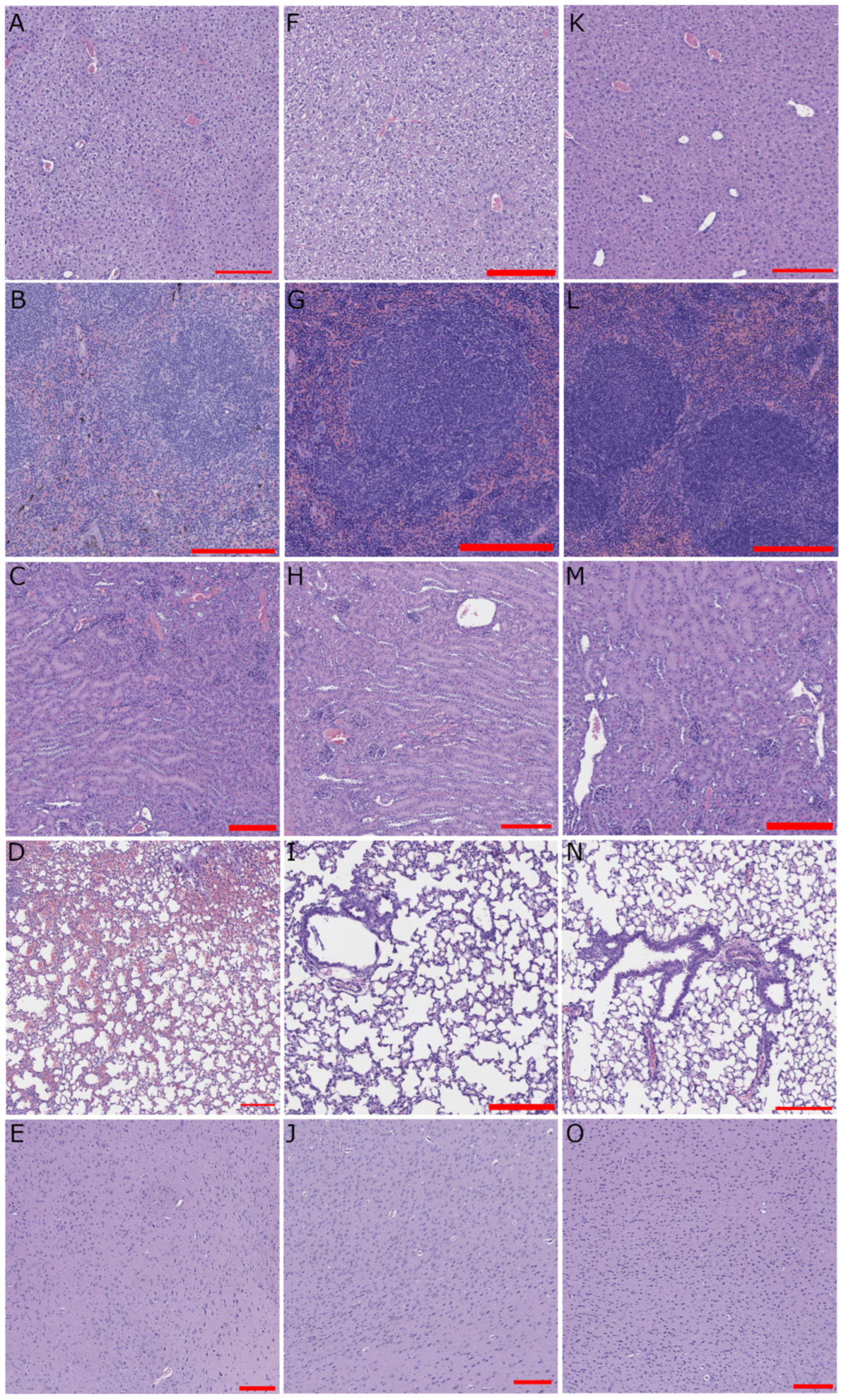
Liver histology revealed diffuse hepatocellular degeneration and necrosis during LUJV infection of STAT-1−/− mice in both subjects meeting early euthanasia criteria (Figure 3A) and those surviving to the planned end of the study at 28 DPI (Figure 3F). Splenic sections from most mice examined had some degree of splenic hemorrhage and parenchymal necrosis (Figure 3B,G,L). Renal infiltrates were observed in STAT-1−/− mice during acute LUJV infection (Figure 3C) and in IFrag−/− mice (Figure 3M). Pulmonary hemorrhage was apparent in STAT-1−/− mice during acute LUJV infection (Figure 3D). Cortex sections appeared normal (Figure 3E,J,O). Overall, lung pathology was a differentiating feature between STAT-1−/− mice that survived compared to those that met early euthanasia criteria.
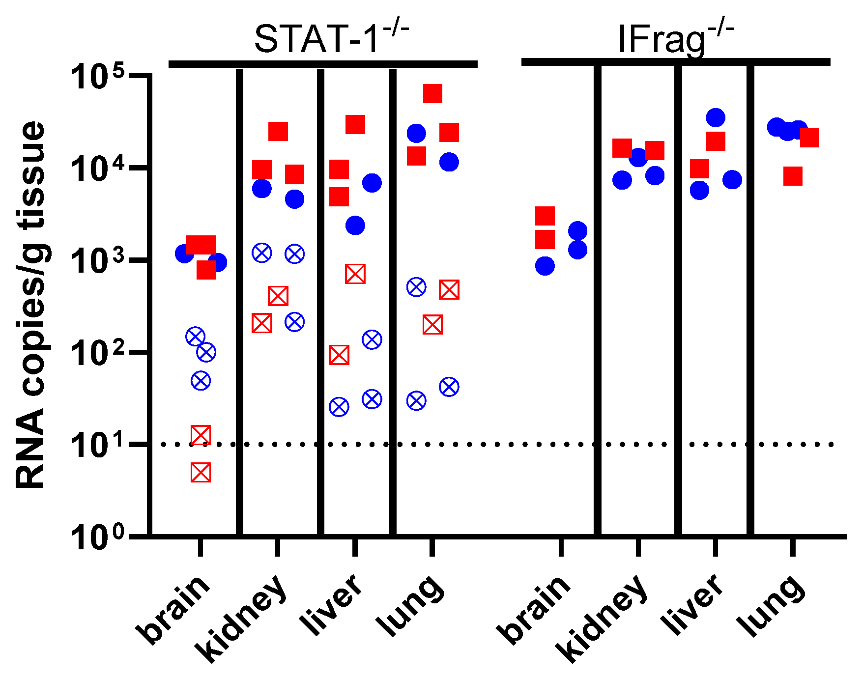
Viral RNA was detected at relatively high levels in kidney, liver, and lung tissue of both STAT-1−/− and IFrag−/− mice surviving to the planned end of the study, with an interesting trend towards the highest viral load being detected in the lung. Markedly less RNA was present in the brain of these subjects. STAT-1−/− mice that succumbed to LUJV infection tended to have a lower viral load present in tissues, with kidney having the most viral load and brain having the least.

4. Discussion

LUJV productively infected STAT-1−/− mice causing lethal disease in half of the infected cohort. The disease caused a febrile illness followed by a period of weight loss and hypothermia (Figure 1). Mice that succumbed to LUJV infection had several hallmarks associated with LHF including low hematocrit, thrombocytopenia, and signs of viral hepatitis (Figure 2, Figure 3 and Figure 4). Interestingly, although STAT-1−/− mice are generally susceptible to arenavirus disease, the pathogenicity of apathogenic-to-human arenaviruses [19,34], and partial lethality of human hemorrhagic fever causing arenaviruses [18,22] highlights that there is not a direct correlation between mortality in humans and STAT-1−/− mice. Conversely, similarly to LASV infection of IFrag−/− mice [23], although there are signs of LUJV viral replication in tissues (Figure 4), IFrag−/− mice do not appear to show overt signs of infection.
LUJV causes severe illness in humans, and in the US it is a risk group 4 agent requiring handling under stringent biosafety level 4 containment. As a result, model development has been limited. This preliminary study utilized small group sizes (n = 10 for STAT-1−/− mice and n = 5 for IFrag−/− mice), which were limited by a combination of vendor availability coinciding with BSL4 schedule, which limits the generalizations that can be drawn from these data. However, these studies form the basis for work to fully characterize LUJV in STAT-1−/− mice, allowing a better a priori estimate of sample size requirements for model refinement in the future. Age- and sex-related differences in LUJV pathogenesis were a feature of disease reported for strain 13 guinea pigs [35]. The small sample size limited the ability to reliably comment on sex-based differences in LUJV pathogenicity in this study.
Given the limited availability of both mice and available BSL4 schedule, a decision was made to use HC data. The decision to use HC data likely introduces cohort bias, and because of this, the hematology and clinical chemistry comparisons must be interpreted with care. However, the magnitude of significance despite the small sample size for many parameters, combined with the expected clinical profiles for arenavirus disease (typically including leukopenia, thrombocytopenia, and other hallmarks of viral hepatitis and hemorrhagic fever related systemic vascular failure) allow us to cautiously interpret these findings as significant. A more comprehensive study including a viral replication time course in tissues, with measurements of infectious virus by plaque assay, is a critical step to validate this model for antiviral therapy efficacy testing. It would also be preferable to describe the hematological and clinical chemistry outcomes compared to in-study controls, and future studies could also potentially include genetic background matched STAT-1 wild-type controls. Future studies could also be designed to probe the nature of the contribution of host responses to disease outcome.
In strain 13 guinea pigs, disease following LUJV appears to be heavily dependent on host immune responses [35]. This finding corroborates evidence that early immunosuppression in the non-human primate (NHP) model leads to mild illness in cynomolgus macaques [17]. In the case of LASV, the matrix protein Z and nucleoprotein antagonize host immunity (as recently reviewed [36]), but relatively little is known about the molecular pathogenicity and host interactions of LUJV. Mouse models are well-suited to the study of host responses to viral infection with many immunological tools available. Although, the immunocompromised nature of STAT-1−/− mice can limit the generalization of immunological findings, this model could still be useful in targeted testing of LUJV induced host responses. Future studies probing these host responses could enable a better understanding of both LUJV molecular pathogenesis and allow insight into the mechanisms of action of antiviral therapies.
This represents the first preliminary description of a commonly available rodent model for LUJV infection. Because LUJV has not re-emerged since the initial outbreak, research funding for LUJV is limited. The two previously described models, although valuable for medical countermeasure development, come with accessibility challenges. Strain 13 guinea pigs are not commercially available and must be maintained in dedicated private breeding colonies. The NHP model of LUJV infection, while useful as an infection model, has limited signs of clinical illness. Limited accessibility of these models creates logistical hurdles making it difficult for first-in-animal screening of medical countermeasures.
Future work to expand the STAT-1−/− mouse model of LHF, particularly increasing sample sizes, conducting dose-ranging experiments to determine if the partially lethal phenotype is related to infectious dose, determining viremia and infectious viral loads in tissues, localizing infection in tissues, and characterizing the host responses to infection are needed to expand this preliminary study. Furthermore, additional time points would allow an understanding of infection kinetics that could inform decisions on the appropriate time-to-treat during antiviral therapy testing. Ultimately, host response characterization would likely be insightful into the comparative molecular pathogenicity of arenaviruses.
Although LUJV is genetically related to LASV, it uses a different receptor from both LASV and the South American hemorrhagic fever causing arenaviruses. This divergence from other hemorrhagic fever arenaviruses, combined with its high lethality in humans makes LUJV an important outgroup for studying arenavirus viral pathogenesis and for testing antiviral compounds with broad arenavirus-family activity. Additionally, the wide availability of reagents and methodology to probe host immune responses in mice could facilitate description of the role of host-immune response in LHF. The STAT-1−/− mouse model provides an accessible animal model for further study and the development of medical countermeasures targeting LHF.

Author Contributions

D.M.J.: Conceptualization, funding acquisition, methodology, formal analysis, investigation, visualization, writing—original draft. S.J.: Investigation, writing—revision. E.D.: Investigation, writing—revision. R.W.C.: Conceptualization, project administration, writing—revision. T.W.G.: Resources, Funding acquisition. J.E.C.: Resources, Funding acquisition. All authors have read and agreed to the published version of the manuscript.

Funding

This work was supported by departmental funds (to T.W.G. and J.E.C.) from the University of Texas Medical Branch, Department of Microbiology and Immunology, and the US Department of Health and Human Services, NIH Grant UC7AI094660 for biosafety level 4 operations in support of the Galveston National Laboratory.

Institutional Review Board Statement

This work was completed with the approval of the University of Texas Medical Branch’s (UTMB) Institutional Animal Care and Use Committee. UTMB’s animal care and use program maintains an assurance in accordance with the Public Health Service Policy on Humane Care and Use of Laboratory Animals (PHS Policy), is United States Department of Agriculture registered, and fully accredited by AAALAC, International since 1995. All work with infectious LUJV was done at A/BSL4 in CDC Select Agent registered lab space in the Galveston National Laboratory.

Informed Consent Statement

Not applicable.

Data Availability Statement

The original contributions presented in this study are included in the article. The raw data supporting the conclusions of this article will be made available by reasonable request directed to the corresponding author.

Acknowledgments

We thank Eduardo Lujan, Cole Rozal, and Jennifer Schwedler for their technical assistance in staining and imaging histological samples.

Conflicts of Interest

The authors declare no conflicts of interest.

Abbreviations

The following abbreviations are used in this manuscript:
LUJVLujo virus (Mammarenavirus lujoense)
LHFLujo hemorrhagic fever
STAT-1Signal transducer and activator of transcription 1
LASVLassa virus (Mammarenavirus lassaense)
LFLassa Fever
LCMVLymphocytic choriomeningitis virus (Mammarenavirus choriomeningitidis)
MOPVMopeia virus (Mammarenavirus mopeiaense)
IPPVIppy virus (Mammarenavirus ippyense)
MOBVMobala virus (Mammarenavirus praomydis)
STAT-1−/−STAT-1 constitutive knockout
IFrag−/−Interferon alpha receptor and interferon gamma receptor double knockout
K2EDTADipotassium ethylenediaminetetraacetic acid
RNARibonucleic acid
qRT-PCRQuantitative reverse transcriptase polymerase chain reaction
HCHistorical controls
BSL4Biosafety level 4
DPIDays post-inoculation
NHPNon-human primate

References

  1. Briese, T.; Paweska, J.T.; McMullan, L.K.; Hutchison, S.K.; Street, C.; Palacios, G.; Khristova, M.L.; Weyer, J.; Swanepoel, R.; Egholm, M. Genetic Detection and Characterization of Lujo Virus, a New Hemorrhagic Fever–Associated Arenavirus from Southern Africa. PLoS Pathog. 2009, 5, e1000455. [Google Scholar] [CrossRef]
  2. Paweska, J.T.; Sewlall, N.H.; Ksiazek, T.G.; Blumberg, L.H.; Hale, M.J.; Lipkin, W.I.; Weyer, J.; Nichol, S.T.; Rollin, P.E.; McMullan, L.K. Nosocomial Outbreak of Novel Arenavirus Infection, Southern Africa. Emerg. Infect. Dis. 2009, 15, 1598. [Google Scholar] [CrossRef]
  3. Richards, G.A.; Weyer, J.; Blumberg, L.H. Viral Haemorrhagic Fevers in South Africa: Review. S. Afr. Med. J. 2015, 105, 748–751. [Google Scholar] [CrossRef] [PubMed][Green Version]
  4. Ceylan, B.; Turhan, V. The Efficacy of Ribavirin in Crimean-Congo Hemorrhagic Fever—Randomized Trials Are Urgently Needed. Int. J. Infect. Dis. 2014, 29, 297–298. [Google Scholar] [CrossRef][Green Version]
  5. Ergonul, O. Evidence Supports Ribavirin Use in Crimean-Congo Hemorrhagic Fever. Int. J. Infect. Dis. 2014, 29, 296. [Google Scholar] [CrossRef][Green Version]
  6. Salam, A.P.; Duvignaud, A.; Jaspard, M.; Malvy, D.; Carroll, M.; Tarning, J.; Olliaro, P.L.; Horby, P.W. Ribavirin for Treating Lassa Fever: A Systematic Review of Pre-Clinical Studies and Implications for Human Dosing. PLoS Neglected Trop. Dis. 2022, 16, e0010289. [Google Scholar] [CrossRef]
  7. Ye, C.; de la Torre, J.C.; Martinez-Sobrido, L. Reverse Genetics Approaches for the Development of Mammarenavirus Live-Attenuated Vaccines. Curr. Opin. Virol. 2020, 44, 66–72. [Google Scholar] [CrossRef]
  8. Paweska, J.T.; van Vuren, P.J.; Weyer, J. 16 Lujo Virus Hemorrhagic Fever. In Viral Hemorrhagic Fevers; Springer: Berlin/Heidelberg, Germany, 2016; pp. 287–300. [Google Scholar]
  9. Raaben, M.; Jae, L.T.; Herbert, A.S.; Kuehne, A.I.; Stubbs, S.H.; Chou, Y.; Blomen, V.A.; Kirchhausen, T.; Dye, J.M.; Brummelkamp, T.R. NRP2 and CD63 Are Host Factors for Lujo Virus Cell Entry. Cell Host Microbe 2017, 22, 688–696.E5. [Google Scholar] [CrossRef]
  10. Cohen-Dvashi, H.; Kilimnik, I.; Diskin, R. Structural Basis for Receptor Recognition by Lujo Virus. Nat. Microbiol. 2018, 3, 1153–1160. [Google Scholar] [CrossRef] [PubMed]
  11. Saito, T.; Hattori, T.; Okuya, K.; Manzoor, R.; Miyamoto, H.; Kajihara, M.; Takada, A. Molecular Mechanisms Underlying the Cellular Entry and Host Range Restriction of Lujo Virus. Mbio 2022, 13, e03060-21. [Google Scholar] [CrossRef] [PubMed]
  12. Ishii, A.; Thomas, Y.; Moonga, L.; Nakamura, I.; Ohnuma, A.; Hang’ombe, B.; Takada, A.; Mweene, A.; Sawa, H. Novel Arenavirus, Zambia. Emerg. Infect. Dis. 2011, 17, 1921. [Google Scholar] [CrossRef]
  13. Ishii, A.; Thomas, Y.; Moonga, L.; Nakamura, I.; Ohnuma, A.; Hang’ombe, B.M.; Takada, A.; Mweene, A.S.; Sawa, H. Molecular Surveillance and Phylogenetic Analysis of Old World Arenaviruses in Zambia. J. Gen. Virol. 2012, 93, 2247–2251. [Google Scholar] [CrossRef]
  14. Moonga, L.C.; Chipinga, J.; Collins, J.P.; Kapoor, V.; Saasa, N.; Nalubamba, K.S.; Hang’ombe, B.M.; Namangala, B.; Lundu, T.; Lu, X.-J. Application of a Sensitive Capture Sequencing Approach to Reservoir Surveillance Detects Novel Viruses in Zambian Wild Rodents. Viruses 2024, 16, 1754. [Google Scholar] [CrossRef]
  15. Genzer, S.C.; Huynh, T.; Coleman-McCray, J.D.; Harmon, J.R.; Welch, S.R.; Spengler, J.R. Hematology and Clinical Chemistry Reference Intervals for Inbred Strain 13/N Guinea Pigs (Cavia porcellus). J. Am. Assoc. Lab. Anim. Sci. 2019, 58, 293–303. [Google Scholar] [CrossRef] [PubMed]
  16. Cosgriff, T.M.; Jahrling, P.B.; Chen, J.P.; Hodgson, L.A.; Lewis, R.M.; Green, D.E.; Smith, J.I. Studies of the Coagulation System in Arenaviral Hemorrhagic Fever: Experimental Infection of Strain 13 Guinea Pigs with Pichinde Virus. Am. J. Trop. Med. Hyg. 1987, 36, 416–423. [Google Scholar] [CrossRef] [PubMed]
  17. Rasmussen, A.L.; Tchitchek, N.; Safronetz, D.; Carter, V.S.; Williams, C.M.; Haddock, E.; Korth, M.J.; Feldmann, H.; Katze, M.G. Delayed Inflammatory and Cell Death Responses Are Associated with Reduced Pathogenicity in Lujo Virus-Infected Cynomolgus Macaques. J. Virol. 2015, 89, 2543–2552. [Google Scholar] [CrossRef]
  18. Yun, N.E.; Ronca, S.; Tamura, A.; Koma, T.; Seregin, A.V.; Dineley, K.T.; Miller, M.; Cook, R.; Shimizu, N.; Walker, A.G. Animal Model of Sensorineural Hearing Loss Associated with Lassa Virus Infection. J. Virol. 2016, 90, 2920–2927. [Google Scholar] [CrossRef] [PubMed]
  19. Johnson, D.M.; Jokinen, J.D.; Lukashevich, I.S. Attenuated Replication of Lassa Virus Vaccine Candidate ML29 in STAT-1-/- Mice. Pathogens 2019, 8, 9. [Google Scholar] [CrossRef]
  20. Johnson, D.M.; Cubitt, B.; Pfeffer, T.L.; de La Torre, J.C.; Lukashevich, I.S. Lassa Virus Vaccine Candidate ML29 Generates Truncated Viral RNAs Which Contribute to Interfering Activity and Attenuation. Viruses 2021, 13, 214. [Google Scholar] [CrossRef] [PubMed]
  21. Bradfute, S.B.; Stuthman, K.S.; Shurtleff, A.C.; Bavari, S. A STAT-1 Knockout Mouse Model for Machupo Virus Pathogenesis. Virol. J. 2011, 8, 300. [Google Scholar] [CrossRef]
  22. Monticelli, S.R.; Kuehne, A.I.; Bakken, R.R.; Coyne, S.R.; Lewis, K.D.; Raymond, J.L.W.; Zeng, X.; Richardson, J.B.; Lapoint, Z.; Williams, J.L. Characterization of a STAT-1 Knockout Mouse Model for Machupo Virus Infection and Pathogenesis. Viruses 2025, 17, 996. [Google Scholar] [CrossRef] [PubMed]
  23. Yun, N.E.; Poussard, A.L.; Seregin, A.V.; Walker, A.G.; Smith, J.K.; Aronson, J.F.; Smith, J.N.; Soong, L.; Paessler, S. Functional Interferon System Is Required for Clearance of Lassa Virus. J. Virol. 2012, 86, 3389–3392. [Google Scholar] [CrossRef] [PubMed]
  24. Rieger, T.; Merkler, D.; Günther, S. Infection of Type I Interferon Receptor-Deficient Mice with Various Old World Arenaviruses: A Model for Studying Virulence and Host Species Barriers. PLoS ONE 2013, 8, e72290. [Google Scholar] [CrossRef] [PubMed]
  25. Kolokoltsova, O.A.; Yun, N.E.; Poussard, A.L.; Smith, J.K.; Smith, J.N.; Salazar, M.; Walker, A.; Tseng, C.-T.K.; Aronson, J.F.; Paessler, S. Mice Lacking Alpha/Beta and Gamma Interferon Receptors Are Susceptible to Junin Virus Infection. J. Virol. 2010, 84, 13063–13067. [Google Scholar] [CrossRef]
  26. Gowen, B.B.; Wong, M.-H.; Larson, D.; Ye, W.; Jung, K.-H.; Sefing, E.J.; Skirpstunas, R.; Smee, D.F.; Morrey, J.D.; Schneller, S.W. Development of a New Tacaribe Arenavirus Infection Model and Its Use to Explore Antiviral Activity of a Novel Aristeromycin Analog. PLoS ONE 2010, 5, e12760. [Google Scholar] [CrossRef]
  27. Lukashevich, I.S. The Search for Animal Models for Lassa Fever Vaccine Development. Expert Rev. Vaccines 2013, 12, 71–86. [Google Scholar] [CrossRef]
  28. Falzarano, D.; Feldmann, H. Vaccines for Viral Hemorrhagic Fevers—Progress and Shortcomings. Curr. Opin. Virol. 2013, 3, 343–351. [Google Scholar] [CrossRef] [PubMed]
  29. Bergeron, É.; Chakrabarti, A.K.; Bird, B.H.; Dodd, K.A.; McMullan, L.K.; Spiropoulou, C.F.; Nichol, S.T.; Albariño, C.G. Reverse Genetics Recovery of Lujo Virus and Role of Virus RNA Secondary Structures in Efficient Virus Growth. J. Virol. 2012, 86, 10759–10765. [Google Scholar] [CrossRef]
  30. Cao, J.; Dong, S.; Liu, Y.; Zhou, M.; Guo, J.; Jia, X.; Zhang, Y.; Hou, Y.; Tian, M.; Xiao, G. Screening and Identification of Lujo Virus Entry Inhibitors from an Food and Drug Administration-Approved Drugs Library. Front. Microbiol. 2021, 12, 793519. [Google Scholar] [CrossRef]
  31. Welch, S.R.; Spengler, J.R.; Genzer, S.C.; Chatterjee, P.; Flint, M.; Bergeron, É.; Montgomery, J.M.; Nichol, S.T.; Albariño, C.G.; Spiropoulou, C.F. Screening and Identification of Lujo Virus Inhibitors Using a Recombinant Reporter Virus Platform. Viruses 2021, 13, 1255. [Google Scholar] [CrossRef]
  32. Johnson, D.M.; Fenton, K.A.; Dobias, N.; Geisbert, T.W.; Cross, R.W. Natural History of Chapare Virus Infection in Strain 13 Guinea Pigs. J. Infect. Dis. 2025, 231, e867–e872. [Google Scholar] [CrossRef]
  33. Comer, J.E.; Escaffre, O.; Neef, N.; Brasel, T.; Juelich, T.L.; Smith, J.K.; Smith, J.; Kalveram, B.; Perez, D.D.; Massey, S. Filovirus Virulence in Interferon α/β and γ Double Knockout Mice, and Treatment with Favipiravir. Viruses 2019, 11, 137. [Google Scholar] [CrossRef] [PubMed]
  34. Jung, S.R.; Ashhurst, T.M.; West, P.K.; Viengkhou, B.; King, N.J.; Campbell, I.L.; Hofer, M.J. Contribution of STAT1 to Innate and Adaptive Immunity during Type I Interferon-Mediated Lethal Virus Infection. PLoS Pathog. 2020, 16, e1008525. [Google Scholar] [CrossRef] [PubMed]
  35. Tailor, N.; Prévost, J.; Soule, G.; Deschambault, Y.; Sloan, A.; Chan, M.; Safronetz, D. Age-and Sex-Associated Differences in Lujo Hemorrhagic Fever Pathogenesis in Strain 13/N Guinea Pigs. PLoS Neglected Trop. Dis. 2025, 19, e0013633. [Google Scholar] [CrossRef] [PubMed]
  36. Jan, S.; Phadke, K.S.; Lam, V.L.; Branda, S.S.; Johnson, D.M. Lassa Virus Protein–Protein Interactions as Mediators of Lassa Fever Pathogenesis. Virol. J. 2025, 22, 52. [Google Scholar] [CrossRef]
Figure 1. STAT-1−/− (top, n = 10, 5 males [blue circles], 5 females [red squares]) and IFrag−/− (bottom, n = 5, 3 males [blue circles], 2 females [red squares]) were challenged with 9.0 × 10³ PFU of LUJV and monitored daily for weight (left), temperature (middle), and survival (right). Weights and temperatures are reported as the percent change from 0 days post-inoculation (DPI). STAT-1−/− subjects that met euthanasia criteria prior to the planned end of the study are graphed separately for weight and temperature and indicated with open symbols. Error bars represent the range of measurements for each study day.
Pathogens 15 00394 g001
Figure 2. Hematology and clinical chemistry values prior to euthanasia. Each closed circle symbol represents data from a single mouse. Data points in red represent subjects that met early euthanasia criteria prior to the planned end of the study. Dashed lines on hematology values represent the typical normal values for healthy mice provided by the manufacturer of the HM5 analyzer. The dashed line on alkaline phosphatase represents the lower limit of detection (LLOD) of 5 U/L; 2 samples each from LUJV infected STAT-1−/− and IFrag−/− mice were below the LLOD and are plotted at the value of 5 U/L. Error bars denote the mean value with standard deviation. Insufficient sample volume for clinical chemistry led to a single LUJV infected IFrag−/− being excluded from this analysis. One LUJV infected STAT-1−/− did not return an alanine aminotransferase value due to sample hemolysis. An unpaired Student’s t-test was used to compare LUJV infected STAT-1−/− to uninfected historical control (HC) STAT-1−/− mice from a prior study [33], * p < 0.05; ** p < 0.01; **** p < 0.0001.
Pathogens 15 00394 g002
Figure 3. Histopathology. Representative images from (AE) a STAT-1−/− LUJV infected mouse that met early euthanasia criteria, (FJ) a STAT-1−/− LUJV infected mouse that survived until the planned end of the study, and (KO) an IFrag−/− LUJV infected mouse. (A,F,K)—liver; (B,G,L)—spleen; (C,H,M)—kidney; (D,I,N)—lung; (E,J,O)—brain. Red scale bars are 200 μm long.
Pathogens 15 00394 g003
Figure 4. Viral quantification in tissues by qRT-PCR. LUJV RNA copies per gram (g) of tissue were quantified with a probe targeting the LUJV nucleoprotein gene. Blue circles represent male mice, and red squares represent female mice. Open symbols with an X are from subjects that met early euthanasia criteria. A dotted line at 10 RNA copies is the lower limit of quantification for the assay.
Pathogens 15 00394 g004
Disclaimer/Publisher’s Note: The statements, opinions and data contained in all publications are solely those of the individual author(s) and contributor(s) and not of MDPI and/or the editor(s). MDPI and/or the editor(s) disclaim responsibility for any injury to people or property resulting from any ideas, methods, instructions or products referred to in the content.