Article

Determination of Phylogroups, Pathotypes and Antibiotic Resistance Profiles of E. coli Isolates from Freshwater and Wastewater in the City of Panama

by
Gabriela A. Rodríguez Guevara
1,†,
Emmanuel Michelangelli
1,†,
Juan R. Medina-Sánchez
1,
Fermín Mejía-Meléndez
1,
Carmen Indira Espino
2,
José E. Moreno P.
3,
Alex O. Martínez Torres
1,4,* and
Jordi Querol-Audí
1,4,5,*
1
Laboratory of Experimental and Applied Microbiology, Universidad de Panamá, Panama City 7096, Panama
2
Department of Human Microbiology, Faculty of Medicine, Universidad de Panamá, Panama City 7096, Panama
3
Bioscience and Biotechnology Doctorate, Technological University of Panamá, Panama City 7095, Panama
4
Sistema Nacional de Investigación (SNI), SENACYT, Panama City 7098, Panama
5
Department of Biochemistry and Nutrition, Faculty of Medicine, Universidad de Panamá, Panama City 7096, Panama
*
Authors to whom correspondence should be addressed.
Both authors contributed equally to this work.
Pathogens 2025, 14(7), 617; https://doi.org/10.3390/pathogens14070617
Submission received: 22 April 2025 / Revised: 1 June 2025 / Accepted: 12 June 2025 / Published: 20 June 2025
(This article belongs to the Special Issue Current Progress on Bacterial Antimicrobial Resistance)

Abstract

:
Untreated water bodies are critical ecological niches where environmental conditions can drive the adaptive evolution of bacterial populations, enabling them to acquire new traits such as antibiotic-resistance genes. Escherichia coli is typically a commensal bacterium but can evolve into a pathogenic form, known as Diarrheagenic E. coli, responsible for both intestinal and extraintestinal diseases. This study focuses on the characterization of E. coli isolates from water samples collected from the Matasnillo River and the influence of the Juan Díaz Wastewater Treatment Plant (WWTP). While isolates from the Matasnillo River were classified as commensal, 18% of the isolates from the WWTP belonged to either phylogroups D or B2. Pathotype analysis revealed the presence of Entero-Toxigenic and Entero-Hemorrhagic E. coli in the WWTP. Moreover, Matasnillo River isolates exhibited resistance mainly to the quinolone ciprofloxacin, whereas those from the WWTP influent showed resistance to multiple broad-spectrum antibiotics. Sequencing analysis revealed the prevalence of the transmissible quinolone resistance qnrB19 among the Matasnillo River isolates and mutations conferring resistance to quinolone in gyrA, parC, and parE. These findings highlight the importance of monitoring antibiotic-resistant bacterial contamination in both freshwater and wastewater to mitigate the risk of the spread of resistant pathogens and potential epidemic outbreaks.

1. Introduction

Waterborne pathogens are a major global health threat, causing over 2.2 million deaths annually, particularly affecting vulnerable populations, such as children under five and the elderly. Contaminated water sources are often reservoirs for a range of infectious agents, including Salmonella sp., Shigella sp., Giardia lamblia, Vibrio cholerae, Cryptosporidium parvum, enteropathogenic strains of Escherichia coli, and enteric viruses [1]. Diarrheal diseases, frequently associated with unsafe drinking water and poor sanitation infrastructure, represent the second leading cause of death in children under five years globally, with an estimated 525,000 fatalities each year [2]. In low-resource settings, where access to clean water and sanitation facilities remains limited, contaminated waters continue to be a key driver of preventable illness and death [3].
E. coli is widely used as an important indicator of fecal contamination in water sources, thus the identification of pathogenic strains is useful in predicting human health risks [4]. Consequently, phylogroup classification is widely used to understand the evolutionary relationships and pathogenic potential of E. coli isolates [5]. Despite their high diversity, commensal and extraintestinal pathogenic E. coli are not phylogenetically separated and the transition to infection is complex [6]. While commensal E. coli populations are prevalent within their host, specific subpopulations exhibit pathogenicity in both humans and animals [7].
Pathogenic E. coli is a highly diverse group of bacteria that can also be classified into several pathotypes based on their virulence factors, mechanisms of infection, host interactions, and clinical manifestations. These include enteropathogenic (EPEC), Shiga toxin-producing (STEC), enteroaggregative (EAEC), enterotoxigenic (ETEC), enteroinvasive (EIEC), and diffusely adherent (DAEC) E. coli [8]. These strains can cause a wide range of diseases, both intestinal (e.g., diarrhea, dysentery) and extra-intestinal (e.g., urinary tract infections, neonatal meningitis). Diarrheagenic E. coli (DEC) strains, in particular, are responsible for a significant proportion—up to 30–40%—of acute diarrheal episodes in children, especially in low- and middle-income countries, where sanitation and hygiene infrastructure is often inadequate [9,10,11]. While the primary mode of transmission is fecal-oral, contamination of water sources by DEC strains further amplifies the risk of disease outbreaks. The persistence of these pathotypes in water systems highlights their potential to spread rapidly.
In Latin America, a high prevalence of different DEC pathotypes has been reported, along with their distribution in each country, especially those present in various water bodies such as rivers, reservoirs, and wastewater treatment influent plants [12]. Even after treatment, effluents from WWTPs often contain high concentrations of these pathogens, which are released into receiving bodies of water that are typically used for irrigation (agricultural, parks, and gardens) and non-potable urban uses (e.g., irrigation of green areas, toilets, and washing) [13]. In fact, several studies have linked the presence of EPEC in contaminated waters to outbreaks of acute diarrhea due to food contamination [14,15,16]. In Latin American countries such as Brazil, Argentina, and Ecuador, EPEC strains were found in domestic animals (including chicken, pigs, lambs, and dogs) and its presence was associated with diarrhea in children [12]. DEC strains, particularly those associated with acute diarrhea, are increasingly exhibiting multidrug resistance, e.g., up to 50% of cases showed resistance to multiple antibiotics in Mexico and Ethiopia [17,18,19] and multidrug resistance DEC prevalence ranged between 18.5% and 97.1% in Asian countries [20,21]. This issue is part of a broader global problem, largely driven by the overuse and misuse of antibiotics in both healthcare and agriculture [22]. Furthermore, the lack of new antibiotic development has worsened the spread of antimicrobial resistance, making it a significant public health threat worldwide.
Given the lack of reports in Panama on environmental pathogenic and antibiotic-resistant E. coli, which could represent a risk to public health, this study aimed to characterize and classify E. coli isolates from two main water sources. One of the sources is the Matasnillo River, a 6 km long inner-city river in Panama City. This river traverses from the north of Panama City drains into the Panama Bay and is subject to pollutants from both industrial and urban environments. The other source is raw wastewater the influent of the Wastewater Treatment Plant of the City (known as the Juan Díaz Wastewater Treatment Plant, WWTP), which receives and treats wastewater from around one million of the city’s inhabitants using aerobic treatment, nutrients removal, sludge treatment, and biogas production systems. By comparing the distribution of phylogroups, pathotypes and antibiotic resistance profiles of E. coli isolates between highly polluted waters (wastewater at the influent of the WWTP) and freshwater (Matasnillo River), the main objective of this research was to evaluate the microbiological quality of the Matasnillo River in order to establish whether circulating waters in the City could represent a potential threat to the population.

2. Materials and Methods

2.1. Sample Collection

This study used a cross-sectional design to gather information regarding antibiotic resistance profiles among E. coli isolates from two different water sources in Panama City. Water samples (1 L each) were collected once in each source as a first screening for environmental and wastewater evaluation.
A surface water sample was collected at the Matasnillo River mouth (Paseo Cinta Costera, Paitilla, Panama City, Panama. 8°58′28.6″ N; 79°31′6.2″ W) in a sterilized glass bottle in the direction of the current. The sample was transported refrigerated to the laboratory and processing was conducted upon arrival (less than 24 h).
Additionally, a collection of 17 cryo-preserved E. coli isolates obtained from a wastewater sample from the influent at the WWTP of Panama City was provided by the Experimental and Applied Microbiology Laboratory of the University of Panama (Universidad de Panama). The same experimental procedure for the isolation and characterization of E. coli from both samples was used (see Section 2.2 and Section 2.3).

2.2. Isolation of E. coli

The isolation of E. coli strains was performed following the protocol established in the Standard Methods for the Examination of Water and Wastewater [23]. Briefly, 1 mL of the water sample was inoculated into 10 mL of Lauryl Tryptose Broth and Durham hood (a total of 10 tubes) and incubated at 37 °C for 24 h. Cultures that showed gas production and turbidity were inoculated into 10 mL of Brilliant Green Bile Broth and Durham hood and incubated at 44.5 °C for 24 h. The media that showed turbidity and gas formation were then streaked onto EMB agar and incubated at 37 °C for 24 h. Colonies that presented a green color with a metallic sheen and a dark center were incubated in EC-MUG broth and Durham hoods at 44.5 °C for 24 h to detect β-Glucuronidase activity. Those that presented turbidity, gas formation, and fluorescence under UV light were considered positive for the presence of E. coli. After streaking these cultures on nutrient agar and incubating at 37 °C for 18 h, isolated colonies were inoculated in nutrient broth and incubated for 18 h at 37 °C. The resulting cultures were cryopreserved in 20% glycerol at −80 °C until use.

2.3. Characterization of E. coli Isolates

The VITEK 2 System® (Biomérieux®, Marcy-l’Étoile, France) was used for bacterial confirmation and antimicrobial susceptibility profile determination, following the protocol established by the manufacturer. Briefly, the isolates were inoculated into nutrient broth and incubated at 37 °C for 24 h. Streaks were then performed on LB agar plates, which were incubated at the same temperature for 18 h. Colony-forming units (CFU) were then transferred using a sterile swab to tubes containing sterile 0.85% saline solution, until an optical density of 0.50–0.63 McFarland was reached. The bacterial suspensions were introduced onto the GN (Gram-negative) cards for bacterial identification and AST cards for antimicrobial susceptibility testing (Biomérieux®, Marcy-l’Étoile, France) and incubated in the VITEK 2 COMPACT®. Identification results and resistance profiling were pre-analyzed by the Advance Expert System (AES Expert, 2.0.0 version).

2.4. DNA Extraction

DNA was extracted using the boiling method. Briefly, E. coli cells were resuspended into nuclease-free water until 0.5 McFarland was reached. The samples were boiled for 10 min and centrifugated at 10,000× g for 10 min. A supernatant containing the DNA was recovered. DNA concentration and purity were determined using a NanoDropTM 2000/2000c Spectrophotometer (Thermo Fisher Scientific, Waltham, MA, USA).

2.5. Phylogenetic Group Determination of the E. coli Isolates

Phylogenetic groups were determined by PCR targeting the yjaA, chuA, and TspE4C2 genes and using the dichotomous Clermont key, as previously described [24]. PCR mixtures contained 10 µL of 2X AmpliTaq GoldTM 360 Master Mix (Thermo Fisher Scientific, Waltham, MA, USA), sample DNA (50 ng), and 0.5 µM of each forward and reverse primer, in a final volume of 20 µL. PCR conditions for target genes were as follows: denaturation for 5 min at 94 °C; 30 cycles of 30 s at 94 °C, 30 s at 55 °C, and 30 s at 72 °C; and a final extension step of 5 min at 72 °C.

2.6. Determination of Pathotypes of E. coli Isolates

Pathotyping was performed by two multiplex PCR protocols, based on the amplification of specific genes previously described [25]. Multiplex was carried out using AmpliTaq GoldTM 360 Master Mix (Thermo Fisher Scientific, Waltham, MA, USA), 0.2 µM of each primer, 100 ng template DNA, in a final volume of 20 µL. Multiplex conditions were as follows: initial denaturation at 95 °C for 5 min, followed by 35 cycles of: denaturation at 95 °C for 30 s, annealing at 60 °C [for multiplex 1 and 55 °C for multiplex 2] for 30 s, extension at 72 °C for 30 s; and a final extension at 72 °C for 5 min. PCR products were run in a 1.5% agarose gel in 1X TBE buffer and visualized under UV light. Target genes for each multiplex and their respective pathotype are listed in Table 1.

2.7. Detection of Quinolone-Resistance Mechanisms

PCR was used to amplify known regions of gyrA, parC, and parE as previously described [26,27]. PCR conditions were as follows: denaturation for 5 min at 94 °C; 30 cycles of 30 s at 94 °C, 30 s at 55 °C for gyrA [56 °C for parC and 58 °C for parE], and 1 min at 72 °C; and a final extension step of 5 min at 72 °C. Amplicon sequences were obtained by Sanger sequencing (Psomagen, Rockville, MD, USA). Chromosomal mutations conferring resistance to quinolones were analyzed by aligning the amino acid sequences with the reference E. coli strain K-12 sub. MG1655. Aligning plots were made using the R package ggmsa v1.13.1 [28].
Additionally, PCR and sequencing were used to detect and characterize quinolone resistance genes qnrB, qnrS, and aac(6′)-Ib-cr using specific primers [29,30]. PCR conditions were as follows: denaturation for 5 min at 94 °C; 30 cycles of 30 s at 94 °C, 30 s at 53 °C for qnrB and qnrS [60 °C for aac(6′)-Ib-cr], and 1 min at 72 °C; and a final extension step of 5 min at 72 °C. DNA sequences were compared with the GenBank database to subtype the antibiotic-resistance genes.

3. Results

3.1. Identification of Phylogroups and Pathotypes

The results showed that all strains isolated from the Matasnillo River (100%, 37/37) belonged to phylogroup A, which is predominantly composed of commensal bacteria while 18% (3/17) of the strains isolated from wastewater samples amplified the chuA gene (Supplementary Figure S1) and were further categorized as strains belonging to phylogroup D (12%, 2/17 isolates) and phylogroup B2 (6%. 1/17 isolates), which are both predominantly pathogenic bacteria. The rest of the isolates (72%; 14/17) belonged to phylogroup A (Figure 1A).
All the strains isolated from the Matasnillo River (37/37) were classified as commensal, as none of the virulence factors were amplified from those samples. In contrast, estB1 gene was detected in 2 out of the 27 strains isolated from the Juan Diaz WWTP (12%), indicating the presence of ETEC (Enterotoxigenic Escherichia coli), 18% (3/17) amplified the stx1 gene corresponding to EHEC (Enterohemorrhagic Escherichia coli), and 70% (12/17) were commensal (Figure 1B, Supplementary Figure S2).

3.2. Antibiotic Resistance Profiles

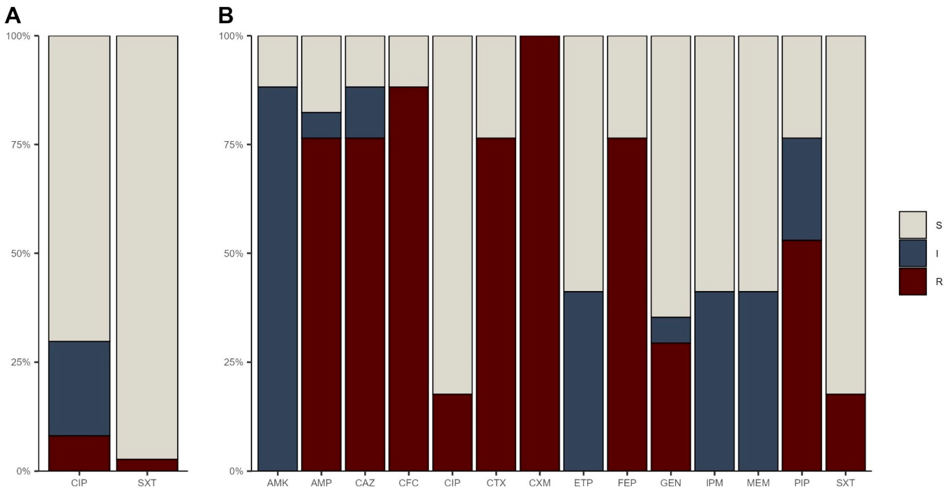
Antibiotic resistance was found in 32% (12/37) of isolates from the Matasnillo River. Resistance to ciprofloxacin was exhibited in 8% (3/37) of isolates, followed by 3% (1/37) to trimethoprim/sulfamethoxazole. Additionally, 22% (8/37) of the isolates displayed intermediate resistance to ciprofloxacin (Figure 2A).
The isolates from the WWTP showed resistance to the following antibiotics: 100% (17/17) to cefuroxime, 88% (15/17) to cefazolin, 76% (13/17) to ampicillin, ceftazidime, cefotaxime, and cefepime, 53% (9/17) to piperacillin-tazobactam, 29% (5/17) to gentamicin, 18% (3/17) to ciprofloxacin, and 18% (3/17) to trimethoprim/sulfamethoxazole. Furthermore, 88% (15/17) exhibited intermediate resistance, including 88% (15/17) to amikacin, 41% (7/17) to ertapenem, meropenem, and imipenem, 23% (4/17) to piperacillin-tazobactam, 12% (2/17) to ceftazidime, 6% (1/17) to gentamicin, and 6% (1/17) to ampicillin (Figure 2B).

3.2.1. Multidrug Resistance Profiles

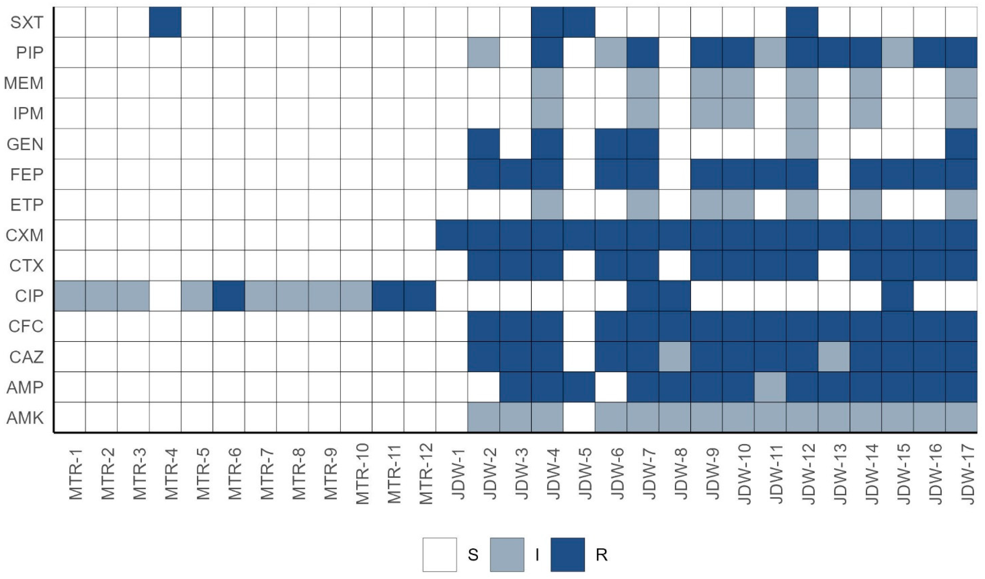
No multidrug-resistant bacteria were detected among the isolates from the Matasnillo River, following the antimicrobial susceptibility profile determination used in this study. In contrast, 94% (16/17) of the E. coli isolates from the WWTP exhibited multidrug resistance (Figure 3), which included resistance to beta-lactams (cephalosporins) and aminoglycosides in combination with quinolones, sulfonamides and carbapenems.
The total of quinolone-resistant isolates belonged to phylogroup A. All the river quinolone-resistant isolates were commensal while 2 isolates from the WWTP sample were ETEC.

3.2.2. Quinolone Resistance Mechanisms

Molecular detection revealed that 50% (7/14) of quinolone-resistant strains carried the qnrB gene (Supplementary Figure S3). All qnrB-positive isolates came from the Matasnillo River. The allele typing analysis of qnrB positives determined the prevalence of allele qnrB19. No other qnrB allele was found and aac(6′)-Ib-cr and qnrS alleles were not detected in any of the quinolone-resistant isolates.
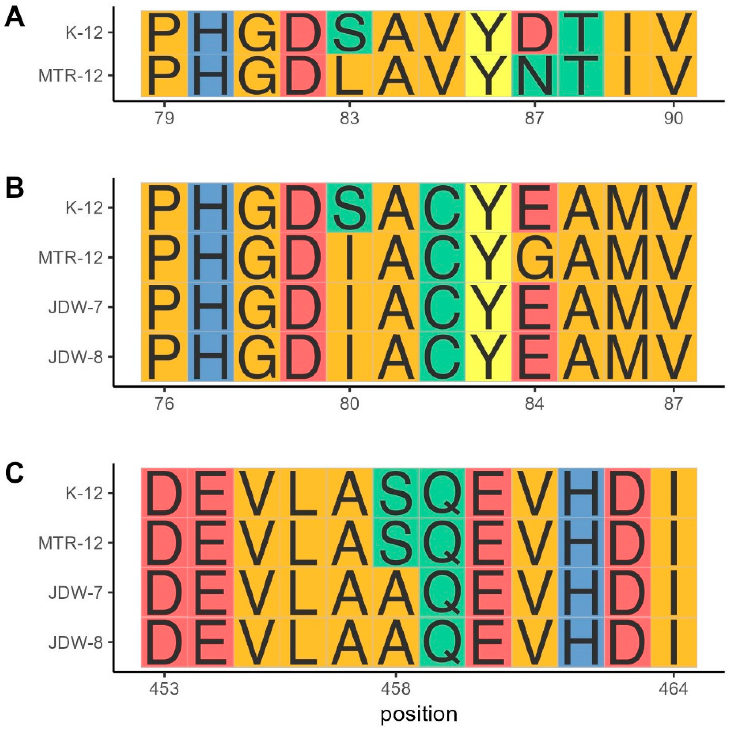
Mutations analysis revealed that 1 river isolate (MTR-12) presented gyrA mutations S83L and D87N (Figure 4A) and parC mutations S80I and E84G (Figure 4B). Two WWTP isolates (JDW-7 and JDW-8) presented both the same parC mutation (S80I, Figure 4B) and parE mutation (S458A, Figure 4C), even though gyrA sequences for these isolates could not be retrieved.

4. Discussion

This study focused on the characterization of Escherichia coli isolates obtained from untreated water sources, aiming to determine their antimicrobial resistance profiles and their phylogenetic and pathogenic groups. A high prevalence of multidrug-resistant isolates was found in the wastewater sample and a distinct quinolone resistance pattern was identified from the Matasnillo River isolates. Furthermore, pathogenic E. coli groups were only detected among wastewater isolates.
In this study, phylogroup A was predominant among the E. coli isolates from both water samples comprising all the isolates from the Matasnillo River and most isolates from the wastewater sample. This phylogroup is highly associated with human sources and most strains are commensal [31]. These findings are consistent with previous characterization of E. coli isolated from fecal-contaminated water sources and sewage [32,33].
Phylogroup A was predominant among the isolates from wastewater and lower but similar proportions of phylogroup D and B2 were detected. This distribution is a worldwide pattern, where phylogroup A is the most common followed by phylogroups D and B2, which usually have overlapping or low-frequency differences across locations [34]. Although phylogroup B2 and D are considered pathogens, phylogroup A is generally considered to represent commensal strains. It is important to note that some isolates within the phylogroup can carry virulence factors and cause disease, including urinary tract infections [35]. The differences between the distribution of virulence factors and phylogenetic groups may be related to geographical differences [36]. Therefore, the presence of commensal strains with quinolone resistance in the Matasnillo River indicates fecal contamination from humans or animals.
We investigated the presence of five clinically significant E. coli pathotypes—EHEC, EPEC, EIEC, ETEC, and DAEC—due to their relevance to public health and their association with outbreaks in Latin America [12]. Currently, little is known about the prevalence of circulating E. coli pathotypes in Panama. Publications about this topic were not found. Furthermore, the presence of pathogens in Panamanian water sources was not determined or published before. All isolates from the Matasnillo River were classified as commensal as none of the targeted virulence factors were detected in these isolates. Nevertheless, commensal E. coli is a potential reservoir for antibiotic resistance genes that can be transferred to other bacteria in the environment and within the host gut [37,38,39], thus continued monitoring of antibiotic resistance in Matasnillo River is crucial to manage and mitigate the spread of resistances in the city.
Despite the prevalence of commensal strains in the raw wastewater sample, EHEC and ETEC pathotypes were detected, posing a risk of infection for the WWTP workers and surrounding populations due to exposure to aerosolized bacteria. Both pathotypes remain important pathogens in Latin American countries. For example, EHEC infections in Argentina were a leading cause of acute renal failure among children and ETEC was associated with diarrheal cases in Mexico, Colombia, and Nicaragua [12]. Although the Shiga toxin producer EHEC-serotype O157 is worldwide predominant [40], the isolation methods suggest that the mentioned EHEC isolates do not belong to this serotype or are atypical O157:H7 since all exhibited β-Glucuronidase-positive phenotype and carried the stx1 gene, as previously reported [41].
Antibiotic resistance in bacteria is a pressing global issue, with members of the Enterobacteriaceae family being particularly concerned. In this study, E. coli isolates from both wastewater and the Matasnillo River exhibited varying resistance profiles. A clear difference in resistance distribution was observed between the two samples where wastewater isolates demonstrated resistance to a broader range of antibiotic families compared to those from river water. Interestingly, both wastewater and river isolates showed resistance to the fluoroquinolone/quinolone antibiotic class. This aligns with clinical surveillance reports from Panama, which highlight fluoroquinolone resistance as particularly widespread among E. coli isolates [42]. Thus, the detection of quinolone-resistant and intermediately resistant isolates in the Matasnillo River underscores the impact of anthropogenic pollution on environmental bacterial populations. This situation increases the risk of waterborne infections and facilitates the dissemination of antibiotic-resistance genes between humans, animals, and the environment, which, again, highlights the importance of continuous microbiological monitoring of surface waters.
Quinolone as well as fluoroquinolones antibiotics and by-products have been detected worldwide in surface waters due to their wide use in human and veterinary medicine along with their limited absorption and excretion into the environment [43]. The presence of this antibiotic class increases the evolution of bacteria and generates resistance as a consequence of selection pressure [44] and can also induce resistance to non-quinolone antibiotics by increasing mutation rates, mutation frequencies, and recombination [45]. The presence of quinolone-resistant bacteria in the Matasnillo River may indicate the presence of these compounds even though the concentration has not been determined, posing a major health risk.
Quinolone resistance in Matasnillo River isolates is primarily conferred by quinolone resistance genes. Previous studies determined that E. coli isolates from aquatic environments are sources of plasmid-mediated quinolone resistance mechanisms such as qnrS, qnrB, aac(6′)-Ib-cr, oqxAB and qepA [46]. Most studies reported a high prevalence of qnrS in E. coli isolates from surface water sources used for drinking, irrigation, and recreational activities [47,48,49]. However, a high prevalence of qnrB was detected among the Matasnillo River isolates while qnrS was not detected. Our findings suggest that the Matasnillo River is a reservoir for qnrB genes and support previous reports proposing that the distribution of predominant quinolone-resistance mechanisms varies over geographical regions [50,51,52]. In this study, the allele qnrB19 is prevalent within the quinolone resistance isolates from the Matasnillo River. In Latin American countries, the same allele was highly prevalent and mainly carried by commensal E. coli and Salmonella spp. isolated from human and water samples [53,54,55]. A similar prevalence of qnrB19 was documented in different geographical regions including Enterobacteria isolated from free-living animals, stock, and food samples [56,57,58]. Moreover, identical qnrB19-carrying plasmids have been found in unrelated Salmonella serotypes and E. coli, suggesting the dissemination of quinolone resistance through horizontal transfer mechanisms [59]. Furthermore, plasmid transmission can lead to chromosome stabilization via transposition as previously reported in chromosomal qnrB19-carrying E. coli isolates [60].
Quinolone-resistance genes qnrB and qnrS were not detected in wastewater isolates. On the contrary, chromosomal mutations in parC and parE genes were identified. Comparably, chromosomal mutations in gyrA, parC, and parE genes have been reported as the primary cause of quinolone resistance rather than resistance genes in E. coli isolated from wastewater samples [61]. Wastewater isolates also exhibited resistance to cephalosporins, which suggests the production of cephalosporinases or extended-spectrum beta-lactamases (ESBL). High rates of ESBL-producing Escherichia coli resistant to quinolones have been also reported from samples collected from wastewater treatment plant influents [62]. Even though this study used a cross-sectional design as a first screening for antibiotic resistance profiles among environmental and wastewater E. coli isolates, our results emphasize the importance of continuous monitoring of the microbiological quality of circulating waters in Panama City.

5. Conclusions

This study has determined the presence of pathogenic and antibiotic-resistant E. coli strains in circulating waters in Panama City, highlighting the need for routine surveillance and stricter monitoring of microbial contaminants in wastewater before environmental discharge.
The isolation, identification, and characterization of E. coli pathotypes are important indicators of contamination when considering water quality. Furthermore, the study of the resistance profiles of these environmental strains is highly relevant for the ongoing monitoring of our surrounding environments, based on the one-health concept.
We recommend conducting periodic studies to assess the evolution of circulating resistant strains that impact public health. We should also consider comparing environmental monitoring isolates with clinical information to establish state actions to curb antibiotic resistance.

Supplementary Materials

The following supporting information can be downloaded at: https://www.mdpi.com/article/10.3390/pathogens14070617/s1, Figure S1: Detection of chuA gene; Figure S2: Results of Multiplex PCR 2 for detection of EHEC, EIEC and ETEC pathotypes; Figure S3: PCR detection of qnrB group.

Author Contributions

Conceptualization, J.Q.-A.; methodology, J.Q.-A., F.M.-M. and C.I.E.; validation and formal analysis, J.Q.-A., J.R.M.-S. and J.E.M.P.; investigation, G.A.R.G. and E.M.; resources, G.A.R.G., E.M., J.E.M.P., A.O.M.T. and J.Q.-A.; data curation, J.R.M.-S. and J.Q.-A.; writing—original draft preparation, G.A.R.G. and E.M.; writing—review and editing, J.Q.-A., J.R.M.-S., J.E.M.P., A.O.M.T. and C.I.E.; supervision, J.Q.-A.; project administration, C.I.E. and J.Q.-A.; funding acquisition, G.A.R.G., E.M., A.O.M.T. and J.Q.-A. All authors have read and agreed to the published version of the manuscript.

Funding

This research was funded by the Vice-Rectorate for Research and Postgraduate Studies (VIP) of the University of Panama (Universidad de Panama, CUFI-2022-CNET-EG-001), the 2022 New Researchers Call (APY-NI-2022-10, SENACYT) and the SNI fund 2038-2022. The Ministry of Environment (MIAMBIENTE, agreement No. 0-26-45-2019).

Data Availability Statement

All the data that supports the findings of this study are available from the corresponding author J.Q.A, upon reasonable request.

Conflicts of Interest

The authors declare no conflicts of interest. The funders had no role in the design of the study; in the collection, analyses, or interpretation of data; in the writing of the manuscript; or in the decision to publish the results.

References

  1. Troeger, C.; Blacker, B.F.; Khalil, I.A.; Rao, P.C.; Cao, S.; Zimsen, S.R.M.; Albertson, S.B.; Stanaway, J.D.; Deshpande, A.; Abebe, Z.; et al. Estimates of the Global, Regional, and National Morbidity, Mortality, and Aetiologies of Diarrhoea in 195 Countries: A Systematic Analysis for the Global Burden of Disease Study 2016. Lancet Infect. Dis. 2018, 18, 1211–1228. [Google Scholar] [CrossRef] [PubMed]
  2. Jiwok, J.C.; Adebowale, A.S.; Wilson, I.; Kancherla, V.; Umeokonkwo, C.D. Patterns of Diarrhoeal Disease among Under-Five Children in Plateau State, Nigeria, 2013–2017. BMC Public Health 2021, 21, 2086. [Google Scholar] [CrossRef] [PubMed]
  3. Gordon, B.; Boisson, S.; Johnston, R.; Trouba, D.J.; Cumming, O. Unsafe Water, Sanitation and Hygiene: A Persistent Health Burden. Bull. World Health Organ. 2023, 101, 551-551A. [Google Scholar] [CrossRef] [PubMed]
  4. Corzo-Ariyama, H.A.; García-Heredia, A.; Heredia, N.; García, S.; León, J.; Jaykus, L.; Solís-Soto, L. Phylogroups, Pathotypes, Biofilm Formation and Antimicrobial Resistance of Escherichia Coli Isolates in Farms and Packing Facilities of Tomato, Jalapeño Pepper and Cantaloupe from Northern Mexico. Int. J. Food Microbiol. 2019, 290, 96–104. [Google Scholar] [CrossRef]
  5. Halaji, M.; Fayyazi, A.; Rajabnia, M.; Zare, D.; Pournajaf, A.; Ranjbar, R. Phylogenetic Group Distribution of Uropathogenic Escherichia Coli and Related Antimicrobial Resistance Pattern: A Meta-Analysis and Systematic Review. Front. Cell. Infect. Microbiol. 2022, 12, 790184. [Google Scholar] [CrossRef]
  6. Morel-Journel, T.; Lehtinen, S.; Cotto, O.; Amia, R.; Clermont, O.; Dion, S.; Figueroa, C.; Martinson, J.; Ralaimazava, P.; Duval, X.; et al. Residence-Colonization Trade-off and Niche Differentiation Enable the Coexistence of Escherichia Coli Phylogroups in Healthy Humans. bioRxiv 2024. [Google Scholar] [CrossRef]
  7. Machado, M.; Panzenhagen, P.; Aburjaile, F.F.; Brenig, B.; da Costa, M.M.; Azevedo, V.A.d.C.; de Souza Figueiredo, E.E.; Conte-Junior, C.A. Evolution of Pathogenic Escherichia Coli Harboring the Transmissible Locus of Stress Tolerance: From Food Sources to Clinical Environments. Sci. Rep. 2025, 15, 5014. [Google Scholar] [CrossRef]
  8. Pokharel, P.; Dhakal, S.; Dozois, C.M. The Diversity of Escherichia Coli Pathotypes and Vaccination Strategies against This Versatile Bacterial Pathogen. Microorganisms 2023, 11, 344. [Google Scholar] [CrossRef]
  9. Saka, H.K.; Dabo, N.T.; Muhammad, B.; García-Soto, S.; Ugarte-Ruiz, M.; Alvarez, J. Diarrheagenic Escherichia Coli Pathotypes From Children Younger Than 5 Years in Kano State, Nigeria. Front. Public Health 2019, 7, 348. [Google Scholar] [CrossRef]
  10. Robert, B.; Richard, J.; Shane, K.; Attila, H.; Tom, S. Monitoring Drinking Water Quality in Nationally Representative Household Surveys in Low- and Middle-Income Countries: Cross-Sectional Analysis of 27 Multiple Indicator Cluster Surveys 2014–2020. Environ. Health Perspect. 2025, 129, 97010. [Google Scholar] [CrossRef]
  11. Abdulhadi, R.; Bailey, A.; Van Noorloos, F. Access Inequalities to WASH and Housing in Slums in Low- and Middle-Income Countries (LMICs): A Scoping Review. Glob. Public Health 2024, 19, 2369099. [Google Scholar] [CrossRef] [PubMed]
  12. Torres, A.G. Escherichia Coli Diseases in Latin America—A ‘One Health’ Multidisciplinary Approach. Pathog. Dis. 2017, 75, ftx012. [Google Scholar] [CrossRef] [PubMed]
  13. Canizalez-Roman, A.; Velazquez-Roman, J.; Valdez-Flores, M.A.; Flores-Villaseñor, H.; Vidal, J.E.; Muro-Amador, S.; Guadrón-Llanos, A.M.; Gonzalez-Nuñez, E.; Medina-Serrano, J.; Tapia-Pastrana, G.; et al. Detection of Antimicrobial-Resistance Diarrheagenic Escherichia Coli Strains in Surface Water Used to Irrigate Food Products in the Northwest of Mexico. Int. J. Food Microbiol. 2019, 304, 1–10. [Google Scholar] [CrossRef] [PubMed]
  14. Aijuka, M.; Santiago, A.E.; Girón, J.A.; Nataro, J.P.; Buys, E.M. Enteroaggregative Escherichia Coli Is the Predominant Diarrheagenic E. Coli Pathotype among Irrigation Water and Food Sources in South Africa. Int. J. Food Microbiol. 2018, 278, 44–51. [Google Scholar] [CrossRef]
  15. Park, J.; Kim, J.S.; Kim, S.; Shin, E.; Oh, K.-H.; Kim, Y.; Kim, C.H.; Hwang, M.A.; Jin, C.M.; Na, K.; et al. A Waterborne Outbreak of Multiple Diarrhoeagenic Escherichia Coli Infections Associated with Drinking Water at a School Camp. Int. J. Infect. Dis. 2018, 66, 45–50. [Google Scholar] [CrossRef]
  16. Lim, M.-A.; Kim, J.-Y.; Acharya, D.; Bajgain, B.B.; Park, J.-H.; Yoo, S.-J.; Lee, K. A Diarrhoeagenic Enteropathogenic Escherichia Coli (EPEC) Infection Outbreak That Occurred among Elementary School Children in Gyeongsangbuk-Do Province of South Korea Was Associated with Consumption of Water-Contaminated Food Items. Int. J. Environ. Res. Public Health 2020, 17, 3149. [Google Scholar] [CrossRef]
  17. Zelelie, T.Z.; Eguale, T.; Yitayew, B.; Abeje, D.; Alemu, A.; Seman, A.; Jass, J.; Mihret, A.; Abebe, T. Molecular Epidemiology and Antimicrobial Susceptibility of Diarrheagenic Escherichia Coli Isolated from Children under Age Five with and without Diarrhea in Central Ethiopia. PLoS ONE 2023, 18, e0288517. [Google Scholar] [CrossRef]
  18. Tapia-Pastrana, G.; Rojas-Bautista, M.; Hernández-Pérez, P.; Santiago-Martínez, O.; Gómez-Rodríguez, L.C.; Terrazas-Luna, V.M.; Montes-Yedra, J.; Bautista-Avendaño, A.A.; García-López, E.S.; Leon-Sicairos, N.; et al. Virulence Genes, Antimicrobial Resistance Profile, Phylotyping and Pathotyping of Diarrheagenic Escherichia Coli Isolated from Children in Southwest Mexico. PLoS ONE 2024, 19, e0300304. [Google Scholar] [CrossRef]
  19. Angulo-Zamudio, U.A.; Gutiérrez-Jiménez, J.; Monroy-Higuera, L.; Flores-Villaseñor, H.; Leon-Sicairos, N.; Velazquez-Roman, J.; Vidal, J.E.; Tapia-Pastrana, G.; Canizalez-Roman, A. Non-Diarrheagenic and Diarrheagenic E. Coli Carrying Supplementary Virulence Genes (SVG) Are Associated with Diarrhea in Children from Mexico. Microb. Pathog. 2021, 157, 104994. [Google Scholar] [CrossRef]
  20. Han, H.; Li, W.; Liu, J.; Zhang, X.; Huo, X.; Sun, Y.; Chen, J.; Fan, R.; Zhang, J.; Chen, Y.; et al. Seven-Year Overview of Antimicrobial Resistance in Diarrheagenic Escherichia Coli from Sporadic Human Diarrhea Cases in 20 Chinese Provinces. One Health Adv. 2024, 2, 29. [Google Scholar] [CrossRef]
  21. Salleh, M.Z.; Nik Zuraina, N.M.N.; Hajissa, K.; Ilias, M.I.; Deris, Z.Z. Prevalence of Multidrug-Resistant Diarrheagenic Escherichia Coli in Asia: A Systematic Review and Meta-Analysis. Antibiotics 2022, 11, 1333. [Google Scholar] [CrossRef] [PubMed]
  22. Mittal, A.K.; Bhardwaj, R.; Mishra, P.; Rajput, S.K. Antimicrobials Misuse/Overuse: Adverse Effect, Mechanism, Challenges and Strategies to Combat Resistance. Open Biotechnol. J. 2020, 14, 107–112. [Google Scholar] [CrossRef]
  23. American Public Health Association; American Water Works Association; Water Environment Federation. Standard Methods for the Examination of Water and Wastewater, 24th ed.; Lipps, W.C., Braun-Howland, E.B., Baxter, T.E., Eds.; American Water Works Association: Washington, DC, USA, 2023. [Google Scholar]
  24. Clermont, O.; Bonacorsi, S.; Bingen, E. Rapid and Simple Determination of the Escherichia Coli Phylogenetic Group. Appl. Environ. Microbiol. 2000, 66, 4555–4558. [Google Scholar] [CrossRef] [PubMed]
  25. Chandra, M.; Cheng, P.; Rondeau, G.; Porwollik, S.; McClelland, M. A Single Step Multiplex PCR for Identification of Six Diarrheagenic E. Coli Pathotypes and Salmonella. Int. J. Med. Microbiol. 2013, 303, 210–216. [Google Scholar] [CrossRef]
  26. Le, T.A.H.; Fabre, L.; Roumagnac, P.; Grimont, P.A.; Scavizzi, M.R.; Weill, F.X. Clonal Expansion and Microevolution of Quinolone-Resistant Salmonella Enterica Serotype Typhi in Vietnam from 1996 to 2004. J. Clin. Microbiol. 2007, 45, 3485–3492. [Google Scholar] [CrossRef]
  27. Yang, F.; Zhang, S.; Shang, X.; Wang, L.; Li, H.; Wang, X. Characteristics of Quinolone-Resistant Escherichia Coli Isolated from Bovine Mastitis in China. J. Dairy Sci. 2018, 101, 6244–6252. [Google Scholar] [CrossRef]
  28. Zhou, L.; Feng, T.; Xu, S.; Gao, F.; Lam, T.T.; Wang, Q.; Wu, T.; Huang, H.; Zhan, L.; Li, L.; et al. Ggmsa: A Visual Exploration Tool for Multiple Sequence Alignment and Associated Data. Brief. Bioinform. 2022, 23, bbac222. [Google Scholar] [CrossRef]
  29. Fernández-Martínez, M.; Miró, E.; Ortega, A.; Bou, G.; González-López, J.J.; Oliver, A.; Pascual, A.; Cercenado, E.; Oteo, J.; Martínez-Martínez, L.; et al. Molecular Identification of Aminoglycoside-Modifying Enzymes in Clinical Isolates of Escherichia Coli Resistant to Amoxicillin/Clavulanic Acid Isolated in Spain. Int. J. Antimicrob. Agents 2015, 46, 157–163. [Google Scholar] [CrossRef]
  30. Salah, F.D.; Soubeiga, S.T.; Ouattara, A.K.; Sadji, A.Y.; Metuor-Dabire, A.; Obiri-Yeboah, D.; Banla-Kere, A.; Karou, S.; Simpore, J. Distribution of Quinolone Resistance Gene (Qnr) in ESBL-Producing Escherichia Coli and Klebsiella spp. in Lomé, Togo. Antimicrob. Resist. Infect. Control 2019, 8, 104. [Google Scholar] [CrossRef]
  31. Aguirre-Sánchez, J.R.; Valdez-Torres, J.B.; del Campo, N.C.; Martínez-Urtaza, J.; del Campo, N.C.; Lee, B.G.; Quiñones, B.; Chaidez-Quiroz, C. Phylogenetic Group and Virulence Profile Classification in Escherichia Coli from Distinct Isolation Sources in Mexico. Infect. Genet. Evol. 2022, 106, 105380. [Google Scholar] [CrossRef]
  32. Redha, M.A.; Al Sweih, N.; Albert, M.J. Virulence and Phylogenetic Groups of Escherichia Coli Cultured from Raw Sewage in Kuwait. Gut Pathog. 2022, 14, 18. [Google Scholar] [CrossRef] [PubMed]
  33. Gomes, L.; Bordalo, A.A.; Machado, A. Characterization of Escherichia Coli Isolates in Recreational Waters: Implications for Public Health and One Health Approach. Water 2024, 16, 2695. [Google Scholar] [CrossRef]
  34. Stoppe, N.d.C.; Silva, J.S.; Carlos, C.; Sato, M.I.Z.; Saraiva, A.M.; Ottoboni, L.M.M.; Torres, T.T. Worldwide Phylogenetic Group Patterns of Escherichia Coli from Commensal Human and Wastewater Treatment Plant Isolates. Front. Microbiol. 2017, 8, 2512. [Google Scholar] [CrossRef] [PubMed]
  35. Denamur, E.; Clermont, O.; Bonacorsi, S.; Gordon, D. The Population Genetics of Pathogenic Escherichia Coli. Nat. Rev. Microbiol. 2021, 19, 37–54. [Google Scholar] [CrossRef]
  36. Hemati, S.; Halimi, S.; Jabalameli, F.; Emaneini, M.; Beigverdi, R. Phylogenetic Group, Antibiotic Resistance, Virulence Gene, and Genetic Diversity of Escherichia Coli Causing Bloodstream Infections in Iran. Front. Microbiol. 2024, 15, 1426510. [Google Scholar] [CrossRef]
  37. Singh, N.S.; Singhal, N.; Kumar, M.; Virdi, J.S. High Prevalence of Drug Resistance and Class 1 Integrons in Escherichia Coli Isolated from River Yamuna, India: A Serious Public Health Risk. Front. Microbiol. 2021, 12, 621564. [Google Scholar] [CrossRef]
  38. Bong, C.W.; Low, K.Y.; Chai, L.C.; Lee, C.W. Prevalence and Diversity of Antibiotic Resistant Escherichia Coli From Anthropogenic-Impacted Larut River. Front. Public Health 2022, 10, 794513. [Google Scholar] [CrossRef]
  39. Tawfick, M.M.; Elshamy, A.A.; Mohamed, K.T.; El Menofy, N.G. Gut Commensal Escherichia Coli, a High-Risk Reservoir of Transferable Plasmid-Mediated Antimicrobial Resistance Traits. Infect. Drug Resist. 2022, 15, 1077–1091. [Google Scholar] [CrossRef]
  40. Correa-Martinez, C.L.; Leopold, S.R.; Köck, R.; Kossow, A.; Bauwens, A.; Mellmann, A. Enterohemorrhagic E. coli (EHEC): Environmental-Vehicle-Human Interface BT—Zoonoses: Infections Affecting Humans and Animals; Sing, A., Ed.; Springer International Publishing: Cham, Switzerland, 2023; pp. 355–372. ISBN 978-3-031-27164-9. [Google Scholar]
  41. Ogura, Y.; Seto, K.; Morimoto, Y.; Nakamura, K.; Sato, M.; Gotoh, Y.; Itoh, T.; Toyoda, A.; Ohnishi, M.; Hayashi, T. Genomic Characterization of β-Glucuronidase–Positive Escherichia coli O157:H7 Producing Stx2a. Emerg. Infect. Dis. J. 2018, 24, 2219. [Google Scholar] [CrossRef]
  42. Moreno, J.; Conte, E.; Tribaldos, M.; Morales, Y.; Zamorano, C.; Gómez, B.; Toro, J. Antibiotic Resistance Profiles in Panama: Trends from 2007 to 2013. Pharm. Pharmacol. Int. J. 2018, 6, 350–355. [Google Scholar] [CrossRef]
  43. Hu, H.; Da, X.; Li, Z.; Li, T.; Zhang, X.; Bian, T.; Jin, Y.; Xu, K.; Guo, Y. Determination and Ecological Risk Assessment of Quinolone Antibiotics in Drinking and Environmental Waters Using Fully Automated Disk-Based SPE Coupled with UPLC-MS/MS. Molecules 2024, 29, 4611. [Google Scholar] [CrossRef] [PubMed]
  44. Bhatt, S.; Chatterjee, S. Fluoroquinolone Antibiotics: Occurrence, Mode of Action, Resistance, Environmental Detection, and Remediation—A Comprehensive Review. Environ. Pollut. 2022, 315, 120440. [Google Scholar] [CrossRef] [PubMed]
  45. Bush, N.G.; Diez-Santos, I.; Sankara Krishna, P.; Clavijo, B.; Maxwell, A. Insights into Antibiotic Resistance Promoted by Quinolone Exposure. Antimicrob. Agents Chemother. 2024, 69, e00997-24. [Google Scholar] [CrossRef] [PubMed]
  46. Amin, M.B.; Saha, S.R.; Islam, M.R.; Haider, S.M.A.; Hossain, M.I.; Chowdhury, A.S.M.H.K.; Rousham, E.K.; Islam, M.A. High Prevalence of Plasmid-Mediated Quinolone Resistance (PMQR) among E. Coli from Aquatic Environments in Bangladesh. PLoS ONE 2021, 16, e0261970. [Google Scholar] [CrossRef]
  47. Amato, M.; Dasí, D.; González, A.; Ferrús, M.A.; Castillo, M.Á. Occurrence of Antibiotic Resistant Bacteria and Resistance Genes in Agricultural Irrigation Waters from Valencia City (Spain). Agric. Water Manag. 2021, 256, 107097. [Google Scholar] [CrossRef]
  48. Amarasiri, M.; Takezawa, T.; Malla, B.; Furukawa, T.; Sherchand, J.B.; Haramoto, E.; Sei, K. Prevalence of Antibiotic Resistance Genes in Drinking and Environmental Water Sources of the Kathmandu Valley, Nepal. Front. Microbiol. 2022, 13, 894014. [Google Scholar] [CrossRef]
  49. Beshiru, A.; Isokpehi, N.A.; Igbinosa, I.H.; Akinnibosun, O.; Ogofure, A.G.; Igbinosa, E.O. Extended-Spectrum Beta-Lactamase (ESBL)- and Non-ESBL Producing Escherichia Coli Surveillance in Surface Water Sources in Edo State, Nigeria: A Public Health Concern. Sci. Rep. 2024, 14, 21658. [Google Scholar] [CrossRef]
  50. Naveen Kumar, V.; Saikumar, C. Prevalence of Quinolone-Resistance Uropathogenic Escherichia Coli in a Tertiary Care Hospital in South Iran. Indian J. Public Health Res. Dev. 2019, 10, 2609–2613. [Google Scholar] [CrossRef]
  51. Zhao, Y.-C.; Sun, Z.-H.; Xiao, M.-X.; Li, J.-K.; Liu, H.; Cai, H.-L.; Cao, W.; Feng, Y.; Zhang, B.-K.; Yan, M. Analyzing the Correlation between Quinolone-Resistant Escherichia Coli Resistance Rates and Climate Factors: A Comprehensive Analysis across 31 Chinese Provinces. Environ. Res. 2024, 245, 117995. [Google Scholar] [CrossRef]
  52. Yanat, B.; Rodríguez-Martínez, J.-M.; Touati, A. Plasmid-Mediated Quinolone Resistance in Enterobacteriaceae: A Systematic Review with a Focus on Mediterranean Countries. Eur. J. Clin. Microbiol. Infect. Dis. 2017, 36, 421–435. [Google Scholar] [CrossRef]
  53. Pallecchi, L.; Riccobono, E.; Mantella, A.; Bartalesi, F.; Sennati, S.; Gamboa, H.; Gotuzzo, E.; Bartoloni, A.; Rossolini, G.M. High Prevalence of Qnr Genes in Commensal Enterobacteria from Healthy Children in Peru and Bolivia. Antimicrob. Agents Chemother. 2009, 53, 2632–2635. [Google Scholar] [CrossRef] [PubMed]
  54. Chen, Z.; Delgado Suárez, E.J.; Bonelli, R.R.; Oliveira, C.J.B.; Moreno-Switt, A.I.; Adell, A.D.; Reyes-Jara, A.; Grim, C.J.; Allard, M.W.; Tallent, S.M.; et al. Exploring the Genomic and Antimicrobial Resistance Tapestry: Comparative Insights into Salmonella Enterica Serotypes Agona, Braenderup, Muenchen, and Panama in Latin American Surface Waters. Microbiol. Spectr. 2024, 13, e01706-24. [Google Scholar] [CrossRef] [PubMed]
  55. Chiluisa Guacho, C.; Gómez Martínez, N.; Vilema Vizuete, E.G.; Dutra Asensi, M. The first report of the qnrB19 and aac(6’)-Ib-cr in isolates uropathogenic Escherichia coli ciprofloxacin-resistant in Ecuador. Interam. J. Health Sci. 2024, 4, 179. [Google Scholar] [CrossRef]
  56. Furmanek-Blaszk, B.; Sektas, M.; Rybak, B. High Prevalence of Plasmid-Mediated Quinolone Resistance among ESBL/AmpC-Producing Enterobacterales from Free-Living Birds in Poland. Int. J. Mol. Sci. 2023, 24, 12804. [Google Scholar] [CrossRef]
  57. Tyson, G.H.; Li, C.; Hsu, C.-H.; Bodeis-Jones, S.; McDermott, P.F. Diverse Fluoroquinolone Resistance Plasmids From Retail Meat E. Coli in the United States. Front. Microbiol. 2019, 10, 2826. [Google Scholar] [CrossRef]
  58. Laurent, P.; Jean-Yves, M.; Agnese, L.; Anne-Kathrin, S.; Nicolas, K.; Patrice, N.; Stefan, S. Antimicrobial Resistance in Escherichia Coli. Microbiol. Spectr. 2018, 6, 10-1128. [Google Scholar] [CrossRef]
  59. Moreno-Switt, A.I.; Pezoa, D.; Sepúlveda, V.; González, I.; Rivera, D.; Retamal, P.; Navarrete, P.; Reyes-Jara, A.; Toro, M. Transduction as a Potential Dissemination Mechanism of a Clonal QnrB19-Carrying Plasmid Isolated from Salmonella of Multiple Serotypes and Isolation Sources. Front. Microbiol. 2019, 10, 2503. [Google Scholar] [CrossRef]
  60. Thanh, H.H.T.; Mayumi, Y.; Manuel, C.; Carlos, B.-C.; Yoshimasa, Y. Chromosomal QnrB19-Carrying Escherichia Coli Isolated from the Stool Sample of a Community Resident in Ecuador. Microbiol. Resour. Announc. 2024, 13, e00046-24. [Google Scholar] [CrossRef]
  61. Park, J.-H.; Kang, J.; Bae, K.-S.; Lee, H.; Kim, J.; Park, E.-R.; Yoon, J.-K.; Lee, S.-H. Evaluation and Characterization of Quinolone-Resistant Escherichia Coli in Wastewater Treatment Plant Effluents. Water 2023, 15, 4040. [Google Scholar] [CrossRef]
  62. Al-Mustapha, A.I.; Tiwari, A.; Laukkanen-Ninios, R.; Lehto, K.-M.; Oikarinen, S.; Lipponen, A.; Pitkänen, T.; Heikinheimo, A.; Heljanko, V.; Johansson, V.; et al. Wastewater Based Genomic Surveillance Key to Population Level Monitoring of AmpC/ESBL Producing Escherichia Coli. Sci. Rep. 2025, 15, 7400. [Google Scholar] [CrossRef]
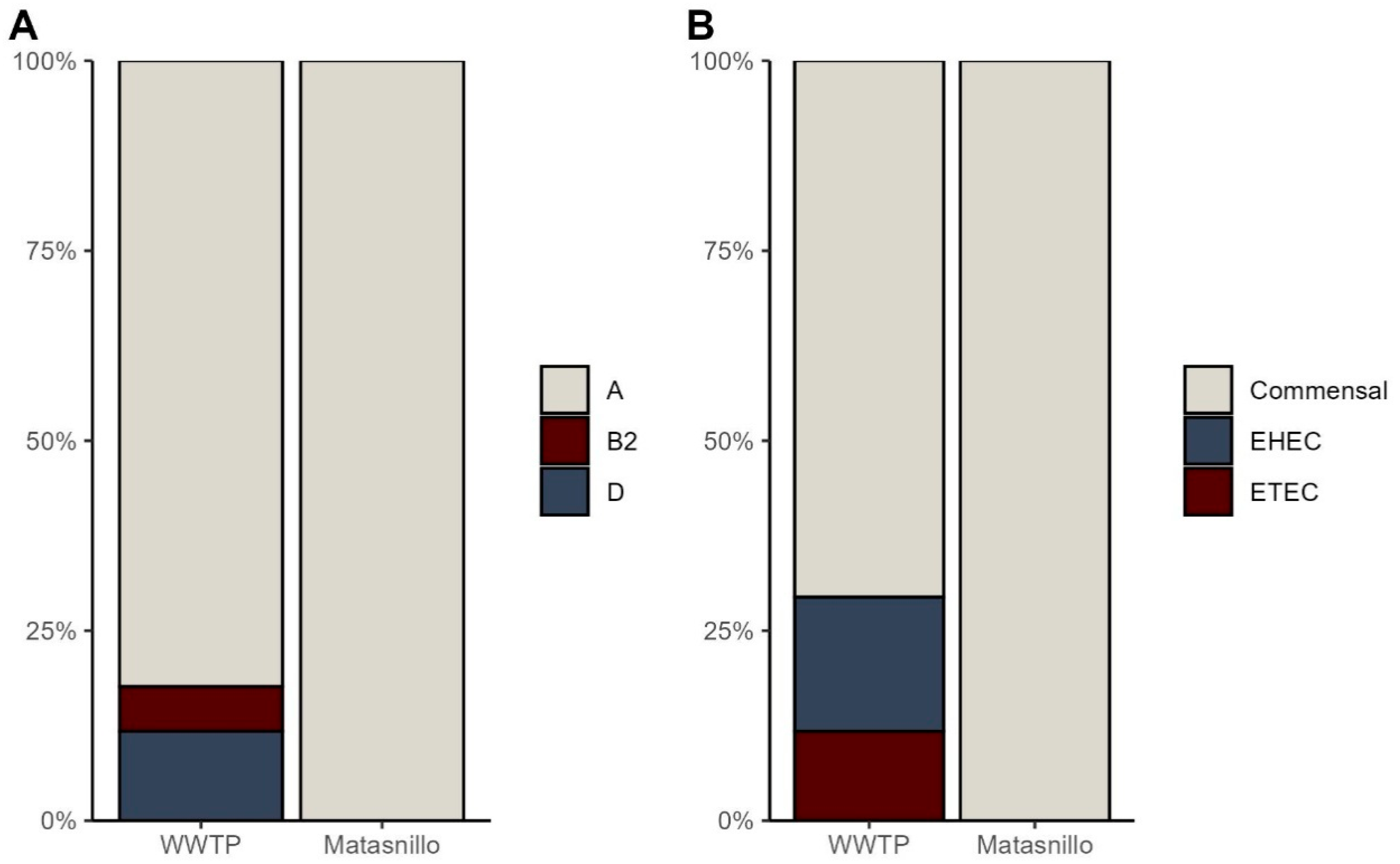
Figure 1. E. coli phylogroups (A) and pathotypes (B) from Juan Díaz WWTP and Matasnillo River.
Pathogens 14 00617 g001
Figure 2. Proportion of antibiotic resistance phenotypes from E. coli strains isolated from (A) the Matasnillo River and (B) the Juan Diaz Wastewater Treatment Plant (WWTP): susceptible (S), intermediate (I), and resistant (R). Antibiotics: amikacin (AMK), ampicillin (AMP), ceftazidime (CAZ), cefazolin (CFC), ciprofloxacin (CIP), cefotaxime (CTX), cefuroxime (CXM), ertapenem (ETP), cefepime (FEP), gentamicin (GEN), imipenem (IPM), meropenem (MEM), piperacillin-tazobactam (PIP) and trimethoprim-sulfamethoxazole (SXT).
Pathogens 14 00617 g002
Figure 3. Antibiotic resistance profiles of E. coli isolates from the Matasnillo River (MTR-1 to MTR-12) and Juan Diaz Wastewater Treatment Plant (JDW-1 to JDW-17). The color fill indicates the resistance phenotype: S = susceptible, I = intermediate, and R = resistant.
Pathogens 14 00617 g003
Figure 4. Amino acids alignments of (A) gyrA, (B) parC, and (C) parE regions and identified mutations conferring resistance to quinolones. Amino acids are colored according to their side-chain chemistry.
Pathogens 14 00617 g004
Table 1. Pathotype multiplex PCR targeting genes.
PathotypeGene TargetMultiplex PCR 1Multiplex PCR 2
EPECeae, bfpBeae, bfpB
EHECeae, stx1, stx2stx2stx1
EIECvirF, ipaHvirFipaH
DAECdaaEdaaE
ETECestB1 estB1
Disclaimer/Publisher’s Note: The statements, opinions and data contained in all publications are solely those of the individual author(s) and contributor(s) and not of MDPI and/or the editor(s). MDPI and/or the editor(s) disclaim responsibility for any injury to people or property resulting from any ideas, methods, instructions or products referred to in the content.