Article

Experimental Research on the Behavior and Crack-Sealing Capability of Masonry Mortars Reinforced with PVA Fibers


1
Faculty of Civil Engineering, Technical University of Cluj-Napoca, EUT+, 28 Memorandumului Street, 400114 Cluj-Napoca, Romania
2
NIRD URBAN-INCERC Cluj-Napoca Branch, National Institute for Research and Development in Construction, Urban Planning and Sustainable Spatial Development, 117 Calea Floresti, 400524 Cluj-Napoca, Romania
3
Mediterranean Agroforestry Institute (IAM), Universitat Politècnica de València, Camino de Vera s/n, 46022 Valencia, Spain
4
Department of Horticulture and Landscape, University of Agricultural Sciences and Veterinary Medicine Cluj-Napoca, 3–5 Manastur Street, 400372 Cluj-Napoca, Romania
5
Department of Geology, Faculty of Biology and Geology, Babeș-Bolyai University, 400084 Cluj-Napoca, Romania
6
NIRD URBAN-INCERC Bucharest Branch, National Institute for Research and Development in Construction, Urban Planning and Sustainable Spatial Development, Sos. Pantelimon, 266, 021652 Bucharest, Romania
*
Author to whom correspondence should be addressed.
Buildings 2026, 16(18), 3697; https://doi.org/10.3390/buildings16183697
Submission received: 31 July 2026 / Revised: 7 September 2026 / Accepted: 12 September 2026 / Published: 16 September 2026

Abstract

The durability of cementitious materials is essential for the development of sustainable construction, and the integration of self-healing mechanisms can reduce damage and maintenance costs. This paper analyzes the self-healing capacity of masonry mortars reinforced with polyvinyl alcohol (PVA) fibers and crystalline additives, when exposed to repeated wet-dry cycles. Four mortar mixtures were investigated, including two control mixtures and two mixtures modified with self-healing agents. The experimental program included determination of density, water absorption, compressive strength, and flexural strength, as well as monitoring the evolution of microcracks during the healing process. The results showed that modified mortars exhibited complete surface crack sealing under the applied wet-dry conditioning regime, including an initial crack width of 346 μm, which was completely sealed within 48 h. The observed crack-sealing behavior may result from the combined contribution of autogenous healing processes, PVA-fiber crack bridging, and the crystalline admixture; however, their individual contributions and possible synergistic effects cannot be distinguished from the present experimental design. The results indicate the potential of the investigated modified mortars to improve crack-sealing performance and contribute to enhanced durability. However, no direct environmental or life-cycle assessment was performed in the present study; therefore, potential sustainability benefits associated with service-life extension and reduced repair demand require further quantitative assessment.

1. Introduction

The durability and sustainability of construction materials represent some of the most significant challenges in modern civil engineering, given the continuous increase in requirements for structural safety, economic efficiency, and reduced environmental impact [1,2,3]. Cement-based materials form the foundation of most civil, industrial, and infrastructure structures, due to their high mechanical strength, technological versatility, and relatively low production costs [4]. However, their long-term performance is limited by the onset of degradation processes caused by the simultaneous action of mechanical stresses and environmental factors [5]. Cracking is one of the most significant mechanisms of deterioration, directly affecting the durability and service life of structures [6].
In recent decades, the criteria used to evaluate the performance of cementitious materials have evolved significantly [7]. Whereas in the past the focus was almost exclusively on developing materials with high mechanical strength, research today is geared toward creating materials capable of combining structural performance with durability, sustainability, and smart functionalities [8,9]. The increasing emphasis on circular economy principles, resource efficiency, and the extension of the service life of buildings has stimulated the development of advanced cementitious materials capable of limiting degradation processes and potentially reducing the frequency of maintenance and rehabilitation interventions [10,11]. Self-healing cementitious materials are particularly relevant in this context because their ability to seal cracks may contribute to maintaining material performance and extending service life. However, such durability-related benefits should not be directly interpreted as evidence of reduced environmental impact, as the overall sustainability of these materials also depends on factors such as binder composition, clinker content, production impacts, and service-life extension [10,11].
As illustrated in Figure 1, the development of cementitious materials has been driven by the gradual evolution of the requirements for modern construction. This evolution began with ensuring stability and mechanical strength, continued with the optimization of durability and structural performance, and in the last two decades, the focus has shifted toward reducing environmental impact and developing smart materials. In this context, self-healing materials represent one of the most promising areas of research, as they help limit the effects of cracking, reduce permeability, and extend the service life of structures [12,13].
In addition to durability, mechanical performance, and sustainability, fire safety represents an important consideration in the development of advanced cementitious materials for building applications. Although cement-based mortars are generally characterized by favorable behavior under fire exposure due to their predominantly inorganic composition, the incorporation of polymeric fibers may influence their response at elevated temperatures. In particular, PVA fibers undergo significant physical and mechanical changes when exposed to elevated temperatures, potentially modifying the pore structure, permeability, crack development, and residual mechanical properties of the cementitious matrix [14,15,16]. The microstructural changes associated with PVA fibers at elevated temperatures may increase the permeability of the cementitious matrix and facilitate vapor transport, thereby contributing to the mitigation of pore-pressure-induced explosive spalling [14,15,16]. Consequently, the fire behavior and post-fire residual performance of PVA-modified cementitious materials represent relevant aspects when assessing their safety and potential application in masonry systems [14,15,16].
Cementitious materials, including masonry mortars, are prone to microcracks as early as the initial stages of service due to drying shrinkage, temperature variations, repeated freeze–thaw cycles, and mechanical stresses [17,18]. Although these microcracks are initially small, they facilitate the penetration of water, carbon dioxide, chloride ions, and sulfates into the material matrix, accelerating degradation processes and reducing long-term structural performance [19,20]. In the case of reinforced elements, these processes lead to the initiation and propagation of reinforcement corrosion, and in the case of masonry mortars, they promote increased permeability, loss of matrix cohesion, and reduced mechanical strength [21,22].
The repairs required to address this deterioration involve significant economic costs and additional consumption of materials and energy, contributing to an increase in the carbon footprint associated with the life cycle of buildings. For this reason, the development of materials capable of limiting crack propagation and autonomously restoring the continuity of the cementitious matrix is a priority area of current research [23,24].
In this context, self-healing cementitious materials have attracted considerable interest due to their ability to autonomously seal microcracks and partially restore the material’s functional properties [25,26]. Autogenous (intrinsic) mechanisms harness the natural processes occurring within the cementitious matrix, such as the delayed hydration of unhydrated cement particles and the precipitation of calcium carbonate in the presence of water [27,28]. In contrast, autonomous (extrinsic) mechanisms utilize additional agents introduced into the material’s composition as early as the preparation phase, such as superabsorbent polymers, crystallization additives, silicate microcapsules, vascular systems, or bacteria, which are activated upon the formation of cracks [29,30,31].
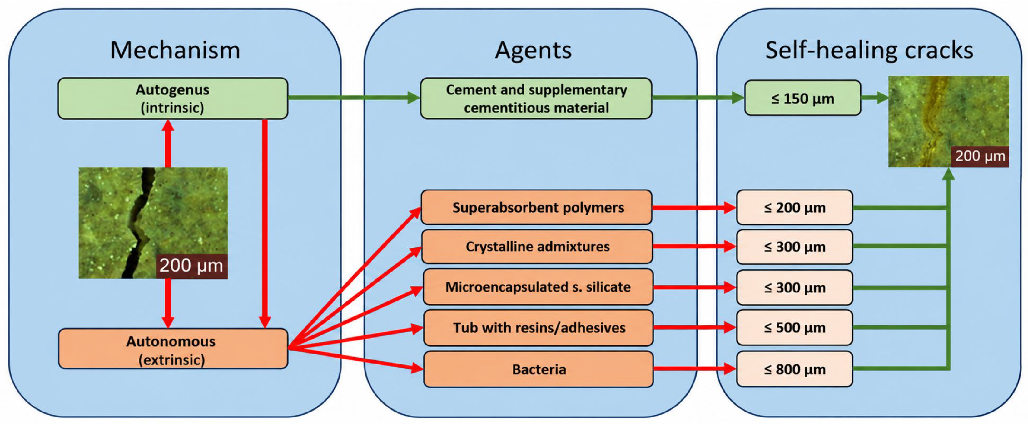
Figure 2 summarizes the main self-healing mechanisms developed to date and highlights the differences between autogenous and autonomous systems. It can be observed that autogenous mechanisms are generally effective for fine cracks with openings of up to approximately 150 μm, while the use of functional agents allows the scope of application to be extended to cracks with openings of up to approximately 800 μm, depending on the type of system used [32,33]. Among the solutions investigated in recent years, the combination of crystallization additives and dispersed reinforcement with polyvinyl alcohol (PVA) fibers is of particular interest, as the two components act complementarily [34,35,36]. Crystallization additives generate insoluble products that reduce permeability and promote the sealing of microcracks, while PVA fibers control crack opening through the crack-bridging effect, reduce stress concentration, and provide a substrate for the nucleation and growth of hydration products [37,38,39,40].
In the recent years, numerous studies have been published on the self-healing mechanisms of cementitious materials, but most of them focus on concrete and on the evaluation of short-term behavior [41,42]. In comparison, the research on masonry mortars modified simultaneously with PVA fibers and crystallization additives is still limited, particularly regarding the evaluation of behavior under repeated wet-dry cycles, the correlation of microstructural phenomena with the evolution of physical and mechanical properties, and the development of mathematical models capable of describing the kinetics of the self-healing process [43,44,45]. Furthermore, there is still insufficient information regarding long-term behavior, the compatibility of these mortars with conventional application technologies, performance under elevated temperatures and fire exposure, and the economic efficiency of their large-scale use [46,47,48,49]. Furthermore, although the behavior of PVA-reinforced cementitious composites at elevated temperatures has received increasing attention, limited information is available regarding the fire response and post-fire residual performance of self-healing masonry mortars incorporating both PVA fibers and crystallization-based additives [14,15,16].
In addition to their potential durability benefits, self-healing cementitious materials may contribute to improved resource efficiency over the service life of construction elements by reducing the frequency of maintenance and repair interventions [50,51]. In principle, fewer repair operations may result in lower consumption of additional materials, energy, and water and may reduce the generation of construction waste [52]. However, these potential environmental benefits depend on the composition of the self-healing material, the environmental impacts associated with its constituent materials, and the actual extension of service life achieved in practice. Therefore, demonstrating an overall sustainability benefit requires a dedicated life-cycle assessment rather than being inferred solely from crack-sealing performance [53,54].
Based on these premises, this study aims to investigate the physical and mechanical behavior and crack-sealing capability of masonry mortars incorporating polyvinyl alcohol (PVA) fibers and a crystalline admixture. The experimental program evaluates the influence of these modifications on selected physical and mechanical properties and investigates the evolution of controlled surface cracks under repeated wet–dry conditioning. Digital microscopy is used to monitor crack-width evolution, while mineralogical characterization is employed to support the interpretation of the products formed within the crack regions. In addition, an empirical mathematical model is used to describe the evolution of crack width as a function of conditioning time. Although fire performance is outside the scope of the present experimental program, the influence of elevated temperatures on the microstructure, residual mechanical properties, and self-healing potential of PVA-modified mortars is identified as an important direction for future investigation [14,15,16].

2. Materials and Methods

2.1. Materials

The experimental program was designed to evaluate the influence of polyvinyl alcohol (PVA) fibers and a crystallization-based waterproofing additive on the physical and mechanical properties and self-healing capacity of masonry mortars. The component materials were selected to be representative of mortars commonly used in construction, while also allowing for the investigation of self-healing mechanisms generated using functional additives [55,56].
The binder used was a CEM I 42.5 R Portland cement type (Holcim România S.A. Aleșd, Bihor County, Romania), in accordance with the requirements of EN 197-1 [57], characterized by rapid development of mechanical strengths and a high clinker content, which is favorable for secondary hydration processes.
CEM I 42.5 R was selected to provide a well-defined and reproducible cementitious matrix and to minimize compositional variability associated with blended cements, thereby facilitating the interpretation of the effects observed within the investigated mortar system. However, CEM I is characterized by a relatively high clinker content and was not selected as a low-carbon binder. Therefore, the present study does not claim an environmental advantage associated with the cement type itself. No life-cycle assessment or embodied-carbon analysis was performed, and the environmental implications of the investigated formulations should consequently be regarded as prospective rather than quantitatively demonstrated. Future studies should evaluate the proposed crack-sealing approach using lower-clinker cements and supplementary cementitious materials and should include dedicated life-cycle assessment.
To improve workability and the behavior of the fresh mortar, hydrated lime paste produced by EL PRIM PRODCOM SRL (Boldești-Scăeni, Prahova County, Romania) was used, adhering to European standards EN 459-1 [58]. The hydrated lime was supplied commercially as a ready-to-use lime paste, pre-dosed and packaged in sealed polyethylene bags, and was incorporated into the mortar as received. No additional preparation or dilution of the lime paste was performed in the laboratory.
Natural siliceous sand from the Holcim Gligorești quarry (Gligorești, Luna Commune, Cluj County, Romania) was used as the fine aggregate, with a maximum grain size of 2 mm, complying with EN 13139 [59]; it was selected for its uniform particle size distribution and low impurity content. The water used to prepare all mixtures was potable water, in accordance with European standard EN 1008 [60] for mortar preparation.
To induce self-healing properties, two functional agents with complementary mechanisms of action were introduced.
The first agent was a crystallization-based waterproofing additive (WP1000, BASF Construction Chemicals Italia S.p.A., Treviso, Italy), added at a rate of 3% of the cement mass. This additive reacts with the cement’s hydration products in the presence of water, generating insoluble crystalline compounds that reduce the permeability of the cementitious matrix and promote the sealing of microcracks [61,62].
The second additive consisted of polyvinyl alcohol (PVA) fibers (KURARAY Europe GmbH, Hattersheim, Germany), 8 mm in length and with a density of 1.30 g/cm3, added at a proportion of 2% by mass relative to the cement. PVA fibers were selected due to their high mechanical strength, excellent adhesion to the cementitious matrix, and ability to control crack propagation through crack-bridging [34,35]. Additionally, they can serve as a support for the nucleation and development of secondary hydration products, indirectly contributing to the efficiency of the self-healing process [63,64].
Based on these materials, four experimental mixtures were designed. Two of these (T1 and T2) served as control mortars, without self-healing agents, while the other two (T3 and T4) included both the crystallization additive and PVA fibers, while maintaining the same compositional base as the control variants.
The mortar compositions are presented in Table 1.
The constituent quantities reported in Table 1 correspond to the amounts used for mortar preparation and were not derived using an absolute-volume mix-design procedure. The lime was used as a commercially supplied ready-to-use lime paste, delivered in sealed polyethylene bags and incorporated into the mortar as received. Therefore, the lime paste contributed both lime solids and water to the mixture. The 40 kg/m3 water content reported in Table 1 refers only to the mixing water added separately and does not represent the total water present in the fresh mortar.
It should be emphasized that T3 and T4 do not differ exclusively in cement content. The increase in cement dosage from 215 to 300 kg/m3 is accompanied by a reduction in lime paste content from 150 to 120 kg/m3, while the nominal amount of added mixing water remains unchanged. Consequently, the nominal water-to-cement ratio based only on the explicitly added water decreases from approximately 0.186 for T3 to 0.133 for T4, while the cement-to-lime-paste mass ratio increases from approximately 1.43 to 2.50. Because the water content of the lime paste was not independently quantified, an effective total water-to-binder ratio cannot be reliably calculated. Therefore, differences between T3 and T4 should be interpreted as resulting from changes in the overall matrix composition rather than being attributed exclusively to cement dosage.
To evaluate the influence of functional additives on mortar performance, all mixtures were examined in terms of their fresh and hardened properties, including consistency, density, water absorption, flexural strength, and compressive strength. For the innovative mixtures (T3 and T4), self-healing capacity was further analyzed by controlled induction of microcracks, monitoring their closure during wet-dry cycles, and correlating the observed phenomena with the results of mineralogical analyses and mathematical and numerical modeling.

2.2. Specimen Preparation

The mortar mixtures were designed based on recommendations from the technical literature and in accordance with the provisions of NE 012/2007–Code of practice for the execution of concrete, reinforced concrete, and prestressed concrete works [65]. All constituent materials were weighed and mixed using a paddle mixer, following the same sequence for adding materials across all experimental variants to ensure the reproducibility of the preparation process.
After homogenization, the fresh mortar was poured into prismatic metal molds measuring 40 × 40 × 160 mm, in two successive layers. Each layer was compacted on a vibrating table to remove entrapped air and achieve as homogeneous a structure as possible. After casting, the specimens were covered to prevent water evaporation and stored for 24 h in a controlled environment at a temperature of 20 ± 2 °C and a relative humidity of approximately 90%.
After demolding, all test specimens were cured in water at a temperature of 20 ± 1 °C for 28 days to ensure the complete development of the hydration processes, following standard practice EN 12390-2 [66].
For each of the control compositions T1 and T2, a total of 30 specimens were prepared for the physical and mechanical tests, whereas 40 specimens were prepared for each of the modified compositions T3 and T4. For each composition, 10 specimens were used for hardened mortar density determination, 10 specimens for water absorption testing, and 10 prismatic specimens for flexural strength testing. Following the flexural strength tests, the two resulting halves of each of the 10 prismatic specimens were used for compressive strength testing, providing 20 test portions for compressive strength determination per composition. For T3 and T4, 10 additional prismatic specimens per composition were prepared for controlled microcrack induction and subsequent crack-sealing monitoring. Crack-sealing monitoring was not performed on the control compositions T1 and T2. The physical and mechanical results reported in this study are presented as mean experimental values. The original replicate-level measurements were not retained in a form that permits a reliable retrospective calculation of standard deviations, confidence intervals, effect sizes, or inferential statistical parameters. Consequently, the comparisons of the physical and mechanical properties presented herein are descriptive, and the observed differences between compositions are interpreted as numerical trends rather than as statistically significant effects.

2.3. Experimental Testing Program

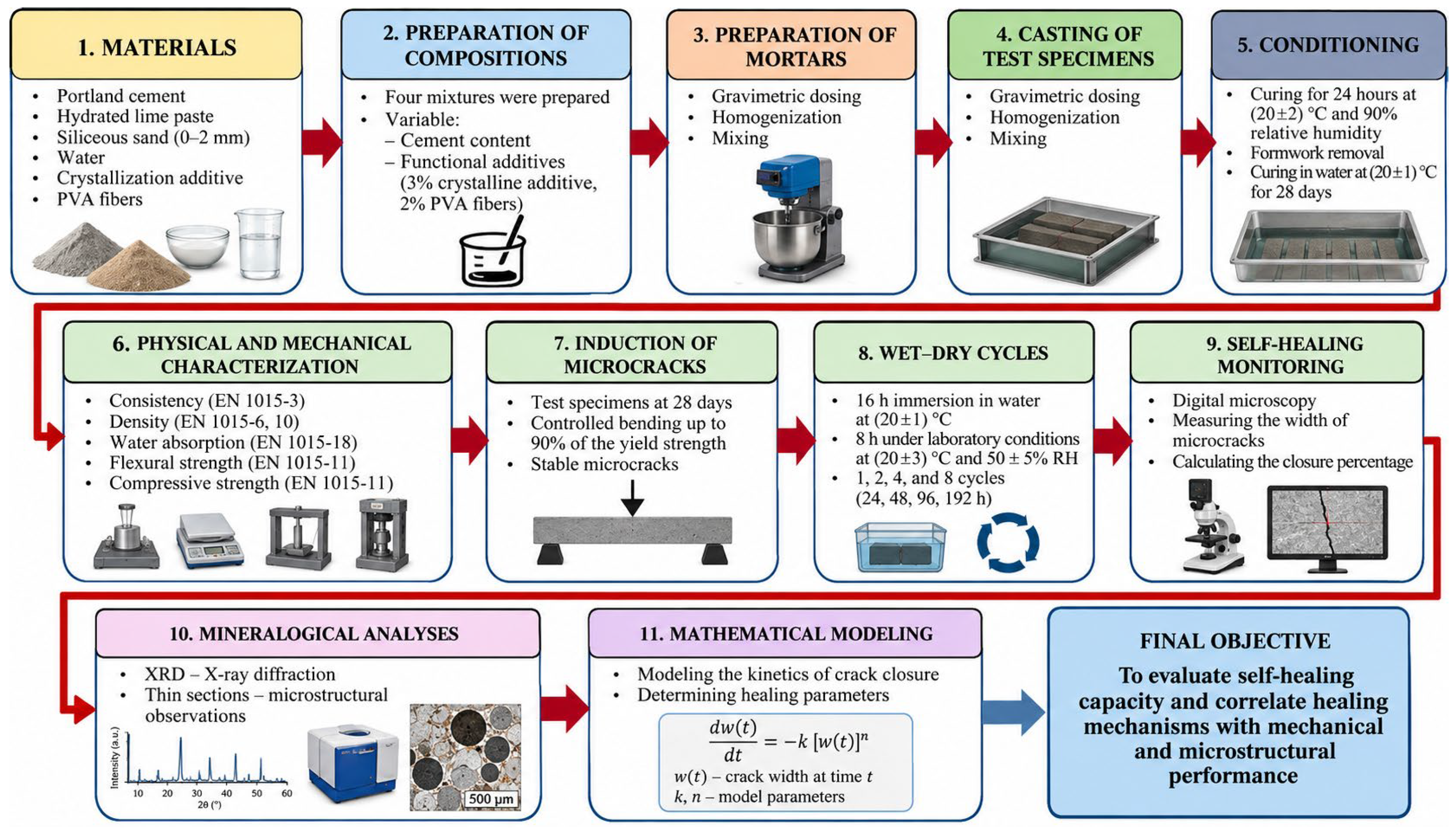
The experimental program was designed to evaluate the influence of polyvinyl alcohol (PVA) fibers and a crystallization-based waterproofing additive on the physical and mechanical properties, self-healing capacity, and microstructural evolution of masonry mortars. The sequence of experimental steps is schematically presented in Figure 3, which highlights the main phases of the research, beginning with the preparation of materials and continuing with physical-mechanical characterization, controlled induction of microcracks, monitoring of the self-healing process, and mineralogical analysis, followed by mathematical modeling of the crack closure phenomenon. Figure 3 also indicates X-ray fluorescence (XRF) as a complementary analytical technique that may be used for elemental/oxide-composition characterization within a broader mineralogical assessment. However, XRF analysis was not performed as part of the experimental program reported in the present study; consequently, no XRF results are presented or discussed herein.
After the test specimens had cured for 28 days, all compositions were characterized in terms of their properties in both the fresh and hardened states. For the innovative variants (T3 and T4), the experimental program was expanded to include the controlled induction of microcracks, exposure to repeated wet-dry cycles, and monitoring of the self-healing process via digital microscopy. In the final stage, the experimental results were correlated with mineralogical analyses and with the mathematical and numerical models developed to describe the self-healing mechanisms.

2.3.1. Determination of Physical and Mechanical Properties

The characterization of the physical and mechanical properties was intended to evaluate the influence of self-healing agents on the behavior of the mortar both in its fresh state and after hardening. All tests were conducted in accordance with the applicable European standards for masonry mortars, using calibrated equipment and standardized procedures.
The consistency of the fresh mortar was determined in accordance with EN 1015-3 [67], using the spreading table method. The final diameter of the mortar cake was measured and used as an indicator of the workability of each composition. This determination was used to characterize and compare the fresh-state consistency of the investigated mortar compositions.
The density of the fresh mortar was determined in accordance with EN 1015-6 [68], immediately after the mixture was prepared, as the ratio of the mass to the volume of the specimen. The density of the hardened mortar was determined after 28 days of curing, in accordance with EN 1015-10 [69], using standardized prismatic specimens.
Water absorption was determined in accordance with EN 1015-18 [70] on specimens dried to constant mass and subsequently immersed in water. The determination of water absorption provided information on the accessible porosity and permeability of the mortar, parameters directly correlated with durability and the effectiveness of self-healing mechanisms [71,72,73,74].
The flexural tensile strength and compressive strength were determined in accordance with EN 1015-11 [75]. The flexural tests were performed on prismatic specimens measuring 40 × 40 × 160 mm, using a three-point loading configuration. The fragments resulting from the bending test were subsequently used to determine compressive strength, thus ensuring a comprehensive evaluation of the mechanical performance of each composition.

2.3.2. Evaluation of Self-Healing Capacity

The self-healing capacity was evaluated exclusively for the modified T3 and T4 compositions, which contain both PVA fibers and a crystallization additive. The experimental methodology aimed to replicate actual operating conditions and quantify the evolution of the microcrack closure process over time [76,77].
To induce microcracks in a controlled manner, the 28-day-old prismatic specimens were subjected to flexural loading. For each investigated composition, the load level used for crack induction was established at 90% of the corresponding mean flexural strength experimentally determined and reported in Table 2. Accordingly, the target nominal flexural stress was 3.15 N/mm2 for T3, corresponding to 90% of its mean flexural strength of 3.50 N/mm2, and 5.47 N/mm2 for T4, corresponding to 90% of its mean flexural strength of 6.08 N/mm2. For the three-point bending configuration, prismatic specimens measuring 40 × 40 × 160 mm were tested using a support span of 100 mm. Based on the 40 × 40 mm specimen cross-section, the target nominal flexural stresses of 3.15 N/mm2 for T3 and 5.47 N/mm2 for T4 corresponded to applied loads of approximately 1.34 kN and 2.33 kN, respectively. This loading level was selected to induce controlled microcracking while avoiding complete failure of the specimens, thereby allowing subsequent monitoring of crack closure during the wet–dry exposure cycles [29,78].
Following controlled cracking and initial crack-width measurement, the specimens were subjected to repeated wet–dry exposure cycles. Each 24 h cycle consisted of 16 h of immersion in water maintained at 20 ± 1 °C, followed by 8 h under laboratory conditions at 20 ± 3 °C and a relative humidity of 50 ± 5%. During the immersion stage, the specimens were positioned with the cracked surface facing upward, without the cracked surface coming into contact with any other object or supporting surface, and the cracked region was fully submerged in water. For microscopic examination, the specimens were removed from the water and maintained under laboratory conditions for approximately 30 min to allow excess surface water to evaporate before crack-width measurements were performed. Thus, microscopic observations were conducted on specimens removed from the water and surface-dried under laboratory conditions, rather than in the immersed state. Microscopic observations and crack-width measurements were performed at predefined monitoring times calculated from the initial crack-width measurement. The observation times were expressed as elapsed time from the initial crack-width measurement, while each complete wet–dry conditioning cycle comprised 16 h of immersion followed by 8 h under laboratory conditions.
The resulting microcracks were analyzed using a Leica DMC2500 digital microscope (Leica Microsystems GmbH, Wetzlar, Germany), with each crack divided into segments so that the entire crack width could be analyzed within the microscope’s field of view. For each segment, the initial maximum width was determined, which served as the reference value for evaluating the effectiveness of the self-healing process.

3. Results

The aim of the experimental evaluation was to investigate the influence of polyvinyl alcohol (PVA) fibers and the crystallization-based waterproofing additive on the physical and mechanical properties, self-healing capacity and microstructural characteristics of masonry mortars. The experimental program sought to identify the relationships between the material composition and the performance achieved, by correlating the experimental results with the physicochemical mechanisms responsible for the self-healing process.
The analysis is structured around four main areas: evaluation of physical and mechanical properties, analysis of the self-healing process’s performance, microstructural characterization, and mathematical modeling of crack closure kinetics. The results are interpreted considering the relevant literature and aim to highlight the contribution of each component of the self-healing system to the mortar’s behavior.

3.1. Physical and Mechanical Properties

Characterizing the physical and mechanical properties is the first step in evaluating the performance of the mortars developed in this study. The analyzed parameters—consistency, density, water absorption, flexural strength, and compressive strength—allow for an assessment of the influence of PVA fibers and the crystallization additive on the internal structure and mechanical behavior of the material.
The experimental results show relatively limited numerical variations in the volumetric properties of the investigated mortars, together with differences in their mechanical behavior and water absorption. Given the absence of replicate-level data suitable for inferential statistical analysis, these differences are interpreted as descriptive experimental trends rather than as statistically significant effects.
The consistency of the fresh mortars, determined using the flow-table method in accordance with EN 1015-3, resulted in flow diameters of 130 mm for T1, 129 mm for T2, 127 mm for T3, and 126 mm for T4. The relatively small variation in the measured flow diameters indicates comparable fresh-state consistency among the investigated mortar compositions.

3.1.1. Density of Fresh and Hardened Mortars

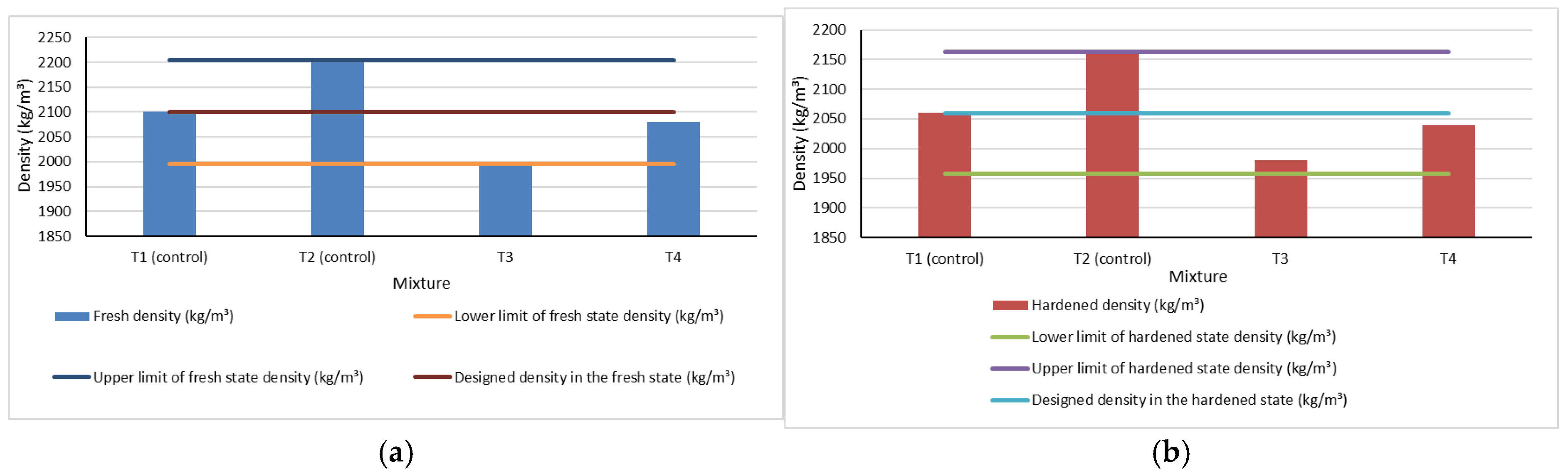
The density of the mortar in its fresh state and after 28 days of curing was determined to evaluate the influence of PVA fibers and the crystallization additive on the material’s volume structure. The experimental values are presented in Figure 4.
The experimental results indicate relatively limited numerical variations in density among the investigated compositions, both in the fresh and hardened states. The density of the fresh mortar ranged from 2000 to 2200 kg/m3, while the density of the hardened mortar ranged from 1980 to 2160 kg/m3. These differences are interpreted descriptively and should not be regarded as evidence of statistically significant differences among the mixtures. Compared with the reference mortars T1 and T2, the modified mortars T3 and T4 exhibited only limited numerical differences in bulk density. Within the descriptive scope of the available data, these results indicate that the incorporation of PVA fibers and the crystalline admixture was not associated with major changes in the measured bulk density. The slight reductions in density can be attributed to the reduced volume occupied by the fibers and their uniform distribution within the cement matrix, without the occurrence of segregation or a significant increase in porosity.
The maintenance of similar density values confirms that the self-healing system does not affect the mortar’s preparation and compaction process. This aspect is important because it demonstrates that the improvement in self-healing performance is not associated with changes in volumetric properties, but rather with microstructural processes that develop after cracks form.
The results are consistent with studies conducted by [9,28,35,46], which showed that PVA fibers and crystalline additives have a minor effect on bulk density, contributing primarily to the control of crack propagation and the development of self-healing mechanisms.
Overall, the mean density values indicate relatively limited numerical differences between the reference and modified mortars. These results are interpreted descriptively and do not establish statistically significant differences among the investigated compositions [34,46,61].
Water absorption is one of the most important indicators used to assess the durability of cement-based materials, as it reflects the degree of connectivity of the capillary network and the material’s ability to allow water and aggressive agents to penetrate the cementitious matrix. In the case of self-healing mortars, this parameter is of particular importance, as the efficiency of the healing process is closely linked to the pore structure and the evolution of microcracks.
The experimental results regarding water absorption are presented in Figure 4, and the determined values are summarized in Table 2.
The results show a consistent numerical reduction in the mean water absorption values for mortars containing PVA fibers and the crystalline admixture. For the mixtures containing 215 kg/m3 of cement, water absorption decreased from 6.61% for the reference mortar T1 to 6.49% for mixture T3, corresponding to a reduction of approximately 1.8%. A similar trend was observed for the mixtures containing 300 kg/m3 of cement, with water absorption decreasing from 6.58% for T2 to 6.45% for T4, corresponding to a reduction of approximately 2.0%. These differences are interpreted descriptively and should not be considered statistically significant in the absence of replicate-level statistical analysis.
Although the numerical differences in water absorption are relatively small, the consistently lower mean values observed for T3 and T4 may indicate changes in the pathways governing liquid-water transport through the mortar. This behavior may be associated with the combined presence of the crystalline admixture and PVA fibers. The crystalline admixture may contribute to the formation of products within pores and microcracks, while PVA fibers may assist in controlling crack development. However, the present water-absorption measurements alone do not allow these mechanisms, or changes in total porosity and capillary pore connectivity, to be directly demonstrated. Therefore, these interpretations should be regarded as possible mechanisms consistent with the observed experimental trends rather than as directly verified effects. The results obtained are consistent with research conducted by [9,28,46], who demonstrated that the introduction of PVA fibers and crystalline additives leads to a reduction in permeability without significant changes in bulk density. Their studies show that the main effect of these components is to optimize the microstructure and develop a less interconnected capillary system, which is conducive to self-healing processes.
Overall, the mean experimental values indicate slightly lower water absorption for the modified mortars T3 and T4 compared with their corresponding reference mixtures. Within the limitations of the available data, this observation is interpreted as a descriptive numerical trend and does not establish a statistically significant improvement in water-transport properties.

3.1.2. Mechanical Properties

The mechanical performance of mortars is an essential criterion in assessing their suitability for use in masonry elements exposed to mechanical stresses and environmental factors. In this study, the flexural strength and compressive strength after 28 days of curing were determined to evaluate the influence of PVA fibers and the crystallization additive on the structural behavior of the material.
Analysis of the results highlights different behaviors of the mortar depending on the type of mechanical load. The mean experimental values indicate different numerical trends depending on the type of mechanical loading. The modified mortars exhibited slightly higher mean flexural strengths and lower mean compressive strengths than their corresponding reference mortars. These differences are interpreted descriptively and should not be considered statistically significant effects.
Resistance to Bending
The results obtained demonstrate the direct influence of cement content on flexural strength. Mortars made with 300 kg/m3 of cement developed higher strengths than those prepared with 215 kg/m3, which highlights the decisive role of the cementitious matrix in absorbing tensile stresses.
Based on the mean experimental values, the modified mortars exhibited moderately higher flexural strengths than their corresponding reference mortars. The mean flexural strength increased from 3.35 N/mm2 for T1 to 3.50 N/mm2 for T3, corresponding to approximately 4.5%, and from 5.73 N/mm2 for T2 to 6.08 N/mm2 for T4, corresponding to approximately 6.1%. These differences represent descriptive numerical trends and were not evaluated for statistical significance.
This behavior may be associated with the crack-bridging action commonly reported for PVA-fiber-reinforced cementitious materials; however, direct fractographic evidence of fiber bridging or pull-out was not obtained in the present study. During bending loading, the fibers limit crack propagation and redistribute stresses within the matrix, delaying the onset of brittle failure and imparting a more stable mechanical response to the mortar. The contribution of the crack-bridging additive is limited at this stage; its effects become significant after cracks form and during the self-healing process.
The moderate numerical increases in mean flexural strength are consistent with trends reported in the literature for fiber-reinforced cementitious materials. However, because inferential statistical analysis could not be performed, the present results should not be interpreted as demonstrating a statistically significant effect of PVA-fiber incorporation on flexural strength.
Compressive Strength
The results regarding compressive strength highlight the dominant influence of cement content on mechanical performance. Mortar T2, prepared with a cement content of 300 kg/m3, developed a compressive strength nearly double that of mortar T1, confirming the decisive role of the binder content in the development of a compact and strong matrix.
Based on the mean experimental values, lower compressive strengths were observed for the modified mortars in both investigated series. The mean compressive strength decreased from 16.88 N/mm2 for T1 to 11.01 N/mm2 for T3 and from 31.87 N/mm2 for T2 to 19.14 N/mm2 for T4. These differences are reported as descriptive numerical trends and should not be interpreted as statistically significant effects.
The reduction in compressive strength may be related to changes in the internal structure associated with fiber incorporation, including possible local discontinuities at the fiber–matrix interface. However, the present study did not include fractographic or dedicated fiber–matrix interface characterization; therefore, this interpretation should be regarded as a possible mechanism rather than a directly demonstrated phenomenon.
Overall, the analysis of the mechanical properties demonstrates that the introduction of PVA fibers and the crystallization additive modify the mortar’s response mechanism. The material exhibits more favorable behavior after cracking initiates and provides the necessary conditions for the effective activation of self-healing mechanisms, an aspect that will be analyzed in detail in the following subsection.

3.2. Crack-Sealing Performance

The crack-sealing process is the main objective of the experimental research and constitutes the essential criterion for evaluating the effectiveness of the system based on PVA fibers and a waterproofing additive through crystallization. Unlike the physical-mechanical properties, which characterize the material’s immediate response to stress, the self-healing process highlights the mortar’s ability to partially restore its microstructural continuity after cracks form and to limit the penetration of water and aggressive agents into the cementitious matrix.
To evaluate this phenomenon, cracks induced under controlled conditions in prismatic specimens were monitored after 28 days of curing. Their evolution was tracked over successive wetting–drying cycles by periodically measuring crack width and determining the closure rate. The methodology employed allowed for the evaluation of both the efficiency of the self-healing process and the crack-sealing kinetics of the investigated T3 and T4 mixtures.
The experimental results were supplemented by microscopic observations and subsequently correlated with the mineralogical and microstructural investigations presented in the following chapters, to identify the mechanisms responsible for the formation of self-healing products. In this way, the analysis is not limited to quantifying the reduction in crack opening, but aims to explain the physicochemical phenomena that control their evolution over time.
Crack-sealing behavior was evaluated for the modified compositions T3 and T4 by monitoring the evolution of controlled cracks during repeated wet–dry exposure. Crack width was periodically measured using optical microscopy, and the degree and rate of closure were determined as functions of exposure time and initial crack width.
It is important to note that the crack-sealing experiment was not performed on cracked reference specimens T1 and T2. Furthermore, no formulations containing only PVA fibers or only the crystalline admixture were investigated. Therefore, the experimental design does not permit quantitative separation of ordinary autogenous healing associated with continued hydration and carbonation from effects potentially associated with PVA fibers or the crystalline admixture. Accordingly, the results presented in this section describe the crack-sealing response of the combined T3 and T4 systems under the investigated exposure conditions and should not be interpreted as direct evidence of superior healing relative to unmodified mortars.
The experimental values regarding the evolution of crack width and closure rates are summarized in Table 3, which forms the basis for the interpretation presented in the following subsections.

3.2.1. Evolution of the Crack Closure in Mortar T3

The T3 mixture was used to evaluate the behavior of a self-healing mortar based on PVA fibers and a crystallization additive, developed on a cementitious matrix with a cement content of 215 kg/m3. This experimental variant serves as a reference for analyzing the influence of cement dosage on the kinetics of the self-healing process and allows for the identification of the mechanisms responsible for crack closure under conditions where a moderate amount of hydration products is available.
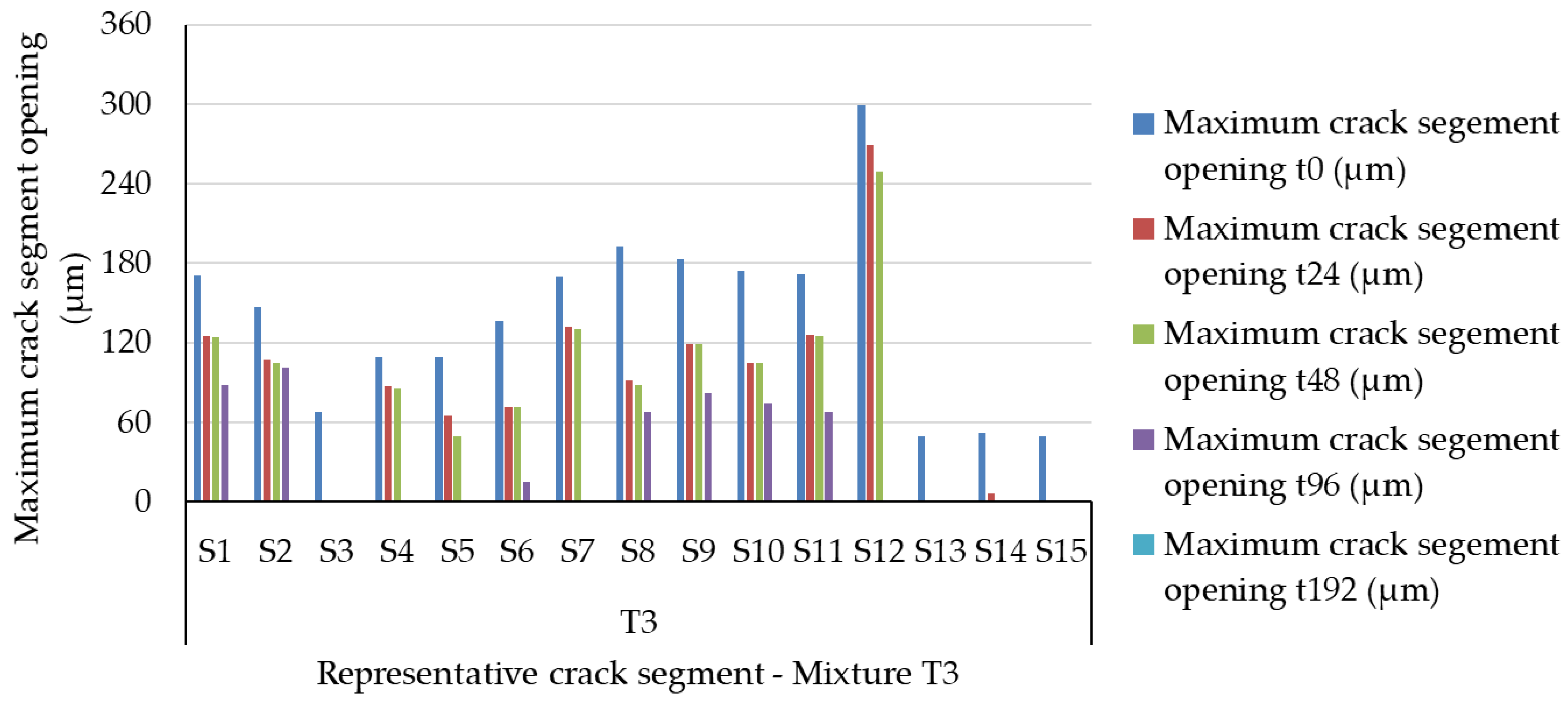
The evolution of the self-healing process is shown in Figure 5, and the characteristic values of crack opening and average closure rates are summarized in Table 3.
Analysis of the results shows that the initial crack width ranged from 49 to 299 μm, with an average value of approximately 139 μm. The investigated cracks therefore covered a relatively broad range of initial widths, including cracks exceeding 200 μm. This range is consistent with crack widths frequently investigated in studies addressing autogenous and stimulated crack-sealing processes in cementitious materials. The experimental results highlight a direct relationship between the initial crack width and the self-healing rate. Cracks with widths less than 100 μm showed the fastest healing, with most being completely sealed after the first or second wetting–drying cycle. Under these conditions, the small distance between the crack faces promotes water retention and the rapid formation of hydration and carbonation products, leading to the complete sealing of the crack.
As the initial crack width increases, the self-healing process gradually slows down. For cracks ranging in size from 100 to 200 μm, the degree of closure exceeded 80% in most cases after approximately 96 h, demonstrating that the system based on PVA fibers and a crystallization additive maintains its effectiveness even for medium-sized cracks. However, the increase in the volume that must be filled by hydration and crystallization products leads to a reduction in the closure rate compared to fine cracks.
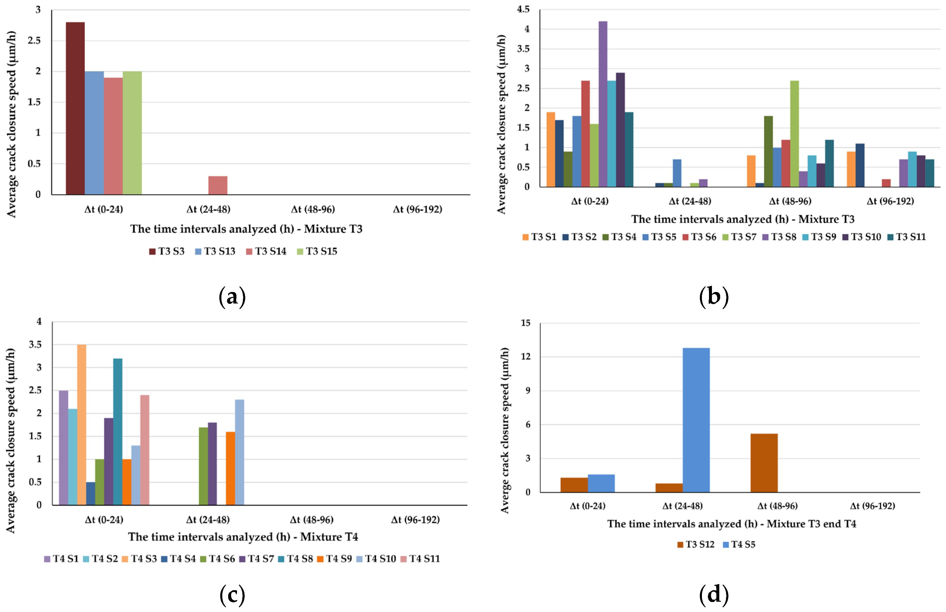
The most representative behavior is observed for the S12 segment, which is characterized by the largest initial crack width (299 μm). In this case, the first 24 h are characterized by a relatively limited reduction in crack width, followed by a marked acceleration of the process between 48 and 96 h, when the average closure rate reaches 5.2 μm/h. This evolution indicates that the self-healing process is not linear but involves an initial activation stage, followed by the accelerated development of the mineral products responsible for sealing the cracks.
Overall, the results obtained for T3 show that the extent and rate of crack closure depended on both the initial crack width and the duration of wet–dry exposure. Narrower cracks generally exhibited faster closure, whereas larger cracks required longer exposure periods. These results provide the experimental basis for comparison with T4. However, the comparison between the two compositions should consider the simultaneous differences in cement content, lime paste content, nominal water-to-cement ratio, and initial crack-width distribution, rather than attributing the observed differences exclusively to cement dosage.

3.2.2. Evolution of the Crack Closure in Mortar T4

The T4 mixture was investigated to evaluate the crack-sealing behavior of the modified mortar system at a different matrix composition. Compared with T3, T4 contains a higher cement content (300 kg/m3) and a lower lime paste content (120 kg/m3), while the nominal amount of added mixing water remains unchanged. Consequently, the two mixtures also differ in their nominal water-to-cement and cement-to-lime-paste ratios. Therefore, the crack-sealing behavior of T4 should be interpreted in relation to the overall matrix composition rather than to cement content alone.
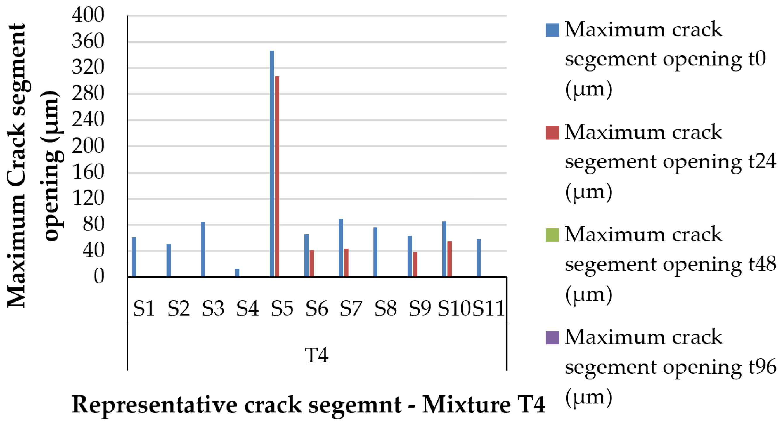
The experimental results regarding the evolution of crack closure are presented in Figure 6, and the characteristic values of crack opening and average closure rates are summarized in Table 3.
Analysis of the results highlights a significant improvement in the self-healing performance compared to the T3 mortar. The initial crack width ranged from 13 to 346 μm, with an average value of approximately 90 μm, which is considerably lower than that determined for the T3 composition. The initial crack-width distribution differed between T3 and T4. Because the two mixtures also differ in cement content, lime paste content, and associated compositional ratios, the differences in initial crack width cannot be attributed to a single compositional parameter.
Most of the segments investigated exhibited initial crack widths below 100 μm, a range in which the self-healing process proceeded with very high efficiency. For these cracks, complete closure was achieved after the first wetting–drying cycle, demonstrating that the mechanisms of secondary hydration and crystallization are activated immediately upon exposure to water.
In contrast to the T3 specimen, remarkable performance was also achieved for cracks with large openings. The most representative example is the S5 segment, characterized by an initial crack opening of 346 μm, the largest value recorded throughout the entire experimental program. After the first 24 h, the reduction in crack width was relatively modest, but in the following period the process accelerated considerably, with the crack closing completely after only 48 h.
This result represents one of the most important findings of the experimental research, demonstrating that the proposed system can close, within a very short time, cracks whose dimensions exceed the limit reported in numerous studies on autogenous self-healing.
For segment S5, the highest interval-based average closure rate was 12.8 μm/h over the 24–48 h observation interval. Because the exact time of complete closure within this interval was not determined, this value should not be interpreted as an instantaneous maximum closure rate.
Under the investigated experimental conditions, T4 exhibited faster crack closure than T3. However, this difference should not be attributed to a single compositional parameter. In addition to the higher cement content, T4 contains a lower amount of lime paste and has different nominal water-to-cement and cement-to-lime-paste ratios. Moreover, because PVA fibers and the crystalline admixture were dosed as percentages of cement mass, their absolute contents were higher in T4 (6.0 and 9.0 kg/m3, respectively) than in T3 (4.3 and 6.45 kg/m3, respectively). The initial crack-width distributions of T3 and T4 were also different. These interrelated variables should therefore be considered when interpreting the differences in crack-sealing kinetics between the two mixtures.
The results obtained are consistent with observations reported in the literature regarding the role of PVA fibers in controlling crack propagation and the influence of crystalline additives on permeability reduction.

3.2.3. Comparative Assessment of Self-Healing Performance

T3 and T4 contain the same types of functional constituents at the same dosages expressed as percentages of cement mass (2% PVA fibers and 3% crystalline admixture), but their absolute contents differ because of the different cement dosages. Consequently, the differences in crack-sealing behavior between T3 and T4 cannot be attributed exclusively to cement content.
The experimental results summarized in Table 3 and illustrated in Figure 7 highlight the main differences between the two compositions investigated.
The Influence of the Initial Opening of Cracks
For both compositions, it was observed that the initial crack width was associated with the time required for crack closure. Cracks with widths less than 100 μm exhibited the highest sealing efficiency, closing completely within 24 to 48 h.
As the crack width increases, the time required for closure also increases; however, the influence of this factor differs between the two compositions investigated.
In the case of T3 mortar, an increase in the initial crack width leads to a progressive reduction in the self-healing rate. Cracks with openings between 100 and 200 μm required approximately 96 h to achieve a high degree of sealing, while for cracks approaching 300 μm, the process extended to 192 h.
In contrast, the T4 mortar exhibited significantly different behavior. Even for cracks with initial widths exceeding 300 μm, the self-healing process was completed after only 48 h, indicating a faster crack-sealing response under the investigated experimental conditions.
Analysis of Self-Healing Speed
A comparison of the experimentally determined closure rates shows differences between the T3 and T4 compositions. For the T3 mortar, the maximum rate determined experimentally was 5.2 μm/h, recorded for segment S12 between 48 and 96 h. In contrast, for the T4 composition, the maximum rate reached 12.8 μm/h, as determined for segment S5 between 24 and 48 h.
Because crack widths were measured at discrete observation times, the exact time of complete closure within each observation interval could not be determined. Consequently, these values represent interval-based average closure rates and should not be interpreted as instantaneous or intrinsic material-specific rates. Direct quantitative comparison of these highest interval-based rates is therefore not used to infer a percentage increase in the intrinsic crack-sealing rate of T4 relative to T3.
The interval-based closure rates varied over the successive observation periods, indicating that crack-width reduction did not proceed at a constant rate under the investigated conditions. However, because measurements were performed at discrete time points, these data do not allow the instantaneous evolution of the crack-sealing rate to be determined.
The Influence of Composition on Process Efficiency
The observed differences in crack-sealing behavior between T3 and T4 cannot be attributed exclusively to cement content, since the two mixtures differ in several interrelated compositional and experimental variables, including the absolute contents of PVA fibers and crystalline admixture.
Increasing the cement dosage results in a greater availability of unhydrated particles and calcium hydroxide, promoting the formation of hydration by-products and the precipitation of calcium carbonate in the crack zone. Consequently, the self-healing process proceeds more rapidly, and the system’s scope of application is extended to cracks with considerably larger openings.
Correlation with Academic Literature
The results obtained in this study are consistent with the mechanisms described in the literature regarding the self-healing of cementitious materials. Studies conducted by [34] have highlighted the role of PVA fibers in controlling crack opening and in developing pseudo-ductile behavior, while research by Dimitri Snoeck and Liberato Ferrara has demonstrated that the efficiency of the process depends directly on the initial size of the cracks and the availability of water required for secondary reactions.
The result demonstrates the potential for using these mortars in masonry elements subjected to repeated wetting and drying cycles.
Conclusions of the Comparative Analysis
The comparative analysis showed that T3 and T4 exhibited different surface crack-sealing behavior under the investigated wet–dry conditioning regime. T4 exhibited faster crack closure than T3; however, this difference cannot be attributed to a single compositional parameter because several interrelated variables differ between the two mixtures.
Accordingly, the present results demonstrate differences in surface crack-sealing behavior between T3 and T4 under the investigated conditions but do not establish an optimal formulation or demonstrate synergistic effects among the individual constituents.

3.2.4. Microscopic Analysis of Self-Healing Mechanisms

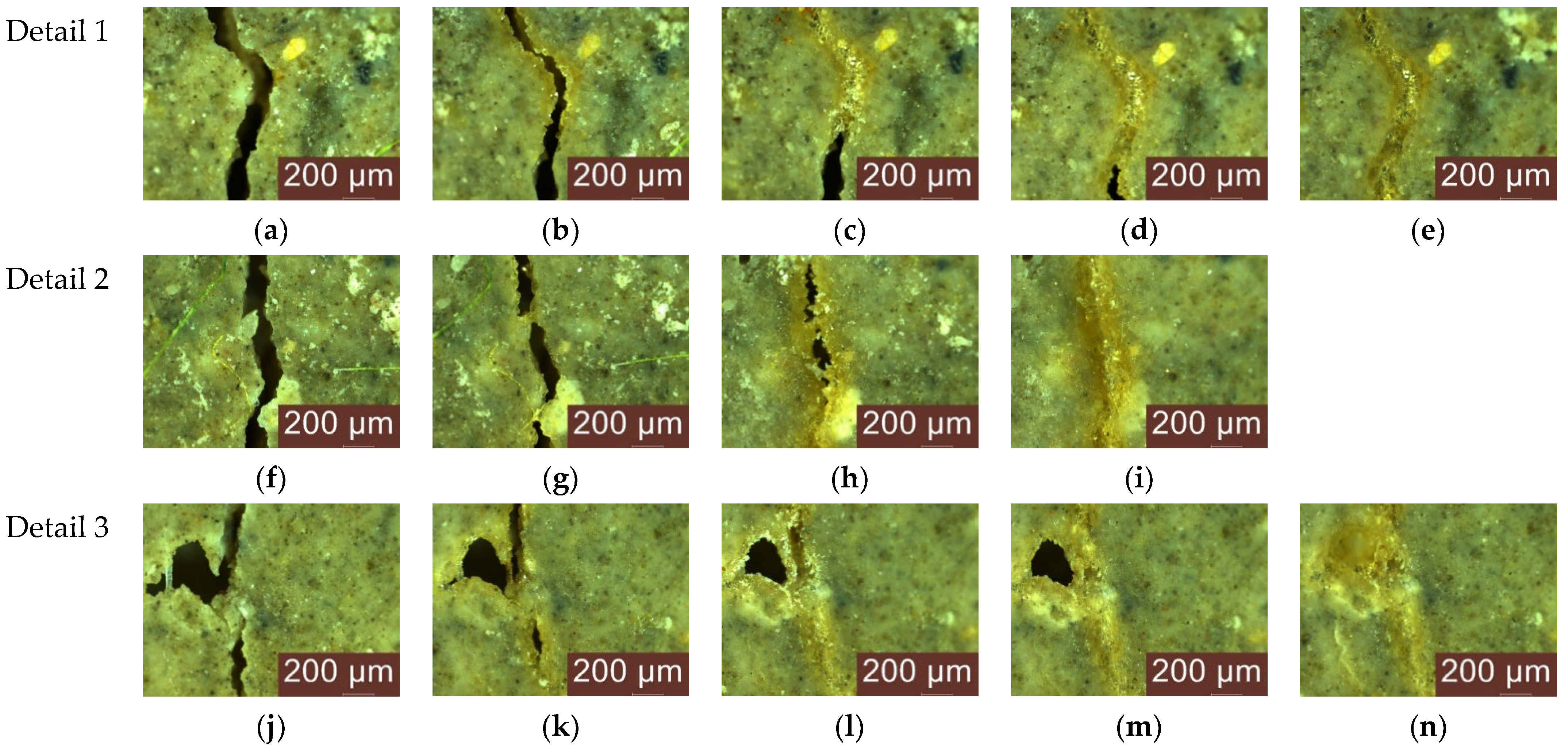
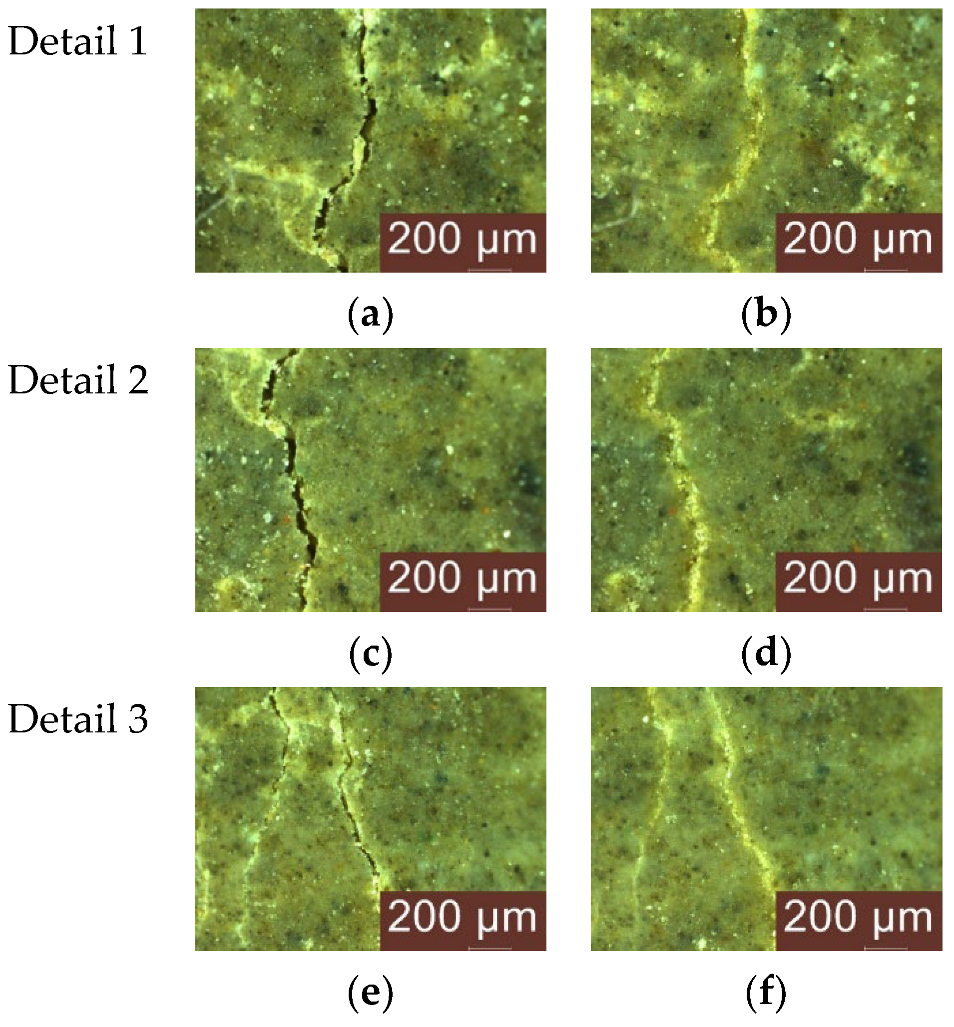
To complement the quantitative crack-width measurements, representative crack segments from T3 and T4 were examined by optical microscopy during successive wet–dry cycles. The images presented in Figure 8 and Figure 9 document morphological changes within the cracked regions and the progressive appearance of solid deposits associated with crack sealing [76].
The image analysis highlights the existence of three successive stages in the self-healing process, common to both compositions investigated, though with different rates of progression.
In the first stage, corresponding to the initial state and the first wetting–drying cycle, the cracks exhibit well-defined edges and clean surfaces, with no visible mineral deposits. After the first 24 h, discontinuous mineral formations appear along the edges of the cracks, indicating the initiation of secondary hydration reactions and the nucleation of the first crystalline products.
In the intermediate stage, corresponding to the 24–96 h interval, the development of these products becomes much more evident. The mineral deposits grow progressively in both volume and continuity, gradually reducing the crack width and forming the first mineral bridges between the two faces of the cracks. At this stage, the differences between the two compositions become evident. For the T3 mortar, the reduction in crack width is gradual, requiring several wet–dry cycles to achieve complete sealing. In contrast, for the T4 composition, the development of mineral products is significantly faster, and after two conditioning cycles, most of the cracks investigated are nearly completely sealed.
The final stage is characterized by the stabilization of the self-healing process and the formation of a continuous mineral structure in the cracked area. In the images corresponding to the final conditioning cycles, it can be observed that the surface of the cracks becomes nearly continuous, and their initial boundaries are difficult to identify, which confirms the effectiveness of the sealing process.
A comparison of the images corresponding to the two compositions highlights the fact that an increase in cement content influences not only the rate of the self-healing process but also the development of the mineral products. In the case of composition T4, the deposits are more uniform and fill the entire cracked space in a shorter time, which explains the higher self-healing rates determined experimentally and this composition’s ability to completely seal cracks with initial openings of up to 346 μm.
The microscopic observations, considered together with the quantitative crack-width measurements, indicate that crack sealing was accompanied by the progressive formation of solid deposits within the crack regions. Secondary hydration and carbonation may contribute to this process, while the presence of PVA fibers and the crystalline admixture may also influence crack development and mineral precipitation. However, the respective contributions of these mechanisms cannot be independently quantified from the present experimental design.
Microscopic observations therefore provide complementary evidence of the progressive development of deposits associated with crack closure. Nevertheless, because the investigated T3 and T4 mixtures contained both PVA fibers and the crystalline admixture, these observations cannot demonstrate a synergistic interaction between the individual components.

3.3. Microstructural and Mineralogical Validation of the Self-Healing Mechanism

The experimental results presented in the previous subsection demonstrated the ability of mortars modified with PVA fibers and a crystallization-based waterproofing additive to autonomously close cracks that developed under loading. However, measuring the reduction in crack width provides information on the efficiency of the process without revealing the nature of the mineral products responsible for sealing the cracks.
To identify the mechanisms governing self-healing, the experimental investigations were supplemented with mineralogical and microstructural analyses. X-ray diffraction (XRD) was used to identify the crystalline phases developed in the cementitious matrix and in the precipitates formed in the crack zone, while microscopic observations allowed for the evaluation of the distribution and morphology of the self-healing products.
Correlating these investigations with the experimental crack-closure results provides complementary information on the mineralogical changes associated with the observed crack-sealing process.

3.3.1. X-Ray Diffraction (XRD) Analysis

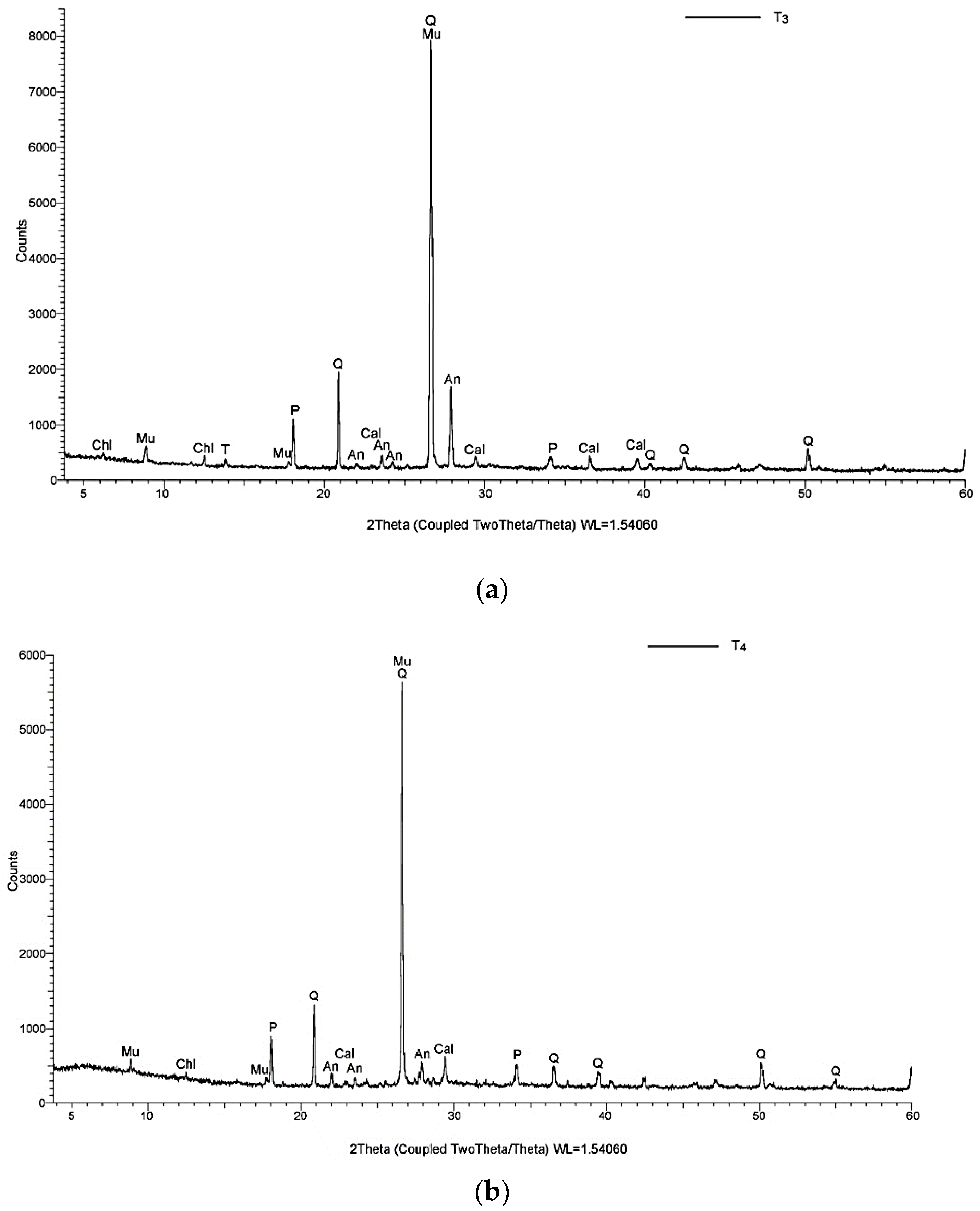
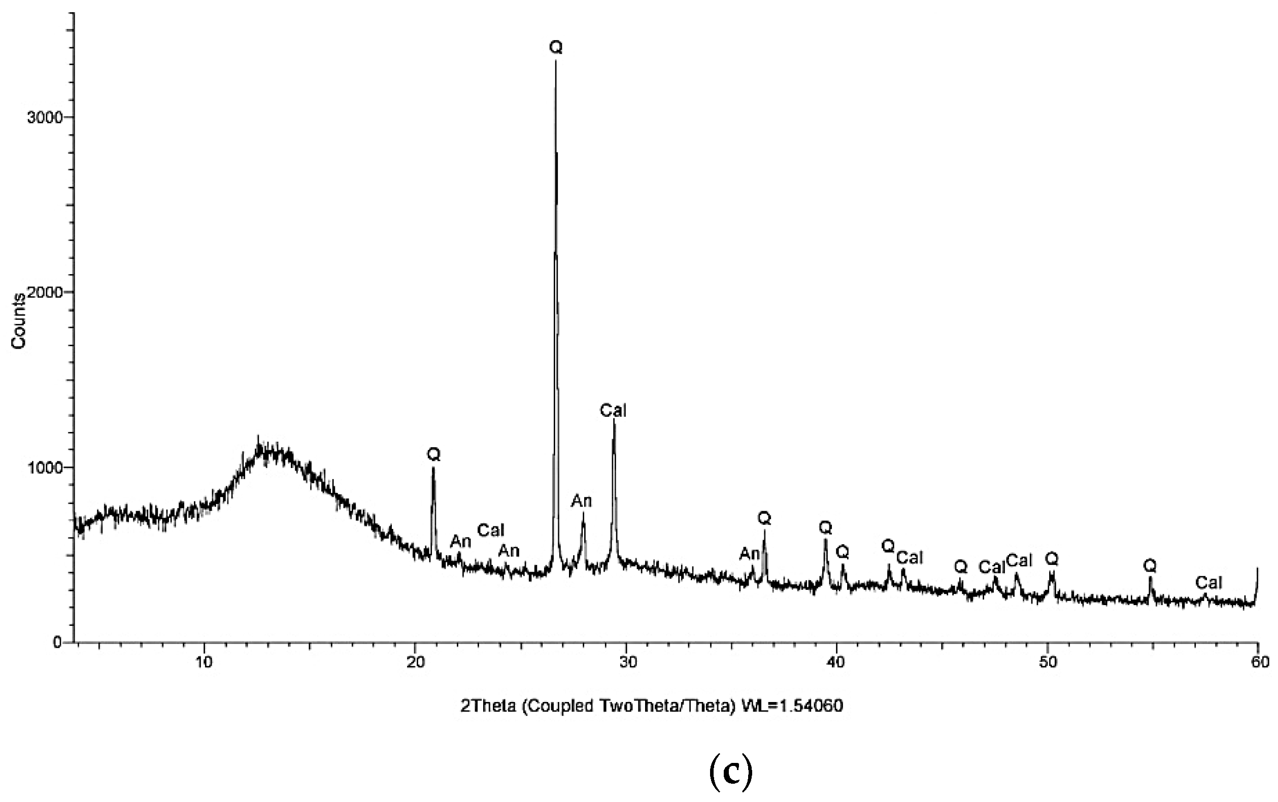
X-ray diffraction (XRD) analysis was performed using a Bruker D8 ADVANCE diffractometer (Bruker AXS GmbH, Karlsruhe, Germany) for the qualitative identification of crystalline phases present in the investigated mortar samples and in the mineral precipitates collected from the crack regions after successive wet–dry exposure cycles. The diffraction patterns were recorded over a 2θ range of approximately 5–60°, using an X-ray wavelength of λ = 1.54060 Å. Phase identification was performed qualitatively based on the characteristic diffraction peaks. No quantitative phase analysis or Rietveld refinement was performed; therefore, the relative abundance of the identified crystalline phases was not quantitatively determined from the diffraction peak intensities. The XRD results obtained for T3, T4, and the crack-region precipitates are presented in Figure 10 and Table 4.
Analysis of the diffractograms revealed the presence of quartz (SiO2), portlandite [Ca(OH)2], calcite (CaCO3), anorthite, albite, muscovite, chlorite, and tobermorite among the crystalline phases identified in the investigated samples. These phases reflect both the initial composition of the material and the mineralogical transformations that occurred during the self-healing process [79,80,81,82,83,84].
Quartz is the predominant phase derived from the mineral aggregates used in the mortar composition, contributing to the dimensional stability of the matrix without directly participating in the self-healing reactions. In contrast, portlandite and calcite are of particular importance in interpreting the mechanisms responsible for crack closure [24,25].
The presence of portlandite confirms the existence of a reserve of calcium hydroxide available for secondary hydration and carbonation reactions [80]. Upon contact with water that has penetrated the crack zone and with carbon dioxide from the surrounding environment, portlandite participates in the formation of calcium carbonate, one of the main products involved in the progressive sealing of cracks.
The diffractograms show reflections characteristic of calcite in the investigated samples, supporting the presence of calcium carbonate among the crystalline phases associated with the crack-sealing process. Portlandite was also identified, consistent with the presence of crystalline products related to cement hydration and subsequent carbonation reactions. Because the XRD investigation was performed for qualitative phase identification only, differences in diffraction peak intensities between T3, T4, and the crack-region precipitates were not used to quantify differences in phase abundance. Accordingly, the XRD results are interpreted as qualitative mineralogical evidence and are considered together with the microscopic observations and crack-width evolution. These results are fully consistent with the experimental observations presented in the previous subsection, where the T4 composition demonstrated the highest self-healing rate and the greatest crack-sealing capacity. Therefore, XRD analysis provides mineralogical confirmation of the performance differences observed between the two compositions and demonstrates that the efficiency of the self-healing process is closely linked to the development of hydration and carbonation products [63,64].
It should be noted that XRD analysis primarily identifies the crystalline phases of the material, while weakly crystalline or amorphous products, such as the C–S–H gel, are revealed only indirectly through general changes in the diffractograms and by correlation with other investigative methods. For this reason, the interpretation of the XRD results is supplemented in the following subsection by microstructural and digital image analysis, which allows for the evaluation of the distribution and morphology of the products formed in the crack zone.
Overall, the mineralogical analysis indicates that the observed crack-sealing process is associated with mineralogical transformations involving secondary hydration and carbonation reactions. Correlation of the XRD results with the microscopic observations and quantitative crack-width evolution provides complementary evidence of mineral precipitation within the crack regions. However, because the experimental design did not include cracked reference mixtures or single-component formulations, these results do not allow the individual contributions or a synergistic interaction between the PVA fibers and the crystalline admixture to be experimentally demonstrated.

3.3.2. Microstructural Characterization Using Optical Microscopy and Digital Image Analysis

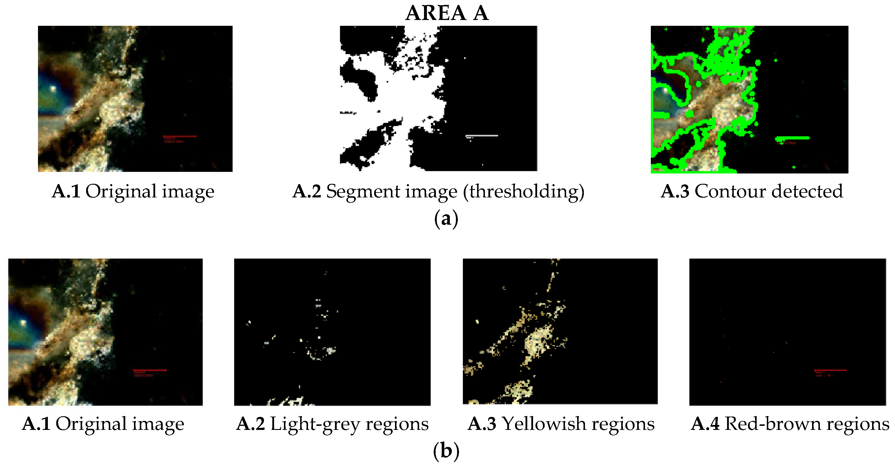
To supplement the mineralogical investigations conducted using X-ray diffraction, microscopic observations were performed on thin sections prepared from the cracked regions of the 40 × 40 × 160 mm prismatic mortar specimens. The thin sections were examined using a Nikon optical microscope (Nikon Corporation, Tokyo, Japan) equipped with a digital imaging system and connected to a computer for image acquisition and analysis. The microscopic examination was used to characterize the morphology and spatial distribution of solid deposits within the crack regions. Calibrated dimensional measurements were performed directly on the acquired microscopic images, and scale bars were retained in the corresponding micrographs.
Unlike XRD analysis, which provides information on the mineralogical composition of the investigated material, optical microscopy provides direct morphological information on the spatial distribution of solid deposits within the crack regions, without permitting definitive mineralogical phase identification based on optical appearance alone. To supplement the mineralogical investigations conducted using X-ray diffraction and to further characterize the deposits formed within the crack regions, microscopic observations were performed on thin sections prepared from the cracked regions of the 40 × 40 × 160 mm prismatic mortar specimens. The thin sections were examined using a Nikon optical microscope (Nikon Corporation, Tokyo, Japan) equipped with a digital imaging system and connected to a computer for image acquisition and analysis. The microscopic examination was used to characterize the morphology and spatial distribution of solid deposits within the crack regions. Calibrated dimensional measurements were performed directly on the acquired microscopic images.
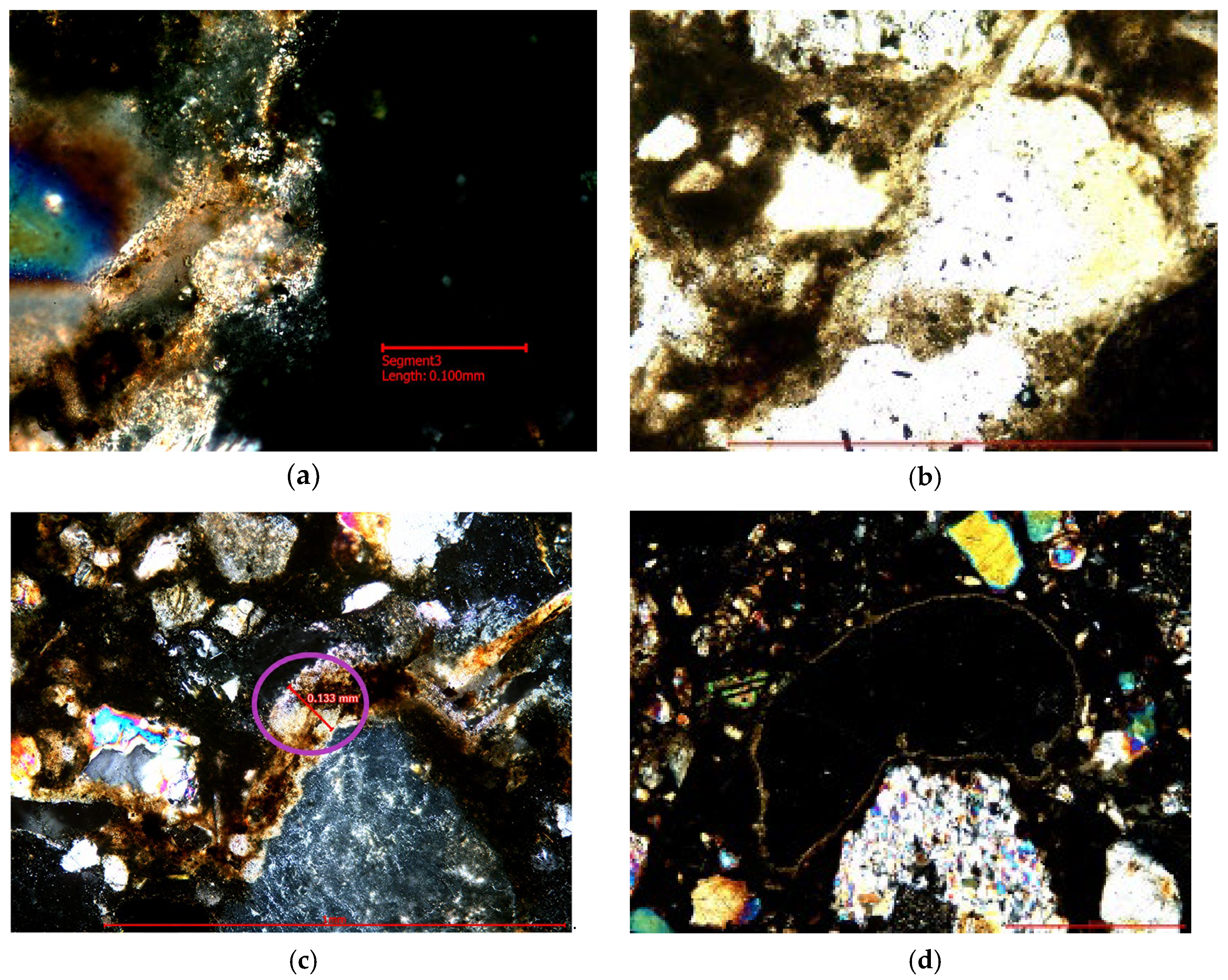
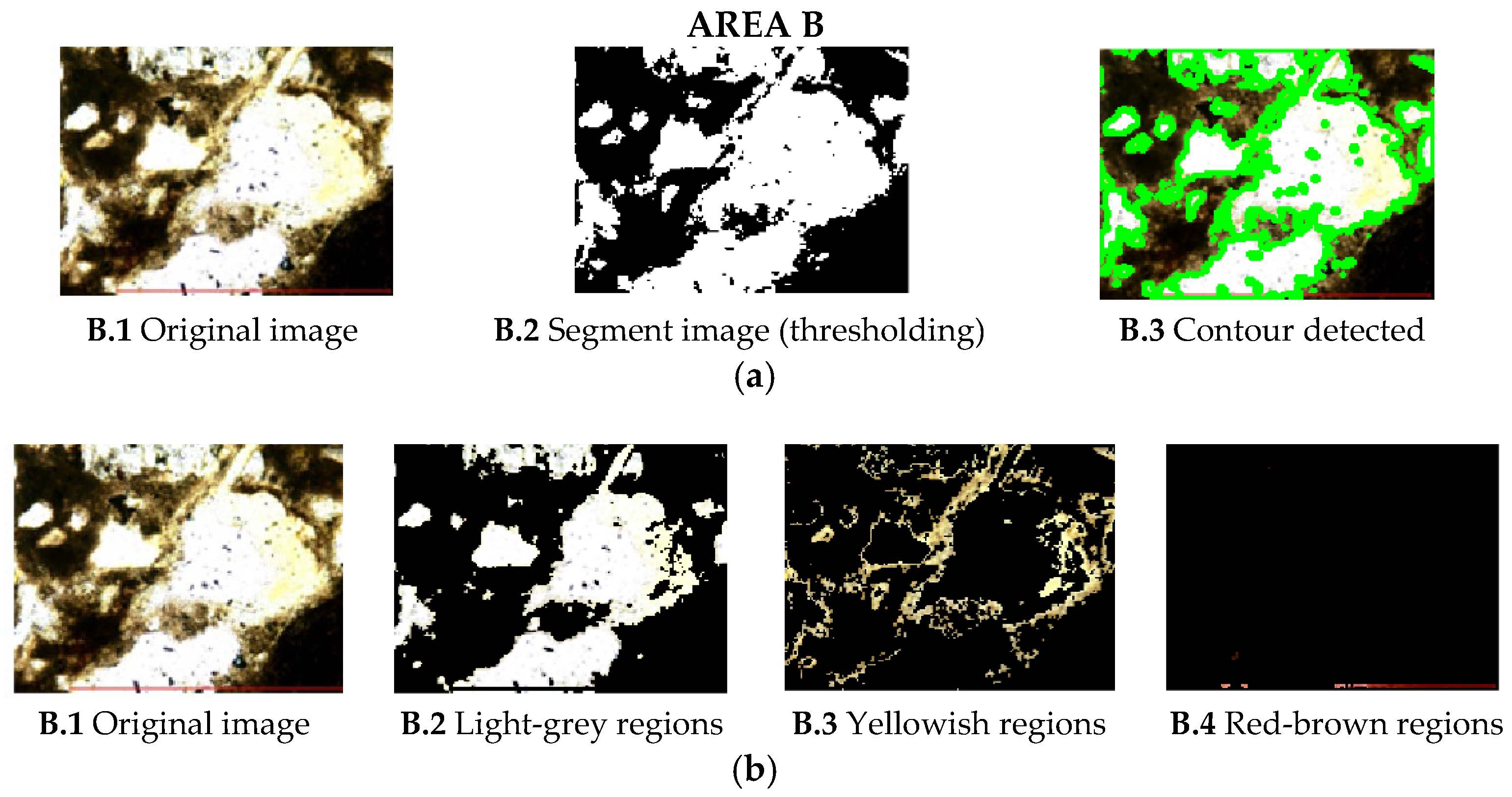
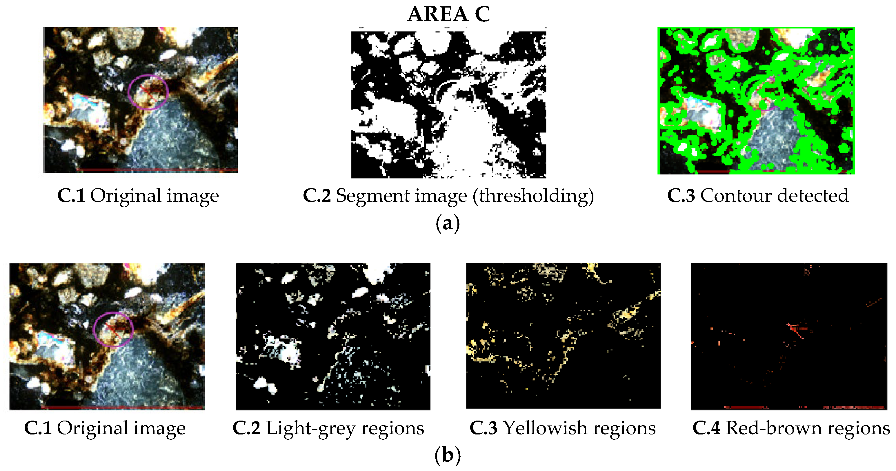
The results of the microscopic observations are presented in Figure 11, which shows representative thin sections prepared from the cracked regions after completion of the wet–dry exposure regime.
The optical images reveal the progressive development of deposits along the crack walls and within the crack opening. Their appearance is consistent with the formation of solid products during wet–dry exposure; however, optical microscopy alone does not permit definitive mineralogical identification of these deposits. Their possible mineralogical nature is therefore discussed only in conjunction with the XRD results and relevant literature.
In the investigated sections, solid deposits can be observed along the crack walls and, locally, across the crack opening. Their progressive development is consistent with the surface crack-sealing behavior observed during the wet–dry exposure.
From a morphological standpoint, the observed deposits appear more concentrated near the crack edges and progressively extend toward the central region of the crack. This spatial distribution indicates the progressive accumulation of solid material within the crack during wet–dry exposure, without allowing the specific nature or formation mechanism of these deposits to be established from optical microscopy alone.
Comparison of the investigated areas shows a broadly similar morphological pattern, characterized by the presence of solid deposits along the crack regions (Figure 12, Figure 13 and Figure 14). These observations indicate that deposit formation was not restricted to a single investigated area; however, the optical microscopy results alone do not allow the specific formation mechanisms or the individual contributions of the mortar constituents to be determined.

3.4. Mathematical Modeling of the Kinetics of the Self-Healing Process

The experimental results presented in Section 3.3 showed that the self-healing process of mortars modified with PVA fibers and a crystallization additive follows a nonlinear evolution, characterized by an accelerated reduction in crack width during the first conditioning cycles, followed by a gradual decrease in the closure rate until a steady state is reached. This behavior is characteristic of processes simultaneously controlled by secondary hydration reactions and calcium carbonate precipitation and was therefore represented in the present study using an empirical exponential fitting model.
An empirical exponential model was adopted to describe the experimentally observed evolution of mean crack width over time:
w(t) = w0 ekt
where w(t) represents the crack opening at time t; w0 represents the initial crack opening; k represents the constant of the self-healing process; and t represents the exposure time, expressed in hours.
The exponential relationship was used as an empirical fitting model to describe the experimentally observed decrease in mean surface crack width with exposure time, rather than as a mechanistic model of the crack-sealing process. The model was fitted to the experimentally determined mean crack-width values obtained at the predefined observation times for each mortar composition. The initial mean crack width was represented by w0, whereas k was determined from the fitting of the experimental crack-width evolution. The parameter k was estimated by fitting the exponential function to the experimental mean crack-width evolution for each composition, using w0 as the experimentally determined initial mean crack width. Accordingly, the fitted parameters characterize the present experimental dataset and should not be interpreted as independently validated predictive parameters.
The model assumes that the closure rate is proportional to the width of the existing crack at a given moment, which leads to an exponential reduction in crack width. The exponential function approaches zero asymptotically and therefore does not predict an exact zero crack width or an exact time of complete closure at a finite exposure time.
Accordingly, the values of 0 μm final crack width and 100% crack closure reported in Table 5 refer to the experimental observation of complete surface crack closure and not to values predicted by the exponential fitting model. Complete closure was assigned when no residual surface crack opening could be identified in the microscopic image at the corresponding observation time. Since a specific instrumental detection limit was not established during the experimental program, no numerical detection threshold is assigned retrospectively.
The empirical fitting was based on the experimental mean crack-width values obtained at the predefined observation times for T3 and T4. The resulting fitting parameters are summarized in Table 5.
Analysis of the parameters presented in Table 5 reveals significant differences between the two mixtures investigated. In the case of mortar T3, the self-healing process is characterized by a gradual progression, with the time required for complete crack closure ranging from 24 to 192 h, depending on the initial crack width. In contrast, for the T4 mixture, all cracks investigated were completely sealed within a maximum of 48 h, even in the case of cracks with initial openings exceeding 300 μm.
The fitted empirical constant k obtained for T4 was approximately twice that obtained for T3, reflecting the faster crack-sealing kinetics observed experimentally for T4. However, this difference cannot be attributed exclusively to cement content because T3 and T4 also differ in lime paste content, associated compositional ratios, and initial crack-width distribution.
For T4, the fitted value k = 0.030 h−1 indicates a faster decrease in mean crack width within the empirical exponential representation than that obtained for T3 (k = 0.014 h−1). However, these fitted constants describe the overall experimental trend and do not represent the experimentally observed time to complete surface crack closure. In particular, complete surface closure observed experimentally for T3 at 192 h and for T4 within 48 h should not be interpreted as zero crack-width values predicted by the exponential model.
Correlation of the experimental results, mineralogical observations, and mathematical model indicates that the observed crack-sealing behavior is associated with several interrelated factors: the development of hydrated phases resulting from secondary hydration and the precipitation of calcium carbonate within the cracks. PVA fibers indirectly contribute to the process’s efficiency by limiting crack opening and providing a favorable substrate for crystal nucleation, while the crystallization additive accelerates the formation of insoluble compounds responsible for sealing the cracks.
The empirical exponential model provides a compact quantitative representation of the observed evolution of mean surface crack width under the investigated wet–dry exposure conditions. Its applicability is restricted to the present experimental dataset, and additional experimental data and independent validation would be required before the model could be used predictively or generalized to other mortar compositions, crack-width ranges, or exposure conditions. The integration of experimental results with mathematical modeling represents one of the original contributions of this research and provides a foundation for the development of complex numerical models of the continuum damage–healing type, capable of simulating the behavior of structures made of cementitious materials with self-healing properties under real operating conditions.

4. Discussion

The results obtained in this study show that the T3 and T4 mortar systems containing both PVA fibers and the crystalline admixture exhibited complete surface crack sealing under the investigated wet–dry conditioning regime. The experimental observations and mineralogical characterization are consistent with the possible contribution of several crack-sealing processes; however, because formulations containing the individual functional agents were not investigated, their separate contributions and any possible synergistic interaction cannot be established from the present experimental design.
Regarding the physical and mechanical properties, the mean experimental values showed relatively limited numerical differences in density and water absorption between the reference and modified mortars. In contrast, the modified mortars exhibited lower mean compressive strengths and slightly higher mean flexural strengths than their corresponding reference mixtures. Because replicate-level data suitable for inferential statistical analysis were not available, these observations should be interpreted as descriptive numerical trends rather than as statistically significant effects. Possible mechanisms associated with these trends are discussed with reference to the literature, but they were not directly verified in the present study.
The results regarding self-healing confirm that the effectiveness of the process is primarily determined by the initial crack width and the mortar composition [46,47]. For the T3 mixture, the self-healing process was highly effective for cracks with openings smaller than approximately 200 μm, while for cracks with larger openings, the time required for complete closure increased significantly. In contrast, the T4 composition demonstrated the ability to completely seal even cracks with initial widths of approximately 350 μm within just 48 h. The complete closure observed for cracks with initial widths exceeding 300 μm is noteworthy within the present experimental dataset. However, direct quantitative comparison with crack-closure limits reported in previous studies should be made with caution because healing conditions, crack-generation procedures, exposure regimes, material compositions, and criteria used to define complete closure differ considerably among studies.
The XRD results identified calcite and portlandite among the crystalline phases present in the investigated materials and crack-region precipitates. These findings are consistent with the possible contribution of carbonation and continued hydration processes to the observed crack sealing.
The observed crack-sealing behavior under wet–dry exposure is consistent with recent findings on the self-healing performance of cementitious materials incorporating crystalline admixtures [85]. The identification of calcite, portlandite, via XRD analysis [86,87], as well as their distribution observed through optical microscopy and digital image analysis [79,86], demonstrates that self-healing is not merely a process of physically sealing cracks, but rather the result of complex mineralogical transformations that lead to the restoration of the continuity of the cementitious matrix [61,62].
The observed dependence of crack-sealing behavior on exposure time and initial crack width is also consistent with previous findings showing that healing duration and initial crack opening are important parameters governing the self-healing performance of cementitious materials incorporating crystalline admixtures [88].
The empirical mathematical model complements the experimental observations by providing a quantitative description of the evolution of mean surface crack width under the investigated conditions. The fitted empirical k values obtained for the two compositions reflect the different trends in mean crack-width reduction observed for T3 and T4. However, these differences should be interpreted in relation to the overall matrix compositions and the different initial crack-width distributions rather than being attributed exclusively to cement content [89,90,91,92].
Comparing the results obtained with those reported in the literature reveals agreement regarding the positive influence of PVA fibers on the control of crack propagation and the development of self-healing mechanisms [34,46].
From a practical standpoint, the results of this research indicate that mortars modified with PVA fibers and a crystallization additive may represent a viable solution for increasing the durability of masonry and concrete structures, particularly in the case of elements exposed to repeated cycles of wetting and drying [50,51]. For masonry applications, the observed surface crack-sealing behavior may be relevant to limiting moisture and aggressive-agent ingress; however, the associated effects on transport properties and long-term durability were not directly evaluated in the present study. For potential applications in reinforced cementitious elements, the influence of crack sealing on reinforcement corrosion and service-life performance should be investigated through dedicated durability and corrosion-related testing. In addition, reducing the need for repair interventions could potentially lower maintenance demand and the environmental impacts associated with rehabilitation activities; however, these potential benefits were not quantified in the present study and would require dedicated life-cycle assessment.
However, the present study has several limitations that should be considered when interpreting the results. First, the experimental program was conducted under controlled laboratory conditions using repeated wet–dry cycles and a limited number of mortar compositions. Under actual service conditions, crack-sealing behavior may be influenced by additional factors, including climatic variations, repeated mechanical loading, salt exposure, and freeze–thaw cycles [17,18].
Fractographic characterization of the specimens after flexural and compressive testing was not performed. Consequently, mechanisms such as fiber bridging, fiber pull-out, crack deflection, and changes in the fiber–matrix interfacial zone could not be directly verified. Therefore, the mechanistic interpretations proposed for the observed mechanical behavior should be regarded as possible explanations consistent with the literature rather than as mechanisms directly demonstrated by the present experimental results.
Moreover, crack-sealing monitoring was performed only for the T3 and T4 mixtures containing both PVA fibers and the crystalline admixture. Because cracked reference mixtures and single-component formulations containing only PVA fibers or only the crystalline admixture were not included, the individual contributions of autogenous healing, PVA fibers, and the crystalline admixture cannot be quantitatively separated based on the present experimental results.
An additional limitation concerns the statistical treatment of the physical and mechanical results. Although multiple specimens were tested and mean values were obtained for the investigated properties, the original replicate-level measurements were not retained in a form that allowed a reliable retrospective calculation of standard deviations, confidence intervals, effect sizes, or inferential statistical tests. Consequently, the comparisons among T1–T4 presented in this study are descriptive and should not be interpreted as demonstrating statistical significance. This limitation highlights the need for future experimental programs to retain and report complete replicate-level datasets and to incorporate appropriate inferential statistical analyses.
Another limitation is related to the use of CEM I 42.5 R as the primary cementitious binder. This cement was selected to provide a well-defined and reproducible Portland-cement matrix for the experimental investigation. Due to its relatively high clinker content, however, it does not represent an optimized binder from the perspective of current cement decarbonization strategies. No life-cycle assessment, embodied-carbon analysis, or other quantitative environmental evaluation was performed in the present study. Therefore, potential sustainability benefits associated with crack sealing, service-life extension, and reduced maintenance requirements should be regarded as prospective benefits rather than experimentally demonstrated environmental advantages. Future studies should investigate the performance of the proposed approach using lower-clinker binders and supplementary cementitious materials and should include a dedicated life-cycle assessment.
Finally, the empirical mathematical model provides a simplified description of the evolution of surface crack width under the investigated conditions and does not explicitly account for the individual physicochemical processes involved or for the interaction between mechanical damage and mineral precipitation. Its applicability beyond the investigated mixtures, crack-width range, and wet–dry conditioning regime therefore requires further validation.
Therefore, future research should extend the experimental program to long-term and more representative exposure conditions and include cracked reference mixtures as well as single-component formulations containing only PVA fibers or only the crystalline admixture. Additional fractographic and microstructural investigations should be performed to directly characterize fiber–matrix interactions and fracture mechanisms. The use of lower-clinker binders and supplementary cementitious materials should also be investigated together with quantitative life-cycle assessment. Furthermore, dedicated durability and corrosion-related testing and more advanced numerical models incorporating mechanical damage, moisture transport, hydration, carbonation, and mineral precipitation would provide a more comprehensive assessment of the long-term performance of these materials.

5. Conclusions

The objective of this research was to investigate the behavior of a masonry mortar modified with polyvinyl alcohol (PVA) fibers and a mass crystallization additive, by evaluating its ability to self-heal microcracks and identifying the physicochemical mechanisms responsible for this process. The experimental approach included determining the physical and mechanical properties, monitoring the evolution of fissures following successive wet–dry cycles, mineralogical characterization by X-ray diffraction (XRD), microstructural analysis by optical microscopy and digital image processing, as well as the development of a mathematical model capable of describing the kinetics of the self-healing process. The integration of these methods has enabled a thorough understanding of the material’s behavior and the mechanisms involved in the self-healing of the cementitious matrix.
The mean experimental values showed relatively limited numerical differences in density and water absorption between the reference and modified mortars. The modified mortars exhibited lower mean compressive strengths and slightly higher mean flexural strengths than their corresponding reference mixtures. Because replicate-level data suitable for inferential statistical analysis were not available, these differences should be interpreted as descriptive numerical trends rather than as statistically significant effects. The possible mechanisms associated with these trends, including fiber–matrix interactions and crack-bridging effects, were not directly verified in the present study and should therefore be regarded as interpretations consistent with the available literature.
Analysis of the self-healing process showed that the effectiveness of crack closure depends on both the mortar composition and the initial size of the cracks. Composition T3, made with 215 kg/m3 of cement, demonstrated effective self-healing for cracks with small and medium openings; however, for larger cracks, the time required for complete closure increased considerably. In contrast, the T4 mixture, containing 300 kg/m3 of cement, demonstrated superior performance, successfully and completely sealing even cracks with initial widths of approximately 346 μm within a period of only 48 h. These results highlight differences in crack-sealing behavior between T3 and T4 under the investigated conditions. However, because the two mixtures differ simultaneously in cement content, lime paste content, associated compositional ratios, and initial crack-width distribution, the observed differences cannot be attributed exclusively to cement dosage.
Qualitative XRD analysis identified calcite, portlandite, chlorite, and tobermorite among the crystalline phases present in the investigated samples and crack-region precipitates. The XRD results, considered together with the optical microscopic observations, are consistent with the occurrence of mineral precipitation within the crack regions during wet–dry exposure. However, the individual contributions of the identified phases, PVA fibers, and the crystalline admixture to the observed crack-sealing process could not be quantitatively determined from the present experimental design.
The determined model parameters indicated faster crack-sealing kinetics for T4 than for T3 under the investigated conditions. However, the difference between the two compositions should be interpreted in relation to their overall compositional differences and initial crack-width distributions rather than as an isolated effect of cement content. The empirical exponential model provided a good fit to the experimental mean crack-width data for T3 and T4 under the investigated conditions, with R2 values of 0.943 and 0.991, respectively.
From a practical standpoint, the results obtained demonstrate that mortars modified with PVA fibers and a crystallization additive represent a promising solution for producing construction materials with high durability and self-healing capabilities. Their ability to limit crack propagation and reduce permeability can lead to a decrease in the penetration of aggressive agents, a slowing of degradation processes, and, consequently, an increase in the service life of structures. A potential reduction in the need for repair interventions may contribute to lower resource consumption and environmental impacts associated with rehabilitation activities; however, these potential sustainability benefits were not quantified in the present study.
The originality of this research lies in the integrated experimental, mineralogical, and mathematical approach used to investigate the behavior of self-healing masonry mortars. Unlike most existing studies, which analyze mechanical performance or mineralogical mechanisms separately, this work demonstrates the direct relationship between material composition, microstructural evolution, and the kinetics of the self-healing process. The proposed methodology provides a coherent framework for the design of smart mortars with superior performance and can serve as the basis for the development of advanced numerical models capable of simultaneously describing the degradation and regeneration processes of cementitious materials under real-world service conditions.

Author Contributions

Conceptualization, T.P.T., I.N.S., A.H. and A.S.; methodology, T.P.T., N.H. and I.N.S.; software, C.M.; validation, C.M., T.P.T., N.H. and A.-T.M.; formal analysis, I.N.S. and N.H.; investigation, I.N.S., A.S.; resources, T.P.T., A.H. and C.M.; data curation, A.-T.M., and N.H.; writing—original draft preparation, I.N.S. and T.P.T.; writing—review and editing, T.P.T., D.-M.M., A.-T.M. and A.S.; visualization, A.-T.M. and D.-M.M.; supervision, C.M., T.P.T., N.H., and A.H.; project administration, C.M., T.P.T. and A.H. All authors have read and agreed to the published version of the manuscript.

Funding

This research received no external funding.

Data Availability Statement

The data supporting the results reported in this study are contained within the article. The original replicate-level data are not available in a form that permits reliable retrospective statistical analysis.

Conflicts of Interest

The authors declare no conflicts of interest.

Abbreviations

The following abbreviations are used in this manuscript:
PVAPolyvinyl alcohol
XRDX-ray diffraction

References

  1. Neville, A.M. Properties of Concrete, 5th ed.; Pearson Education Limited: Harlow, UK, 2011. [Google Scholar]
  2. Mehta, P.K.; Monteiro, P.J.M. Concrete: Microstructure, Properties, and Materials, 4th ed.; McGraw-Hill Education: New York, NY, USA, 2014. [Google Scholar]
  3. Taylor, H.F.W. Cement Chemistry, 2nd ed.; Thomas Telford Publishing: London, UK, 1997. [Google Scholar]
  4. Scrivener, K.L.; John, V.M.; Gartner, E.M. Eco-efficient cements: Potential economically viable solutions for a low-CO2 cement-based materials industry. Cem. Concr. Res. 2018, 114, 2–26. [Google Scholar] [CrossRef] [Scilit]
  5. Gartner, E.; Sui, T. Alternative cement clinkers. Cem. Concr. Res. 2018, 114, 27–39. [Google Scholar] [CrossRef] [Scilit]
  6. Habert, G.; Miller, S.A.; John, V.M.; Provis, J.L.; Favier, A.; Horvath, A.; Scrivener, K.L. Environmental impacts and decarbonization strategies in the cement and concrete industries. Nat. Rev. Earth Environ. 2020, 1, 559–573. [Google Scholar] [CrossRef] [Scilit]
  7. Scrivener, K.L.; Juilland, P.; Monteiro, P.J.M. Advances in understanding hydration of Portland cement. Cem. Concr. Res. 2015, 78, 38–56. [Google Scholar] [CrossRef] [Scilit]
  8. Bullard, J.W.; Jennings, H.M.; Livingston, R.A.; Nonat, A.; Scherer, G.W.; Schweitzer, J.S.; Scrivener, K.L.; Thomas, J.J. Mechanisms of cement hydration. Cem. Concr. Res. 2011, 41, 1208–1223. [Google Scholar] [CrossRef] [Scilit]
  9. Van Tittelboom, K.; De Belie, N. Self-healing in cementitious materials—A review. Materials 2013, 6, 2182–2217. [Google Scholar] [CrossRef] [Scilit]
  10. De Belie, N.; Gruyaert, E.; Al-Tabbaa, A.; Antonaci, P.; Baera, C.; Bajare, D.; Darquennes, A.; Davies, R.; Ferrara, L.; Jefferson, T.; et al. A review of self-healing concrete for damage management of structures. Adv. Mater. Interfaces 2018, 5, 1800074. [Google Scholar] [CrossRef] [Scilit]
  11. Rooij, M.R.D.; Van Tittelboom, K.; De Belie, N.; Schlangen, E. (Eds.) Self-Healing Phenomena in Cement-Based Materials: State-of-the-Art Report of RILEM Technical Committee 221-SHC; Springer: Dordrecht, The Netherlands, 2013. [Google Scholar] [CrossRef] [Scilit]
  12. Schlangen, E.; Joseph, C. Self-healing processes in concrete. In Self-Healing Materials: Fundamentals, Design Strategies, and Applications; Ghosh, S.K., Ed.; Wiley-VCH: Weinheim, Germany, 2009; pp. 141–182. [Google Scholar]
  13. Hearn, N. Self-sealing, autogenous healing and continued hydration: What is the difference? Mater. Struct. 1998, 31, 563–567. [Google Scholar] [CrossRef] [Scilit]
  14. Liu, J.C.; Tan, K.H. Mechanism of PVA fibers in mitigating explosive spalling of engineered cementitious composite at elevated temperature. Cem. Concr. Compos. 2018, 93, 235–245. [Google Scholar] [CrossRef] [Scilit]
  15. Wang, D.; Yang, Y.; Yu, Z.; Liu, R. Mechanical properties of PVA fiber reinforced cementitious composites (PVA-FRCC) after high temperature. Case Stud. Constr. Mater. 2024, 21, e04020. [Google Scholar] [CrossRef] [Scilit]
  16. Ge, C.; Chen, X.; Gong, Y.; Kong, X.; Chen, F. Effect of high temperature on micro-structure and mechanical properties of fiber-reinforced cement-based composites. Crystals 2024, 14, 778. [Google Scholar] [CrossRef] [Scilit]
  17. Edvardsen, C. Water permeability and autogenous healing of cracks in concrete. ACI Mater. J. 1999, 96, 448–454. [Google Scholar] [CrossRef] [Scilit]
  18. Reinhardt, H.W.; Jooss, M. Permeability and self-healing of cracked concrete as a function of temperature and crack width. Cem. Concr. Res. 2003, 33, 981–985. [Google Scholar] [CrossRef] [Scilit]
  19. Granger, S.; Loukili, A.; Pijaudier-Cabot, G.; Chanvillard, G. Experimental characterization of the self-healing of cracks in an ultra-high-performance cementitious material: Mechanical tests and acoustic emission analysis. Cem. Concr. Res. 2007, 37, 519–527. [Google Scholar] [CrossRef] [Scilit]
  20. Jacobsen, S.; Marchand, J.; Hornain, H. SEM observations of the microstructure of frost-deteriorated and self-healed concretes. Cem. Concr. Res. 1995, 25, 1781–1790. [Google Scholar] [CrossRef] [Scilit]
  21. Şahmaran, M.; Keskin, S.B.; Ozerkan, G.; Yaman, I.O. Self-healing of mechanically loaded self-consolidating concretes with high volumes of fly ash. Cem. Concr. Compos. 2008, 30, 872–879. [Google Scholar] [CrossRef] [Scilit]
  22. Yang, Y.; Lepech, M.D.; Yang, E.H.; Li, V.C. Autogenous healing of engineered cementitious composites under wet–dry cycles. Cem. Concr. Res. 2009, 39, 382–390. [Google Scholar] [CrossRef] [Scilit]
  23. Homma, D.; Mihashi, H.; Nishiwaki, T. Self-healing capability of fibre reinforced cementitious composites. J. Adv. Concr. Technol. 2009, 7, 217–228. [Google Scholar] [CrossRef] [Scilit]
  24. Qureshi, T.S.; Al-Tabbaa, A. Self-healing of drying shrinkage cracks in cement-based materials incorporating reactive MgO. Smart Mater. Struct. 2016, 25, 084004. [Google Scholar] [CrossRef] [Scilit]
  25. Huang, H.; Ye, G.; Damidot, D. Characterization and quantification of self-healing behaviors of microcracks due to further hydration in cement paste. Cem. Concr. Res. 2013, 52, 71–81. [Google Scholar] [CrossRef] [Scilit]
  26. Huang, H.; Ye, G.; Damidot, D. Effect of blast furnace slag on self-healing of microcracks in cementitious materials. Cem. Concr. Res. 2014, 60, 68–82. [Google Scholar] [CrossRef] [Scilit]
  27. Sisomphon, K.; Copuroglu, O.; Koenders, E.A.B. Self-healing of surface cracks in mortars with expansive additive and crystalline additive. Cem. Concr. Compos. 2012, 34, 566–574. [Google Scholar] [CrossRef] [Scilit]
  28. Ferrara, L.; Krelani, V.; Carsana, M. A fracture testing-based approach to assess crack healing of concrete with and without crystalline admixtures. Constr. Build. Mater. 2014, 68, 535–551. [Google Scholar] [CrossRef] [Scilit]
  29. Ferrara, L.; Van Mullem, T.; Alonso, M.C.; Antonaci, P.; Borg, R.P.; Cuenca, E.; Jefferson, A.; Ng, P.L.; Peled, A.; Roig-Flores, M.; et al. Experimental characterization of the self-healing capacity of cement-based materials and its effects on the material performance: A state of the art report by COST Action SARCOS WG2. Constr. Build. Mater. 2018, 167, 115–142. [Google Scholar] [CrossRef] [Scilit]
  30. Roig-Flores, M.; Moscato, S.; Serna, P.; Ferrara, L. Self-healing capability of concrete with crystalline admixtures in different environments. Constr. Build. Mater. 2015, 86, 1–11. [Google Scholar] [CrossRef] [Scilit]
  31. Cuenca, E.; Tejedor, A.; Ferrara, L. A methodology to assess crack-sealing effectiveness of crystalline admixtures under repeated cracking–healing cycles. Constr. Build. Mater. 2018, 179, 619–632. [Google Scholar] [CrossRef] [Scilit]
  32. Pazderka, J.; Hájková, E. Crystalline admixtures and their effect on selected properties of concrete. Acta Polytech. 2016, 56, 306–311. [Google Scholar] [CrossRef] [Scilit]
  33. Li, V.C. On engineered cementitious composites: A review of the material and its applications. J. Adv. Concr. Technol. 2003, 1, 215–230. [Google Scholar] [CrossRef] [Scilit]
  34. Li, V.C.; Wang, S.; Wu, C. Tensile strain-hardening behavior of polyvinyl alcohol engineered cementitious composite. ACI Mater. J. 2001, 98, 483–492. [Google Scholar] [CrossRef] [Scilit]
  35. Li, V.C.; Wu, C.; Wang, S.; Ogawa, A.; Saito, T. Interface tailoring for strain-hardening polyvinyl alcohol-engineered cementitious composite. ACI Mater. J. 2002, 99, 463–472. [Google Scholar] [CrossRef] [Scilit]
  36. Li, V.C. Engineered cementitious composites: Material, structural, and durability performance. In Concrete Construction Engineering Handbook, 2nd ed.; Nawy, E.G., Ed.; CRC Press: Raton, FL, USA, 2008. [Google Scholar]
  37. Li, V.C. Engineered Cementitious Composites: Bendable Concrete for Sustainable and Resilient Infrastructure; Springer: Berlin/Heidelberg, Germany, 2019. [Google Scholar] [CrossRef] [Scilit]
  38. Kanda, T.; Li, V.C. Interface property and apparent strength of high-strength hydrophilic fiber in cement matrix. J. Mater. Civ. Eng. 1998, 10, 5–13. [Google Scholar] [CrossRef] [Scilit]
  39. Redon, C.; Li, V.C.; Wu, C.; Hoshiro, H.; Saito, T.; Ogawa, A. Measuring and modifying interface properties of PVA fibers in ECC matrix. J. Mater. Civ. Eng. 2001, 13, 399–406. [Google Scholar] [CrossRef] [Scilit]
  40. Li, V.C.; Leung, C.K.Y. Steady-state and multiple cracking of short random fiber composites. J. Eng. Mech. 1992, 118, 2246–2264. [Google Scholar] [CrossRef] [Scilit]
  41. Bentur, A.; Mindess, S. Fibre Reinforced Cementitious Composites, 2nd ed.; Taylor & Francis: London, UK, 2006. [Google Scholar]
  42. Naaman, A.E. Engineered steel fibers with optimal properties for reinforcement of cement composites. J. Adv. Concr. Technol. 2003, 1, 241–252. [Google Scholar] [CrossRef] [Scilit]
  43. Banthia, N.; Gupta, R. Hybrid fiber reinforced concrete: Fiber synergy in high-strength matrices. Mater. Struct. 2004, 37, 707–716. [Google Scholar] [CrossRef] [Scilit]
  44. Snoeck, D.; De Belie, N. From straw in bricks to modern use of microfibres in cementitious composites for improved autogenous healing—A review. Constr. Build. Mater. 2015, 95, 774–787. [Google Scholar] [CrossRef] [Scilit]
  45. Snoeck, D.; De Belie, N. Repeated autogenous healing in strain-hardening cementitious composites by using wet–dry cycles. Cem. Concr. Compos. 2016, 69, 95–103. [Google Scholar] [CrossRef] [Scilit]
  46. Snoeck, D.; Van Tittelboom, K.; Steuperaert, S.; Dubruel, P.; De Belie, N. Self-healing cementitious materials by the combination of microfibres and superabsorbent polymers. J. Intell. Mater. Syst. Struct. 2014, 25, 13–24. [Google Scholar] [CrossRef] [Scilit]
  47. Kan, L.L.; Shi, H.S.; Sakulich, A.R.; Li, V.C. Self-healing characterization of engineered cementitious composite materials. ACI Mater. J. 2010, 107, 617–624. [Google Scholar] [CrossRef] [Scilit]
  48. Herbert, E.N.; Li, V.C. Self-healing of microcracks in engineered cementitious composites under a natural environment. Materials 2013, 6, 2831–2845. [Google Scholar] [CrossRef] [Scilit]
  49. Şahmaran, M.; Li, V.C. Durability properties of micro-cracked ECC containing high volumes of fly ash. Cem. Concr. Res. 2009, 39, 1033–1043. [Google Scholar] [CrossRef] [Scilit]
  50. Sahmaran, M.; Yildirim, G.; Erdem, T.K. Self-healing capability of cementitious composites incorporating different supplementary cementitious materials. Cem. Concr. Compos. 2013, 35, 89–101. [Google Scholar] [CrossRef] [Scilit]
  51. Arandigoyen, M.; Alvarez, J.I. Pore structure and mechanical properties of cement–lime mortars. Cem. Concr. Res. 2007, 37, 767–775. [Google Scholar] [CrossRef] [Scilit]
  52. Lanas, J.; Pérez Bernal, J.L.; Bello, M.A.; Alvarez-Galindo, J.I. Mechanical properties of natural hydraulic lime-based mortars. Cem. Concr. Res. 2004, 34, 2191–2201. [Google Scholar] [CrossRef] [Scilit]
  53. Hall, C.; Hoff, W.D. Water Transport in Brick, Stone and Concrete, 2nd ed.; CRC Press: Boca Raton, FL, USA, 2012. [Google Scholar]
  54. Hall, C. Water sorptivity of mortars and concretes: A review. Mag. Concr. Res. 1989, 41, 51–61. [Google Scholar] [CrossRef] [Scilit]
  55. Hall, C.; Yau, M.H.R. Water movement in porous building materials—IX. The water absorption and sorptivity of concretes. Build. Environ. 1987, 22, 77–82. [Google Scholar] [CrossRef] [Scilit]
  56. Martys, N.S.; Ferraris, C.F. Capillary transport in mortars and concrete. Cem. Concr. Res. 1997, 27, 747–760. [Google Scholar] [CrossRef] [Scilit]
  57. EN 197-1:2011; Cement—Part 1: Composition, Specifications and Conformity Criteria for Common Cements. European Committee for Standardization (CEN): Brussels, Belgium, 2011.
  58. EN 459-1:2015; Building Lime—Part 1: Definitions, Specifications and Conformity Criteria. European Committee for Standardization (CEN): Brussels, Belgium, 2015.
  59. EN 13139:2002; Aggregates for Mortar. European Committee for Standardization (CEN): Brussels, Belgium, 2002.
  60. EN 1008:2002; Mixing Water for Concrete—Specification for Sampling, Testing and Assessing the Suitability of Water. European Committee for Standardization (CEN): Brussels, Belgium, 2002.
  61. Lo Monte, F.; Ferrara, L. Self-healing characterization of ultra-high-performance fiber-reinforced cementitious composites with crystalline admixture: Experimental assessment through a multi-test/multi-parameter approach. Constr. Build. Mater. 2021, 283, 122579. [Google Scholar] [CrossRef] [Scilit]
  62. Oliveira, A.S.; Gomes, O.F.M.; Ferrara, L.; Fairbairn, E.M.R.; Toledo Filho, R.D. Microstructural characterization of self-healing products in cementitious systems containing crystalline admixture in the short- and long-term. Cem. Concr. Compos. 2022, 126, 104369. [Google Scholar] [CrossRef] [Scilit]
  63. Tsampali, E.; Stefanidou, M. The role of crystalline admixtures in the long-term healing process of fiber-reinforced cementitious composites. J. Build. Eng. 2022, 60, 105164. [Google Scholar] [CrossRef] [Scilit]
  64. Li, H.F.; Yu, Q.Q.; Zhang, K.; Wang, X.Y.; Liu, Y.; Zhang, G.Z. Effect of curing environments on the self-healing capacity of mortars incorporating crystalline admixture. Case Stud. Constr. Mater. 2022, 17, e01713. [Google Scholar] [CrossRef] [Scilit]
  65. NE 012-1:2007; Code of Practice for the Execution of Concrete, Reinforced Concrete and Prestressed Concrete Works—Part 1: Production of Concrete. Ministry of Development, Public Works and Housing: Bucharest, Romania, 2007.
  66. EN 12390-2:2019; Testing Hardened Concrete—Part 2: Making and Curing Specimens for Strength Tests. European Committee for Standardization (CEN): Brussels, Belgium, 2019.
  67. EN 1015-3:1999; Methods of Test for Mortar for Masonry—Part 3: Determination of Consistence of Fresh Mortar by Flow Table. European Committee for Standardization (CEN): Brussels, Belgium, 1999.
  68. EN 1015-6:1998; Methods of Test for Mortar for Masonry—Part 6: Determination of Bulk Density of Fresh Mortar. European Committee for Standardization (CEN): Brussels, Belgium, 1998.
  69. EN 1015-10:1999; Methods of Test for Mortar for Masonry—Part 10: Determination of Dry Bulk Density of Hardened Mortar. European Committee for Standardization (CEN): Brussels, Belgium, 1999.
  70. EN 1015-18:2002; Methods of Test for Mortar for Masonry—Part 18: Determination of Water Absorption Coefficient Due to Capillary Action of Hardened Mortar. European Committee for Standardization (CEN): Brussels, Belgium, 2002.
  71. Neville, A.M. The confused world of sulfate attack on concrete. Cem. Concr. Res. 2004, 34, 1275–1296. [Google Scholar] [CrossRef] [Scilit]
  72. Scrivener, K.L.; Snellings, R.; Lothenbach, B. (Eds.) A Practical Guide to Microstructural Analysis of Cementitious Materials; CRC Press: Boca Raton, FL, USA, 2016. [Google Scholar]
  73. Bish, D.L.; Post, J.E. (Eds.) Modern Powder Diffraction; Mineralogical Society of America: Washington, DC, USA, 1989. [Google Scholar]
  74. Klug, H.P.; Alexander, L.E. X-Ray Diffraction Procedures for Polycrystalline and Amorphous Materials, 2nd ed.; Wiley: New York, NY, USA, 1974. [Google Scholar]
  75. EN 1015-11:2019; Methods of Test for Mortar for Masonry—Part 11: Determination of Flexural and Compressive Strength of Hardened Mortar. European Committee for Standardization (CEN): Brussels, Belgium, 2019.
  76. Wang, X.; Ding, Z.; Zhang, Y.; Xu, Y. Self-healing of high-performance engineered cementitious materials with crystalline admixture in seawater environments. J. Build. Eng. 2023, 63, 105472. [Google Scholar] [CrossRef] [Scilit]
  77. Alemu, A.S.; Choi, J.I.; Nguyễn, H.H.; Choi, S.; Suh, J.I.; Lee, B.Y.; Kim, H.K. On crack healing in fiber-reinforced cementitious composites incorporating a mineral-based healing agent and superabsorbent polymer: Evaluation using a modified permeability test method. Cem. Concr. Compos. 2023, 141, 105111. [Google Scholar] [CrossRef] [Scilit]
  78. Zhang, G.-Z.; Ma, X.; Liu, Y. Study on the Synergistic Effect of Superabsorbent Polymer and Crystalline Admixture on Self-Healing Performance of Mortar Based on Image Binarization Method. Buildings 2023, 13, 2953. [Google Scholar] [CrossRef] [Scilit]
  79. Boca, A. Contribuții Privind Reabilitarea Ansamblurilor Structurale Romanice din Transilvania. Ph.D. Thesis, Technical University of Cluj-Napoca, Cluj-Napoca, Romania, 2024. [Google Scholar]
  80. Morandeau, A.; Thiéry, M.; Dangla, P. Investigation of the carbonation mechanism of CH and C–S–H in terms of kinetics, microstructure changes and moisture properties. Cem. Concr. Res. 2014, 56, 153–170. [Google Scholar] [CrossRef] [Scilit]
  81. Auroy, M.; Poyet, S.; Le Bescop, P.; Torrenti, J.M.; Charpentier, T.; Moskura, M.; Bourbon, X. Impact of carbonation on unsaturated water transport properties of cement-based materials. Cem. Concr. Res. 2015, 74, 44–58. [Google Scholar] [CrossRef] [Scilit]
  82. Niu, M.; Wang, Y.; He, X.; Miao, D.; Li, G. Enhancing the self-healing capability of ternary repair cementitious composites incorporating fibers and crystalline admixtures. Constr. Build. Mater. 2025, 483, 141744. [Google Scholar] [CrossRef] [Scilit]
  83. Van Mullem, T.; De Belie, N.; Maddalena, R. From crack closure to durability improvement of self-healing cementitious materials. Dev. Built Environ. 2025, 24, 100812. [Google Scholar] [CrossRef] [Scilit]
  84. Wen, X.; Pagcaliwagan, A.M.; Hou, H.; Yin, X. Advancements in self-healing concrete: Materials, mechanisms, characterization methods, and applications. Alex. Eng. J. 2025, 131, 104–124. [Google Scholar] [CrossRef] [Scilit]
  85. Duan, L.; Zhang, Y. Crack self-healing of cementitious materials with crystalline admixtures. J. Wuhan Univ. Technol.-Mater. Sci. Ed. 2025, 40, 1660–1671. [Google Scholar] [CrossRef] [Scilit]
  86. Lothenbach, B.; Scrivener, K.; Hooton, R.D. Supplementary cementitious materials. Cem. Concr. Res. 2011, 41, 1244–1256. [Google Scholar] [CrossRef] [Scilit]
  87. Richardson, I.G. The calcium silicate hydrates. Cem. Concr. Res. 2008, 38, 137–158. [Google Scholar] [CrossRef] [Scilit]
  88. Doostkami, H.; Formagini, S.; Serna, P.; Roig-Flores, M. Effects of healing start time and duration on conventional and high-performance concretes incorporating SAP, crystalline admixture, and sepiolite: A comparative study. Case Stud. Constr. Mater. 2024, 20, e02835. [Google Scholar] [CrossRef] [Scilit]
  89. Wu, H.; Zhao, Y.; Chen, X.; Li, S.; Zhao, Y.; Sun, Q. Effect of crystalline admixture and superabsorbent polymer on the self-healing performance of cement-based composites. J. Asian Archit. Build. Eng. 2024, 23, 1265–1277. [Google Scholar] [CrossRef] [Scilit]
  90. Ammar, M.A.; Chegenizadeh, A.; Budihardjo, M.A.; Nikraz, H. The effects of crystalline admixtures on concrete permeability, durability, and self-healing performance. Buildings 2024, 14, 3000. [Google Scholar] [CrossRef] [Scilit]
  91. Borçato, A.G.; Medeiros-Junior, R.A. Self-healing in metakaolin-based geopolymers incorporating crystalline admixtures and expansive agents. Constr. Build. Mater. 2024, 418, 135391. [Google Scholar] [CrossRef] [Scilit]
  92. Shams, M.A.; Bheel, N.; Abid, M.M.; Alraeeini, A.S.; Almaliki, A.H.; Dodo, Y.A.; Benjeddou, O. Exploring fracture energy in engineered cementitious composites with different PVA-fiber volume fractions. Int. J. Concr. Struct. Mater. 2025, 19, 66. [Google Scholar] [CrossRef] [Scilit]
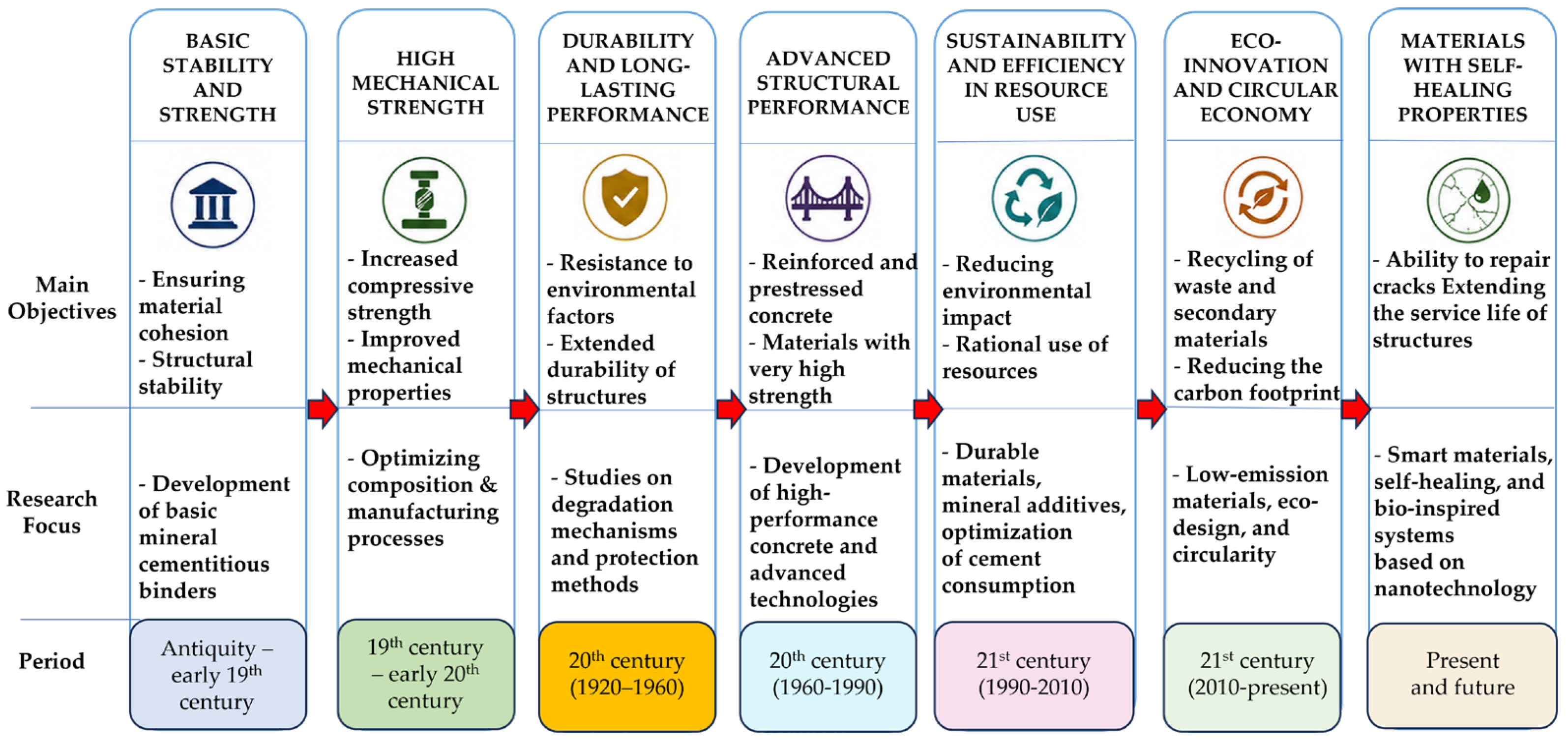
Figure 1. The evolution of performance criteria for cementitious materials toward eco-innovative materials with self-healing properties.
Buildings 16 03697 g001
Figure 2. Classification of the main self-healing mechanisms, the agents used, and the maximum sizes of cracks that can be sealed.
Buildings 16 03697 g002
Figure 3. Experimental workflow adopted in the present study, together with complementary analytical techniques applicable to the broader characterization of cementitious materials.
Buildings 16 03697 g003
Figure 4. Density of mortar in the fresh state (a) and after hardening, 28 days after preparation (b).
Buildings 16 03697 g004
Figure 5. Crack-closure evolution for the T3 composition at 0, 24, 48, 96, and 192 h. The t192 series is retained in the legend although no corresponding bars are visible because all crack segments reached a measured opening of 0 μm at 192 h.
Buildings 16 03697 g005
Figure 6. Crack-closure evolution for the T4 composition at 0, 24, 48, and 96 h. All monitored crack segments reached a measured opening of 0 μm by 48 h; therefore, no visible bars are present for t48 and t96. At the subsequent t192 observation, the crack width remained 0 μm for all investigated segments and is therefore not separately represented.
Buildings 16 03697 g006
Figure 7. Comparison of the crack-sealing performance of mixtures T3 and T4: (a) T3, initial crack openings <100 μm; (b) T3, initial crack openings ≥100 μm, excluding the representative large-opening segment S12; (c) T4, initial crack openings <100 μm; (d) comparison of the representative large-opening segments T3 S12 (299 μm) and T4 S5 (346 μm).
Buildings 16 03697 g007
Figure 8. Details of microscopic evolution of the self-healing process for T3 mortar during successive wet–dry cycles. (a,f,j) Initial phase; (b,g,k) after 24 h—(1 wet-dry cycle); (c,h,l) after 48 h—(2 wet-dry cycles); (d,i,m) after 96 h—(4 wet-dry cycles); (e,n) after 192 h—(8 wet-dry cycles) complete closure. For Detail 2, imaging was discontinued after complete crack closure had been observed; therefore, no image is shown at 192 h.
Buildings 16 03697 g008
Figure 9. Details of the microscopic evolution of the self-healing process for T4 mortar during successive wet–dry cycles: (a,c,e) initial phase; (b,d,f) after 24 h (1 wet–dry cycle).
Buildings 16 03697 g009
Figure 10. XRD diffractograms for: (a) composition T3; (b) composition T4; (c) the mineral precipitate deposited in the crack after successive wet–dry cycles.
Buildings 16 03697 g010aBuildings 16 03697 g010b
Figure 11. Representative optical microscopic images of thin sections prepared from the cracked regions of the mortar specimens after completion of the wet–dry exposure regime: (ad) representative microstructural features and solid deposits observed within and along the investigated crack regions. Scale bars and measurement annotations are shown in the individual micrographs.
Buildings 16 03697 g011
Figure 12. Workflow for the digital analysis of microscopic images of Area A. (a) Contour-based segmentation: A.1, original microscopic image; A.2, segmented image obtained by thresholding; A.3, detected contour. (b) Color-based differentiation: A.1, original microscopic image; A.2, light-grey regions; A.3, yellowish regions; A.4, red-brown regions. Color filtering was used exclusively as an image-processing tool for differentiating optically distinct regions and not for mineralogical phase identification.
Buildings 16 03697 g012
Figure 13. Workflow for the digital analysis of microscopic images of Area B. (a) Contour-based segmentation: B.1, original microscopic image; B.2, segmented image obtained by thresholding; B.3, detected contour. (b) Color-based differentiation: B.1, original microscopic image; B.2, light-grey regions; B.3, yellowish regions; B.4, red-brown regions. Color filtering was used exclusively as an image-processing tool for differentiating optically distinct regions and not for mineralogical phase identification.
Buildings 16 03697 g013
Figure 14. Workflow for the digital analysis of microscopic images of Area C. (a) Contour-based segmentation: C.1, original microscopic image with the selected region of interest indicated; C.2, segmented image obtained by thresholding; C.3, detected contour. (b) Color-based differentiation: C.1, original microscopic image with the selected region of interest indicated; C.2, light-grey regions; C.3, yellowish regions; C.4, red-brown regions. Color filtering was used exclusively as an image-processing tool for differentiating optically distinct regions and not for mineralogical phase identification.
Buildings 16 03697 g014
Table 1. Mortar Composition (per cubic meter).
MixtureCement
(kg)
Lime
Paste
(kg)
Sand
(kg)
Water
(kg)
Crystalline Admixtures (% Relative to the Cement Content)PVA
(% Relative to the Cement Content)
T1 (control)215150150040--
T2 (control)300120150040--
T321515015004032
T430012015004032
Table 2. Physico-mechanical properties of the T1–T4 compositions.
MixtureFresh
Density
(kg/m3)
Hardened
Density
(kg/m3)
Water
Absorption
(%)
Compressive Strength
(N/mm2)
Flexural Strength (N/mm2)
T1 (control)210020606.6116.883.35
T2 (control)220021606.5831.875.73
T3200019806.4911.013.50
T4208020406.4519.146.08
Note: The values reported in the table represent mean experimental values. Comparisons among mixtures are descriptive; inferential statistical significance was not assessed because replicate-level data were not available for retrospective statistical analysis.
Table 3. Self-healing performance, analyzed for the crack with maximum opening.
MixturesRepresentative Crack SegmentMaximum Crack Segment OpeningAverage Crack Closure Speed
w0
(µm)
w24
(µm)
w48
(µm)
w96
(µm)
w192
(µm)
v(0–24)
(µm/h)
v(24–48)
(µm/h)
v(48–96)
(µm/h)
v(96–192)
(µm/h)
T3S11711251248801.90.00.80.9
S214710710510101.70.10.11.1
S36800002.80.00.00.0
S41098785000.90.11.80.0
S51096549001.80.71.00.0
S613671711502.70.01.20.2
S7170132130001.60.12.70.0
S819392886804.20.20.40.7
S91831191198202.70.00.80.9
S101741051057402.90.00.60.8
S111721261256801.90.01.20.7
S12299269249001.30.85.20.0
S134900002.00.00.00.0
S145260001.90.30.00.0
S154900002.00.00.00.0
T4S16100002.50.00.00.0
S25100002.10.00.00.0
S38400003.50.00.00.0
S41300000.50.00.00.0
S53463070001.612.80.00.0
S666410001.01.70.00.0
S789440001.91.80.00.0
S87600003.20.00.00.0
S963380001.01.60.00.0
S1085550001.32.30.00.0
S115800002.40.00.00.0
Table 4. Crystalline phases identified by qualitative X-ray diffraction (XRD) in the investigated mortar samples and crack-region precipitates, together with their interpretative relevance to the observed crack-sealing process.
Identified
Phase
Chemical
Formula
Interpretative Relevance to Crack SealingXRD
Identification
QuartzSiO2Aggregate-related crystalline phase; no direct contribution to the observed crack-sealing process was established.
PortlanditeCa(OH)2Hydration-related crystalline phase that may provide calcium species involved in subsequent carbonation reactions.
CalciteCaCO3Crystalline carbonation product whose presence is consistent with mineral precipitation observed in the crack regions and may contribute to crack sealing.
Calcium Silicate Hydrate (C–S–H)Hydration-related product that may contribute to local matrix densification; however, its contribution to crack sealing was not quantified in the present study.
AnorthiteCaAl2Si2O8Aggregate-related crystalline phase; no specific contribution to crack sealing was established.
AlbiteNaAlSi3O8Aggregate-related crystalline phase with no specific crack-sealing role established in the present study.
MuscoviteKAl2(AlSi3O10)(OH)2Aggregate-related phyllosilicate phase; no specific contribution to crack sealing was established.
Chlorite (Chl)-Aggregate-related phyllosilicate phase; no specific contribution to crack sealing was established in the present study.
Tobermorite (T) Hydrated calcium silicate phase identified by qualitative XRD; its individual contribution to the observed crack-sealing process was not quantified.
Note: ✓ indicates that the corresponding crystalline phase was identified by qualitative XRD analysis. The interpretative relevance assigned to the identified phases is based on their known behavior in cementitious systems and on the relevant literature. The qualitative XRD analysis performed in the present study was used only for phase identification and does not establish the relative abundance or individual quantitative contribution of these phases to the observed crack-sealing process.
Table 5. Experimental crack-sealing parameters and exponential-model fitting parameters for the T3 and T4 mixtures.
ParameterSymbolT3T4
Number of analyzed segmentsn1511
Minimum initial crack width (µm)w0,min4913
Maximum initial crack width (µm)w0,max299346
Average initial crack width (µm) w 0 ¯ 138.790.2
Average final crack width (µm) w f ¯ 00
Complete closure interval (h)t(c) 24–19224–48
Average closing rate in the first 24 h (µm/h)v0–242.161.91
Highest interval-based average closure rate (µm/h)vmax5.2012.80
Final degree of crack sealing (%)H100100
Exponential model constant (h−1) *k0.0140.030
Coefficient of determination *R20.9430.991
* The parameters k and R2 were determined by fitting the exponential model to the experimental mean values of crack width.
Disclaimer/Publisher’s Note: The statements, opinions and data contained in all publications are solely those of the individual author(s) and contributor(s) and not of MDPI and/or the editor(s). MDPI and/or the editor(s) disclaim responsibility for any injury to people or property resulting from any ideas, methods, instructions or products referred to in the content.